Subsampling Methods for Fast Electron Backscattered Diffraction Analysis
Abstract
Despite advancements in electron backscatter diffraction (EBSD) detector speeds, the acquisition rates of 4-Dimensional (4D) EBSD data, i.e., a collection of 2-dimensional (2D) diffraction maps for every position of a convergent electron probe on the sample, is limited by the capacity of the detector. Such 4D data enables computation of, e.g., band contrast and Inverse Pole Figure (IPF) maps, used for material characterisation. In this work we propose a fast acquisition method of EBSD data through subsampling 2-D probe positions and inpainting. We investigate reconstruction of both band contrast and IPF maps using an unsupervised Bayesian dictionary learning approach, i.e., Beta process factor analysis. Numerical simulations achieve high quality reconstructed images from 10% subsampled data.
I Introduction
Electron backscatter diffraction (EBSD) is a scanning electron microscopy based technique used for the characterization of microstructures, providing crystallographic information such as grain morphology and crystal orientation [1].
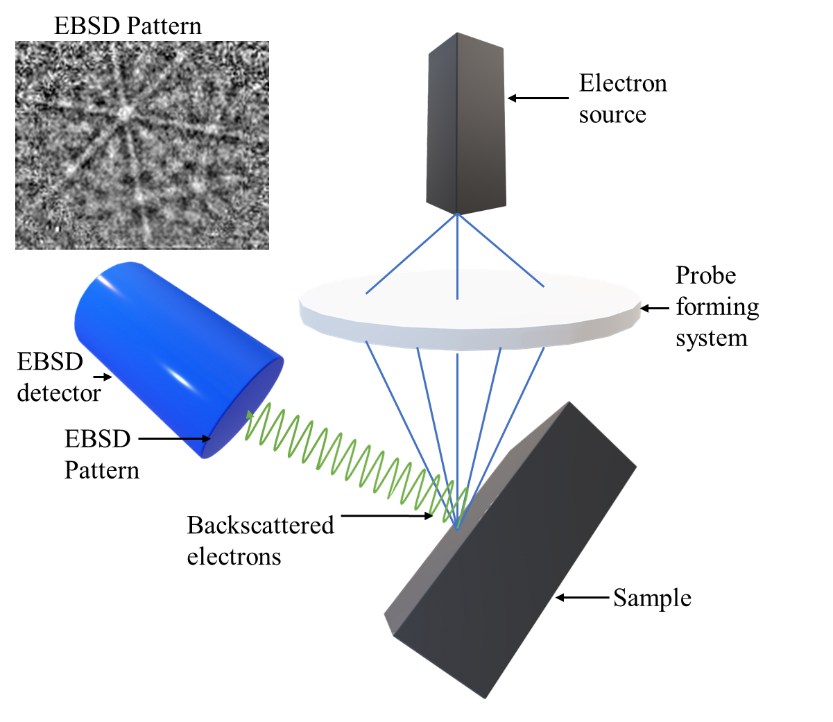
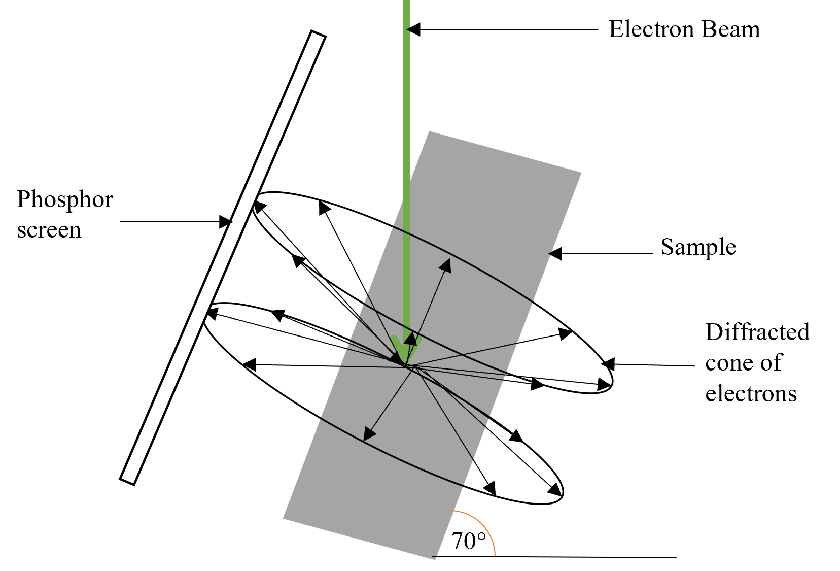
As illustrated in 1, the sample is scanned by the electron beam, with a 2-dimensional (2D) EBSD pattern being taken at each scanned location, producing a 4-dimensional (4D) data-set. EBSD patterns are characteristic of the phase and orientation of a crystalline material, containing multiple bands from the strongest reflecting planes, as demonstrated by 2 Prior to EBSD acquisition, some knowledge of the sample is required, such as the chemical composition, which allows the correct dictionaries to be used for the indexing step. Indexing is performed on each pattern as it is acquired based on the dictionaries provided. Indexing allows the bands in the EBSD patterns to be identified and characterised based on the known reflections of the possible sample structures, allowing the orientations and phases to be identified [2]. Once indexing has been completed, maps of the sample can be created to visualise the sample structure. In these indexed maps each pixel corresponds to a probe position, not to be confused with the pixel positions within an acquired EBSD pattern.
Since the first observation of a backscattered diffraction pattern in 1928 [3] acquisition speed and processing time has significantly improved, going from maximum acquisition speeds of approximately 1500 patterns per second in 2015 using a charge coupled device detector to speeds reach 6000 patterns per second with the implementation of the complementary metal oxide superconductor detector [4]. Automated indexing is commonplace in commercial systems allowing bands in patterns to be easily identified and indexed. Despite these innovations, the speed of EBSD is limited by the acquisition of patterns.


Inspired by compressive sensing theory [5, 6], it is believed that EBSD data acquisition speeds could be improved without any significant advances in detector technology being necessary. In recent studies, compressive sensing has been found to be effective in numerous microscopy applications, notably in scanning transmission electron microscopy (STEM) [7, 8, 9, 10]. Through subsampling signals, data can be acquired at a greater speed and reconstructed using an inpainting algorithm, which could significantly improve the speed of acquisition and processing with regards to EBSD detection. Moreover, in the cases of beam sensitive materials, the effects of beam damage would be greatly reduced.
In this paper we introduce a new EBSD acquisition approach based on subsampling probe positions; resulting in an incomplete 4D EBSD data. Indexing such data, however, yields band contrast and IPF maps with missing values associated with unsampled probe positions. Our approach is to inpaint the indexed images using Beta Process Factor Analysis (BPFA), i.e., a joint blind dictionary learning and sparse coding. Our inpainting approach is based on a fast GPU implementation of BPFA; allowing significantly faster data acquisition without any significant advances in detector technology being necessary.
II Methods
Assume an EBSD detector with pixels recording the measurements from scanning electron probes. Let be the discretised 4-dimensional representation of the EBSD dataset, with representing each virtual image corresponding to a pair of detector pixel coordinates with being the total number of pixels in the detector. We assimilate each to its vectorised version , where is the total number of probe positions. We now introduce the subsampling of probe positions to reduce the acquisition time and beam damage. This amounts to subsampling virtual images where the number of acquired spatial locations in the virtual image is given as acquired over the subsampling set with cardinality . This defines our acquisition model as,
| (1) |
for every virtual image where is a mask operator with if and otherwise. The notable takeaway is that is the same for all virtual images.
Once the measurements have been acquired, indexing is performed, there is no impact on the indexing step through probe subsampling: missing probe positions are ignored during indexing. The subsampled data is then inpainted using BPFA, i.e., a blind dictionary learning approach. For a full explanation of this recovery method refer to Section 2.2 of [10].
III Simulations
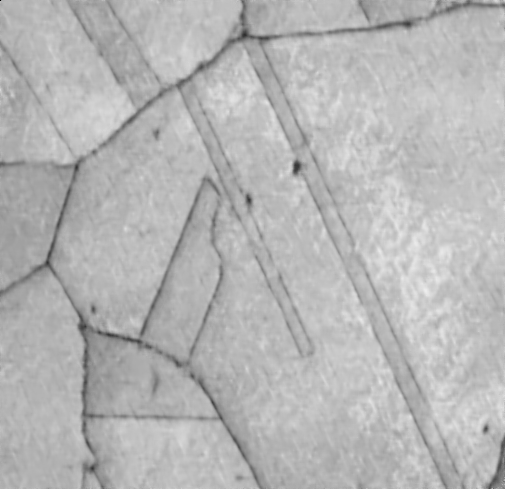
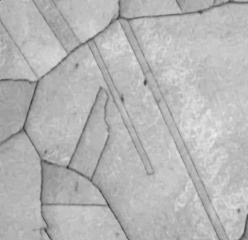
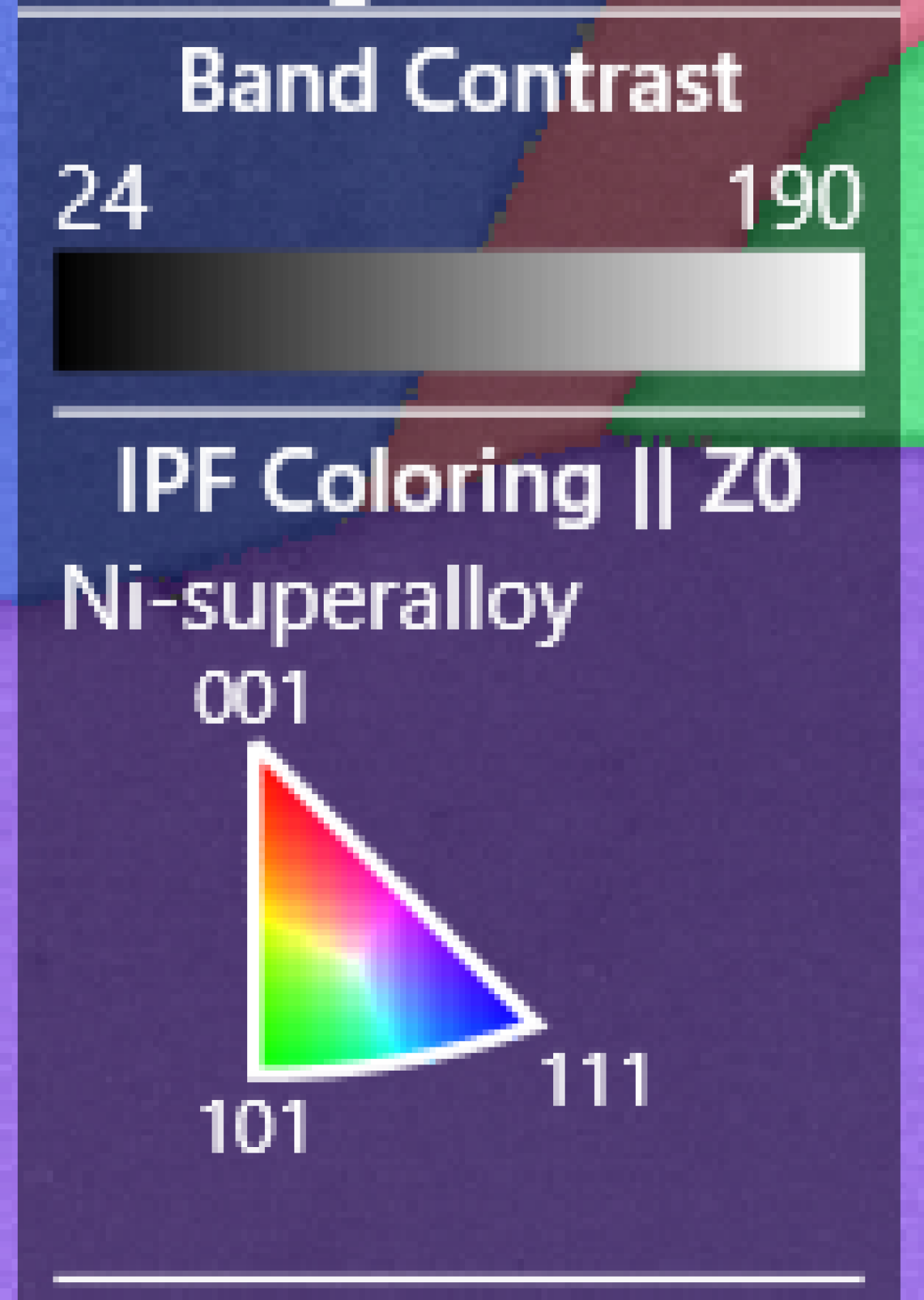
Presented here is one sub-sampling application using a 1024 x 704 pixel EBSD data set of Ni-superalloy. This data was acquired at an acquisition speed of approximately 1600 patterns per second, giving an acquisition time of 7 minutes 24 seconds.
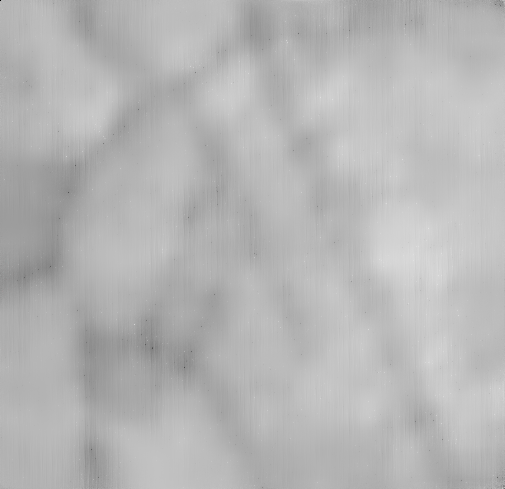
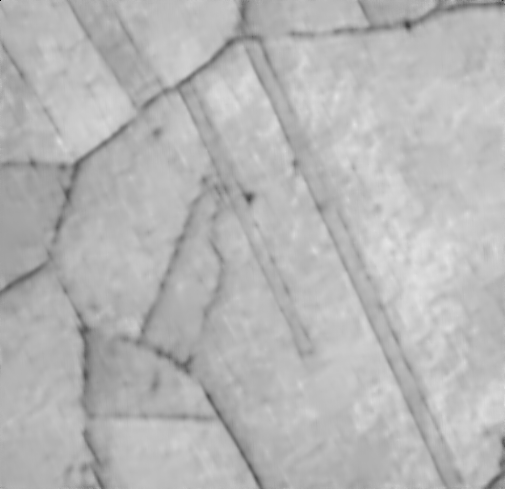
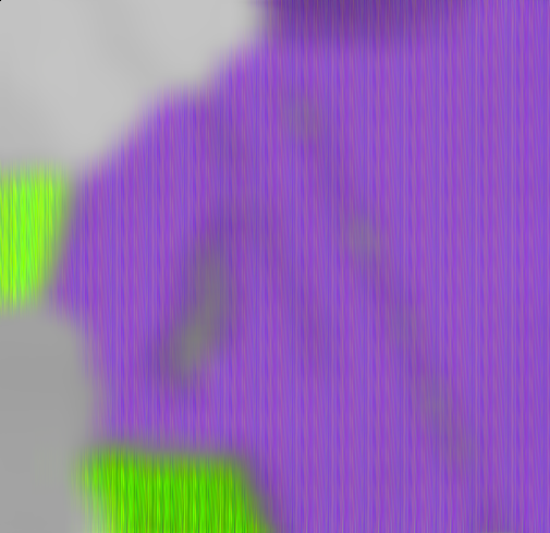
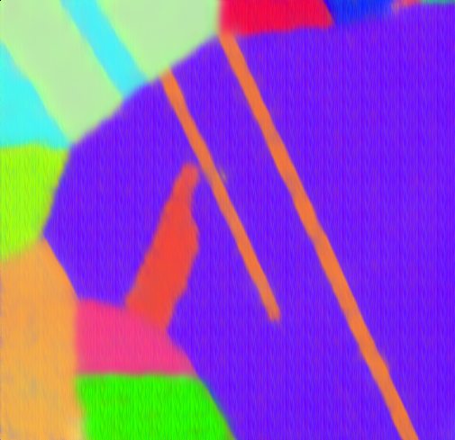
The inpainting algorithm investigated was BPFA [11], with band contrast, and IPF maps being investigated. A 512 x 512 pixel subset of the data was subsampled and reconstructed, shown in Figure 3. The structural similarity (SSIM) results of the reconstructions are shown in 4. The IPF map had a significantly lower SSIM values at 1 and 5 % sampling ratios. Meanwhile, the band contrast map had more consistent SSIM values, ranging from 0.69 to 0.86 from sampling ratios of 1 to 25 %. Acquisition speed of the data is theoretically proportional to the sampling ratio, with a sampling ratio of 10% taking 10% of the acquisition time of a full scan.
[width=1]Images/plot_ssim
IV Conclusions
A proof of concept for subsampling scanning probe positions in the context of EBSD data acquisition using scanning electron microscopy have been discussed. Further investigation into this method will allow for the extension of reconstruction into additional map types. A second subsampling scanning probe position technique will also be investigated, inpainting the EBSD patterns which can then be indexed alongside the acquired patterns to produce a full 4D dataset.
References
- [1] J. I. Goldstein, D. E. Newbury, J. R. Michael, N. W. Ritchie, J. H. J. Scott, and D. C. Joy, Scanning electron microscopy and X-ray microanalysis. Springer, 2017.
- [2] N. C. K. Lassen, “Automated determination of crystal orientations from electron backscattering patterns,” Ph.D. dissertation, The Technical University of Denmark, 1994.
- [3] A. J. Schwartz, M. Kumar, B. L. Adams, and D. P. Field, Electron backscatter diffraction in materials science, 2nd ed. New York: Springer, 2009.
- [4] J. Goulden, P. Trimby, and A. Bewick, “The benefits and applications of a cmos-based ebsd detector,” Microscopy and Microanalysis, vol. 24, pp. 1128–1129, 2018.
- [5] E. J. Candès, J. Romberg, and T. Tao, “Robust uncertainty principles: Exact signal reconstruction from highly incomplete frequency information.” IEEE Transactions on information theory, vol. 52(2), pp. 489–509, 2006.
- [6] D. L. Donoho, “Compressed sensing,” IEEE Transactions on information theory, vol. 52(4), pp. 1289–1306, 2006.
- [7] N. D. Browning, J. Castagna, A. I. Kirkland, A. Moshtaghpour, D. Nicholls, A. W. Robinson, J. Wells, and Y. Zheng., “The advantages of sub-sampling and inpainting for scanning transmission electron microscopy,” Applied Physics Letters, vol. 122(5), p. 050501, 2023.
- [8] D. Nicholls, J. Wells, A. Stevens, Y. Zheng, J. Castagna, and N. D. Browning, “Sub-sampled imaging for stem: Maximising image speed, resolution and precision through reconstruction parameter refinement.” Ultramicroscopy, vol. 233, p. 113451, 2022.
- [9] D. Nicholls, A. W. Robinson, J. Wells, A. Moshtaghpour, A. I. K. M. Bahri, and N. D. Browning, “Compressive scanning transmission electron microscopy,” ICASSP 2022-2022 IEEE International Conference on Acoustics, Speech and Signal Processing (ICASSP), pp. 1586–1590, 2022.
- [10] D. Nicholls, J. Wells, A. W. Robinson, A. Moshtaghpour, M. Kobylynska, R. A. Fleck, A. I. Kirkland, and N. D. Browning, “A targeted sampling strategy for compressive cryo focused ion beam scanning electron microscopy,” arXiv preprint arXiv:2211.03494, 2022.
- [11] J. Paisley and L. Carin, “Nonparametric factor analysis with beta process priors.” Proceedings of the 26th annual international conference on machine learning, pp. 777–784, 2009.