OpenClinicalAI: An Open and Dynamic Model for Alzheimer’s Disease Diagnosis
Abstract
Although Alzheimer’s disease (AD) cannot be reversed or cured, timely diagnosis can significantly reduce the burden of treatment and care. Current research on AD diagnosis models usually regards the diagnosis task as a typical classification task with two primary assumptions: 1) All target categories are known a priori; 2) The diagnostic strategy for each patient is consistent, that is, the number and type of model input data for each patient are the same. However, real-world clinical settings are open, with complexity and uncertainty in terms of both subjects and the resources of the medical institutions. This means that diagnostic models may encounter unseen disease categories and need to dynamically develop diagnostic strategies based on the subject’s specific circumstances and available medical resources. Thus, the AD diagnosis task is tangled and coupled with the diagnosis strategy formulation. To promote the application of diagnostic systems in real-world clinical settings, we propose OpenClinicalAI for direct AD diagnosis in complex and uncertain clinical settings. This is the first powerful end-to-end model to dynamically formulate diagnostic strategies and provide diagnostic results based on the subject’s conditions and available medical resources. OpenClinicalAI combines reciprocally coupled deep multiaction reinforcement learning (DMARL) for diagnostic strategy formulation and multicenter meta-learning (MCML) for open-set recognition. The experimental results show that OpenClinicalAI achieves better performance and fewer clinical examinations than the state-of-the-art model. Our method provides an opportunity to embed the AD diagnostic system into the current health care system to cooperate with clinicians to improve current health care.
keywords:
Real-world clinical setting , Alzheimer’s disease , diagnose , AI , deep learning[label1]organization=Key Lab of Education Blockchain and Intelligent Technology, Ministry of Education, Guangxi Normal University,city=Guilin, country=China
[label2]organization=Guangxi Key Lab of Multi-Source Information Mining and Security, Guangxi Normal University,city=Guilin, country=China
[label4]organization=University of Chinese Academy of Sciences,country=China
[label3]organization=Guilin Medical University,city=Guilin, country=China
[label6]organization=Department of Physiology and Pathophysiology, Capital Medical University,country=China
[label5]organization=International Open Benchmark Council
1 Introduction
Alzheimer’s disease is an incurable disease that heavily burdens our society (the total cost of caring for individuals with AD or other dementias is estimated at billion) [1, 2, 3, 4, 5]. Early and accurate diagnosis of AD is crucial for effectively managing the disease and has the potential to save nearly trillion in medical and care costs. [1, 4]. However, it is estimated that million of the million people with dementia worldwide have not received a timely and accurate diagnosis due to limited medical resources, availability of experts, etc. [6]. Artificial intelligence (AI), as one of the technologies with the most potential to improve medical services, is widely employed in AD diagnosis research [7, 8]. Daniel et al. [9] utilized plasma metabolites as inputs for Extreme Gradient Boosting (XGBoost) and demonstrated their potential in diagnosing AD. Xin et al. [10] proposed an approximate rank pooling method to transform D nuclear magnetic resonance images (MRI) into D images, followed by the utilization of a D convolutional neural network (CNN) for AD classification. Qiu et al. [11] reported a multimodal diagnosis framework that used CNN to capture the critical features of MRI images and then used Categorical Boosting (CatBoost) to detect the presence of AD and cognitively normal (CN) from demographics, medical history, neuroimaging, neuropsychological testing, and functional assessments.
As Fig. 1 (a) shows, the AD diagnosis models in previous works are designed for the closed clinical setting with the following primary assumptions: (1) all categories of subjects are known a priori (subject categories are aligned in training and test sets by inclusion and exclusion criteria); (2) the same diagnostic strategies are used to diagnose all subjects (Delete subjects with missing data of a certain type or fill in missing data for subjects in data preprocessing); and (3) all medical institutions are capable of executing the prescribed diagnostic strategies [12, 10, 13]. This makes the AD diagnosis task an independent typical classification task. However, as Fig. 1 (b) shows, the real-world clinical setting is open with uncertainty and complexity: (1) The categories of subjects in real-world clinical settings are not all known in advance and may include unknown categories that do not appear during the development of AI models. This implies that the test set may contain subject categories not present in the training set. This transforms AD diagnosis into an open-set recognition problem instead of it being a conventional classification problem. (2) Each subject is unique, and there is no one-size-fits-all diagnosis strategy. This results in variations in the amount and types of data across different subjects. (3) The conditions of medical institutions vary and are not known in advance; e.g., positron emission tomography (PET) is available in some hospitals but most hospitals in underdeveloped areas are not equipped with PET. Thus, the AD diagnosis task is an open-set recognition task tangled and coupled with the formulation of diagnosis strategies.
a
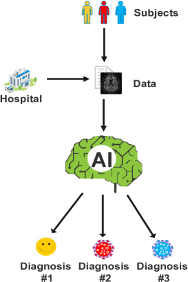
b
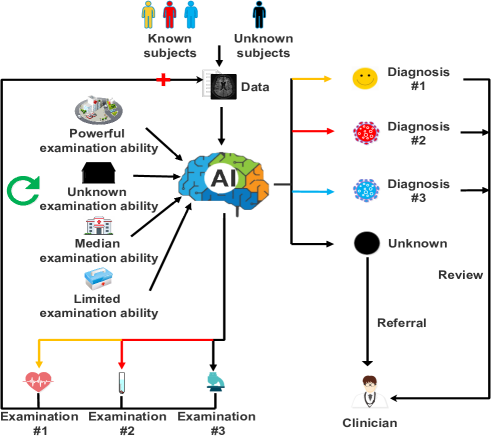
Currently, to tackle the challenges in the real-world clinical setting, open-set recognition technology has emerged in various fields [14]. However, open-set recognition only considers unknown categories while overlooking the complexities of subjects and the constraints posed by medical resources. In the real-world clinical setting, this hinders the application of open-set recognition technology to AD diagnosis. Therefore, the critical problem is how to simultaneously handle the uncertainty and complexity of subjects and medical resources in AD diagnosis within the real-world clinical setting.
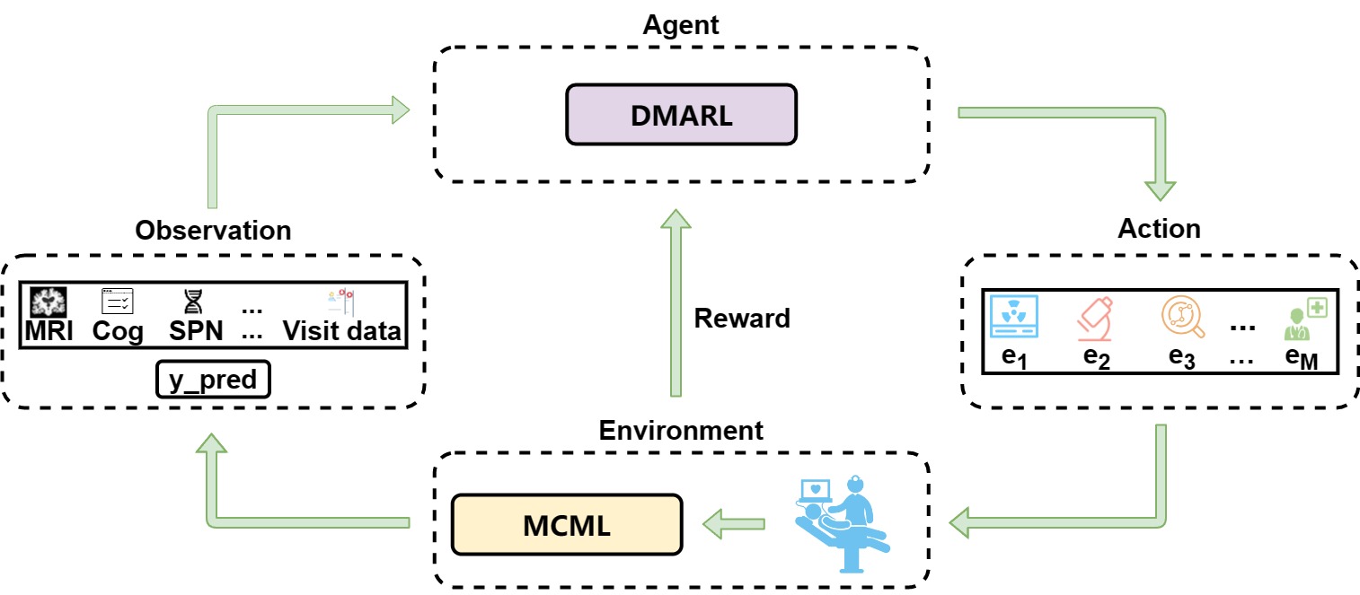
In this paper, we explore the AD diagnosis task from a new perspective of both subjects and medical institutions and redefine AD diagnosis as an open, dynamic real-world clinical setting recognition problem. Specifically, clinicians first formulate a preliminary diagnosis strategy based on the individual’s condition and available medical resources after enquiring about the basic information involving the subject. Second, the subjects undergo examinations in accordance with the diagnostic strategy. Third, the model merges all available information, categorizes subjects into prespecified or unknown categories, or adjusts the diagnostic strategy and returns to the second step. To realize this approach, as illustrated in Fig. 2, we propose OpenClinicalAI, an open and dynamic deep learning model to directly diagnose subjects in complex and uncertain real-world clinical settings. OpenClinicalAI comprises two tangled and coupled modules: deep multiaction reinforcement learning (DMARL) and multicenter meta-learning (MCML). MCML utilizes AutoEncoder and meta-learning techniques to diagnose subjects based on the subject’s data obtained from the diagnostic strategy formulated by DMARL. It serves as an environmental simulator, providing feedback to DMARL. DMARL is a multitask learning model that functions as an agent which dynamically adjusts the subject’s diagnosis strategy and
acquires fresh examination data based on rewards from MCML and the subject’s current examination data.
To summarize, our contributions are as follows:
-
OpenClinicalAI is the first end-to-end coupling model designed specifically for direct AD diagnosis in complex and uncertain real-world clinical settings. It dynamically develops different diagnosis strategies according to different subject situations and different examination abilities of medical institutions on the test set. The framework of OpenClinicalAI is domain-independent and can be extended to other diseases to promote the development of real-world clinical setting diagnosis.
-
The newly designed DMARL enables OpenClinicalAI to dynamically adjust the diagnosis strategy according to the subject’s current data, and MCML provides feedback on classification information and rewards. Its novelty lies in designing a multitask model for selecting the next clinical examinations for a subject and avoiding a step-by-step form of reinforcement learning.
-
The novel MCML promotes open-set recognition of AD based on the diagnosis strategy from DMARL and provides disease classification information and rewards for DMARL. It uses AutoEncoders to retain more general features for open set recognition and uses clustering to divide subjects of the same category into multiple subcategories. This improves the accuracy of meta-learning algorithms in calculating subject similarity, thereby providing unknown subject recognition. In addition, it is also used as an environmental simulator to evaluate the rewards of the diagnostic strategies formulated by DMARL, and the disease classification information is used as the input of DMARL to help dynamically formulate diagnostic strategies.
-
In the closed clinical setting, OpenClinicalAI shows a comparable performance to the current state-of-the-art model in terms of the AUC (area under the receiver operating characteristic curve) metric. At the same time, less than of subjects are required to have an MRI image. In the real-world clinical setting, our model outperforms the current state-of-the-art model by: (1) an absolute increase of and (AD diagnosis and cognitively normal diagnosis) in AUC and 30.09% and in sensitivity; and (2) an absolute reduction of MRI for subjects. In addition, it has in sensitivity for identifying unknown categories of subjects.
2 Related Work
AD diagnosis model. With progress in machine learning and deep learning, AD diagnostic models have gained significant attention in the medical community. AD diagnosis studies can be divided into nonimage-based, image-based, and multimodal-based studies. Aljovic et al. [15] designed a linear feed-forward (FF) neural network that used biomarkers (albumin ratio, AP, AP, tau-total, and tau-phospho) as input to classify Alzheimer’s disease. This work proved that it is possible to use biomarker data for AD discrimination. Lu et al. [16] trained a -dimensional sex classifier Inception-ResNet-V as a base model in transfer learning for AD diagnosis. They then made it suitable for -dimensional MRI inputs and achieved high accuracy in a large collection of brain MRI samples ( scans from participants). Qiu et al. [12] proposed an interpretable multimodal deep learning framework that used a fully convolutional network (FCN) to perform MRI image feature extraction and then used a traditional multilayer perceptron (MLP) with multimodal inputs (image and text features) to classify the disease and generate disease probability maps. Many studies have shown that the performance of multimodal models is better than that of single-modal models because different modal information can complement each other to help diagnose AD.
Open-set recognition. Various open-set recognition (OSR) techniques have been proposed to solve the problem of unknown categories in the real world. They can be divided into discriminant and generative models [14]. Scheirer et al. [17] proposed a preliminary solution, a -vs.-Set machine, which sculpts a decision space from the marginal distances of a 1-class or binary SVM with a linear kernel. Bendale et al. [18] introduced a model called layer-OpenMax, which estimates the probability of an input being from an unknown class to adapt deep networks for open-set recognition. Oza et al. [19] proposed a deep neural network-based model—CAE, which uses class-conditioned autoencoders with novel training and testing methodologies. Yang et al. [20] proposed a model based on a generative adversarial network (GAN) to address open-set human activity recognition without manual intervention during the training process. Geng et al. [21] proposed a collective decision-based OSR framework (CD-OSR) by slightly modifying the hierarchical Dirichlet process (HDP). This method aimed to extend existing OSR for new class discovery while considering correlations among the testing instances. Perera et al. [22] utilized self-supervision and augmented the input image to learn richer features to improve separation between classes. These methods tend to use powerful generative models to mimic novel patterns.
Deep reinforcement learning. Deep learning is already widely applied in the medical field for prognosis prediction and treatment recommendation. Saboo et al. [23] simulated the progression of Alzheimer’s disease (AD) by integrating differential equations (DEs) and reinforcement learning (RL) with domain knowledge. DEs serve as an emulator, providing the relationships between some (but not all) factors associated with AD. The trained RL model acts as a proxy to extract the missing relationship by optimizing the objective function over time. The model uses baseline ( year) characteristics from aggregated and real data to personalize a patient’s -year AD progression. Quan et al. [24] proposed an interpretable deep reinforcement learning model to reconstruct compressed sensing MRI images. Komorowski et al. [25] developed an artificial intelligence (AI) clinician model using a reinforcement learning agent. This model extracts implicit knowledge from patient data based on the lifetime experience of human clinicians and analyzes numerous treatment decisions, including mostly suboptimal decisions, to learn optimal treatment strategies. Experiments have shown that, on average, the value of treatments selected by the AI clinician model is reliably higher than that of human clinicians.
Deep reinforcement learning (DRL) is a combination of deep learning and reinforcement learning algorithms that learn and optimize decision-making strategies through interactions with the environment. However, current research often treats disease diagnosis as a one-step task, where the model inputs patient information and obtains a classification. This approach has led to a neglect of the potential application of DRL in diagnostic tasks.
3 Problem Formulation
In this work, the diagnosis of AD involves dynamically developing diagnostic strategies based on the subject’s situation and the medical institution, ultimately determining whether the subject belongs to the unknown category. To address the dynamic interaction process between subjects and medical institutions, we propose a reinforcement learning model, namely, OpenClinicalAI, to solve this problem. The detailed definition of the AD diagnosis problem is as follows:
Agent: The agent is capable of perceiving the state of the external environment and the rewards and uses this information to learn and make decisions. In this problem, we employ the DMARL model as the agent, which takes observation of the environment as input and formulates recommended next clinical examinations based on the subject’s conditions and the medical institution.
Environment: The environment refers to all objects external to the intelligent agent, whose observation is influenced by the actions of the agent and provides corresponding rewards to the agent. In our problem, the environment encompasses three components: the MCML model, subjects, and medical institutions. Medical institutions conduct clinical examinations on subjects according to the action suggested by the agent. The data generated by these clinical examinations serve as input for the MCML model, which generates intermediate diagnostic results. The environment provides feedback to the intelligent agent using the diagnostic intermediate results and the latest clinical data while simultaneously calculating the corresponding reward.
Observation: The agent’s observation of the environment include the intermediate diagnostic results from MCML and the currently available clinical data of the subjects.
Action: The agent interacts with the environment via actions where the actions include types of clinical examinations. An action may consist of one or more clinical examinations.
Reward: The reward is the bonus that an agent gets once it takes an action in the environment at the given time step . In this paper, the reward is measured as the degree of improvement in the probability of intermediate diagnostic results obtained by the MCML model after taking an action.
Discount factor (): The discount factor measures the importance of future rewards to the agent in the current state. In this paper, represents the set of hyperparameters of the OpenClinicalAI model. During the gradient descent process, the model will adjust these hyperparameters to obtain the maximum expected reward.
We use to represent clinical examinations performed for Alzheimer’s disease, to represent the data obtained by a subject undergoing the corresponding clinical examination, and to represent the time step series of the model, where is determined by the model according to the conditions of the subject and the medical institution. Using to represent the data for subjects contained in the dataset, where represents the clinical examination data that has up to the time step with , , , . Using represents the original data of . indicates the next clinical examination recommended by the model (which contains clinical examinations) at the time step, where , , , . is used to represent the clinical examination data obtained by the subject after executing . Then, update the data .
In particular, we call the set of actions selected by the model in to time steps a diagnostic strategy , and the set of diagnostic strategies is represented by , where represents the number of diagnostic strategies generated based on the combination of clinical examinations contained by the subject data, which is determined by the model based on the subject and the medical institution. Therefore, we use to represent the data obtained by after executing the diagnostic strategy . In addition, as an input of the MCML model will obtain the corresponding diagnosis , and as an input of the MCML model will obtain the corresponding intermediate diagnosis . Thus, the observation of the time step is , and the reward after executing action is .
Based on the aforementioned definitions, is a sequence of states, actions, and rewards that are generated by the model’s interaction with over a time step of duration
4 Methodology
4.1 Data preparation
For each subject, if there is more than one visit, each visit is considered to be an independent sample. Multiple categories of data are generated at each visit based on the diagnostic strategy. This results in each sample usually containing multiple types of data. As for medical images, we first convert the data from DICOM format to NIFTI format using the dcmnii library. Then, we perform image registration using the ANTs library [26, 27, 28]. Next, we convert the D image into D slices and transform the image from grayscale to RGB. Finally, we utilize a pretrained model called DenseNet to extract features from the D slices [29]. For genetic data, we extract single nucleotide polymorphisms (SNPs) that are highly related to AD and represent each SNP using a one-hot encoding method [30, 31, 32]. To accommodate the varying dimensions of each data category, the number of data categories included in each visit, and the number of past visits for each subject, we use a unified data representation framework, as illustrated in Supplementary Figure. S1. In this framework, we use an array with a shape of to represent the examination category in the subject’s visit. The shape of our data is , where n represents the number of data categories for the subject.
4.2 OpenclinicalAI
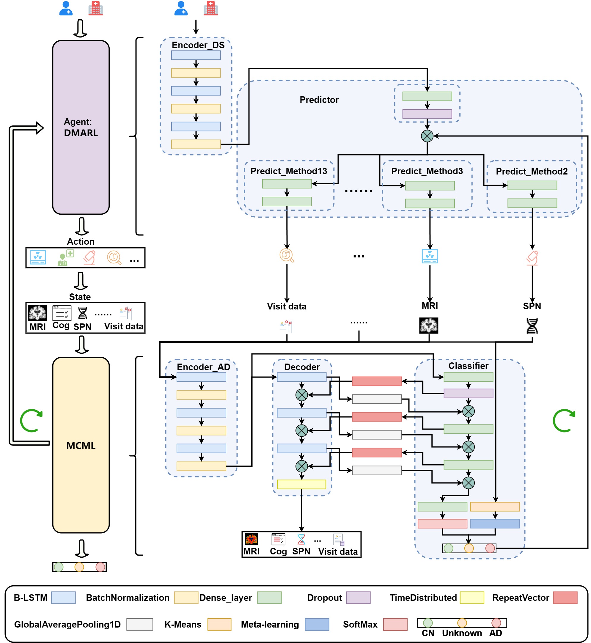
As shown in Fig. 3, OpenclinicalAI is based on the status of subjects and medical institutions to directly formulate diagnostic strategies and provide accurate diagnostic results. This approach enables the system to effectively handle the inherent uncertainty and complexity of real-world clinical settings. This model is composed of two coupled elements: (1) DMARL (refer to Section 4.2.2) combines encoder, multitask learning, and deep reinforcement learning to dynamically adapt the subject’s diagnostic strategy based on the specific circumstances of the hospital, subject, and feedback from MCML. (2) MCML (refer to Section 4.2.1) takes the data generated by DMARL’s predictions as input, employs AutoEncoder to extract more general features, calculates more precise sample-to-class center distances, and integrates meta-learning techniques to facilitate AD identification in open clinical environments. In summary, the objective of OpenclinicalAI can be formulated as follows:
| (1) | ||||
where and indicate the predicted class probability and true label of the subject, respectively. Similarly, and denote the subject’s original input features and reconstructed features, respectively. , , , and correspond to the hyperparameters of each loss, while signifies the weight of the network. Specifically, is the softmax cross-entropy function, represents the regularized loss function, utilizes uncertainty to weigh losses in a multitask learning scenario, and captures the reconstruction loss of .
4.2.1 MCML for AD recognition and environment simulation
The architecture of MCML is inspired by Generative Discriminative Feature Representations [22] and meta-learning-based OpenMax [18]. As depicted in Fig. 3, MCML combines the advantages of the previous two works. In addition, (1) MCML further improves the retention of generalized features by fostering information interaction between each layer of the decoder and classifier. This mitigates the risk of the classifier solely focusing on the most relevant features for classification. (2) MCML utilizes k-means clustering to partition subjects of the same category into multiple subtypes. It then calculates the distance between subjects and the center of the nearest subtype. This approach reduces the likelihood of atypical subjects being misclassified as unknown categories due to their substantial distance from the category center. Specifically, MCML comprises three components: , , and . These components classify subjects into AD, CN, and unknown categories based on specific diagnostic strategies. The is structured as a mirror image of the to enable the model to capture more subject-specific characteristics associated with AD and CN classification. The consists of three dense layers and a SoftMax/OpenMax layer which facilitates subject classification in the open clinical setting. In summary, the loss function for the MCML module is formulated as follows:
| (2) |
where denotes the probability assigned to the subject’s cognitive state prediction, and the cross entropy loss is employed to quantify the disparity between the subject’s current cognitive state, determined after an examination, and the cognitive state predicted by the MCML model. The score modifier is based on [18], and represents the probability that the sample belongs to an unknown category.
4.2.2 DMARL for diagnostic strategy development
As depicted in Fig. 3, the DMARL module consists of an encoder, , and a predictor, . As an agent, DMARL takes the observation of the subject and the feedback of MCML as inputs. To effectively incorporate variable-length clinical data based on different diagnostic strategies, we employ a bidirectional long short-term memory (B-LSTM) network in the encoding pathway. comprises three layers of B-LSTM units. The primary objective of DMARL is to utilize existing medical resources, subject observations, and the classification confidence from MCML to predict the subsequent clinical examinations for each subject and dynamically formulate an optimal diagnostic strategy. To integrate the output of with the classification confidence from MCML, consists of dense layers. Furthermore, to determine the appropriate clinical examination for each subject, the model incorporates independent sigmoid classifiers. In addition, due to the lack of labels for the next clinical examination, DMARL will be trained according to the rewards of MCML. Thus, the loss function for the DMARL module is expressed as follows:
| (3) |
The sum of losses is calculated by evaluating recommended subtasks () for examination, where is an observation noise scalar of the output of ith examination [33].
4.3 Reward of the diagnosis strategy
Although there have been significant efforts to improve the interpretability and internal logic of deep learning, understanding the behavior of deep learning models remains challenging [34, 35]. We do not know whether the diagnosis strategy of the AI model needs to be consistent with that of an human expert . Therefore, in this work, we train the DMARL module based on the reward generated by MCML. The ultimate goal of OpenclinicalAI is to accurately identify AD patients using MCML. The subsequent examination for each subject is determined by whether it leads to a higher predicted probability for the correct category and lower predicted probabilities for other categories. The reward of MCML is calculated using Algorithm 1.
4.4 Model training
The training process is divided into two stages. In the first stage, the procedure is as follows: (1) For each , the diagnostic strategy set is generated according to examinations contained in the data . It should be noted that the types and number of clinical examinations are not the same between subjects since the diagnosis strategy will change dynamically according to different subjects and available medical resources. (2) Every is considered to be an independent sample and forms the dataset , where represents the true diagnostic category label of . (3) The MCML model is trained and tested on , and the intermediate diagnosis result is obtained. In the second stage, the process is as follows: (1) For every , we obtain the reward and by Algorithm 1, and form the dataset . (2) We then train and test the DMARL on .
4.5 Prediction
To deliver the final diagnosis result, it is imperative to dynamically adjust the diagnosis strategy. The prediction process is delineated in Algorithm 2.
5 Results
5.1 Human subjects
Data used in the preparation of this article were obtained from the Alzheimer’s Disease Neuroimaging Initiative (ADNI) dataset (http://adni.loni.usc.edu). We included subjects from ADNI , ADNI GO, ADNI , and ADNI for visits based on data availability. Detailed information can be found in Section S1. The characteristics of the subjects are shown in Table 1. All the models will be verified in both a closed clinical setting and a real-world clinical setting. The configurations of the closed clinical setting and real-world clinical setting are as follows:
| Data set | Training set | Validation set | Test set | ||
| Age | 54-59.9 | 80 | 36 | 2 | 59 |
| 60-69.9 | 596 | 246 | 10 | 442 | |
| 70-70.9 | 1048 | 528 | 46 | 695 | |
| 80-80.9 | 395 | 213 | 14 | 259 | |
| 90-91.9 | 6 | 1 | 1 | 4 | |
| Missing | 2 | 1 | 0 | 1 | |
| Gender | Female | 1130 | 560 | 44 | 785 |
| Male | 997 | 465 | 29 | 675 | |
| Educate | 8-10 | 40 | 18 | 2 | 23 |
| 11-13 | 353 | 176 | 13 | 243 | |
| 14-16 | 823 | 403 | 26 | 558 | |
| 17-20 | 900 | 424 | 32 | 628 | |
| Missing | 11 | 4 | 0 | 8 | |
| Ethnic category | Hisp/Latino | 73 | 32 | 5 | 49 |
| Not Hisp/Latino | 2042 | 986 | 67 | 1404 | |
| Missing | 12 | 7 | 1 | 7 | |
| Racial category | Asian | 40 | 20 | 0 | 25 |
| Black | 88 | 41 | 5 | 57 | |
| Hawaiian/Other PI | 2 | 0 | 0 | 2 | |
| More than one | 25 | 10 | 0 | 18 | |
| White | 1964 | 954 | 68 | 1350 | |
| Am Indian/Alaskan | 4 | 0 | 0 | 4 | |
| Missing | 4 | 0 | 0 | 4 | |
| Marriage | Married | 1618 | 805 | 59 | 1100 |
| Never_married | 73 | 30 | 3 | 48 | |
| Widowed | 238 | 114 | 8 | 165 | |
| Divorced | 191 | 75 | 3 | 141 | |
| Missing | 7 | 1 | 0 | 6 | |
| Category | AD | 740 | 587 | 44 | 109 |
| CN | 589 | 466 | 31 | 92 | |
| MCI | 1082 | 0 | 0 | 1082 | |
| SMC | 280 | 0 | 0 | 280 |
5.1.1 Closed world setting
In the closed world setting, of AD and CN subjects were used for the training set, of AD and CN subjects were used for the validation set, and of AD and CN subjects were used for the test set.
5.1.2 Real world setting
A total of subjects with visits were included in our work. A subject during a visit may require different categories of examination. Every combination of those examinations represents a diagnostic strategy. Thus, for the data of subject, 443,795 strategies were generated. These AD and CN subjects were randomly assigned to the training, validation, and test sets. The training set contained subjects with visits and generated strategies. In the training set, subjects with visits were AD and developed strategies, and subjects with visits were CN and generated strategies. The validation set contained subjects with visits and generated strategies. In the validation set, subjects with visits were AD and develop strategies, and subjects with visits were CN and generated strategies. The test set contained subjects with visits. In the test set, subjects with visits were AD, subjects with visits were CN, subjects with visits were MCI (mild cognitive impairment), and subjects with visits were SMC (significant memory concern). Notably, the dataset contains multiple visits for a subject’s progression from CN to AD.
We note that the lack of examination data was able to simulate the lack of the executive ability of the examination by the medical institution. It is logically equivalent to missed examinations in medical institutions. All subjects with labels containing at least one of the above categories of information were considered in this study.
5.2 Comparison methods
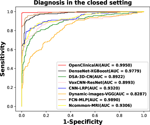
We validated the effectiveness of OpenclinicalAI by comparing its performance with recent work: image-based models (DSA-3D-CNN (2016) [36], VoxCNN-ResNet (2017) [37], CNN-LRP (2019) [13], Dynamic-image-VGG (2020) [10], Ncommon-MRI (2022) [11], DenseNet-XGBoost) and multimodal input models (FCN-MLP (2020) [12]). In addition, the transfer learning-based model DenseNet-XGBoost was the previous state-of-the-art baseline model, since among the recent AI diagnosis studies, the transfer learning framework of the pretrained model achieved state-of-the-art performance in many diagnosis tasks based on medical images [38, 39, 40, 41, 42].
5.3 Experimental setup
The model was optimized using mini-batch stochastic gradient descent with Adam and a base learning rate of [43]. All comparison models were constructed and trained according to the needs of AD diagnosis tasks and their official codes under the same settings. The experiments were conducted on a Linux server equipped with Tesla P and Tesla P GPUs.
| Model | AUC(95% CI) | Accuracy(95% CI) | Sensitivity(95% CI) | Specificity(95% CI) |
|---|---|---|---|---|
| DSA-3D-CNN [36] | 89.23%(87.91%-90.56%) | 84.20%(82.76%-85.60%) | 77.05%(74.55%-79.49%) | 89.00%(87.46%-90.66%) |
| VoxCNN-ResNet [37] | 90.64%(89.31%-91.80%) | 85.24%(83.76%-86.56%) | 80.76%(78.44%-83.04%) | 88.65%(86.96%-90.25%) |
| CNN-LRP [13] | 93.21%(92.11%-94.16%) | 82.36%(80.72%-83.76%) | 90.68%(88.91%-92.27%) | 75.97%(73.57%-78.21%) |
| Dynamic-image-VGG [10] | 83.22%(81.58%-84.81%) | 73.42%(71.71%-75.12%) | 85.24%(83.11%-87.34%) | 64.41%(61.98%-66.89%) |
| Ncomms2022-MRI [11] | 93.06%(92.04%-94.05%) | 87.40%(86.12%-88.68%) | 82.53%(80.26%-84.71%) | 91.08%(89.66%-92.57%) |
| DenseNet-XGBoost | 97.79%(97.30%-98.27%) | 93.12%(92.12%-94.08%) | 91.09%(89.40%-92.90%) | 94.53%(93.35%-95.64%) |
| FCN-MLP [12] | 97.28%(96.71%-97.81%) | 90.72%(89.56%-91.84%) | 92.26%(90.59%-93.76%) | 89.55%(87.89%-91.10%) |
| OpenClinicalAI | 99.50%(99.11%-99.80%) | 96.52%(95.76%-97.20%) | 91.86%(90.13%-93.37%) | 100%(100%-100%) |
-
1
CI = confidence interval. To evaluate the evaluation index of the AI model, a non-parametric bootstrap method was applied to calculate the CI for the evaluation index [44]. We calculated CI for every evaluation index. We randomly sampled cases from the test set and evaluated the AI model by the sampled set for every evaluation index. repeated trials were executed, and values of the evaluation index were generated. The CI was obtained by the and percentiles of the distribution of the evaluation index values.
| Model | AUC(95% CI) | Sensitivity(95% CI) | |||
|---|---|---|---|---|---|
| AD | CN | AD | CN | unknown | |
| DSA-3D-CNN-Thr | 78.90%(74.91%-82.71%) | 66.72%(63.33%-70.07%) | 75.93%(68.66%-82.92%) | 85.56%(80.12%-90.50%) | 6.49%(5.51%-7.63%) |
| DSA-3D-CNN-OpenMax | 78.03%(74.03%-81.62%) | 66.86%(63.41%-70.38%) | 60.29%(51.97%-68.38%) | 72.63%(65.86%-79.25%) | 30.26%(28.46%-32.22%) |
| VoxCNN-ResNet-Thr | 76.02%(71.66%-80.07%) | 66.42%(63.01%-69.77%) | 72.82%(65.41%-80.00%) | 85.20%(80.09%-90.15%) | 7.05%(6.00%-8.12%) |
| VoxCNN-ResNet-OpenMax | 76.33%(72.32%-79.97%) | 64.12%(60.58%-67.60%) | 59.86%(51.67%-67.39%) | 65.02%(58.06%-72.13%) | 29.14%(27.33%-31.05%) |
| CNN-LRP-Thr | 82.93%(79.43%-86.25%) | 67.92%(64.76%-71.14%) | 89.51%(83.82%-94.24%) | 71.59%(64.67%-78.02%) | 6.36%(5.34%-7.45%) |
| CNN-LRP-OpenMax | 82.82%(78.96%-86.18%) | 67.91%(64.72%-70.91%) | 85.29%(78.73%-90.90%) | 58.13%(50.80%-65.13%) | 20.16%(15.11%-23.89%) |
| Dynamic-image-VGG-Thr | 71.75%(67.55%-75.64%) | 63.07%(59.66%-66.53%) | 85.25%(79.35%-90.72%) | 64.16%(57.29%-70.93%) | 0.04%(0.00%-0.13%) |
| Dynamic-image-OpenMax | 70.75%(66.62%-74.84%) | 63.28%(59.76%-66.63%) | 79.19%(72.35%-85.82%) | 57.28%(50.26%-64.40%) | 14.26%(12.78%-15.77%) |
| Ncomms2022-MRI-Thr | 81.15%(77.64%-84.44%) | 69.32%(65.94%-72.40%) | 79.13%(72.43%-85.48%) | 87.84%(82.98%-92.59%) | 10.34%(9.14%-11.64%) |
| Ncomms2022-MRI-OpenMax | 81.21%(77.55%-84.74%) | 69.33%(66.11%-72.46%) | 63.98%(56.12%-71.87%) | 68.57%(62.05%-75.28%) | 38.14%(36.13%-40.25%) |
| DenseNet-XGBoost-Thr | 84.00%(80.55%-87.28%) | 87.79%(85.06%-90.25%) | 54.83%(46.04%-63.01%) | 33.33%(26.63%-39.79%) | 88.88%(87.53%-90.18%) |
| FCN-MLP-Thr | 91.71%(89.42%-93.78%) | 74.16%(71.11%-76.98%) | 91.93%(86.79%-96.15%) | 90.00%(85.11%-94.08%) | 14.64%(13.22%-16.09%) |
| FCN-MLP-OpenMax | 92.55%(90.26%-94.53%) | 74.30%(71.45%-77.04%) | 73.15%(65.67%-80.45%) | 66.47%(59.45%-73.17%) | 57.95%(55.81%-60.03%) |
| OpenClinicalAI-G | 85.54%(83.30%-87.70%) | 82.65%(80.05%-85.30%) | 93.28%(88.66%-96.92%) | 76.84%(70.79%-82.75%) | 34.33%(32.21%-36.30%) |
| OpenClinicalAI | 95.02%(93.04%-96.62%) | 99.27%(98.54%-99.81%) | 84.92%(78.91%-90.51%) | 81.27%(75.51%-86.67%) | 93.96%(92.90%-94.92%) |
| Model | Base | Cog | CE | Neur | FB | PE | Blood | Urine | MRI | FDG | AV45 | Gene | CSF | |
|---|---|---|---|---|---|---|---|---|---|---|---|---|---|---|
| closed world setting | DSA-3D-CNN [36] | 0 | 0 | 0 | 0 | 0 | 0 | 0 | 0 | 594 (122) | 0 | 0 | 0 | 0 |
| VoxCNN-ResNet [37] | 0 | 0 | 0 | 0 | 0 | 0 | 0 | 0 | 594 (122) | 0 | 0 | 0 | 0 | |
| CNN-LRP [13] | 0 | 0 | 0 | 0 | 0 | 0 | 0 | 0 | 594 (122) | 0 | 0 | 0 | 0 | |
| Dynamic-image-VGG [10] | 0 | 0 | 0 | 0 | 0 | 0 | 0 | 0 | 594 (122) | 0 | 0 | 0 | 0 | |
| Ncomms2022-MRI [11] | 0 | 0 | 0 | 0 | 0 | 0 | 0 | 0 | 594 (122) | 0 | 0 | 0 | 0 | |
| DenseNet-XGBoost | 0 | 0 | 0 | 0 | 0 | 0 | 0 | 0 | 594(122) | 0 | 0 | 0 | 0 | |
| FCN-MLP [12] | 716 | 716 | 0 | 0 | 0 | 0 | 0 | 0 | 594 (122) | 0 | 0 | 0 | 0 | |
| OpenClinicalAI | 716 | 216 | 145 | 114 | 144 | 137 | 34 | 32 | 71 | 28 | 28 | 1 | 0 | |
| real world setting | DSA-3D-CNN-Thr | 0 | 0 | 0 | 0 | 0 | 0 | 0 | 0 | 4609(744) | 0 | 0 | 0 | 0 |
| DSA-3D-CNN-OpenMax | 0 | 0 | 0 | 0 | 0 | 0 | 0 | 0 | 4609(744) | 0 | 0 | 0 | 0 | |
| VoxCNN-ResNet-Thr | 0 | 0 | 0 | 0 | 0 | 0 | 0 | 0 | 4609(744) | 0 | 0 | 0 | 0 | |
| VoxCNN-ResNet-OpenMax | 0 | 0 | 0 | 0 | 0 | 0 | 0 | 0 | 4609(744) | 0 | 0 | 0 | 0 | |
| CNN-LRP-Thr | 0 | 0 | 0 | 0 | 0 | 0 | 0 | 0 | 4609(744) | 0 | 0 | 0 | 0 | |
| CNN-LRP-OpenMax | 0 | 0 | 0 | 0 | 0 | 0 | 0 | 0 | 4609(744) | 0 | 0 | 0 | 0 | |
| Dynamic-image-VGG-Thr | 0 | 0 | 0 | 0 | 0 | 0 | 0 | 0 | 4609(744) | 0 | 0 | 0 | 0 | |
| Dynamic-image-VGG-OpenMax | 0 | 0 | 0 | 0 | 0 | 0 | 0 | 0 | 4609(744) | 0 | 0 | 0 | 0 | |
| Ncomms2022-MRI-Thr | 0 | 0 | 0 | 0 | 0 | 0 | 0 | 0 | 4609(744) | 0 | 0 | 0 | 0 | |
| Ncomms2022-MRI-OpenMax | 0 | 0 | 0 | 0 | 0 | 0 | 0 | 0 | 4609(744) | 0 | 0 | 0 | 0 | |
| DenseNet-XGBoost | 0 | 0 | 0 | 0 | 0 | 0 | 0 | 0 | 4609(744) | 0 | 0 | 0 | 0 | |
| FCN-MLP-Thr | 5353 | 5353 | 0 | 0 | 0 | 0 | 0 | 0 | 4609(744) | 0 | 0 | 0 | 0 | |
| FCN-MLP-OpenMax | 5353 | 5353 | 0 | 0 | 0 | 0 | 0 | 0 | 4609(744) | 0 | 0 | 0 | 0 | |
| OpenClinicalAI-G | 5353 | 2921 | 2483 | 2261 | 2388 | 2245 | 510 | 228 | 1533 | 684 | 509 | 347 | 232 | |
| OpenClinicalAI | 5353 | 5353 | 2146 | 2051 | 2021 | 1986 | 450 | 154 | 1663 | 681 | 444 | 449 | 240 | |
-
1.
The in the column of MRI means that of all the samples required to provide MRI, only samples provided information, and 122 samples did not.
5.4 The performance in the closed clinical setting
As shown in Table 2 and Fig. 4 (a), in general, the performance of multimodal model (such as AUC score of FCN-MLP achieving [ CI -], accuracy score achieving [ CI -]) is better than that of single-modal model (such as AUC score of CNN-LRP achieving [ CI -], accuracy score achieving [ CI -]). The OpenClinicalAI achieves the state-of-the-art performance with the AUC score of [ CI -] and the accuracy score of [ CI -] in the closed setting. Considering the confidence interval of the model, OpenClincialAI does not show significant improvement compared with other multimodal-based models. This indicates that the task of diagnosing AD in a closed clinical setting is not very challenging, and there is not much room for improvement [7, 8].
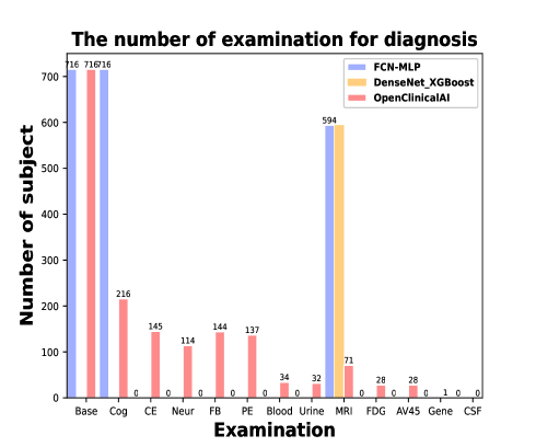
The essential improvement from the state-of-the-art model to OpenClinicalAI is that the latter can dynamically develop personalized diagnosis strategies according to specific subjects and medical institutions. As shown in Table 4 and Fig. 4 (b), less than of the subjects require a nuclear magnetic resonance scan, and most of the subjects only require less demanding examination, such as cognitive examination. We conclude that OpenClinicalAI can avoid unnecessary examinations for subjects and is suitable for medical institutions with varying examination facilities 111Different hospitals have various clinical settings, such as community hospitals without nuclear magnetic resonance machines and large hospitals with multiple facilities. Of the samples in the test set, only samples had MRI data, and the remaining samples were simply discarded because they could not be used as inputs to the corresponding MRI model.
a
b
5.5 The performance of AD diagnosis in the real-world clinical setting
To apply the model to the real-world clinical setting, the model trained in a closed clinical setting usually needs to set a threshold or add OpenMax or use the generated pattern to identify samples of unknown categories [14]. As shown in Table 3 and Fig. 5, in general, the performance of all models has declined significantly (AUC score decline for AD was in the range of [-], accompanied by a very low sensitivity to unknown), but the multimodal model is still better than the single-modal model. OpenClinicalAI achieves state-of-the-art performance (AUC score to AD reaching [ CI -], sensitivity to unknown reaching [ CI -]). In addition, except for our models, all models have high recognition accuracy for samples of known categories and significantly low recognition accuracy for samples of unknown categories (for example, the sensitivity of FCN-MLP-Thr to AD is [ CI -], the sensitivity to CN is [ CI -], and the sensitivity to unknown was [ CI -]), or vice versa (the sensitivity of DenseNet-XGBoost-Thr to unknown is [ CI -]).
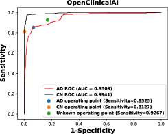
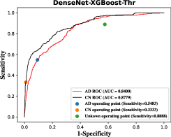
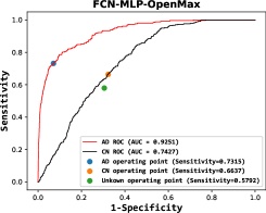
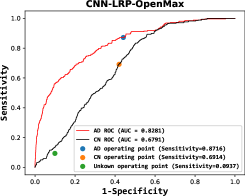
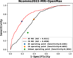
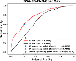
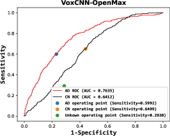
Compared to the state-of-the-art model, OpenClinicalAI demonstrates a significant improvement in the AUC of identification of AD subjects and the AUC of identification of CN subjects . It is worth noting that, as shown in Fig. 5 (a), OpenClinicalAI has a substantial improvement in the sensitivity of AD, CN, and unknown operating points (the sensitivity of OpenClinicalAI to unknown is [ CI -]).
For a state-of-the-art model, such as DenseNet-XGBoost, the sensitivity of known (AD and CN) subjects is low, the sensitivity of AD is just , and the sensitivity of CN is just . This indicates that most known subjects will be marked as unknown and sent to a clinician for diagnosis. Moreover, the sensitivity of unknown subjects is , meaning of unknown subjects will be misdiagnosed. In addition, the DenseNet-XGBoost model requires that every subject has a nuclear magnetic resonance scan, and hence every medical institution that deploys the baseline model must be equipped with a nuclear magnetic resonance apparatus.
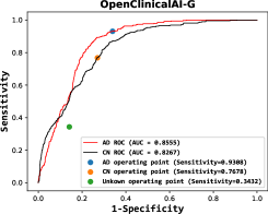
For OpenClinicalAI-G, which does not have an OpenMax mechanism but instead uses a generative-discriminative mechanism, the sensitivity for known (AD and CN) subjects is as good as that of OpenClinicalAI with an OpenMax mechanism [22] (the sensitivity of OpenClinicalAI-G to AD is [ CI -], and the sensitivity to CN is [ CI -]). In contrast, the sensitivity of unknown subjects is much worse than that of OpenClinicalAI with an OpenMax mechanism (the sensitivity of OpenClinicalAI-G to unknown subjects is [ CI -]). This means that most unknown subjects will be misdiagnosed, which is unacceptable in real-world settings. The generative model of open set recognition is not very helpful for AD diagnosis in a real-world clinical setting. In addition, as shown in Table 3, the combination of the OpenMax mechanism and traditional model cannot help AD diagnosis in a real-world clinical setting.
In contrast, OpenClinicalAI diagnoses most of the known (AD and CN) subjects correctly, marks most of the rest as unknown, and sends them to the clinician for further diagnosis. In addition, most unknown subjects are correctly identified, and the misdiagnosis of unknown subjects is only . This means that OpenClinicalAI has significant potential application value for implementation in real-world settings. In addition, as shown in Fig. 5 (i), similar to the behaviors of OpenClinicalAI in the closed setting, OpenClinicalAI can develop and adjust diagnosis strategies for every subject dynamically in the real-world setting. Only a small portion of subjects require a nuclear magnetic resonance scan and more costly (both in terms of economy and potential harm) examinations.
5.6 Development of diagnostic strategies in the real-world clinical setting
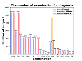
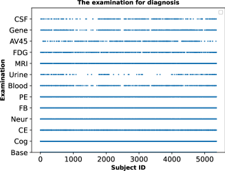
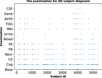
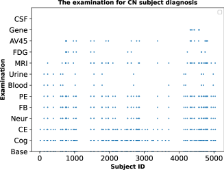
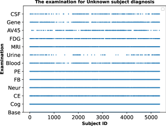
Unlike the current mainstream AD diagnostic models in which all subjects require a nuclear magnetic resonance scan, OpenClinicalAI develops personalized diagnostic strategies for each subject. For every subject, first, it will acquire the base information of the subject. Second, it will give a final diagnosis or receive other examination information according to the current data of the subject. Third, the previous step is repeated until the diagnosis is finalized or there is no further examination. As shown in Fig. 6 (a), the diagnosis strategies of subjects are not the same (as shown in Supplementary Table S2). Our model dynamically develops diagnosis strategies according to different subject situations and all examination abilities of medical institutions in the test set (as shown in Supplementary Table S3). For the known (AD and CN) subjects, as shown in Fig. 6 (b) and (c), most of the subjects require low-cost examinations (such as cognition examination (CE)). A small portion of subjects require high-cost examinations (such as cerebral spinal fluid analysis (CSF)). For unknown subjects, as shown in Fig. 6 (d), different from the diagnosis of known (AD and CN) subjects, identifying unknown subjects is more complex and more dependent on high-cost examinations. The reason is that according to the mechanism of OpenClinicalAI, it will do its best to distinguish whether the subject belongs to the known categories. When it fails, it will mark the subject as unknown. This means that the unknown subject will undergo more examinations. The details of the high-cost examination requirements are as follows: (1) of unknown subjects require a nuclear magnetic resonance scan (that of the known subject is ). (2) of unknown subjects require a positron emission computed tomography scan with -FDG (that of the known subject is ). (3) of unknown subjects require a positron emission computed tomography scan with AV (that of the known subject is ). (4) of unknown subjects require a gene analysis (that of the known subject is ). (5) of unknown subjects require a cerebral spinal fluid analysis (that of the known subject is ).
5.7 Potential clinical applications
OpenClinicalAI enables the AD diagnosis system to be implemented in uncertain and complex clinical settings thereby reducing the workload of AD diagnosis and minimizing the cost to subjects.
To identify the known (AD and CN) subjects with high confidence, the operating point of OpenClinicalAI runs with a high decision threshold (). For the test set, OpenClinicalAI achieves an accuracy value of , an AD sensitivity value of , and a CN sensitivity value of while retaining an unknown sensitivity value of . In addition, it can cooperate with the senior clinician to identify the rest of the known subjects, which are not marked as any known kinds (AD or CN). In this work, of AD subjects and of CN subjects are marked as unknown and sent to senior clinicians for diagnosis. This is significant for undeveloped areas, since it is a promising way to connect developed and undeveloped areas to reduce workload, improve overall medical services, and promote medical equity. To minimize the subject cost and maximize the subject benefit, our method dynamically develops personalized diagnosis strategies for the subject according to the subject’s situation and existing medical conditions.
OpenClinicalAI judges whether it can finalize the subject’s diagnosis according to the currently obtained information of subjects. If the current data are insufficient to establish a high confidence diagnosis, it will provide recommendations for the most appropriate next steps. This approach effectively tackles the issue of overtesting, resulting in reduced costs for subjects while maximizing the benefits they receive. For the test set, different diagnosis strategies are applied to the subject by OpenClinicalAI (as shown in Table S2). The details of the high-cost examination are as follows:
(1) of subjects require a nuclear magnetic resonance scan. (2) of subjects require a positron emission computed tomography scan with -FDG. (3) of subjects require a positron emission computed tomography scan with AV. (4) of subjects require a gene analysis. (5) of subjects require a cerebral spinal fluid analysis.
For the medical institution, before the system recommends an examination for a subject, OpenClinicalAI will inquire whether the medical institution can execute the examination. Suppose the medical institution cannot perform the examination. In this case, OpenClinicalAI will recommend other examinations until the current information of the subject is enough to support it to make a diagnosis or until all common examinations have been suggested and the subject is marked as unknown. This enables OpenClinicalAI to be deployed in different medical institutions with varying examination capabilities. In this study, OpenClinicalAI conducted subject diagnoses for examination conditions that could potentially be encountered in a health care facility (Table S3). Additionally, OpenClinicalAI made adjustments to diagnostic strategies for subjects in the test set who lacked the necessary information to receive examination recommendations. This suggests that the medical facility may have been incapable of performing the recommended examinations.
6 Conclusion
After comparing the performance of state-of-the-art models for AD diagnosis in both closed clinical and real-world settings, we noticed that the models that performed exceptionally well in the closed clinical setting did not maintain the same level of effectiveness in the real-world setting. This suggests it is time to switch attention from algorithmic research in closed clinical setting settings to systematic study in real-world settings while focusing on the challenge of tackling the uncertainty and complexity of real-world settings. In this work, we have proposed a novel open, dynamic machine learning framework to allow the model to directly address uncertainty and complexity in the real-world setting. The resulting AD diagnostic system demonstrates great potential to be implemented in real-world settings with different medical environments to reduce the workload of AD diagnosis and minimize the cost to the subject.
Although many AI diagnostic systems have been proposed, how to embed these systems into the current health care system to improve medical services remains an open issue [45, 46, 47, 48]. OpenClinicalAI provides a reasonable way to embed the AI system into the current health care system. OpenClinicalAI can collaborate with clinicians to improve the clinical service quality, especially the clinical service quality of undeveloped areas. On the one hand, OpenClinicalAI can directly deal with the diagnosis task in an uncertain and complex real-world setting. On the other hand, OpenClinicalAI can diagnose typical patients of known subjects while sending those challenging or atypical patients of known subjects to clinicians for diagnosis. Although AI technology is different from traditional statistics, the model of the AI system still learns patterns from training data. The model can easily learn patterns from typical patients, but it can be challenging to learn patterns for atypical patients. Thus, every atypical and unknown patient is especially needed to be treated by clinicians. In this work, most of the known subjects are diagnosed by OpenClinicalAI, and the rest are marked as unknown and sent to the senior clinician.
Although OpenClinicalAI is promising for impacting future research on the diagnosis system, several limitations remain. First, prospective clinical studies of the diagnosis of Alzheimer’s disease will be required to prove the effectiveness of our system. Second, the data collection and processing are required to follow the standards of the ADNI.
7 CRediT authorship contribution statement
Yunyou Huang: Conceptualized, Methodology, Model design, Coding, Data curation, Writing the original draft. Xiaoshuang Liang: Writing review, Data curation. Suqin Tang: Writing review, Data curation. Li Ma: Writing review, Data curation. Fan Zhang: Data curation. Fan Zhang: Data curation. Xiuxia Miao: Data curation, Software. Xiangjiang Lu: Data curation, Software. Jiyue Xie: Data curation, Software. Zhifei Zhang and Jianfeng Zhan: Supervision, Conceptualization, Funding acquisition, Project administration, Writing review & editing.
8 Declaration of competing interests
The authors declare no competing financial interest.
9 Data availability
The data from the Alzheimer’s Disease Neuroimaging Initiative were used under license for the current study. Applications for access to the dataset can be made at http://adni.loni.usc.edu/data-samples/access-data/. All original code has been deposited at the website https://www.benchcouncil.org/BenchCouncil and is publicly available when this article is published.
10 Acknowledgment
We thank Weibo Pan and Fang Li for downloading the raw datasets from the Alzheimer’s Disease Neuroimaging Initiative. This work is supported by the Standardization Research Project of Chinese Academy of Sciences (No. BZ201800001 to J.Z. ), the Project of Guangxi Science and Technology (No. GuiKeAD20297004 to Y.H. ), and the National Natural Science Foundation of China (No. 61967002 and No. U21A20474 to S.T. ).
References
- htt [2022] 2022 alzheimer’s disease facts and figures, Alzheimer’s & Dementia 18 (2022) 700–789.
- Hebert et al. [2001] L. E. Hebert, L. A. Beckett, P. A. Scherr, D. A. Evans, Annual incidence of alzheimer disease in the united states projected to the years 2000 through 2050, Alzheimer Disease & Associated Disorders 15 (2001) 169–173.
- Hebert et al. [2013] L. E. Hebert, J. Weuve, P. A. Scherr, D. A. Evans, Alzheimer disease in the united states (2010–2050) estimated using the 2010 census, Neurology 80 (2013) 1778–1783.
- Association et al. [2018] A. Association, et al., 2018 alzheimer’s disease facts and figures, Alzheimer’s & Dementia 14 (2018) 367–429.
- Frigerio et al. [2019] C. S. Frigerio, L. Wolfs, N. Fattorelli, N. Thrupp, I. Voytyuk, I. Schmidt, R. Mancuso, W.-T. Chen, M. E. Woodbury, G. Srivastava, et al., The major risk factors for alzheimer’s disease: age, sex, and genes modulate the microglia response to a plaques, Cell reports 27 (2019) 1293–1306.
- Prince et al. [2018] M. Prince, R. Bryce, C. Ferri, World alzheimer report 2011: The benefits of early diagnosis and intervention (2018).
- Tanveer et al. [2020] M. Tanveer, B. Richhariya, R. Khan, A. Rashid, P. Khanna, M. Prasad, C. Lin, Machine learning techniques for the diagnosis of alzheimer’s disease: A review, ACM Transactions on Multimedia Computing, Communications, and Applications (TOMM) 16 (2020) 1–35.
- Mahajan et al. [2020] S. Mahajan, G. Bangar, N. Kulkarni, Machine learning algorithms for classification of various stages of alzheimer’s disease: A review, Machine Learning 7 (2020).
- Stamate et al. [2019] D. Stamate, M. Kim, P. Proitsi, S. Westwood, A. Baird, A. Nevado-Holgado, A. Hye, I. Bos, S. J. Vos, R. Vandenberghe, et al., A metabolite-based machine learning approach to diagnose alzheimer-type dementia in blood: Results from the european medical information framework for alzheimer disease biomarker discovery cohort, Alzheimer’s & Dementia: Translational Research & Clinical Interventions 5 (2019) 933–938.
- Xing et al. [2020] X. Xing, G. Liang, H. Blanton, M. U. Rafique, C. Wang, A.-L. Lin, N. Jacobs, Dynamic image for 3d mri image alzheimer’s disease classification, in: European Conference on Computer Vision, Springer, 2020, pp. 355–364.
- Qiu et al. [2022] S. Qiu, M. I. Miller, P. S. Joshi, J. C. Lee, C. Xue, Y. Ni, Y. Wang, D. Anda-Duran, P. H. Hwang, J. A. Cramer, et al., Multimodal deep learning for alzheimer’s disease dementia assessment, Nature communications 13 (2022) 1–17.
- Qiu et al. [2020] S. Qiu, P. S. Joshi, M. I. Miller, C. Xue, X. Zhou, C. Karjadi, G. H. Chang, A. S. Joshi, B. Dwyer, S. Zhu, et al., Development and validation of an interpretable deep learning framework for alzheimer’s disease classification, Brain 143 (2020) 1920–1933.
- Böhle et al. [2019] M. Böhle, F. Eitel, M. Weygandt, K. Ritter, Layer-wise relevance propagation for explaining deep neural network decisions in mri-based alzheimer’s disease classification, Frontiers in aging neuroscience 11 (2019) 194.
- Geng et al. [2020] C. Geng, S.-j. Huang, S. Chen, Recent advances in open set recognition: A survey, IEEE transactions on pattern analysis and machine intelligence (2020).
- Aljović et al. [2016] A. Aljović, A. Badnjević, L. Gurbeta, Artificial neural networks in the discrimination of alzheimer’s disease using biomarkers data, in: 2016 5th Mediterranean Conference on Embedded Computing (MECO), IEEE, 2016, pp. 286–289.
- Lu et al. [2022] B. Lu, H.-X. Li, Z.-K. Chang, L. Li, N.-X. Chen, Z.-C. Zhu, H.-X. Zhou, X.-Y. Li, Y.-W. Wang, S.-X. Cui, et al., A practical alzheimer’s disease classifier via brain imaging-based deep learning on 85,721 samples, Journal of Big Data 9 (2022) 1–22.
- Scheirer et al. [2012] W. J. Scheirer, A. de Rezende Rocha, A. Sapkota, T. E. Boult, Toward open set recognition, IEEE transactions on pattern analysis and machine intelligence 35 (2012) 1757–1772.
- Bendale and Boult [2016] A. Bendale, T. E. Boult, Towards open set deep networks, in: Proceedings of the IEEE conference on computer vision and pattern recognition, 2016, pp. 1563–1572.
- Oza and Patel [2019] P. Oza, V. M. Patel, C2ae: Class conditioned auto-encoder for open-set recognition, in: Proceedings of the IEEE/CVF Conference on Computer Vision and Pattern Recognition, 2019, pp. 2307–2316.
- Yang et al. [2019] Y. Yang, C. Hou, Y. Lang, D. Guan, D. Huang, J. Xu, Open-set human activity recognition based on micro-doppler signatures, Pattern Recognition 85 (2019) 60–69.
- Geng and Chen [2020] C. Geng, S. Chen, Collective decision for open set recognition, IEEE Transactions on Knowledge and Data Engineering 34 (2020) 192–204.
- Perera et al. [2020] P. Perera, V. I. Morariu, R. Jain, V. Manjunatha, C. Wigington, V. Ordonez, V. M. Patel, Generative-discriminative feature representations for open-set recognition, in: Proceedings of the IEEE/CVF Conference on Computer Vision and Pattern Recognition, 2020, pp. 11814–11823.
- Saboo et al. [2021] K. Saboo, A. Choudhary, Y. Cao, G. Worrell, D. Jones, R. Iyer, Reinforcement learning based disease progression model for alzheimer’s disease, in: M. Ranzato, A. Beygelzimer, Y. Dauphin, P. Liang, J. W. Vaughan (Eds.), Advances in Neural Information Processing Systems, volume 34, Curran Associates, Inc., 2021, pp. 20903–20915.
- Zhang et al. [2021] Q. Zhang, Q. Du, G. Liu, A whole-process interpretable and multi-modal deep reinforcement learning for diagnosis and analysis of alzheimer’s disease∗, Journal of Neural Engineering 18 (2021) 066032. URL: https://dx.doi.org/10.1088/1741-2552/ac37cc. doi:10.1088/1741-2552/ac37cc.
- Komorowski et al. [2018] M. Komorowski, L. A. Celi, O. Badawi, A. C. Gordon, A. A. Faisal, The artificial intelligence clinician learns optimal treatment strategies for sepsis in intensive care, Nature medicine 24 (2018) 1716–1720.
- Darkner [2013] S. Darkner, Fdg-pet template mni152 1mm, 2013.
- Smith et al. [2004] S. M. Smith, M. Jenkinson, M. W. Woolrich, C. F. Beckmann, T. E. Behrens, H. Johansen-Berg, P. R. Bannister, M. De Luca, I. Drobnjak, D. E. Flitney, et al., Advances in functional and structural mr image analysis and implementation as fsl, Neuroimage 23 (2004) S208–S219.
- Seneca et al. [2011] N. Seneca, C. Burger, I. Florea, Improved spatial normalization using pet templates of [18f] florbetapir brain uptake of control and ad subjects (2011) 1–3.
- Huang et al. [2017] G. Huang, Z. Liu, L. Van Der Maaten, K. Q. Weinberger, Densely connected convolutional networks, in: Proceedings of the IEEE conference on computer vision and pattern recognition, 2017, pp. 4700–4708.
- Lambert et al. [2013] J.-C. Lambert, C. A. Ibrahim-Verbaas, D. Harold, A. C. Naj, R. Sims, C. Bellenguez, G. Jun, A. L. DeStefano, J. C. Bis, G. W. Beecham, et al., Meta-analysis of 74,046 individuals identifies 11 new susceptibility loci for alzheimer’s disease, Nature genetics 45 (2013) 1452–1458.
- Kunkle et al. [2019] B. W. Kunkle, B. Grenier-Boley, R. Sims, J. C. Bis, V. Damotte, A. C. Naj, A. Boland, M. Vronskaya, S. J. Van Der Lee, A. Amlie-Wolf, et al., Genetic meta-analysis of diagnosed alzheimer’s disease identifies new risk loci and implicates a, tau, immunity and lipid processing, Nature genetics 51 (2019) 414–430.
- Desikan et al. [2017] R. S. Desikan, C. C. Fan, Y. Wang, A. J. Schork, H. J. Cabral, L. A. Cupples, W. K. Thompson, L. Besser, W. A. Kukull, D. Holland, et al., Genetic assessment of age-associated alzheimer disease risk: development and validation of a polygenic hazard score, PLoS medicine 14 (2017) e1002258.
- Kendall et al. [2018] A. Kendall, Y. Gal, R. Cipolla, Multi-task learning using uncertainty to weigh losses for scene geometry and semantics, in: Proceedings of the IEEE conference on computer vision and pattern recognition, 2018, pp. 7482–7491.
- Zhang et al. [2020] Y. Zhang, Q. V. Liao, R. K. Bellamy, Effect of confidence and explanation on accuracy and trust calibration in ai-assisted decision making, in: Proceedings of the 2020 Conference on Fairness, Accountability, and Transparency, 2020, pp. 295–305.
- Linardatos et al. [2021] P. Linardatos, V. Papastefanopoulos, S. Kotsiantis, Explainable ai: A review of machine learning interpretability methods, Entropy 23 (2021) 18.
- Hosseini-Asl et al. [2018] E. Hosseini-Asl, M. Ghazal, A. Mahmoud, A. Aslantas, A. M. Shalaby, M. F. Casanova, G. N. Barnes, G. Gimel’farb, R. Keynton, A. El-Baz, Alzheimer’s disease diagnostics by a 3d deeply supervised adaptable convolutional network, Frontiers in bioscience (Landmark edition) 23 (2018) 584–596.
- Korolev et al. [2017] S. Korolev, A. Safiullin, M. Belyaev, Y. Dodonova, Residual and plain convolutional neural networks for 3d brain mri classification, in: 2017 IEEE 14th international symposium on biomedical imaging (ISBI 2017), IEEE, 2017, pp. 835–838.
- Lee et al. [2019] H. Lee, S. Yune, M. Mansouri, M. Kim, S. H. Tajmir, C. E. Guerrier, S. A. Ebert, S. R. Pomerantz, J. M. Romero, S. Kamalian, et al., An explainable deep-learning algorithm for the detection of acute intracranial haemorrhage from small datasets, Nature biomedical engineering 3 (2019) 173–182.
- Esteva et al. [2017] A. Esteva, B. Kuprel, R. A. Novoa, J. Ko, S. M. Swetter, H. M. Blau, S. Thrun, Dermatologist-level classification of skin cancer with deep neural networks, Nature 542 (2017) 115.
- Tschandl et al. [2020] P. Tschandl, C. Rinner, Z. Apalla, G. Argenziano, N. Codella, A. Halpern, M. Janda, A. Lallas, C. Longo, J. Malvehy, et al., Human–computer collaboration for skin cancer recognition, Nature Medicine 26 (2020) 1229–1234.
- Poplin et al. [2018] R. Poplin, A. V. Varadarajan, K. Blumer, Y. Liu, M. V. McConnell, G. S. Corrado, L. Peng, D. R. Webster, Prediction of cardiovascular risk factors from retinal fundus photographs via deep learning, Nature Biomedical Engineering 2 (2018) 158–164.
- Kermany et al. [2018] D. S. Kermany, M. Goldbaum, W. Cai, C. C. Valentim, H. Liang, S. L. Baxter, A. McKeown, G. Yang, X. Wu, F. Yan, et al., Identifying medical diagnoses and treatable diseases by image-based deep learning, Cell 172 (2018) 1122–1131.
- Kingma and Ba [2015] D. P. Kingma, J. Ba, Adam: A method for stochastic optimization, in: ICLR (Poster), 2015.
- Efron and Tibshirani [1994] B. Efron, R. J. Tibshirani, An introduction to the bootstrap, CRC press, 1994.
- McKinney et al. [2020] S. M. McKinney, M. Sieniek, V. Godbole, J. Godwin, N. Antropova, H. Ashrafian, T. Back, M. Chesus, G. S. Corrado, A. Darzi, et al., International evaluation of an ai system for breast cancer screening, Nature 577 (2020) 89–94.
- Kuo and Chiu [2021] C.-Y. Kuo, H.-M. Chiu, Application of artificial intelligence in gastroenterology: Potential role in clinical practice, Journal of Gastroenterology and Hepatology 36 (2021) 267–272.
- Schneider and Agus [2021] J. Schneider, M. Agus, Reflections on the clinical acceptance of artificial intelligence, in: Multiple Perspectives on Artificial Intelligence in Healthcare, Springer, 2021, pp. 103–114.
- Bullock et al. [2020] J. Bullock, A. Luccioni, K. H. Pham, C. S. N. Lam, M. Luengo-Oroz, Mapping the landscape of artificial intelligence applications against covid-19, Journal of Artificial Intelligence Research 69 (2020) 807–845.