Photothermal Microscopy & Spectroscopy with Nanomechanical Resonators
Abstract
In nanomechanical photothermal absorption spectroscopy and microscopy, the measured substance becomes a part of the detection system itself, inducing a nanomechanical resonance frequency shift upon thermal relaxation. Suspended, nanometer-thin ceramic or 2D material resonators are innately highly-sensitive thermal detectors of localized heat exchanges from substances on their surface or integrated into the resonator itself. Consequently, the combined nanoresonator-analyte system is a self-measuring spectrometer and microscope: responding to a substance’s transfer of heat over the entire spectrum for which it absorbs, according to the intensity it experiences. Limited by their own thermostatistical fluctuation phenomena, nanoresonators have demonstrated sufficient sensitivity for measuring trace analyte as well as single particles and molecules with incoherent light or focused and unfocused coherent light. They are versatile in their design, support various sampling methods and hyphenation with other spectroscopic methods, and are capable in a wide range of applications including fingerprinting, separation science, and surface sciences.
keywords:
nanoelectromechanical systems, nanomechanical photothermal spectroscopy, nanomechanical resonators, optomechanical resonators, photothermal sensing, photothermal microscopyTU Wien] Institute of Sensor and Actuator Systems, TU Wien, Gusshausstrasse 27-29, 1040 Vienna, Austria. \abbreviationsNEMS, IR, UV
1 Introduction
Absorption spectroscopy is employed in species identification and investigations into key properties, function, and behavior of molecules and nanoparticles. Traditionally, absorption spectroscopy has been performed on molecular ensembles, where degrees of freedom, species variability, such as isomerization, and interaction with the environment screen the intrinsic, response of the individual molecules. In heterogeneous samples, there can be significant variations in the size, shape, and composition of the particles, affecting their properties and, thus, their function and behavior in their environment. This motivates research in the absorption response at the single molecule and single nanoparticle level. Developments in photothermal spectroscopy have facilitated the measurement of ever-lessening analyte concentrations as well as sub-diffraction-limited localization of single nanoparticles and molecules.1, 2, 3 As a result, photothermal spectroscopy has transcended the capabilities of contemporary absorption spectroscopy techniques to provide a more detailed understanding of heterogeneous samples, with significant potential applications in a wide range of fields, including material science, catalysis, nanotechnology, and biophysics.
Photothermal absorption spectroscopy incorporates a wide-ranging set of methods based upon the measurement of a substance’s wavelength-dependent absorption by way of its thermal energy transfer to its environment upon relaxation. In general, the advantages of photothermal spectroscopy have been duly established: it is a label-free approach, unaffected by scattered light, and a direct measurement of the sample’s absorption, whose cross-section scales with the first power of a particle’s volume, as compared to the second order in scattering.4, 5 This perspective article addresses a particular fledging, photothermal method, which relies on the thermally-induced mechanical resonance detuning in nanoresonators.
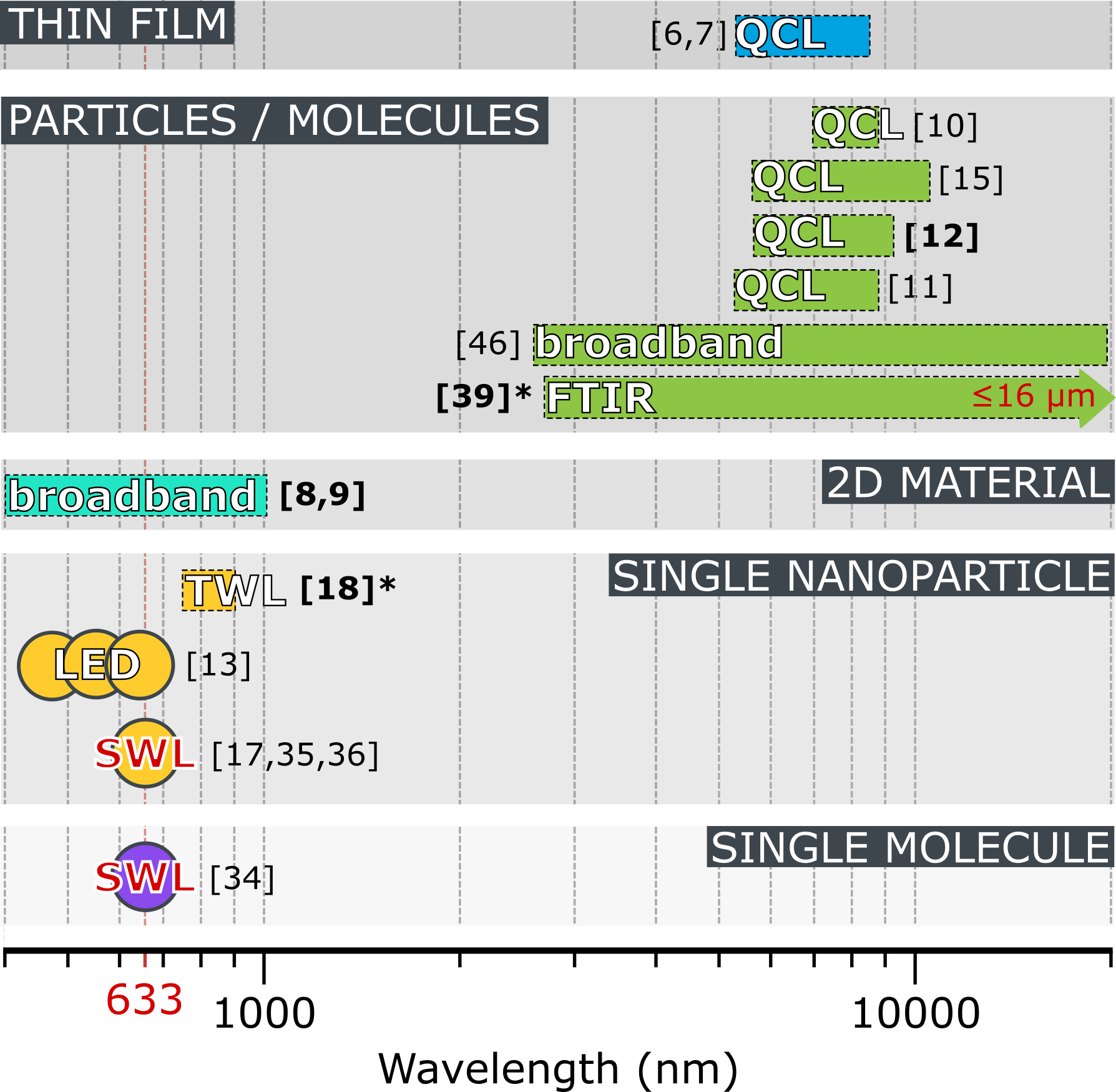
In contrast to the contemporary thermo-optical methods, which can be performed in liquid, matrix, or host cell, the nanomechanical frequency-detuning method does not require a thermo-refractive medium and must be performed in a gaseous or low pressure environments, typically of mbar. A vacuum lessens deleterious air damping and convective heat transfer, allowing for a sensitive detection paradigm for numerous applications, some exclusive to low pressure environments. Optical complexities, such as precise optical overlap, modulation of a heating laser are requirements often considered synonymously with photothermal spectroscopy. Thermo-optical studies regularly require precise focal depth of a probing laser relative to a heating laser as well as diffraction, screening, and filtering of light. However, with nanomechanical photothermal sensing, the ”substrate” on which the sample is placed acts as the thermal detector. Hence, it does not share these requirements for its basic function, and is siutable for focused or unfocused, coherent or incoherent light, to spectroscopically analyze thin films,6, 7 2D materials,8, 9 surface-adsorbed chemical species,10, 11, 12 explosives,10 nanoparticle ensembles,13, 14, 15 and individual single nanoparticles.16, 17, 18. It is worth noting, that micro-fabrication methods used to make nanoresonators have also inspired suspended microchannel resonators (SMRs), which enable photothermal resonance-tuning detection in microfluidics. SMRs are capable of single cell and single protein mass detection and have duly demonstrated sensitivities in hundreds of nJ for absorption spectroscopy in the static, surface-stress tuning mode and also in frequency-tuning, dynamic mode.19, 20 However, the primary thrust of this perspective is to introduce a wider community to the accomplishments and direction of research in nanomechanical resonators on the path of ever-increasing sensitivity toward single-molecule absorption spectromicroscopy.
Though explorations into NEMS have led down many avenues of cutting-edge research, only tens of applications showcase their potential for photothermal spectroscopy (see Figure 1), but greater consideration is being given to this research as of late. Nonetheless, the concept of spectroscopy by changes in stress in a resonant structure are not new, having been introduced in 1969 for the design of nanomechanical thermal infrared detectors.21 The concept of photothermal spectroscopy with microstructures began with bimaterial cantilevers, whose \qty100\pico thermal sensitivity, dependent upon quasi-static thermal stress-induced bending, already showed great promise in the early 1990s.22, 23 Due to the relatively long thermal time constants, these quasi-static deflection measurements have to be performed at low frequencies of a few tens to hundreds of Hz, a frequency range that typically is strongly affected by 1/f noise.24 Conversely, nanomechanical resonators, such as strings, drumheads, or trampolines, are highly sensitive to temperature variations, as seen in the shift of their resonant frequency. The higher operation frequencies in the kHz to MHz regime is one of the big advantages of photothermal sensing with nanomechanical resonators.
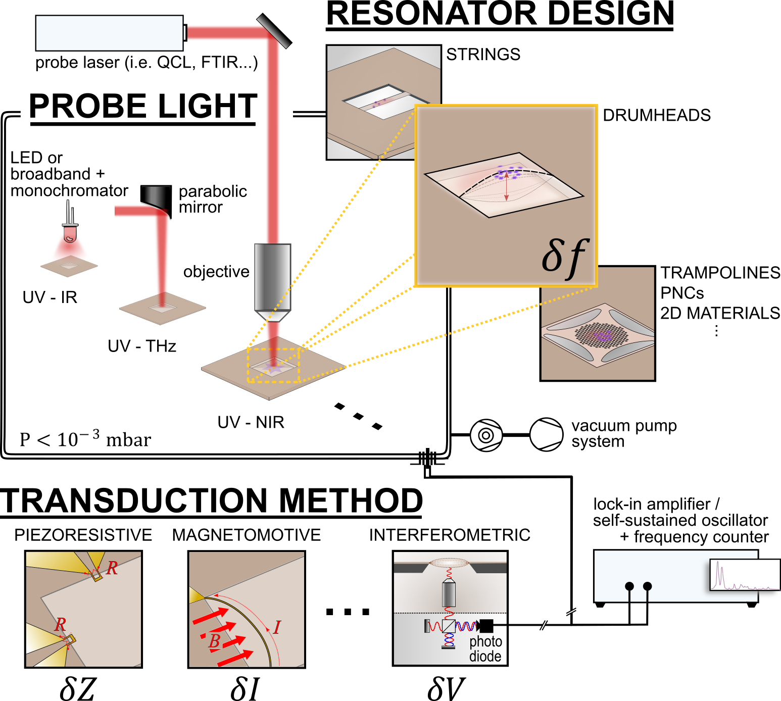
The fundamental principle, which allows nanomechanical systems to be highly sensitive spectrometers is straightforward: light, which is absorbed by the sample, anywhere from UV to THz, is dissipated as heat into the thin resonator upon thermal relaxation. This induces a frequency shift, measured optically by interferometry or electrically, by a variety of methods (see Figure 2). Resonating, nanometer-thick microstructures, such as strings, drumheads, or trampolines, are exceptionally sensitive to local changes in temperature, inducing a change in tensile stress and ultimately limited by fundamental, statistical thermal fluctuations including that described by the fluctuation dissipation theorem.25, 26 Though the weight and interplay of these manifestations in the frequency noise is still an ongoing investigation;Zhang2023demonstration, 27, 28, 29 the extent of heat dissipation and thermal interfaces in these thin, suspended structures are clearly definable, as in the case of a point-source heating the resonator, which will be introduced in the discussion of Fundamental Principles & Limitations3. To this end, the ultimate goal is to operate nanomechanical photothermal sensors at the ultimate thermal noise limit to create, on the one hand, broadband detectors for electromagnetic radiation 30, 31, 32, 33, and on the other hand, single-molecule absorption spectromicroscopes.
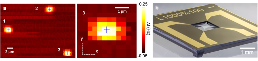
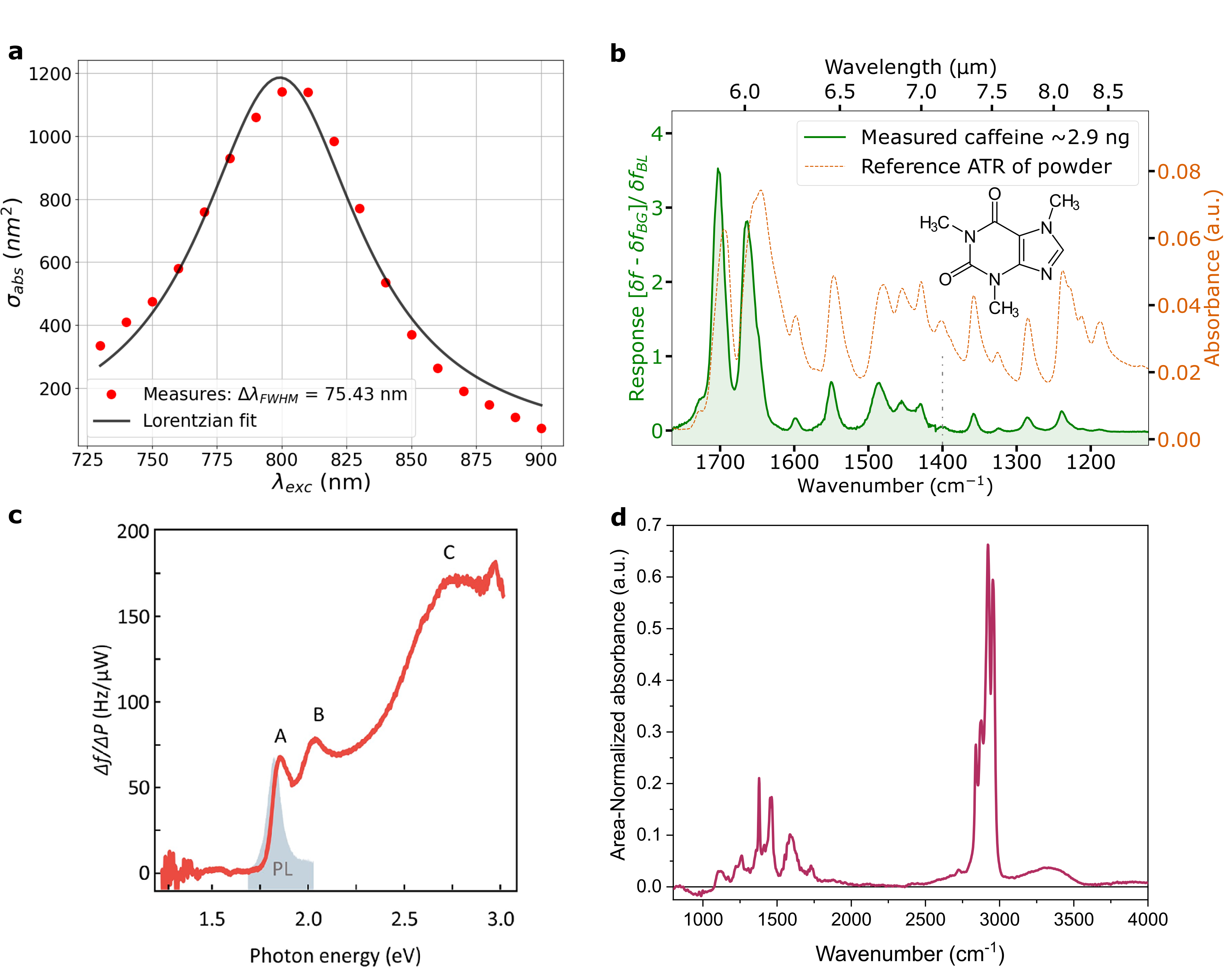
Focused light enables photothermal microscopy in addition to spectroscopy, localizing particles far below the diffraction limit and allowing for their characterization. Individual, fluorescing Atto633 molecules have been localized on SiN drumhead resonators by Chien, et. al (see Figure 3a).34 With SiN trampolines (Figure 3b), the same authors spin-coated 200 nm diameter gold nanoparticles and subsequently positioned them with the aid of an atomic force microscopy (AFM) tip.35 Then, they were localized photothermally with a 633 nm laser with 3 resolution. Likewise, trampoline resonators verified the polarization dependence of the photothermal response of single 50 nm-long gold nanorods and revealed their specific absorption spectra (see Figure 4a).35, 18 Nano-trampolines also enabled analysis of carbon content of direct-write plasmonic Au nanostructures and verification of enhancement localized surface plasmon fields between two direct-write bowtie structures.36 Metal nanoparticles with various other shapes were probed on SiN strings.37, 17 The nanoresonators’ response to thermal transfer from the plasmonically heated particles allowed the authors to obtain precise values of their absorption cross-sections.
Beyond visible wavelengths, ventures into the infrared (IR) spectrosocpy began with absorbed trace concentrations of explosive mixtures, identified by their spectral fingerprints in the lower IR region. In a pilot study with less than 100 pg of explosives, absorbed light from a quantum cascade laser (QCL) induced nanomechanical bending of cantilevers upon absorption,38 inspiring further, resonant nanostring detection of 42 fg, or 190 attomoles of RDX.10 Infrared spectroscopy of nanomechanical resonators using dispersed IR light through a monochromator made its debut in a study by Yamada et. al. on SiN strings.14 The string resonators were capable of detecting their own absorption spectrum and that of aerosol sampled polyvinylpyrrolidone (PVP). The sensitivity of the resonators was even sufficient to measure the IR spectra of organic compounds coated on nanoparticles. The estimated limit of detection was 44 fg for obtaining the IR fingerprint spectra of nanoengineered materials. Soon after, Other studies with SiN strings affirmed their sensitivity, achieving similar signal strengths and resolutions equal to that of ATR-FTIR with far less analyte.15 Their versatility in application was demonstrated with spectral characterization of Tadalafil drug and PVP thin films with a quantum cascade laser (QCL).6, 7 The signal-to-noise ratio (SNR) for 200 nm polymer films was almost six times higher than with FTIR-ATR, and 20 nm-thick layers with a SNR of 307.
This handful of applications indisputably demonstrates nanoresonators’ capability as direct, in situ spectrometers, whose wavelength range is only constrained by the analyte absorption spectrum. Nanomechanical resonators have shown capability for use in tandem with various optical methods, most recently including Fourier Transform Infrared Spectrometry.39 In the same vein, nanomechanical photothermal spectroscopy can be used to monitor chemiphysical processes occurring on the resonator surface or the physical behavior of the resonator itself. Some examples of such orthogonal processes are desorption and phase transitions.40, 6, 41 Such spectral ”eavesdropping” during thermal desorption was demonstrated in 2014, as the absorption spectra of a mere 2 fg (that is 190 attomoles) of RDX condensate was measured on silicon nitride nanostrings.10 A recent study by Luhmann et al. built upon this concept with thermal desorption of various mass loads of aerosol-impacted caffeine and theobromine, hyphenating NEMS-based thermal desorption with IR spectroscopy (NEMS-IR-TD).12 In harmony with mass sensing capability of the nanoresonator, the authors obtained characteristic thermal programmed desorption (TPD) dynamical traces of the analyte and condensates in addition to their spectra. The spectrum, as compared to FTIR-ATR spectra of caffeine powder is shown in Figure 4b. With the aid of a Peltier element for temperature control, an analysis of time-dependent spectra during isothermal desorption allowed for separation of the spectra of species with differing desorption energies by time-resolved spectral analysis. This highlights the method’s potential use in separation science. Along these lines, a growing body of literature is supporting the idea that the current utility of nanomechanical photothermal spectroscopy extends to studies in material physics and potentially to surface physics.
Two recent, significant contributions by Kirchhof et al. bear archetypical significance toward this point. In their first study,8 they obtain the in situ absorption spectra of 2D materials, from 400 to 1 (see Figure 4c), integrated into the resonator itself. These structures represent extended single molecules, or atoms, for which a measurement of the simultaneously reflected light is then corroborated to obtain the most precise, and conceivably accurate, determination of a 2D material’s dielectric function to date. This demonstrates nanomechanical photothermal spectroscopy at the fundamental limit for 2D materials, as the substance being measured is entirely the photodetector itself. Their second study,9 demonstrates spectral measurements along with a noise-equivalent power (NEP) of 890 of silicon nitride drumhead resonators, on which the 2D materials were integrated, at room temperature. This sensitivity is on the same order of the commercially-available silicon avalanche photodiodes at maximum sensitivity but over a much broader wavelength range. The hybrid 2D-material-resonator systems readily yield the excitonic transition of , plasmonic modes and intraband transitions of a plasmonic supercrystal, and dipole-dipole excited state transitions in . Spectra of the latter material is said to have been attained with a mere 1 incident radiation, and the SiN drumhead suffers not even a two-fold reduction in responsivity at intermediate temperatures down to 4 .
In a broader aspect, the faculty of nanomechanical photothermal sensing is also merited by its versatility of implementation in diverse applications, which can be performed in conjunction with its photothermal capabilities. This includes its full compatibility with the Fourier transform infrared absorption spectroscopy (FTIR) concept, pioneered by Invisible-Light Labs GmbH.39 Figure 4d shows one such preliminary result of 124 ng of aerosol-impacted polypropylene nanoparticle dispersion, with 50 nm diameter (Lab261 PP50), measured with NEMS photothermal spectroscopy as compared to a standard ATR-FTIR spectrum.
2 Sample Preparation on Nanomechanical Resonators
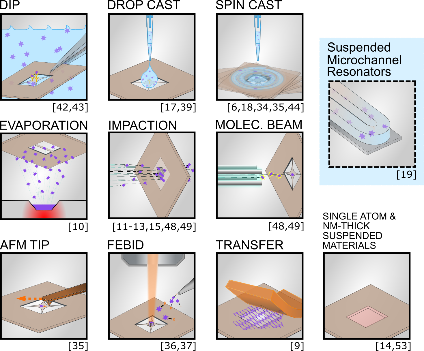
Nanometer-thick suspended structures facilitate a variety of sampling methods already employed in chemistry, life sciences, and surface science and materials (see Figure 5). Though many of these applications do not involve spectroscopy, the resonator takes on the role of a substrate, which can subsequently be used for detection. Fabricated in their diced wafer section, or chip, the resonators can be functionalized, passivated,12 or left bare then dipped into a liquid suspension in the same way that microstructure detectors have been.42, 43 Analyte can also be drop-casted or spin-coated onto the resonator-chip.34, 35, 44, 17, 6, 18 Single nanoparticles, molecules, and thin films have been detected following these basic sampling procedures, and spin-coated nanoparticles can be subsequently positioned with the aid of the cantilever tip of an atomic force microscope (AFM).35 Aerosol impaction is appropriate for airborne analyte, nebulized solutions, or suspensions.45, 12, 11, 46, 15, 47 In vacuum, suspended nanostructures are suitable for adsorption of evaporated analyte or that produced by electrospray ionization and subsequently directed or focused into a molecular beam as in mass spectrometry.10, 48, 49 For studies of the physical properties of materials, transfer by stamping or even focused electron-beam induced deposition can be considered a means of sampling.37, 36, 8, 9
Nanomechanical photothermal spectroscopy can, therefore, be applied to a vast range of substance distributions, from thin films to single molecules. The majority of these cases allow for pressures just below mbar, which is achievable within minutes using a roughing pump. Though the analyte in these studies are in solid phase upon detection, an exception is found in suspended microchannel resonators (SMRs), microresonators with nanometer-thick walled channels hold pico- to femtoliters of solution (highlighted in Figure 5). In this case, photothermal absorption spectroscopy in the dynamic mode, or resonant mode, follows the same principles of frequency tuning. Although the exchange of heat occurs indirectly through the solvent, this method is capable of single-particle, virus, or cell detection.19
3 Fundamental Principles & Limitations
When used as photothermal detectors, nanomechanical resonators are unique in that the analyte becomes a part of the detector system, coupled through thermal energy transfer. The power responsivity of the detector/substance system, having wavelength-dependent absorptance relates the relative, or fractional, frequency shift to the incident power according to
| (1) |
The caveat is that, in all resonators, higher responsivity also increases sensitivity to thermal noise. The tens of studies mentioned in this perspective do not all represent geometries that have been fully optimized for the reduction of this noise, as often, their applications require compensations in the resonator design for reasons such as improving aerosol sampling efficiency. Nonetheless, a SNR of just above 400 was achievable for the strongest spectral signatures of 120 pg of indomethacin by such structures,11 Similarly, a SNR of 337 was determined for a passivation layer, Trimethylchlorsilane, with a surface density of 0.3 fg/m2.12
Regardless of these impressive signal-to-noise ratios, nanoresonators’ full potential as spectrometers has not been fully explored. Though the description of noise processes in mechanical resonators and circuits are well-established25 and adapted to nanoresonators for guiding their optimization,26 the interplay of these processes and their dependence on the geometry of the structure is a developing discourse.28, 50, 51 Nonetheless, an advantage of suspended nanoresonators is their endless variety of forms: strings, drumheads, trampolines, and more intricate structures, including physics-driven or topologically optimized structures such as phononic crystals and spiderwebs optimized by machine learning.52, 53, 54 Likewise, variations in fabrication, such as the reduction in stress by oxygen plasma tuning,55 and in material, such as graphene,56 yield resonators more responsive to thermal exchanges. Other considerations include the resonator’s physical interaction with the incoming light; where, for example, absorption by the resonator itself can be reduced compared to the analyte by reducing and adjusting its thickness, allowing for constructive transmission for a preferred band of wavelengths.57 For methods hyphenated with nanomechanical photothermal spectroscopy, ultimate sensitivity is certainly not always necessary, but applications such as single-molecule spectromicroscopy call for optimization to reduce the various forms of noise.
3.1 Advantage of Photothermal Absorption Measurements
Despite the complexities, which thermal noise imparts to these highly sensitive detectors, one can appreciate photothermal spectroscopy and its superior detection strategy for analyte on, or part of, the nanomechanical resonator compared to even balanced transmission spectroscopy (see Figure 6). Assuming a non-scattering, non-luminescent sample, the power of indicent light () the sample receives is equivalent to the sum of the transmitted (), absorbed (), and reflected () powers:
| (2) |
This distribution of the initial power can be described relatively, by dividing equation (2) by the incident power. That is
| (3) |
where transmittance (), absorptance (), and reflectance (). It is important to note that absorptance should not be confused with absorbance, which is defined as the logarithm of . Typically, when analyzing small samples, the amount of absorbed light is significantly smaller than the transmitted light .
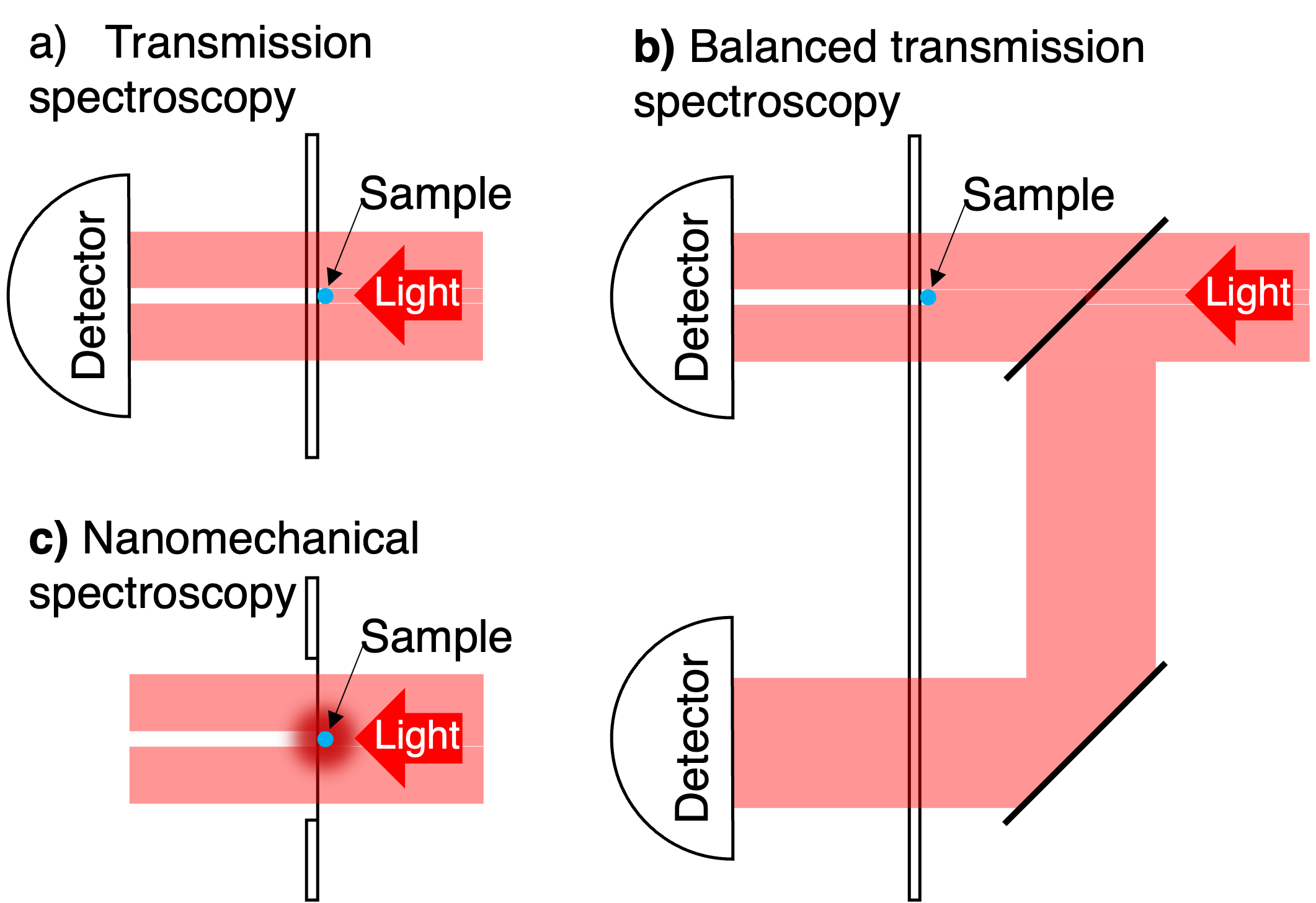
In transmission spectroscopy, as depicted in Figure 6a, the sample is irradiated with probing light, and the sample’s wavelength-dependent absorption is inferred from either the transmitted or the reflected light. A schematic of a transmission-based measurement setup is depicted in Figure 6a. In this scenario, reflectance is typically much smaller than transmittance , independent of the sensitivity of the detector that is used to measure the transmitted IR light. Assuming the probe light source has a relative intensity noise with units [Hz-1] and that all powers are measured within the same system bandwidth. Then the signal-to-noise ratio with units [Hz1/2] for the single-ended detection scheme (Figure 6a) is given by
| (4) |
For a typically small absorptance , this inevitably results in a small SNR. The situation gets even worse for light sources with large relative intensity noise as with IR QCLs, for example, which can be suppressed with a balanced detection scheme.58
Balanced detection is a solution to eliminate correlated (non-quantum) intensity noise of the probing light source: the probing IR light is split into a probing and an identical reference beam where the light is not interacting with the sample itself. While balanced transmission measurements are technically more complex than simple transmission measurements, the resulting SNR is strongly enhanced, allowing even for the detection of single-molecule absorption. 59 Ultimately, the SNR of a balanced detection scheme is vulnerable to quantum noise, such as shot noise.60 A schematic of a balanced transmission scheme (Figure 6b) is shown in Fig. 6b. Subtracting the signal of the probing beam from the reference signal removes the signal due to the power fluctuations
| (5) |
This equation shows, that the SNR improves compared to (4) and it scales directly with the inverse of the relative intensity noise of the light source.
In the case of a photothermal measurement, as depicted in Fig. 6c, the SNR is given by
| (6) |
This equation shows that a photothermal measurements offer the same SNR as a balanced detection scheme (5). In nanomechanical photothermal sensing, the sensitivity is solely dictated by the relative intensity noise of the light source, which can be due to thermal, electronic, or ultimately shot noise. Due to the typically long thermal time constant of nanomechanical resonators in the ms range, the low-frequency relative intensity noise of the light source is relevant and should be as low as possible.
On the one hand, to fully benefit from the improved SNR (6), the nanomechanical resonator needs to be sensitive enough to resolve the intensity fluctuations of the light source , where NEP is the noise equivalent power with units of [W/Hz1/2]. On the other hand, if the sensitivity is too low to resolve the intensity fluctuations , the signal-to-noise-ratio is given by
| (7) |
The NEP of nanomechanical resonators is discussed in the next section.
3.2 Noise-Equivalent Power
While photon detectors can reach single photon sensitivity, the sensitivity of thermal detectors, such as bolometers or pyroelectric detectors, is typically limited by electronic noise, including Johnson noise.61, 62 However, mechanical thermal detectors operate differently as they do not rely on electric detection principles to detect irradiated power. Consequently, nanomechanical detectors are not limited by electronic noise, a unique feature enabling enhanced sensitivity.21
The signal from the resonator is the relative frequency shift in time, , and the resonator detector’s noise-equivalent power is specified by a white noise spectral density (), and its optothermal power responsivity () to that noise:
| (8) |
NEP has units of [W/Hz1/2], since is in units of [Hz-1] and with units [W-1].63
The power responsivity, , is an intrinsic property of a resonator, describing its frequency response to absorbed power according to
| (9) |
where is the fractional frequency response to a change in temperature, and is the thermal conductance. The power responsivity has a low-pass behavior, dropping for power fluctuations with frequencies faster than the resonator’s thermal time constant , given heat capacity, .
According to the definition of the NEP in equation (8), the sensitivity of a system can be improved by minimizing the fractional frequency noise and maximizing the responsivity. To understand this better, we look to the power responsivity (9), which is directly proportional to the responsivity to temperature changes in the resonator. In general, this parameter is influenced by two resonator material parameters: the temperature-induced softening, which affects the Young’s modulus, and thermal expansion, affecting the tensile stress.63
Specifically, in stress-free structures such as beams, plates, and cantilevers, the dominant photothermal effect is the temperature-induced softening. On the other hand, in stressed structures such as strings and drumheads, thermal expansion leading to stress reduction plays a more significant role. For a comparison of these two effects, consider a slender beam and a string, whose temperature responsivity for an even increase in temperature can then be described by and ,63 respectively. Here, is the Youngs’ modulus, is the tensile stress, is the material’s temperature coefficient of Young’s modulus, and is the thermal expansion coefficient. Typically, the temperature and thermal expansion coefficients are of the same order of magnitude. The temperature responsivity of strings is enhanced by a factor of , which is generally much larger than unity for most materials. Therefore, the responsivity of strings and drumheads, which can be further optimized by stress-tuning,34 is significantly larger than that of beams and plates. This enhanced responsivity makes strings and drumheads more suitable for photothermal sensing applications.
In addition to maximizing temperature responsivity, another factor that plays a role in optimizing the responsivity (9) is the minimization of the thermal conductance , where the vacuum environment eliminates contributions due to convection. Here, and are the conductances due to conductive and radiative heat transfer, respectively. Strings and drumheads are naturally suited for achieving a high thermal isolation (small ) due to their large aspect ratios (lateral size, or length to thickness). In fact, it has been demonstrated that larger silicon nitride drumheads reach a regime where radiative heat transfer dominates over conductive heat transfer ().28, 29
According to Stefan-Boltzmann’s law for small temperature variations, the radiative heat conductance is given by ,25 with the surface area , the Stefan-Boltzmann constant , temperature , and the emissivity . As a fundamental physical process, which cannot be completely eliminated, even in materials with very low thermal conductivity, radiative heat transfer establishes the lower limit of the overall achievable heat conductance. Therefore, in order to achieve high thermal isolation, leading to high sensitivity in photothermal detectors, it is necessary to minimize radiative heat transfer. This can be achieved by choosing a resonator material with the very low emissivity . Amorphous dielectrics, such as thin-film silicon nitride are excellent candidates with a low emissivity of the order of for typical 50 nm-thin structures.28, 29
The second factor in equation (8) guiding the NEP is related to the fractional frequency () noise. Consider the common case for which electronic readout noise is negligible. Really, the NEP is the sum of uncorrelated fractional-frequency noise sources based on thermomechanical and temperature fluctuations 26
| (10) |
results from the thermomechanical amplitude vibration of the nanomechanical resonator driven by its own thermal energy. This amplitude noise, in turn, manifests itself in the frequency noise via amplitude-to-phase noise translation. For the optimal case that transduction noise is insignificant and the measurement filter bandwidth is smaller than the peak width () of the resonator with negligible damping, it reduces to a white noise source with the power spectral density64
| (11) |
where is the quality factor, is the vibrational amplitude of the resonator, and is the power spectral density of the thermomechanical displacement noise at the resonance frequency. Equation (11) shows that the influence of thermomechanical noise can be minimized by driving the nanomechanical resonator to the maximal vibrational amplitude limit, at the onset of nonlinearity.
comes from thermostatistical fluctuations of the resonator temperature according to the fluctuation-dissipation theorem, which directly produces frequency noise. For a lumped-element model with a concentrated mass linked to a thermal reservoir via a thermal conductance , it can be simplified to 25, 26
| (12) |
In the case that is dominated by conductive heat transfer, the origin of can be explained as temperature fluctuations due to the resistance in the conductive heat transfer. In the other case that the resonator is fully in the radiative heat transfer regime, the origin of can be understood as temperature fluctuations created by the statistical nature of emitted and received thermal photons. Both the thermomechanical and temperature fluctuation noise can be potentially suppressed by cooling. Zhang2023demonstration
In the special case that heat transfer by conduction is negligible, for structures with sufficient surface area such that heat transfer due to radiation dominates (), thermal frequency fluctuations will also likely dominate (). As a result, the NEP (8) with (9) and (12) reduces to
| (13) |
This expression shows that NEP can be optimized by minimizing and by lowering the temperature of the resonator. However, the exact prediction of NEP remains uncertain as both the thermomechanical 65, 27, 64 and temperature fluctuations Zhang2023demonstration remain an active subject of investigation.
3.3 Sensitivity Limit of a String Resonator
Certain nanomechanical resonators are capable of single molecule and single particle spectroscopy and microscopy. Their expected ultimate sensitivity limit is characterized by the NEP, described in (8). To understand its dependencies, the contributing noise mechanisms in (10) have to be dissected. For simplicity, we consider the sensitivity limit of a string resonator estimated by the interplay of fundamental thermophysical noise mechanisms rooted in the equipartition theorem. A nanostring resonator will have the parameters of length , a cross-sectional area , a mass density , a pre-stress , and a Young’s modulus .
The first phenomenon contributing to the NEP, the thermomechanical fractional frequency noise (11) at resonance comes to 63
| (14) |
Here, is the effective mass of the resonator with the eigenfrequency for a specific mode .
As discussed above, can be minimized by maximizing the coherent vibrational amplitude of the resonator. The string resonator’s geometrical nonlinearity determines the maximal amplitude, given by66
| (15) |
where is the effective Duffing nonlinearity parameter, which for a string is given by .63
A second, separate, thermal phenomenon is the thermal fluctuation fractional frequency noise (12), which scales inversely with the thermal conductance , comprising conductive and radiative heat transfer. This model is for a lumped-element system, which requires the use of an effective thermal conductance for the string. Since thermal fluctuations happen all along the length of the string, the effective can be derived by averaging the thermal resistance over the entire string length
| (16) |
where is the thermal conductivity of the string’s material. The conductance due to thermal radiation () is given by the Stefan-Boltzmann law. Accounting for radiation of both the top and bottom surfaces of the string and assuming a linear temperature field,63 the total effective thermal conductance becomes
| (17) |
where is the Stefan-Boltzmann constant, is the emissivity, and the string width.
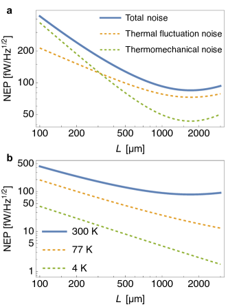
An ”interplay” of these two phenomena contributing to the NEP with differing string lengths is modeled for typical SiN 50 nm-thick resonators in Figure 7a. It can be seen that for longer strings, at room temperature the thermomechanical noise is not ubiquitously the primary noise source, as often suspected65. For longer strings, thermal fluctuations constitute the frequency noise limit with a minimal \qty100\femto/^1/2 at room temperature. The temperature plays a major role in this sensitivity as well, yielding NEP values of just a few femtowatt at 4 K for longer strings (seen in Figure 7b). Thermal fluctuation noise has a quadratic temperature dependence and becomes negligible for temperatures below room temperature and thermomechanical noise becomes the limit. In regard to sensitivity, within these dependencies and parameters are hidden the full breadth and capability of the nanomechanical photothermal spectroscopy method.
In summary, the fundamental thermally-generated fluctuation limitation of nanoscale resonators demarcates the incomparable sensitivities of NEMS resonators to temperature-dependent changes in stress. This requires a careful implementation of the transduction of a resonator’s motion to retain this fundamental limit. That is, the frequency noise in the process of transduction must be lower than fluctuation dissipation noise. Then, further improvements in sensitivity rely upon reducing the factors which govern the thermal fluctuation noise. The performance of dynamic applications will inherently require striking a balance between the speed of the resonator and its sensitivity, as these two factors are inversely proportional.
4 Future Applications
From thin films, to surfaces, and down to single molecule spectromicroscopy, nanomechanical resonators possess wide-ranging capability still to be explored. Immediate applications include characterization of 2D materials8, 9 and various plasmonic structures,36, 17. Considering nanomechanical resonators can operate in UHV and at cryogenic temperatures potentially places these detectors at the forefront of other fields of research. Though applications in cutting-edge heterogeneous catalysis, for example, and quantum systems are on the horizon, the study of functional nanomaterials will continue to be consequential, a springboard into these fields. Even interactions on materials in thermal contact with the resonator, such as thin layers or nanoparticles, may also be observed. Principally, this method is susceptible to all forms of spectroscopy, which can be performed in free-space. As the polarization-dependence has already been shown for single gold nanorods,35, 18 one immediate example is circular dichroism spectroscopy of surface-adsorbed molecules or particles, especially relevant to pharmacology. Other applications, mentioned here are clearly within our reach, outlining a rich and exciting future for nanomechanical absorption spectroscopy.
4.1 Analysis of functional nanomaterials
Functional nanomaterials, such as luminescent nanoparticles and nano-scale catalysts, are some of the most consequential advancements of nanotechnology. For example, quantum dots have emerged at the forefront of many imaging, sensing, and photonic applications. Moreover, metal nanoparticles and nanoclusters not only allow for higher catalytic efficiency but enable deeper exploration into the rich landscape of heterogeneous catlaysis.67 On the single-particle level, particles from the same batch are most certainly heterogeneous in their size, shape, and surface characteristics. This is a crucial concern in fundamental research related to materials chemistry and surface science. In particular, for atomically precise metal nanoclusters, differences of a single atom significantly affect catalytic reaction mechanisms and optical properties.68 Furthermore, it is often challenging to scale up nanomaterial synthesis, and characterization of such materials is only possible on the scale of a single particle.
Single-particle microscopy, such as atomic-force microscopy or electron microscopy, has been crucial in understanding the heterogeneity of nanoparticles and has facilitated functional materials development.69 Likewise, specific optical properties of single nanoparticles are a vital feature in many applications. For metal species, optical properties are strongly correlated with particle size and structure. The corresponding absorption bands are located in the UV-Vis and will generally shift to a lower wavelength for decreasing particle size.70 Therefore, UV-Vis spectroscopy is an essential characterization method for synthesized metal nanoparticles.67, 70 But because standard UV-Vis spectroscopy can only measure the average absorption signal of the particle ensemble, the sample has to be separated and purified with, e.g., size-exclusion chromatography (SEC).71, 72 Whereas, nanomechanical photothermal spectroscopy is capable of enabling the distinction between individual particles’ distinctive features and provide direct information about sample heterogeneity without separation steps. The method allows for spectroscopy across the full spectral range, where IR in combination with UV-Vis absorption can be used to gain chemical information of a nanomaterial, monitor ligand exchange reactions, if enough sample material is available 71, and investigate luminescent nanoparticles as well as chiral nanomaterials.73
Table 1 compares the previously-measured, highly fluorescent Atto 633 (with a signal-to-noise ratio of 70)34 with other single molecules and particles with their attenuation coefficients, cross-sections, and heat dissipation ratios. The single-particle sensitivity will allow the characterization of precious, scarce nanomaterials, which are difficult to fabricate, and nanoresonator spectrometers are also a robust platform for probing single thermoplasmonic and dipole-dipole interactions with their dependencies, which have a significant impact on their function in numerous applications from medicine to light harvesting.74, 75 Metallic nanoparticles themselves have been crucial instruments in the spectroscopy of attached or neighboring analyte and have demonstrated enhancement of absorption in multiple solar cell configurations.76 Plasmonic-enhanced spectroscopy on a thermally sensitive nanoresonator, however, will enable plasmon-enhanced absorption spectroscopy of even non-fluorescing trace substances and single molecules, improving the signal-to-noise ratio and allowing for investigations into their spectral dependencies on surface interactions.
4.2 Analysis of single biomolecules
In the single-molecule sensitivity regime, a sample can be identified by way of its individual specific absorption coefficient. The amount of light a sample species attenuates at a specific wavelength is determined by its molecular composition, and can be represented by its molar attenuation coefficient . In proteins, UV light’s attenuation at 280 nm can primarily be attributed to the three amino acids tryptophan (Trp), tyrosine (Tyr), and cysteine (Cys). Hence, a proteins’ molar attenuation coefficient can be estimated from the sum of the number of individual contributions of these three amino acids 77
| (18) |
The molar attenuation coefficient is highly specific to the amino acid composition of an individual protein.77 Hence, the UV absorption from a single protein can be used as a molecule-specific fingerprint, much in the same way the mid-IR spectral fingerprint is used to identify a myriad of molecular species.
IR bands not only serve an identification function, but a quantification function for proteins. It has been shown that the attenuation signal at the amide I band (6 µm) correlates strongly with the amino acid count of a protein.78, 79 The amide I signal is created by the sum of all peptide bonds that link consecutive amino acids. Therefore, from the amide I attenuation of an individual protein, it is possible to estimate the number of amino acids and identify the protein.
Concerted effort is being directed toward applications such as proteomics to find alternatives for protein identification with improved sensitivity.80 Acquiring chemical fingerprints, in addition to mass, allows for a multi-physical analysis, which provides an enhanced knowledge base for the identification and characterization of individual proteins and protein complexes. Here, nanomechanical single-molecule UV-Vis combines with single-molecule IR absorption spectroscopy to create a promising new method for protein identification and biomolecular analysis, in general.
| Molar | Atten. | Heat | |||
|---|---|---|---|---|---|
| Spectral | attenuation | cross- | dissipation | ||
| wavelength | coefficient | section | ratio | Ref | |
| [M-1 cm-1] | [m2] | ||||
| Hepatitis B virus protein | UV (280 nm) | 7,320,000 | 2.8E-18 | 0.8 | 81 |
| 4 nm Au nanoparticle | Vis (506 nm) | 3,600,000 | 1.4E-18 | 1 | 82 |
| BSA protein | IR (6 µm) | 190,000 | 7.3E-20 | 1 | 83 |
| Atto 633 | Vis (633 nm) | 130,000 | 5.0E-20 | 0.38 | 34 |
| BSA protein | UV (280 nm) | 44,000 | 1.7E-20 | 0.8 | 77 |
| Lysozyme protein | UV (280 nm) | 38,000 | 1.5E-20 | 0.8 | 77 |
| Lysozyme protein | IR (6 µm) | 36,000 | 1.4E-20 | 1 | 83 |
| Hepatitis B virus monomer | UV (280 nm) | 30,500 | 1.2E-20 | 0.8 | 81 |
| Virus RNA nucleotide | UV (260 nm) | 8,000 | 3.0E-21 | 1 | 81 |
| Insulin protein | UV (280 nm) | 6,000 | 2.3E-21 | 0.8 | 77 |
| Tryptophan (amino acid) | UV (280 nm) | 5,500 | 2.1E-21 | 0.8 | 77 |
| Virus RNA nucleotide | UV (280 nm) | 4,000 | 1.5E-21 | 1 | 81 |
| Tyrosine (amino acid) | UV (280 nm) | 1,490 | 5.7E-22 | 0.86 | 77 |
| Single peptide bond | IR (6 µm) | 312 | 1.2E-22 | 1.0 | 84 |
| Cysteine (amino acid) | UV (280 nm) | 125 | 4.8E-23 | N/A | 77 |
4.3 Fingerprinting and separation science
The published limits of detection for non-fluorescent analyte in the range of pg to fg of non-fluorescent analyte testify to the high sensitivity of the nanomechanical photothermal spectroscopy method. Fundamentally, measurement of such trace amounts can be accomplished with unfocused light, which minimally requires a single optical element, attesting to the simplicity of its implementation. Although, nanomechanical resonators function only in low-pressure environments, they are naturally well-suited for applications in surface science. They have already proven to be an effective way to analyze and monitor thin films and their phase transitions.89, 7 Furthermore, spectral ”eavesdropping” can forseeably reveal information about a molecule’s interactions with the surface of the resonator or vicariously through a nanoparticle on its surface. This includes monitoring of isomeric transitions and binding affinities of trace substances down to the single molecule level, adding valuable insight into surface reaction processes. This method may also avail itself to surface-assisted laser desorption of analyte,90, 91 which adsorbs directly on the mechanically resonant material on another layer placed on its surface. In this way, the influence of individual chemical groups of a chemical species can be monitored spectrally during isothermal desorption while being probed with narrow-linewidth IR light, for example. At visible wavelengths, scanning focused light would potentially enable surface sampling mass spectrometry imaging directly on the resonator.91, 92 After all, the method provides time-dependent spectral data in harmony with mass exchange, which carries with it valuable information regarding trends in desorption energies, as demonstrated in Luhmann et al.12. This work also demonstrates that, in mixtures, species can be differentiated and identified simply by singular value decomposition or by global spectro-temporal and, potentially, principal component analyses during isothermal desorption. This avenue of application leaves much yet to be explored but reveals a unique direction for separation science, where temperature control and chemical composition of the surface can be adapted for the separation of diverse analyte mixtures.
5 Challenges
However, as with many technologies at the cutting edge of research, real-world implementation remains the primary challenge in regard to commercial and industrial applications for trace substance identification and quality control. The primary challenge lies in the impediments of vacuum systems; they are bulky and have some pump-down time and venting time. Nonetheless, the trade-off between processing time and sensitivity does not matter for methods such as when nanomechanical IR absorption spectroscopy of thin films was compared to ATR-FTIR measurements.15 At other times, conventional methods introduce artefacts in the spectra which are not seen in nanomechanical photothermal spectroscopy, and the vacuum environment will be a necessary trade-off, especially at the single-molecular level.6 Nonetheless, for trace substance analysis, with an apt system for sample exchange, the speed at which measurements are performed can be improved. Alternatively, at the expense of optimal sensitivity due to ballistic losses, many first-stage pumps can reach an ultimate partial pressure of one order of magnitude beyond mbar in air, which can be reached in a few minutes.93 At these pressures, however, a stabilized vacuum or a compensation mechanism for frequency drift is required.
With the potential for sub-nanometer localization resolution at visible wavelengths, nanomechanical photothermal spectroscopy shows potential to resolve the whole range of particulate matter, which threatens global health and the ecological environment,94 and spectromectrically distinguishing between surface-adsorbed particulates.95 This requires effective and accurate sampling mechanisms and sample-to-measurement tracking. Such accounting is necessary for all cases where native concentrations are desired, as analyte can be lost either in the process of sampling on resonator chips or between this and the measurement process. At low pressures, where temperature control of the chip cannot be used to mitigate loss of analyte, rates of desorption from remaining analyte after pump-down can be used to extrapolate to a near-expected initial concentration on the resonator.40, 12
6 Conclusions
The nanomechanical photothermal spectroscopy method provides a means to measure thermal relaxation of electronically and vibrationally excited substances down to the single molecule level. The substance being measured, whether as a thin layer, adsorbed molecule, or a suspended material, becomes part of the detection mechanism itself. Nanoresonators’ versatility in fabrication, design, sampling method, and probing light characteristics has been expanding the definition of photothermal spectroscopy beyond that conventionally associated with the opto-thermal effect and merits further explorations into trace species identification, homogeneous characterization, and numerous, real-time, simultaneous or hyphenated studies. To date, studies have demonstrated that thin ceramic and 2D material resonators are capable of photothermally probing by an array of light sources and techniques: from electronic absorption of a single particle by directed, incoherent light to focused light with sub-diffraction-limited localization of nanoparticles and even FTIR of highly-dilute nebulized, aerosol-impacted solutions. Geometry and surface structure variability, along with the optical simplicity, makes this method highly adaptable. Though measurements must be performed in vacuo, this does not necessarily mean that throughput must always be sacrificed due to pump-down and venting times. Furthermore, the thermostatistically-limited sensitivity of nanoresonators allows for accuracy beyond contemporary fingerprinting methods, and cutting edge research in surface sciences and heterogeneous catalysis requires low-pressure environments, for which spectroscopy by nanoresonators is ideal. As nanomechanical photothermal spectroscopy is inevitably leading toward single-molecule spectroscopy, there is, likewise, room for increased sensitivity where 2D materials and phononic crystals are concerned. Nonetheless, in the gap between the single molecule limit and the current state-of-the-art remains a wealth of ground-breaking applications waiting to be explored and perfected.
References
- Adhikari et al. 2020 Adhikari, S.; Spaeth, P.; Kar, A.; Baaske, M. D.; Khatua, S.; Orrit, M. Photothermal Microscopy: Imaging the Optical Absorption of Single Nanoparticles and Single Molecules. ACS Nano 2020, 14, 16414–16445
- Adhikari and Orrit 2022 Adhikari, S.; Orrit, M. Progress and perspectives in single-molecule optical spectroscopy. Journal of Chemical Physics 2022, 156
- Shimizu et al. 2022 Shimizu, H.; Chen, C.; Tsuyama, Y.; Tsukahara, T.; Kitamori, T. Photothermal spectroscopy and micro/nanofluidics. Journal of Applied Physics 2022, 132
- Bialkowski et al. 2019 Bialkowski, S. E.; Astrath, N. G. C.; Proskurnin, M. A. Photothermal Spectroscopy Methods; Wiley, 2019
- van Dijk et al. 2006 van Dijk, M. A.; Tchebotareva, A. L.; Orrit, M.; Lippitz, M.; Berciaud, S.; Lasne, D.; Cognet, L.; Lounis, B. Absorption and scattering microscopy of single metal nanoparticles. Physical Chemistry Chemical Physics 2006, 8, 3486–3495
- Casci Ceccacci et al. 2019 Casci Ceccacci, A.; Cagliani, A.; Marizza, P.; Schmid, S.; Boisen, A. Thin Film Analysis by Nanomechanical Infrared Spectroscopy. ACS Omega 2019, 4, 7628–7635
- Samaeifar et al. 2019 Samaeifar, F.; Casci Ceccacci, A.; Bose Goswami, S.; Hagner Nielsen, L.; Afifi, A.; Zór, K.; Boisen, A. Evaluation of the solid state form of tadalafil in sub-micron thin films using nanomechanical infrared spectroscopy. International Journal of Pharmaceutics 2019, 565, 227–232
- Kirchhof et al. 2022 Kirchhof, J. N.; Yu, Y.; Antheaume, G.; Gordeev, G.; Yagodkin, D.; Elliott, P.; De Araújo, D. B.; Sharma, S.; Reich, S.; Bolotin, K. I. Nanomechanical Spectroscopy of 2D Materials. Nano Letters 2022, 22, 8037–8044
- Kirchhof et al. 2023 Kirchhof, J. N.; Yu, Y.; Yagodkin, D.; Stetzuhn, N.; de Araújo, D. B.; Kanellopulos, K.; Manas-Valero, S.; Coronado, E.; van der Zant, H.; Reich, S. et al. Nanomechanical absorption spectroscopy of 2D materials with femtowatt sensitivity. 2023, 0–7
- Biswas et al. 2014 Biswas, T.; Miriyala, N.; Doolin, C.; Liu, X.; Thundat, T.; Davis, J. Femtogram-scale photothermal spectroscopy of explosive molecules on nanostrings. Analytical chemistry 2014, 86, 11368–11372
- Kurek et al. 2017 Kurek, M.; Carnoy, M.; Larsen, P. E.; Nielsen, L. H.; Hansen, O.; Rades, T.; Schmid, S.; Boisen, A. Nanomechanical Infrared Spectroscopy with Vibrating Filters for Pharmaceutical Analysis. Angewandte Chemie 2017, 129, 3959–3963
- Luhmann et al. 2023 Luhmann, N.; West, R. G.; Lafleur, J. P.; Schmid, S. Nanoelectromechanical Infrared Spectroscopy with In Situ Separation by Thermal Desorption: NEMS-IR-TD. ACS Sensors 2023, 8, 1462–1470
- Larsen et al. 2013 Larsen, T.; Schmid, S.; Villanueva, L. G.; Boisen, A. Photothermal Analysis of Individual Nanoparticulate Samples Using Micromechanical Resonators. ACS Nano 2013, 7, 6188–6193
- Yamada et al. 2013 Yamada, S.; Schmid, S.; Larsen, T.; Hansen, O.; Boisen, A. Photothermal infrared spectroscopy of airborne samples with mechanical string resonators. Analytical Chemistry 2013, 85, 10531–10535
- Andersen et al. 2016 Andersen, A. J.; Yamada, S.; Pramodkumar, E. K.; Andresen, T. L.; Boisen, A.; Schmid, S. Nanomechanical IR spectroscopy for fast analysis of liquid-dispersed engineered nanomaterials. Sensors and Actuators, B: Chemical 2016, 233, 667–673
- Ramos et al. 2018 Ramos, D.; Malvar, O.; Davis, Z. J.; Tamayo, J.; Calleja, M. Nanomechanical plasmon spectroscopy of single gold nanoparticles. Nano Letters 2018, 18, 7165–7170
- Rangacharya et al. 2020 Rangacharya, V. P.; Wu, K.; Larsen, P. E.; Thamdrup, L. H. E.; Ilchenko, O.; Hwu, E.-T.; Rindzevicius, T.; Boisen, A. Quantifying Optical Absorption of Single Plasmonic Nanoparticles and Nanoparticle Dimers Using Microstring Resonators. ACS Sensors 2020, 5, 2067–2075
- Kanellopulos et al. 2023 Kanellopulos, K.; West, R. G.; Schmid, S. Nanomechanical Photothermal Spectroscopy of Individual Nanorods. in preparation: https://arxiv.org/abs/2305.05287 2023,
- Pastina and Villanueva 2020 Pastina, A. D.; Villanueva, L. G. Suspended micro/nano channel resonators: a review. Journal of Micromechanics and Microengineering 2020, 30, 18
- Miriyala et al. 2016 Miriyala, N.; Khan, M. F.; Thundat, T. Thermomechanical behavior of a bimaterial microchannel cantilever subjected to periodic IR radiation. Sensors and Actuators, B: Chemical 2016, 235, 273–279
- Cary 1969 Cary, H. H. Infrared radiation detector employing tensioned foil to receive radiation. 1969; US Patent 3457412A
- Barnes et al. 1994 Barnes, J. R.; Stephenson, R. J.; Welland, M. E.; Gerber, C.; Gimzewski, J. K. Photothermal spectroscopy with femtojoule sensitivity using a micromechanical device. Nature 1994, 372, 79–81
- Barnes et al. 1994 Barnes, J. R.; Stephenson, R. J.; Woodburn, C. N.; O’Shea, S. J.; Welland, M. E.; Rayment, T.; Gimzewski, J. K.; Gerber, C. A femtojoule calorimeter using micromechanical sensors. Review of Scientific Instruments 1994, 65, 3793–3798
- Varesi et al. 1997 Varesi, J.; Lai, J.; Perazzo, T.; Shi, Z.; Majumdar, A. Photothermal measurements at picowatt resolution using uncooled micro-optomechanical sensors. Applied Physics Letters 1997, 71, 306–308
- Vig and Kim 1999 Vig, J. R.; Kim, Y. Noise in microelectromechanical system resonators. IEEE transactions on ultrasonics, ferroelectrics, and frequency control 1999, 46, 1558–1565
- Cleland and Roukes 2002 Cleland, A. N.; Roukes, M. L. Noise processes in nanomechanical resonators. Journal of Applied Physics 2002, 92, 2758–2769
- Sadeghi et al. 2020 Sadeghi, P.; Demir, A.; Villanueva, L. G.; Kahler, H.; Schmid, S. Frequency fluctuations in nanomechanical silicon nitride string resonators. Physical Review B 2020, 102
- Piller et al. 2020 Piller, M.; Sadeghi, P.; West, R. G.; Luhmann, N.; Martini, P.; Hansen, O.; Schmid, S. Thermal radiation dominated heat transfer in nanomechanical silicon nitride drum resonators. Applied Physics Letters 2020, 117, 034101
- Zhang et al. 2020 Zhang, C.; Giroux, M.; Nour, T. A.; St-Gelais, R. Radiative heat transfer in freestanding silicon nitride membranes. Physical Review Applied 2020, 14, 024072
- Blaikie et al. 2019 Blaikie, A.; Miller, D.; Alemán, B. J. A fast and sensitive room-temperature graphene nanomechanical bolometer. Nature Communications 2019, 10, 4726
- Piller et al. 2022 Piller, M.; Hiesberger, J.; Wistrela, E.; Martini, P.; Luhmann, N.; Schmid, S. Thermal IR detection with nanoelectromechanical silicon nitride trampoline resonators. IEEE Sensors Journal 2022, 1066 – 1071
- Zhang et al. 2013 Zhang, X. C.; Myers, E. B.; Sader, J. E.; Roukes, M. L. Nanomechanical Torsional Resonators for Frequency-Shift Infrared Thermal Sensing. Nano Letters 2013, 13, 1528–1534
- Qian et al. 2019 Qian, Z.; Rajaram, V.; Kang, S.; Rinaldi, M. High figure-of-merit NEMS thermal detectors based on 50-nm thick AlN nano-plate resonators. Applied Physics Letters 2019, 115, 261102
- Chien et al. 2018 Chien, M.-H.; Brameshuber, M.; Rossboth, B. K.; Schütz, G. J.; Schmid, S. Single-molecule optical absorption imaging by nanomechanical photothermal sensing. Proceedings of the National Academy of Sciences 2018, 115, 11150–11155
- Chien and Schmid 2020 Chien, M.-H.; Schmid, S. Nanoelectromechanical photothermal polarization microscopy with 3 Å localization precision. Journal of Applied Physics 2020, 134501
- Chien et al. 2021 Chien, M. H.; Shawrav, M. M.; Hingerl, K.; Taus, P.; Schinnerl, M.; Wanzenboeck, H. D.; Schmid, S. Analysis of carbon content in direct-write plasmonic Au structures by nanomechanical scanning absorption microscopy. Journal of Applied Physics 2021, 129
- Schmid et al. 2014 Schmid, S.; Wu, K.; Larsen, P. E.; Rindzevicius, T.; Boisen, A. Low-Power Photothermal Probing of Single Plasmonic Nanostructures with Nanomechanical String Resonators. Nano Letters 2014, 14, 2318–2321
- Kim et al. 2013 Kim, S.; Lee, D.; Liu, X.; Van Neste, C.; Jeon, S.; Thundat, T. Molecular recognition using receptor-free nanomechanical infrared spectroscopy based on a quantum cascade laser. Scientific Reports 2013, 3, 1–6
- 39 Lafleur, J. P.; Schmid, S.; Luhman, N.; Bešić, H.; Penn, T.; Vukićević, V.; Timarac Popović, J.; Hiesberger, J.; Geniş, D. Ö. Invisible-Light Labs. \urlhttps://www.invisible-light-labs.com/
- Shakeel et al. 2018 Shakeel, H.; Wei, H.; Pomeroy, J. M. Measurements of enthalpy of sublimation of Ne, N2, O2, Ar, CO2, Kr, Xe, and H2O using a double paddle oscillator. Journal of Chemical Thermodynamics 2018, 118, 127–138
- Karl et al. 2018 Karl, M.; Larsen, P. E.; Rangacharya, V. P.; Hwu, E. T.; Rantanen, J.; Boisen, A.; Rades, T. Ultrasensitive Microstring Resonators for Solid State Thermomechanical Analysis of Small and Large Molecules. Journal of the American Chemical Society 2018, 140, 17522–17531
- Bagheri et al. 2014 Bagheri, M.; Chae, I.; Lee, D.; Kim, S.; Thundat, T. Selective detection of physisorbed hydrocarbons using photothermal cantilever deflection spectroscopy. Sensors and Actuators, B: Chemical 2014, 191, 765–769
- Raiteri et al. 2001 Raiteri, R.; Grattarola, M.; Butt, H.-J.; Skládal, P. Micromechanical cantilever-based biosensors. 2001, 79
- Chien and Schmid 2020 Chien, M.-H.; Schmid, S. Towards nanoelectromechanical photothermal localization microscopy. 2020,
- Schmid et al. 2013 Schmid, S.; Kurek, M.; Boisen, A. Towards airborne nanoparticle mass spectrometry with nanomechanical string resonators. Micro- and Nanotechnology Sensors, Systems, and Applications V 2013, 8725, 872525
- Yamada et al. 2013 Yamada, S.; Schmid, S.; Larsen, T.; Hansen, O.; Boisen, A. Photothermal Infrared Spectroscopy of Airborne Samples with Mechanical String Resonators. Analytical Chemistry 2013, 85, 10531–10535
- Luhmann et al. 2023 Luhmann, N.; Özer Genis, D.; Lafleur, J. P.; Schmid, S. Environmental Aerosol Analysis by Nanomechanical Photothermal Infrared Spectroscopy. in preparation 2023,
- Naik et al. 2009 Naik, A. K.; Hanay, M. S.; Hiebert, W. K.; Feng, X. L.; Roukes, M. L. Towards single-molecule nanomechanical mass spectrometry. Nature Nanotechnology 2009, 4, 445–450
- Roukes and Makarov 2019 Roukes, M. L.; Makarov, A. A. Integrated Hybrid NEMS Mass Spectrometry. 2019
- Snell et al. 2022 Snell, N.; Zhang, C.; Mu, G.; Bouchard, A.; St-Gelais, R. Heat Transport in Silicon Nitride Drum Resonators and its Influence on Thermal Fluctuation-Induced Frequency Noise. Physical Review Applied 2022, 17, 1
- Zhang and St-Gelais 2023 Zhang, C.; St-Gelais, R. Demonstration of frequency stability limited by thermal fluctuation noise in silicon nitride nanomechanical resonators. Appl. Phys. Lett. 2023, 193501
- Sadeghi et al. 2020 Sadeghi, P.; Tanzer, M.; Luhmann, N.; Piller, M.; Chien, M. H.; Schmid, S. Thermal Transport and Frequency Response of Localized Modes on Low-Stress Nanomechanical Silicon Nitride Drums Featuring a Phononic-Band-Gap Structure. Physical Review Applied 2020, 14, 1
- Kirchhof et al. 2021 Kirchhof, J. N.; Weinel, K.; Heeg, S.; Deinhart, V.; Kovalchuk, S.; Höflich, K.; Bolotin, K. I. Tunable Graphene Phononic Crystal. Nano Letters 2021, 21, 2174–2182
- Shin et al. 2022 Shin, D.; Cupertino, A.; de Jong, M. H.; Steeneken, P. G.; Bessa, M. A.; Norte, R. A. Spiderweb Nanomechanical Resonators via Bayesian Optimization: Inspired by Nature and Guided by Machine Learning. Advanced Materials 2022, 34
- Luhmann et al. 2017 Luhmann, N.; Jachimowicz, A.; Schalko, J.; Sadeghi, P.; Sauer, M.; Foelske-Schmitz, A.; Schmid, S. Effect of oxygen plasma on nanomechanical silicon nitride resonators. Applied Physics Letters 2017, 111
- Steeneken et al. 2021 Steeneken, P. G.; Dolleman, R. J.; Davidovikj, D.; Alijani, F.; Van Der Zant, H. S. Dynamics of 2D material membranes. 2D Materials 2021, 8
- King and Milosevic 2012 King, S. W.; Milosevic, M. A method to extract absorption coefficient of thin films from transmission spectra of the films on thick substrates. 2012
- Akhgar et al. 2020 Akhgar, C. K.; Ramer, G.; Żbik, M.; Trajnerowicz, A.; Pawluczyk, J.; Schwaighofer, A.; Lendl, B. The next generation of IR spectroscopy: EC-QCL-based mid-IR transmission spectroscopy of proteins with balanced detection. Analytical Chemistry 2020, 92, 9901–9907
- Celebrano et al. 2011 Celebrano, M.; Kukura, P.; Renn, A.; Sandoghdar, V. Single-molecule imaging by optical absorption. Nature Photonics 2011, 5, 95–98
- Kukura et al. 2010 Kukura, P.; Celebrano, M.; Renn, A.; Sandoghdar, V. Single-molecule sensitivity in optical absorption at room temperature. The Journal of Physical Chemistry Letters 2010, 1, 3323–3327
- Rogalski 2019 Rogalski, A. Infrared and Terahertz Detectors, Third Edition; CRC Press, 2019
- las 2019 Noise in Pyroelectric IR Detectors. 2019; \urlwww.lasercomponents.com
- Schmid et al. 2023 Schmid, S.; Villanueva, L. G.; Roukes, M. L. Fundamentals of nanomechanical resonators, 2nd edition; Springer International Publishing AG Switzerland, 2023
- Demir 2021 Demir, A. Understanding fundamental trade-offs in nanomechanical resonant sensors. Journal of Applied Physics 2021, 129
- Sansa et al. 2016 Sansa, M.; Sage, E.; Bullard, E. C.; Gély, M.; Alava, T.; Colinet, E.; Naik, A. K.; Villanueva, L. G.; Duraffourg, L.; Roukes, M. L. et al. Frequency fluctuations in silicon nanoresonators. Nature Nanotechnology 2016, 11, 552–558
- Lifshitz and Cross 2008 Lifshitz, R.; Cross, M. C. Nonlinear dynamics of nanomechanical and micromechanical resonators. Reviews of nonlinear dynamics and complexity 2008, 1
- Liu and Corma 2018 Liu, L.; Corma, A. Metal Catalysts for Heterogeneous Catalysis: From Single Atoms to Nanoclusters and Nanoparticles. Chemical Reviews 2018, 118, 4981–5079
- Garcia et al. 2020 Garcia, C.; Truttmann, V.; Lopez, I.; Haunold, T.; Marini, C.; Rameshan, C.; Pittenauer, E.; Kregsamer, P.; Dobrezberger, K.; Stöger-Pollach, M. et al. Dynamics of Pd Dopant Atoms inside Au Nanoclusters during Catalytic CO Oxidation. The Journal of Physical Chemistry C 2020, 124, 23626–23636
- Zhou et al. 2020 Zhou, J.; Chizhik, A. I.; Chu, S.; Jin, D. Single-particle spectroscopy for functional nanomaterials. Nature 2020, 579, 41–50
- Vázquez-Vázquez et al. 2009 Vázquez-Vázquez, C.; Bañobre-López, M.; Mitra, A.; López-Quintela, M. A.; Rivas, J. Synthesis of Small Atomic Copper Clusters in Microemulsions. Langmuir 2009, 25, 8208–8216
- Truttmann et al. 2020 Truttmann, V.; Herzig, C.; Illes, I.; Limbeck, A.; Pittenauer, E.; Stöger-Pollach, M.; Allmaier, G.; Bürgi, T.; Barrabés, N.; Rupprechter, G. Ligand engineering of immobilized nanoclusters on surfaces: ligand exchange reactions with supported Au11(PPh3)7Br3. Nanoscale 2020, 12, 12809–12816
- 72 The Dynamic Structure of Au38(SR)24 Nanoclusters Supported on CeO2 upon Pretreatment and CO Oxidation
- Warning et al. 2021 Warning, L. A.; Miandashti, A. R.; McCarthy, L. A.; Zhang, Q.; Landes, C. F.; Link, S. Nanophotonic Approaches for Chirality Sensing. ACS Nano 2021, 15, 15538–15566, PMID: 34609836
- Yu et al. 2019 Yu, H.; Peng, Y.; Yang, Y.; Li, Z. Y. Plasmon-enhanced light–matter interactions and applications. npj Computational Materials 2019, 5, 1–14
- Mathew and Shyju 2022 Mathew, J.; Shyju, T. S. Plasmon-Enhanced Efficiency of DSSC and Hybrid Nano Catalysis Applications. Topics in Catalysis 2022, 65, 1719–1732
- Kundu and Patra 2017 Kundu, S.; Patra, A. Nanoscale strategies for light harvesting. Chemical Reviews 2017, 117, 712–757
- Pace et al. 1995 Pace, C. N.; Vajdos, F.; Fee, L.; Grimsley, G.; Gray, T. How to measure and predict the molar absorption coefficient of a protein. Protein science 1995, 4, 2411–2423
- Strug et al. 2014 Strug, I.; Utzat, C.; Cappione, A.; Gutierrez, S.; Amara, R.; Lento, J.; Capito, F.; Skudas, R.; Chernokalskaya, E.; Nadler, T. Development of a univariate membrane-based mid-infrared method for protein quantitation and total lipid content analysis of biological samples. Journal of analytical methods in chemistry 2014, 2014
- De Meutter and Goormaghtigh 2021 De Meutter, J.; Goormaghtigh, E. Amino acid side chain contribution to protein FTIR spectra: impact on secondary structure evaluation. European Biophysics Journal 2021, 50, 641–651
- Timp and Timp 2020 Timp, W.; Timp, G. Beyond mass spectrometry, the next step in proteomics. Science Advances 2020, 6, eaax8978
- Porterfield and Zlotnick 2010 Porterfield, J. Z.; Zlotnick, A. A simple and general method for determining the protein and nucleic acid content of viruses by UV absorbance. Virology 2010, 407, 281–288
- Liu et al. 2007 Liu, X.; Atwater, M.; Wang, J.; Huo, Q. Extinction coefficient of gold nanoparticles with different sizes and different capping ligands. Colloids and Surfaces B: Biointerfaces 2007, 58, 3–7
- Schwaighofer et al. 2021 Schwaighofer, A.; Akhgar, C. K.; Lendl, B. Broadband laser-based mid-IR spectroscopy for analysis of proteins and monitoring of enzyme activity. Spectrochimica Acta Part A: Molecular and Biomolecular Spectroscopy 2021, 253, 119563
- Rahmelow et al. 1998 Rahmelow, K.; Hübner, W.; Ackermann, T. Infrared absorbances of protein side chains. Analytical biochemistry 1998, 257, 1–11
- Teale and Weber 1957 Teale, F.; Weber, G. Ultraviolet fluorescence of the aromatic amino acids. Biochemical Journal 1957, 65, 476
- Ghisaidoobe and Chung 2014 Ghisaidoobe, A. B.; Chung, S. J. Intrinsic tryptophan fluorescence in the detection and analysis of proteins: a focus on Förster resonance energy transfer techniques. International journal of molecular sciences 2014, 15, 22518–22538
- Peon and Zewail 2001 Peon, J.; Zewail, A. H. DNA/RNA nucleotides and nucleosides: direct measurement of excited-state lifetimes by femtosecond fluorescence up-conversion. Chemical physics letters 2001, 348, 255–262
- Lakowicz 2006 Lakowicz, J. R. Principles of fluorescence spectroscopy; Springer, 2006
- Karl et al. 2020 Karl, M.; Thamdrup, L. H.; Rantanen, J.; Boisen, A.; Rades, T. Temperature-modulated micromechanical thermal analysis with microstring resonators detects multiple coherent features of small molecule glass transition. Sensors (Switzerland) 2020, 20, 1–14
- Abdelhamid and Wu 2016 Abdelhamid, H. N.; Wu, H.-F. Gold nanoparticles assisted laser desorption/ionization mass spectrometry and applications: from simple molecules to intact cells. Analytical and Bioanalytical Chemistry 2016, 408, 4485–4502
- Huang et al. 2022 Huang, H.; Ouyang, D.; Lin, Z. A. Recent Advances in Surface-Assisted Laser Desorption/Ionization Mass Spectrometry and Its Imaging for Small Molecules. Journal of Analysis and Testing 2022, 6, 217–234
- Wong et al. 2023 Wong, K. F. C.; Greatorex, R. E.; Gidman, C. E.; Rahman, S. Surface-sampling mass spectrometry to study. 2023, 0, 1–13
- You et al. 2021 You, A.; Be, M. A. Y.; In, I. Opto-mechanical metamaterial nanobolometer. 2021, 126101
- Ault and Axson 2017 Ault, A. P.; Axson, J. L. Atmospheric Aerosol Chemistry: Spectroscopic and Microscopic Advances. Analytical Chemistry 2017, 89, 430–452
- Gottschalk et al. 2023 Gottschalk, F.; Debray, B.; Klaessig, F.; Park, B.; Lacome, J. M.; Vignes, A.; Portillo, V. P.; Vázquez-Campos, S.; Hendren, C. O.; Lofts, S. et al. Predicting accidental release of engineered nanomaterials to the environment. Nature Nanotechnology 2023, 412–418