ECG-QA: A Comprehensive Question Answering Dataset Combined With Electrocardiogram
Abstract
Question answering (QA) in the field of healthcare has received much attention due to significant advancements in natural language processing. However, existing healthcare QA datasets primarily focus on medical images, clinical notes, or structured electronic health record tables. This leaves the vast potential of combining electrocardiogram (ECG) data with these systems largely untapped. To address this gap, we present ECG-QA, the first QA dataset specifically designed for ECG analysis. The dataset comprises a total of 70 question templates that cover a wide range of clinically relevant ECG topics, each validated by an ECG expert to ensure their clinical utility. As a result, our dataset includes diverse ECG interpretation questions, including those that require a comparative analysis of two different ECGs. In addition, we have conducted numerous experiments to provide valuable insights for future research directions. We believe that ECG-QA will serve as a valuable resource for the development of intelligent QA systems capable of assisting clinicians in ECG interpretations.
1 Introduction
In recent years, significant advancements in natural language processing have revolutionized the field of question answering (QA) in a wide range of domains. Previous works have demonstrated the great potential of QA systems in various domains, where they have been combined with different modalities such as images [1, 32, 15, 8] or tables with images [24, 13]. Concurrently, QA systems have also been explored in the healthcare domain, including visual QA with chest X-ray [11, 14], clinical-note-based QA [20], and QA over structured electronic health record (EHR) data [28, 12]. These pioneering efforts have successfully bridged the gap between general-domain QA and the medical field, unlocking new possibilities to improve healthcare outcomes and enhance medical decision-making processes.
Despite this remarkable progress, there is a noticeable absence of datasets that combine electrocardiogram (ECG) data with question answering. As a fundamental diagnostic tool in cardiology, ECG provides critical insights into the electrical activity of the heart and plays an important role in detecting various cardiac conditions [3, 22, 33]. Consequently, integrating ECG data with QA systems holds tremendous potential to improve the interpretation of cardiac data, leading to more accurate diagnoses and personalized treatment plans.
To this end, we present ECG-QA111The dataset is available at https://github.com/Jwoo5/ecg-qa, licensed under CC-BY-4.0 license., a novel QA dataset that incorporates ECG data for question answering tasks. To the best of our knowledge, ECG-QA is the first dataset that combines QA and ECG, opening up new avenues for integrating multi-modal machine learning with cardiac healthcare.
The main contributions of this work are threefold:
-
•
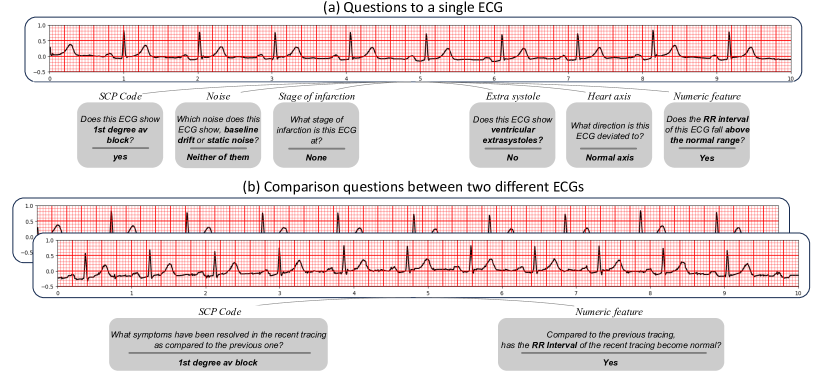
We propose the ECG-QA dataset, a diverse collection of questions focused on ECG interpretation and analysis (See Figure 1). This dataset introduces the novel concept of incorporating question answering into the realm of ECG analysis, making it a valuable resource for developing and evaluating QA systems in the context of cardiology.
-
•
To cover more complex yet clinically critical questions, we include questions that require comparative analysis of two ECGs (See Figure 1 (b)). This inclusion brings a new degree of complexity, as addressing these comparison questions extends beyond the conventional scope of ECG analysis using machine learning. By incorporating these types of questions, we not only address the real-world needs of medical professionals but also broaden the potential applications of machine learning in ECG analysis.
-
•
We provide a benchmark for QA models, including recent large language models (LLMs), on the ECG-QA dataset, promoting further research and encouraging the development of novel methods to leverage ECG signals for question answering tasks. We believe that ECG-QA will serve as a valuable resource in advancing machine learning applications in cardiology and improving medical decision-making processes.
2 Related works
Medical QA datasets
QA systems have been extensively explored in the healthcare domain, catering to the specific needs and challenges of medical data. However, most existing medical QA datasets are primarily based on clinical texts, medical images, or structured EHR tables. For example, Pampari et al. [20] proposed the emrQA dataset, consisting of question-answer pairs derived from unstructured clinical notes. In addition, Kovaleva et al. [11] and Liu et al. [14] proposed datasets for visual QA using X-ray images, aiming to investigate intelligent interactive systems for radiology. Meanwhile, in the field of QA over structured EHR data, Wang et al. [28] and Lee et al. [12] have developed datasets called MIMICSQL and EHRSQL respectively, which consist of questions and their corresponding SQL queries. While these healthcare QA systems demonstrate the potential of leveraging medical data with QA to improve healthcare outcomes, there is currently no dedicated QA dataset specifically designed for ECG data despite its widespread use in diagnosing cardiovascular conditions and monitoring patients’ heart health.
Electrocardiogram
Previous studies in the field of ECG have predominantly focused on using ECG data for diagnostic purposes such as identifying various heart diseases. For instance, Nejedly et al. [18] proposed an ensemble of residual networks with attention modules to classify cardiac diseases using ECGs, which won first place in the PhysioNet/Computing in Cardiology Challenge 2021 [21]. At the same challenge, Han et al. [6] achieved second place by utilizing SE-WRN [31], which is a combination of wide residual network [30] and squeeze-and-excitation modules [7]. Furthermore, several works [4, 10, 19] studied self-supervised learning with ECGs to improve performances on the cardiac arrhythmia classification task. These works concentrate on classifying diagnoses based on a single ECG, and do not consider the significance of comparing two ECGs, despite its importance in clinical contexts. For example, by detecting resolved symptoms after some treatments, the medical practitioners can assess the effectiveness of the treatments and evaluate the progress of the patient’s condition. To reflect this clinical reality, we have included questions that involve the comparison of two ECGs within the ECG-QA dataset, making our dataset unique and valuable.
| Question type | Attribute type | Example template question |
| Single-Verify | SCP Code | Does this ECG show symptoms of non-specific ST changes? |
| Noise | Does this ECG show baseline drift in lead I? | |
| Stage of infarction | Does this ECG show early stage of myocaridal infarction? | |
| Extra systole | Does this ECG show ventricular extrasystoles? | |
| Heart axis | Does this ECG show left axis deviation? | |
| Numeric feature | Does the RR interval of this ECG fall within the normal range? | |
| Single-Choose | SCP Code | Which symptom does this ECG show, conduction disturbance or hypertrophy? |
| Noise | Which noise does this ECG show, baseline drift or static noise? | |
| Stage of infarction | Which stage of infarction is this ECG at, early stage of myocardial infarction or late stage of myocardial infarction? | |
| Extra systole | Which kind of extra systoles does this ECG show, ventricular extrasystoles or supraventricular extrasystoles? | |
| Heart axis | Which cardiac axis does this ECG show, left axis deviation or right axis deviation? | |
| Numeric feature | Which range does the RR interval of this ECG fall in, below the normal range or within the normal range? | |
| Single-Query | SCP Code | What form-related symptoms does this ECG show? |
| Noise | What kind of noises does this ECG show in lead I? | |
| Stage of infarction | What stage of infarction is this ECG at? | |
| Extra systole | What kind of extra systoles does this ECG show? | |
| Heart axis | What direction is this ECG deviated to? | |
| Numeric feature | What range does the RR interval of this ECG fall in? | |
| Comparison- -Consecutive-Verify | SCP Code | Compared to the previous tracing, has left ventricular hypertrophy been resolved in the recent tracing? |
| Numeric feature | Compared to the previous tracing, has the PR interval of the recent tracing become normal? | |
| Comparison- -Consecutive-Query | SCP Code | What symptoms have been resolved in the recent tracing as compared to the previous one? |
| Numeric feature | What numeric features of the recent tracing now have become normal compared to the previous one? | |
| Comparison- -Irrelevant-Verify | SCP Code | Compared to the first ECG, has atrial fibrillation been newly detected in the second ECG? |
| Numeric feature | Compared to the first ECG, has the P duration of the second ECG changed to an abnormal value? | |
| Comparison- -Irrelevant-Query | SCP Code | What symptoms still remain in the second ECG as compared to the first ECG? |
| Numeric feature | What numeric features of the second ECG are now considered abnormal values as compared to the first ECG? |
3 Dataset Construction
We constructed the ECG-QA dataset upon the PTB-XL dataset [26]222This dataset is licensed under CC-BY-4.0 license., which offers comprehensive metadata regarding ECGs annotated by expert cardiologists.
This metadata covers a wide range of information including ECG reports, diagnostic statements, diagnosis likelihoods, and signal-specific properties.
To ensure the high quality of our dataset, we performed additional filtering on the original PTB-XL dataset.
Specifically, we selected ECGs that were marked with a validated_by_human tag set to True, which indicates the validation by a human cardiologist, and excluded ECGs that had empty reports.
As a result, the ECG-QA dataset was constructed using samples of 10-second ECGs from the PTB-XL dataset.
In addition, we split the samples into training and test sets according to a 8:2 ratio based on their patient IDs before generating QA samples to prevent the overlapping of ECGs between training and test sets.
We again split the training samples into training and validation sets by a 9:1 ratio, yielding 7.2:0.8:2.0 training-validation-test distribution.
3.1 Question template
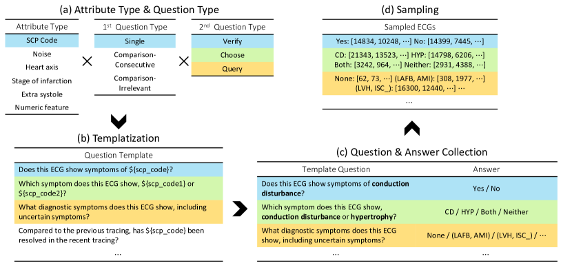
To generate QA samples, we start by creating the question templates to collect questions, answers, and their corresponding ECGs. Because the questions are fully derived from these templates, it is important to define templates that are not only diverse but also clinically meaningful. To achieve this goal, we extracted the relevant attributes from the PTB-XL metadata to determine the content of the questions (i.e., attribute types) and categorized the questions into several question types. Then, we combined these types to construct the template questions, and additionally generated template paraphrases to add lexical diversity to our dataset. As a result, we defined a total of 70 templates as shown in Supplementary A.2, and we also provide the example questions derived from these templates in Table 1. All the processes of designing question templates have been validated by a board-certified medical expert from the Department of Critical Care and Emergency Medicine in terms of clinical utility. The detailed processes of each step are described in the following subsections, as well as visualized in Figure 2 (a) and (b).
3.1.1 Attribute type extraction
SCP code
The PTB-XL dataset provides SCP codes for each ECG sample, consisting of 71 different ECG symptoms that adhere to the SCP-ECG v0.4 that preceded the current SCP-ECG standard [23].
These attributes are composed of form-related (e.g., inverted T-waves), rhythm-related (e.g., sinus arrhythmia), and diagnostic symptoms (e.g., non-specific ischemic) along with additional 5 superclasses for diagnostic labels.
Given that detecting cardiac symptoms is a primary objective in many ECG studies [18, 6, 4, 10, 19], we included questions that inquire about various ECG symptoms in the ECG-QA dataset.
To ensure the dataset quality, we excluded attributes with a low number of positive ECG samples in the test split, such as WPW (wolf-parkinson-white syndrome), resulting in a final selection of 64 attributes, including the 5 superclasses.
Furthermore, we developed a regular expression parser to extract the grounded lead position of form-related symptoms from the ECG reports (See Supplementary B.1).
This enables us to include questions in the ECG-QA dataset that specifically address the leads in which symptoms are detected (e.g., Does this ECG show symptoms of inverted T-waves in lead I?), making the ECG-QA dataset more comprehensive.
Noise
Considering that ECG measurements involve placing electrodes on specific body surfaces, it is inevitable to encounter various signal interferences such as baseline drift, which can be caused by patient movement or machine issues. Therefore, it becomes crucial to differentiate these interferences from the original ECG signals during analysis. To reflect this aspect in the ECG-QA dataset, we leveraged the signal noise information available in the PTB-XL metadata. This information is provided as a string indicating the specific lead positions where each noise is detected (e.g., “v1-v6” or “i-iii”). We parsed these strings to identify the exact lead positions associated with four different types of noises: Baseline drift, Static noise, Burst noise, and Electrode problems.
Stage of infarction
Since identifying the stage of myocardial infarction (MI) helps healthcare professionals determine the most appropriate management strategies by assessing the risk profile for the patient, we also have considered the stage of infarction as an important attribute in the ECG-QA dataset.
In the PTB-XL dataset, it distinguishes the stage of MI into six levels including intermediate stages: "I," "I-II," "II," "II-III," "III," and "Unknown."
In addition, because there are two fields indicating the stage of infarction (infarction_stadium1 and infarction_stadium2), the statements could be potentially multiple.
For the sake of simplicity, we simplified the stages into 4 levels by regarding the intermediate stages ("I-II" and "II-III") as their "lower" stage ("I" and "II").
Then, we used the second statement if there are multiple entries at a time.
After defining an additional stage called "None" for those who do not have MI, we could derive five attributes for the stage of infarction.
Extra systole
Since extra systoles can be a sign of underlying cardiac conditions or abnormalities, it is also important to detect them to evaluate the patient’s heart health. To address the presence of extra systoles in the ECG-QA dataset, we utilized the relevant annotations provided in the PTB-XL metadata, which includes information about the occurrence of different types of extra systoles: Extrasystoles, Ventricular extrasystoles, and Supraventricular extrasystoles.
Heart axis
The heart axis provides valuable information about the direction of the heart’s electrical activity during each cardiac cycle. In the ECG-QA dataset, we have considered the heart axis as another crucial attribute since it is an important parameter that can help in diagnosing certain cardiac conditions. Although the PTB-XL metadata includes heart axis information, we did not utilize it because it does not specify the actual numerical values of the heart axis. Instead, we manually calculated the heart axis degrees by employing an external tool, NeuroKit2 [16] 333This software is licensed under MIT license.. Then, we classified them into four categories following the conventional standards: Normal heart axis, Left axis deviation, Right axis deviation, and Extreme axis deviation.
Numeric feature
The ECG-QA dataset also incorporates numeric features that provide further insights into the cardiac signals. Similar to the heart axis, we used NeuroKit2 to calculate the numeric values for these features since the PTB-XL dataset does not explicitly provide such information. Specifically, we extracted the locations of P, Q, R, S, and T waves for each beat present in lead II, and computed six different numeric features: RR interval, P duration, PR interval, QRS duration, QT interval, and QT corrected. Given that a 10-second ECG recording typically contains multiple beats and thus multiple numeric values for each feature, we represented each feature using its median value. This approach helps to minimize the impact of abnormal contractions, such as ventricular premature contractions, on the calculated values. Additionally, we categorized each numeric value as below, within, or above the normal range, where the normal range criteria are described in Supplementary B.2, derived from a previous study [27]. These ranges serve as a useful reference for assessing the numerical measurements and identifying potential abnormalities in the cardiac signals.
3.1.2 Question type definition
We have defined two different types of questions. The first type pertains to the type of ECGs associated with a question, which can be categorized as follows: 1) Single, which refers to questions involving a single ECG; 2) Comparison-Consecutive, which involves comparison questions between two consecutive ECGs from the same patient; and 3) Comparison-Irrelevant, which involves comparison questions between two irrelevant ECGs from different patients. Although Comparison-Irrelevant questions may not seem realistic in a clinical setting, we included these questions since they can help to reinforce a machine’s comprehension ability and be utilized for model evaluation when comparing two different ECGs. In addition, inspired by GQA [8], the second type of question refers to the main function it should perform. These can be categorized as follows: 1) Verify, which corresponds to yes/no questions; 2) Choose, which applies to questions where the selection is made from two given options; and 3) Query, which are open-ended questions that seek to retrieve specific attributes.
By combining these two types, we could derive a total of 9 possible question types. However, we did not include the combinations of Comparison and Choose types since it seemed unnatural to select from two given options when comparing different ECGs. Similarly, with regards to the attribute types, we only considered SCP code and Numeric feature for comparison questions because these two attribute types are providing the most informative features when comparing two ECGs.
3.1.3 Paraphrase generation
To enhance the lexical diversity of the ECG-QA dataset, we manually curated paraphrases for each question template based on the machine-generated candidates by utilizing OpenAI’s ChatGPT. We ensured that the questions in the test split were not included in the training set to evaluate the generalizability of the QA models on different lexical variations. The detailed procedure for generating paraphrases and its results are presented in Supplementary A.3.
3.2 QA sample collection
As shown in Figure 2 (b) and (c), we collected questions by plugging the corresponding attributes into the placeholder that existed in the question templates.
For example, a question template “Does this ECG show symptoms of ${scp_code}?” can be transformed into “Does this ECG show symptoms of conduction disturbance?”
We further gathered the corresponding answers for each question and paired them to create (question, answer) pairs.
Then, for each (question, answer) pair, we again randomly sampled the corresponding ECGs from the candidate ECGs.
In each split, the candidate ECGs can be 1) all the single ECGs for Single questions; 2) all the (ECG1, ECG2) pairs where ECG1 and ECG2 are the consecutive ECGs from the same patient for Comparison-Consecutive questions; and 3) all the (ECG1, ECG2) combinations where ECG1 and ECG2 have different patient IDs for Comparison-Irrelevant questions.
After we finally replaced the template question with randomly selected paraphrases that matched the corresponding question’s template, the process of collecting QA samples was complete.
The detailed sampling strategies for different question types are described in Supplementary B.3.
After all these processes, the ECG-QA dataset consists of 267,539 training samples, 64,663 validation samples, and 82,146 test samples, which cover various types of attributes and questions. More detailed statistics of the dataset are described in Supplementary B.4.
4 Experiments
Task formulation
We formulate QA task as a multi-label classification over all possible answer options that exist in the ECG-QA dataset. The answer labels are composed of 88 attributes from the six attribute sets, 12 lead positions (i.e., lead I - lead V6), and 3 answers for Verify questions (yes, no, not sure), leading to a total of 103 answers. Note that we processed “None” answer as an empty label.
Baselines
We implemented the following QA baselines: M3AE† [2], MedViLL† [17], Fusion Transformer, Blind Transformer (seeing questions only), and Deaf Transformer (seeing ECGs only). Because the original implementations of M3AE and MedViLL were intended to pre-train images with texts, we modified them to be applied to ECGs instead of images and pre-trained them using ECG data, as marked with †. Additionally, similar to the per Q-type prior in VQA [1], we include a prior model, per Q-type majority, which outputs only the most frequent answer for each question type in the test split. More details about each model implementation including training hyperparameters are described in Supplementary C.1.1.
4.1 Evaluation metrics
Exact match accuracy
To calculate the exact match accuracy, we applied a threshold value of 0.5 to each score in the model’s output vector, , which gives a multi-hot vector of length 103. Then, we compare the output vector with the ground truth answer vector. If the two vectors are exactly the same, we assign a score of 1; otherwise, we assign a score of 0. To obtain the overall accuracy, we sum the scores for all the test questions and divide the aggregated score by the total number of questions, yielding the percentage of questions that were answered exactly.
AUROC
While the exact match accuracy is a useful metric, it may not fully capture the model’s performance since it does not consider partial credits, especially in the case of Query questions that require consideration of much more attributes. To provide a more comprehensive evaluation, we employ the area under ROC curve (AUROC) as another metric. When calculating AUROC, we adopt a cautious approach by only considering the "valid" answer candidates for each question. This approach aims to prevent overestimation, as the model might naturally assign lower scores to "invalid" answer options. For example, in a question like “Which noise does this ECG show, baseline drift or static noise?”, we exclusively consider the scores of the answer options baseline drift and static noise. We collect scores for each answer option over all the samples and compute macro-averaged AUROC among the answer options.
4.2 Upper bound experiments
In the field of clinical medicine, even experienced medical practitioners cannot be entirely certain when making crucial decisions, such as diagnosing a patient’s condition. Similarly, the ECG-QA dataset can also suffer from this inherent uncertainty even though we extracted attributes from the existing annotations made by expert cardiologists. As 100% accuracy is unlikely to attain due to the inherent uncertainty, we aim to estimate the upper bound performance a model can achieve with our dataset, and use it as a reference when evaluating the model performance.
Within our dataset, we speculate that questions of the Single-Verify type necessitate basic perceptual abilities while other question types can be solved by logically combining these perceptual abilities. For example, Single-Choose questions can be answered by verifying the presence of each attribute in the given two options, and similarly, Single-Query questions can be solved by verifying the presence of each element within the specified attribute set. Consequently, we hypothesize that achieving high performance on the whole ECG-QA dataset is unlikely without a high level of perceptual ability. Based on this hypothesis, we can estimate the upper bound performances for the whole ECG-QA dataset by measuring the upper bound performances of the Single-Verify samples. To this end, we designed the following experiments.
We convert all the Single QA samples (Single-Verify, Single-Choose, Single-Query) in the training set into the format that ECG classification models can process, and train the classification models using the converted training samples. Similarly, after converting the Single-Verify samples in the test set, we estimate the upper bound performance by measuring performances on the converted Single-Verify test samples. Then, we compare this upper bound with Single-Verify performances of QA models to show how much the QA baselines can be improved in terms of their perceptual ability. The detailed process of converting QA samples into ECG classification format is described in Supplementary C.1.2.
For these experiments, we employ powerful ECG classification models classifying all the individual attributes present in the Single-Verify samples. The models we use include a Transformer-based model pre-trained with the W2V+CMSC+RLM [19] method, Resnet with Attention [18], and SE-WRN [6]. In addition, to present the maximized upper bound, we derive another model that takes only the maximum score among the three models for each attribute, which is denoted as MAX. Detailed model implementations and training configurations are presented in Supplementary C.1.2.
4.3 Modeling with LLMs
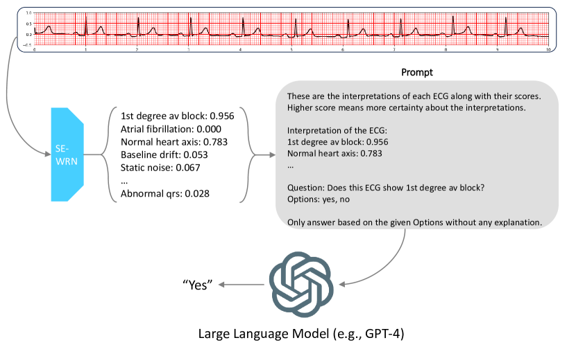
As for one of the future research directions with our dataset, we further investigated the possibility of leveraging LLMs for ECG-QA. Inspired by ChatCAD [29], for each QA sample, we transformed ECGs into text descriptions using the output from the trained upper bound model (SE-WRN) and forwarded them to several OpenAI’s GPT models (gpt-4, gpt-3.5-turbo, text-davinci-003)444As all of these experiments were conducted prior to June 7th, 2023, the associated models were referencing the earlier legacy versions. More precisely, at the time of the experiments, gpt-4 corresponded to gpt-4-0314, and gpt-3.5-turbo matched with gpt-3.5-turbo-0301. along with the corresponding question. Due to the restricted quota of OpenAI’s api usage policy, we randomly sampled 10% from the ECG-QA test set and conducted experiments only once for each LLM model. The detailed processes including prompts that we used are described in Supplementary C.1.3.
4.4 Results
QA results
The baseline results are presented in Table 2. We also report test performances for different attribute types in Supplementary C.2. As expected, Blind and Deaf Transformer exhibit poor performance while other models all achieve higher scores compared to the prior model (per Q-type majority), indicating that our dataset cannot be solved by solely seeing each question and ECG separately. Furthermore, among the top three models (M3AE†, MedViLL†, and Fusion Transformer), the pre-trained models (M3AE† and MedViLL†) outperform Fusion Transformer, which demonstrates the potential advantages of utilizing novel multi-modal pre-training methods for our dataset. Additionally, the lower performance of Choose or Query questions compared to Verify questions suggests that the primary challenges in our dataset lie on a model’s ability to learn logical and set operations based on the basic perceptual abilities that can be acquired from Verify questions.
Upper bound results
The results of the upper bound experiments are reported in Table 4 and 4. When we compare SE-WRN, which is the best upper bound model, with the best QA model, M3AE†, we can see that the perceptual ability of baseline models can be improved by p and p in terms of EM accuracy and AUROC, respectively. Moreover, when comparing with MAX, which is expected to show a higher upper bound, the differences are increased to p in EM accuracy and p in AUROC. We believe these upper bound results can serve as a useful yardstick for assessing the basic perceptual ability required for more complicated questions such as Choose or Query.
LLM modeling results
The results of the experiments with LLMs are presented in Table 5. Interestingly, the performance of all the GPT models did not surpass that of the QA baseline model. We speculate that this is due to two primary reasons: 1) the upper bound model (i.e. SE-WRN) fails to accurately extract necessary information, and 2) some questions were too complicated to be answered with a zero-shot prompt. Since LLMs fully rely on the ECG classification model for interpreting the ECGs, their performance inevitably depends on the capabilities of the ECG classification model. However, we cannot guarantee that SE-WRN is such a perfect model that always outputs accurate interpretations, because it has been trained with a limited set of QA training set to measure only the upper bound of the perceptual ability. Therefore, we expect significant performance gains if we have a strong classification model that can extract all the existing information from an ECG, and fine-tune LLMs with our QA training set (i.e., applying instruction learning to LLMs).
| Question Type | per Q-type majority | SE-WRN + gpt-4 | SE-WRN + gpt-3.5-turbo | SE-WRN + text-davinci-003 | M3AE† [2] |
| S-Verify | |||||
| S-Choose | |||||
| S-Query | |||||
| CC-Verify | |||||
| CC-Query | |||||
| CI-Verify | |||||
| CI-Query | |||||
| S: Single, CC: Comparison-Consecutive, CI: Comparison-Irrelevant | |||||
5 Conclusion
In this work, we present ECG-QA, the first QA dataset that incorporates ECG data for question answering tasks. Our dataset is designed to ensure clinical relevance and has been validated by an ECG expert. We created carefully designed question templates, which leverage clinically meaningful attributes extracted from the PTB-XL dataset, to generate a diverse collection of questions, including those that require the comparison of two ECGs. We believe that our dataset has the potential to significantly advance the field of ECG question answering research and contribute to the improvement of clinical practice in analyzing ECG data.
As for the future research directions with ECG-QA, one of the promising avenues is the exploration of multi-modal LLMs that can simultaneously process both ECG signals and natural language. While there is extensive work on LLMs that combine vision and language, there has been limited research on models that integrate signal processing with natural language. We believe our dataset can serve as an excellent testbed for such models.
6 Limitation
Despite our best efforts to create the current version of the dataset, there are some limitations as follows.
Small number of ECGs
Due to the limited number of ECGs available in the original dataset (PTB-XL), our dataset was constructed using a relatively small number of ECGs (16k), which leads that questions involving too rare symptoms (e.g., Wolf-Parkinson-White syndrome) could not be included. To provide more diverse combinations of ECGs and questions by incorporating questions regarding very rare attributes, we are planning to employ another dataset that is larger than the PTB-XL dataset such as MIMIC-IV-ECG [5], which is planned to be released in late 2023.
Upper-bound of the dataset
As mentioned in Section 4.2, given the intricacies of the medical field, even medical experts cannot provide 100% accurate diagnoses for all questions. Thus, the upper-bound of the dataset itself is not expected to be 100%. To address this, we conducted experiments demonstrating the estimated upper-bound for each question type and attribute.
Old version of SCP-ECG standard
Despite SCP-ECG v3.0 being the latest version, the metadata of the original dataset, PTB-XL, follows the SCP-ECG v0.4 standard. Consequently, in ECG-QA, we were constrained to categorize various symptoms based on the SCP-ECG v0.4 standard. However, after investigating how the SCP codes in SCP-ECG v3.0 are categorized, we found that there is only a little difference between SCP-ECG v0.4 and v3.0 regarding the SCP codes used in PTB-XL. Among the SCP codes used in PTB-XL, only one SCP code (BIGU, bigeminal pattern - unknown origin, SV or Ventricular) has a different representation, which has changed to "SVBIG" (supraventricular bigeminy BIGU bigeminal pattern - unkown origin, SV or Ventricular) in SCP-ECG v3.0. The rest of the SCP codes have maintained their codes and definitions intact in SCP-ECG v3.0. Therefore, we believe that the impact of the differences between the two versions will not be significant in the ECG-QA dataset.
Automatic generation of paraphrases
Although the paraphrases were manually curated, the initial candidates were automatically generated by ChatGPT, which may not be an optimal strategy. We expect that paraphrases could have been more diverse if we had involved medical practitioners in manually generating paraphrases.
Acknowledgments and Disclosure of Funding
This work was supported by Institute of Information & communications Technology Planning & Evaluation (IITP) grant (No.2019-0-00075), National Research Foundation of Korea (NRF) grant (NRF-2020H1D3A2A03100945), and Korea Medical Device Development Fund grant (Project Number: 1711138160, KMDF_PR_20200901_0097), funded by the Korea government (MSIT, MOTIE, MOHW, MFDS).
References
- Antol et al. [2015] Stanislaw Antol, Aishwarya Agrawal, Jiasen Lu, Margaret Mitchell, Dhruv Batra, C Lawrence Zitnick, and Devi Parikh. Vqa: Visual question answering. In Proceedings of the IEEE international conference on computer vision, pages 2425–2433, 2015.
- Chen et al. [2022] Zhihong Chen, Yuhao Du, Jinpeng Hu, Yang Liu, Guanbin Li, Xiang Wan, and Tsung-Hui Chang. Multi-modal masked autoencoders for medical vision-and-language pre-training. In Medical Image Computing and Computer Assisted Intervention–MICCAI 2022: 25th International Conference, Singapore, September 18–22, 2022, Proceedings, Part V, pages 679–689. Springer, 2022.
- Fye [1994] W Bruce Fye. A history of the origin, evolution, and impact of electrocardiography. The American journal of cardiology, 73(13):937–949, 1994.
- Gopal et al. [2021] Bryan Gopal, Ryan Han, Gautham Raghupathi, Andrew Ng, Geoff Tison, and Pranav Rajpurkar. 3kg: contrastive learning of 12-lead electrocardiograms using physiologically-inspired augmentations. In Machine Learning for Health, pages 156–167. PMLR, 2021.
- [5] Brian Gow, Tom Pollard, Larry A Nathanson, Alistair Johnson, Benjamin Moody, Chrystinne Fernandes, Nathaniel Greenbaum, Seth Berkowitz, Dana Moukheiber, Parastou Eslami, et al. Mimic-iv-ecg-diagnostic electrocardiogram matched subset.
- Han et al. [2021] Hyeongrok Han, Seongjae Park, Seonwoo Min, Hyun-Soo Choi, Eunji Kim, Hyunki Kim, Sangha Park, Jinkook Kim, Junsang Park, Junho An, et al. Towards high generalization performance on electrocardiogram classification. In 2021 Computing in Cardiology (CinC), volume 48, pages 1–4. IEEE, 2021.
- Hu et al. [2018] Jie Hu, Li Shen, and Gang Sun. Squeeze-and-excitation networks. In Proceedings of the IEEE conference on computer vision and pattern recognition, pages 7132–7141, 2018.
- Hudson and Manning [2019] Drew A Hudson and Christopher D Manning. Gqa: A new dataset for real-world visual reasoning and compositional question answering. In Proceedings of the IEEE/CVF conference on computer vision and pattern recognition, pages 6700–6709, 2019.
- Kingma and Ba [2014] Diederik P Kingma and Jimmy Ba. Adam: A method for stochastic optimization. arXiv preprint arXiv:1412.6980, 2014.
- Kiyasseh et al. [2021] Dani Kiyasseh, Tingting Zhu, and David A Clifton. Clocs: Contrastive learning of cardiac signals across space, time, and patients. In International Conference on Machine Learning, pages 5606–5615. PMLR, 2021.
- Kovaleva et al. [2020] Olga Kovaleva, Chaitanya Shivade, Satyananda Kashyap, Karina Kanjaria, Joy Wu, Deddeh Ballah, Adam Coy, Alexandros Karargyris, Yufan Guo, David Beymer Beymer, Anna Rumshisky, and Vandana Mukherjee Mukherjee. Towards visual dialog for radiology. In Proceedings of the 19th SIGBioMed Workshop on Biomedical Language Processing, pages 60–69, Online, July 2020. Association for Computational Linguistics. doi: 10.18653/v1/2020.bionlp-1.6. URL https://aclanthology.org/2020.bionlp-1.6.
- Lee et al. [2022] Gyubok Lee, Hyeonji Hwang, Seongsu Bae, Yeonsu Kwon, Woncheol Shin, Seongjun Yang, Minjoon Seo, Jong-Yeup Kim, and Edward Choi. Ehrsql: A practical text-to-sql benchmark for electronic health records. Advances in Neural Information Processing Systems, 35:15589–15601, 2022.
- Li et al. [2022] Yongqi Li, Wenjie Li, and Liqiang Nie. MMCoQA: Conversational question answering over text, tables, and images. In Proceedings of the 60th Annual Meeting of the Association for Computational Linguistics (Volume 1: Long Papers), pages 4220–4231, Dublin, Ireland, May 2022. Association for Computational Linguistics. doi: 10.18653/v1/2022.acl-long.290. URL https://aclanthology.org/2022.acl-long.290.
- Liu et al. [2021] Bo Liu, Li-Ming Zhan, Li Xu, Lin Ma, Yan Yang, and Xiao-Ming Wu. Slake: A semantically-labeled knowledge-enhanced dataset for medical visual question answering. In 2021 IEEE 18th International Symposium on Biomedical Imaging (ISBI), pages 1650–1654. IEEE, 2021.
- Lu et al. [2018] Pan Lu, Lei Ji, Wei Zhang, Nan Duan, Ming Zhou, and Jianyong Wang. R-vqa: learning visual relation facts with semantic attention for visual question answering. In Proceedings of the 24th ACM SIGKDD International Conference on Knowledge Discovery & Data Mining, pages 1880–1889, 2018.
- Makowski et al. [2021] Dominique Makowski, Tam Pham, Zen J. Lau, Jan C. Brammer, François Lespinasse, Hung Pham, Christopher Schölzel, and S. H. Annabel Chen. NeuroKit2: A python toolbox for neurophysiological signal processing. Behavior Research Methods, 53(4):1689–1696, feb 2021. doi: 10.3758/s13428-020-01516-y. URL https://doi.org/10.3758%2Fs13428-020-01516-y.
- Moon et al. [2022] Jong Hak Moon, Hyungyung Lee, Woncheol Shin, Young-Hak Kim, and Edward Choi. Multi-modal understanding and generation for medical images and text via vision-language pre-training. IEEE Journal of Biomedical and Health Informatics, 26(12):6070–6080, 2022.
- Nejedly et al. [2021] Petr Nejedly, Adam Ivora, Radovan Smisek, Ivo Viscor, Zuzana Koscova, Pavel Jurak, and Filip Plesinger. Classification of ecg using ensemble of residual cnns with attention mechanism. In 2021 Computing in Cardiology (CinC), volume 48, pages 1–4. IEEE, 2021.
- Oh et al. [2022] Jungwoo Oh, Hyunseung Chung, Joon-myoung Kwon, Dong-gyun Hong, and Edward Choi. Lead-agnostic self-supervised learning for local and global representations of electrocardiogram. In Conference on Health, Inference, and Learning, pages 338–353. PMLR, 2022.
- Pampari et al. [2018] Anusri Pampari, Preethi Raghavan, Jennifer Liang, and Jian Peng. emrqa: A large corpus for question answering on electronic medical records. arXiv preprint arXiv:1809.00732, 2018.
- Reyna et al. [2021] Matthew Reyna, Nadi Sadr, Annie Gu, Erick Andres Perez Alday, Chengyu Liu, Salman Seyedi, Amit Shah, and Gari Clifford. Will two do? varying dimensions in electrocardiography: the PhysioNet/Computing in Cardiology Challenge 2021. Computing in Cardiology 2021, 48:1–4, 2021.
- Rosiek and Leksowski [2016] Anna Rosiek and Krzysztof Leksowski. The risk factors and prevention of cardiovascular disease: the importance of electrocardiogram in the diagnosis and treatment of acute coronary syndrome. Therapeutics and clinical risk management, pages 1223–1229, 2016.
- Rubel et al. [2016] Paul Rubel, Danilo Pani, Alois Schloegl, Jocelyne Fayn, Fabio Badilini, Peter W Macfarlane, and Alpo Varri. Scp-ecg v3. 0: An enhanced standard communication protocol for computer-assisted electrocardiography. In 2016 Computing in Cardiology Conference (CinC), pages 309–312. IEEE, 2016.
- Talmor et al. [2021] Alon Talmor, Ori Yoran, Amnon Catav, Dan Lahav, Yizhong Wang, Akari Asai, Gabriel Ilharco, Hannaneh Hajishirzi, and Jonathan Berant. Multimodalqa: Complex question answering over text, tables and images. arXiv preprint arXiv:2104.06039, 2021.
- Vaswani et al. [2017] Ashish Vaswani, Noam Shazeer, Niki Parmar, Jakob Uszkoreit, Llion Jones, Aidan N Gomez, Łukasz Kaiser, and Illia Polosukhin. Attention is all you need. In Advances in neural information processing systems, pages 5998–6008, 2017.
- Wagner et al. [2022] Patrick Wagner, Nils Strodthoff, Ralf-Dieter Bousseljot, Wojciech Samek, and Tobias Schaeffter. PTB-XL, a large publicly available electrocardiography dataset (version 1.0.3). PhysioNet, 2022. doi: https://doi.org/10.13026/x4td-x982.
- Wang et al. [2009] Li-ping Wang, Mi Shen, Jia-fei Tong, and Jun Dong. An uncertainty reasoning method for abnormal ecg detection. In 2009 IEEE International Symposium on IT in Medicine & Education, volume 1, pages 1091–1096. IEEE, 2009.
- Wang et al. [2020] Ping Wang, Tian Shi, and Chandan K Reddy. Text-to-sql generation for question answering on electronic medical records. In Proceedings of The Web Conference 2020, pages 350–361, 2020.
- Wang et al. [2023] Sheng Wang, Zihao Zhao, Xi Ouyang, Qian Wang, and Dinggang Shen. Chatcad: Interactive computer-aided diagnosis on medical image using large language models. arXiv preprint arXiv:2302.07257, 2023.
- Zagoruyko and Komodakis [2016] Sergey Zagoruyko and Nikos Komodakis. Wide residual networks. arXiv preprint arXiv:1605.07146, 2016.
- Zhong et al. [2019] Xian Zhong, Oubo Gong, Wenxin Huang, Lin Li, and Hongxia Xia. Squeeze-and-excitation wide residual networks in image classification. In 2019 IEEE International Conference on Image Processing (ICIP), pages 395–399. IEEE, 2019.
- Zhu et al. [2016] Yuke Zhu, Oliver Groth, Michael Bernstein, and Li Fei-Fei. Visual7w: Grounded question answering in images. In Proceedings of the IEEE conference on computer vision and pattern recognition, pages 4995–5004, 2016.
- Zimetbaum and Josephson [2003] Peter J Zimetbaum and Mark E Josephson. Use of the electrocardiogram in acute myocardial infarction. New England Journal of Medicine, 348(10):933–940, 2003.
Supplementary Material
Appendix A Full list of templates
A.1 Attribute type descriptions
Individual attributes belonging to each attribute set used in ECG-QA are described in Table 6. For a detailed description of the attributes in SCP code, which are presented as abbreviations, please refer to the PTB-XL dataset 111https://physionet.org/content/ptb-xl/1.0.3/.
| Name | Description | Num. | Attributes |
| SCP code | SCP-ECG statements (symptoms) | NORM, STTC, MI, HYP, CD, NDT, NST_, DIG, LNGQT, IMI, ASMI, LVH, LAFB, ISC_, IRBBB, 1AVB, IVCD, ISCAL, CRBBB, CLBBB, ILMI, LAO/LAE, AMI, ALMI, ISCIN, INJAS, LMI, ISCIL, LPFB, ISCAS, INJAL, ISCLA, RAO/RAE, ILBBB, IPLMI, ISCAN, IPMI, INJIN, INJLA, PMI, INJIL, ABQRS, PVC, STD_, VCLVH, QWAVE, LOWT, NT_, PAC, LPR, INVT, LVOLT, HVOLT, TAB_, STE_, SR, AFIB, STACH, SARRH, SBRAD, PACE, BIGU, AFLT, SVTAC | |
| Noise | Signal artifacts | Baseline drift, Static noise, Burst noise, Electrode problems | |
| Stage of infarction | Stage of myocardial infarction | 5 | None, Unknown, Early, Middle, Late |
| Extra systole | Extra systoles | Extrasystoles, Ventricular extrasystoles, Supraventricular extrasystoles | |
| Heart axis | Direction of heart axis | Left axis deviation, Right axis deviation Extreme axis deviation, Normal heart axis | |
| Numeric feature | Numeric features | RR interval, P duration, PR interval, QRS duration, QT interval, QT corrected |
A.2 Question templates
A total of 70 question templates is reported in Table 20.
A.3 Paraphrases
The overall procedure of generating paraphrases is as follows:
-
1.
20 candidate paraphrases per template question were automatically generated by OpenAI’s ChatGPT with the following prompt:
-
2.
Based on the machine-generated paraphrases, we manually refined them to ensure the high quality of paraphrases. Specifically, we filtered out the paraphrases that deviated too much from the original question and manually revised them if the specific medical term we were targeting had changed.
-
3.
Then, we randomly selected seven paraphrases for training and validation splits, and three paraphrases for the test split.
As a result, the final paraphrases for each question template are presented in Table 22.
Appendix B Dataset construction details
B.1 Regular expressions for parsing lead positions of form-related SCP codes
To utilize grounded lead information of form-related SCP codes, we defined a parser based on regular expressions, which can be shown in Table 16, 17, and 18. During the parsing process, we utilized the Google Cloud Translation API to translate the ECG reports into English, as the majority of the original reports were written in German.
B.2 Numeric feature extraction
To extract numeric values including heart axis degrees, we utilized NeuroKit2 [16]. The specific method to extract these attributes are described in the following paragraphs.
Heart axis
To calculate heart axis degrees, we extracted the magnitude of R peaks from the lead I and lead aVF for each heartbeat existed in the ECG. Then, we computed heart axis degrees by the following equation:
where is the number of heartbeats in the ECG, and is the magnitude value of the -th R peak in the lead . To ensure accurate calculations, we did not process the samples with any noises in lead I or lead aVF and restricted them not to be sampled from the relevant questions regarding the heart axis. Based on the calculated heart axis degrees, we categorized them into four standard classes as follows:
Numeric feature
For the numeric features such as RR interval or PR interval, we extracted the locations of P, Q, R, S, and T waves for each beat present in lead II, and computed the following six numeric features:
-
•
RR interval: The interval seconds between consecutive R peaks.
-
•
P duration: The interval seconds between P onset and P offset.
-
•
PR interval: The interval seconds between P onset and R onset.
-
•
QRS duration: The interval seconds between Q peak and S peak.
-
•
QT interval: The interval seconds between Q peak and T offset.
-
•
QT corrected:
Similar to Heart axis, we did not process neither the samples with any noises in lead II nor the samples that have less than seven R peaks detected in lead II. Again, we categorized the calculated values into below, within, or above the normal range where the normal range criteria are presented in Table 7.
| Numeric Feature | Below the normal range | Within the normal range | Above the normal range |
| RR interval | |||
| P duration | - | ||
| PR interval | |||
| QRS duration | |||
| QT interval | |||
| QT corrected |
B.3 Sampling strategy
During the sampling process, we sampled more negative samples than positive samples to reflect the clinical reality where normal (negative) cases are much more frequent than abnormal (positive) cases. The sampling size for each question is presented in Table 8. To avoid excessively unbalanced sampling, we set a limit on the number of negative samples if there are too few positive samples, ensuring it does not exceed five times the number of positive samples. For example, if there are 30 positive samples for Single-Verify question in the training set, we sample 150 negative samples for that question instead of 200 negative samples. Different sampling strategies for each question type are described in the following paragraphs.
| Question Type | Train | Validation | Test |
| (Pos / Neg) | (Pos / Neg) | (Pos / Neg) | |
| S-Verify | |||
| S-Choose | |||
| S-Query | |||
| CC-Verify | |||
| CC-Query | |||
| CI-Verify | |||
| CI-Query |
Verify questions
Typically there are two answer options (e.g., yes/no), but we included an additional answer option "not sure" especially for diagnostic labels of SCP codes to utilize the likelihood information given by the PTB-XL metadata. Specifically, in the PTB-XL metadata, diagnostic SCP codes are annotated along with their likelihood, indicating the certainty of the diagnoses on a scale of [0, 15, 35, 50, 80, 100]. Considering that the likelihood was derived from keywords in the ECG report and set to zero where no relevant keyword was available (i.e., statements with no adjectives such as “non-specific st-t wave changes” set to 0 likelihood), we classified 0 and 100 as a "certain" diagnosis, and any other value as an "uncertain" diagnosis. Accordingly, when a question asks to verify the presence of a specific diagnostic SCP code in an ECG, the answer is "not sure" if the ECG has been labeled with that SCP code with a likelihood of [15, 35, 50, 80].
Additionally, for questions related to a specific grounded lead position, we defined two types of negative samples: "hard negative" and "soft negative" to add complexity to the dataset. Specifically, "hard negative" samples have the corresponding attribute in other leads but not in the inquired lead, while "soft negative" samples do not have the attribute at all. We sampled at most half of the negative samples from the "hard negative" samples, and the remaining from the "soft negative" samples. By doing so, we intended the QA models to be able to deduce the grounding lead information of specific attributes.
Choose questions
Choose questions involve two attributes, and the answer depends on whether each attribute exists in the ECG, which leads to four possible answer options for each question. For these questions, we considered the absence of both attributes in the ECG as a negative sample, and the presence of at least one attribute as a positive sample. We balanced out the negative samples by adjusting their number to be no more than five times the maximum number of samples among the three positive options.
Query questions
Similar to Choose questions, we regarded negative samples for Query questions as the samples that do not have any of the attributes in the inquired attribute set. For example, if a question asks “What kind of noises does this ECG show?”, the negative samples are defined as the ECGs that do not have any noises such as Baseline drift or Static noise. Then, as aforementioned, we restricted the number of negative samples to maintain balance.
| Question type | Train | Validation | Test |
| S-Verify | |||
| S-Choose | |||
| S-Query | |||
| CC-Verify | |||
| CC-Query | |||
| CI-Verify | |||
| CI-Query | |||
| Total |
| Attribute type | Train | Validation | Test |
| SCP code | |||
| Noise | |||
| Stage of infarction | |||
| Extra systole | |||
| Heart axis | |||
| Numeric feature | |||
| Total |
B.4 Dataset statistics
Appendix C Experimental details
C.1 Implementation details
C.1.1 QA Baselines
The training configurations for QA baselines along with model configurations and resource information are reported in Table 11. We used Adam [9] optimizer for all the models.
-
1.
per Q-type majority: This is a prior model that outputs only the most frequent answer in the test split for each question type.
-
2.
M3AE† [2]: A multi-modal architecture of ECGs and texts based on Transformer Encoder [25], pre-trained with Masked Language Modeling (MLM), Masked Image Modeling (MIM), and Image Text Matching (ITM) tasks. The architecture comprises three components: 1) separated uni-modal encoders, 2) a multi-modal fusion module, and 3) separated uni-modal decoders for pre-training tasks. Considering the characteristics of the signal data, we employed several 1-d convolutional layers to embed ECGs instead of using a linear layer in the original implementation. Otherwise, we followed the same configurations as the original paper including model architecture and pre-training hyperparameters, except for batch size and learning rate. We used batch size, and e- learning rate for the pre-training. In the fine-tuning phase, for Comparison questions that need to see two ECGs, we concatenate the two ECGs and forwarded them to the uni-modal encoder (the 1st component) to get ECG embeddings.
-
3.
MedViLL† [17]: Fusion Transformer (see below) pre-trained with MedViLL [17] methodology. This method implements multi-modal pre-training with ECGs and texts, consisting of MLM and ITM tasks. When pre-training, we followed the same configurations introduced in the original paper such as MLM ratio, except for batch size and learning rate. We used batch size, and e- learning rate for the pre-training.
-
4.
Fusion Transformer: Both the questions and (potentially two) ECGs are concatenated and forwarded into the Transformer Encoder after embedding ECGs with several convolutional layers. To encode questions, we tokenize questions to subword tokens following the BERT tokenizer and encode them using the embedding layer of BERT. For Single questions which involve only one ECG, the second ECG is padded by zeros. Then, we separately average-pool each of the modalities, which yields two ECG vectors ( ) and a single question vector ( ). For Single questions, is set to . We concatenate these vectors and perform multi-label classification task after projecting them onto the final answer space ( ).
-
5.
Deaf Transformer: Only the ECGs are forwarded to the Transformer Encoder after being embedded by several convolutional layers. We average-pool the output vectors for each of ECGs, which results in two ECG vectors ( ). Similar to the Fusion Transformer, for Single questions, the second ECG is padded by zeros, and is set to . Then, we concatenate two vectors and project them onto the final answer space ( ).
-
6.
Blind Transformer: Only the questions are forwarded to the Transformer Encoder. Similar to Fusion Transformer, we follow BERT to tokenize and encode questions. We average-pool the output vectors to get a single question vector, and project the vector onto the final answer space ( ).
| Name | M3AE† [2] | MedViLL† [17] | Fusion Transf. | Deaf Transf. | Blind Transf. |
| Model configurations | |||||
| Conv layers | [(, , )] | [(, , )] 4 | [(, , )] | [(, , )] | N/A |
| Transformer layers | N/A | ||||
| Hidden dimension | |||||
| Attention heads | N/A | ||||
| Training configurations | |||||
| Training step | |||||
| Local batch size | |||||
| Total batch size | |||||
| Gradient accumulation step | 4 | 8 | 8 | 4 | 1 |
| Learning rate | e- | e- | e- | e- | e- |
| LR scheduler | Tri(, , ) | Tri(, , ) | Tri(, , ) | Tri(, , ) | Tri(, , ) |
| Resources | |||||
| GPU device | A6000 | RTX 3090 | RTX 3090 | RTX 3090 | RTX 3090 |
| VRAM | 48GB | 24GB | 24GB | 24GB | 24GB |
| Training time | hours | hours | hours | hours | hours |
| Conv layers: [(channel size, kernel size, stride)] N | |||||
| Total batch size: Local batch size Gradient accumulation step Number of GPU devices | |||||
| Tri(x, y, z): warmup ratio (x), hold ratio (y), exponential decay ratio (z), final lr decaying scale= | |||||
| A6000: NVIDIA RTX A6000 | |||||
| RTX 3090: NVIDIA GeForce RTX 3090 | |||||
C.1.2 Upper bound models
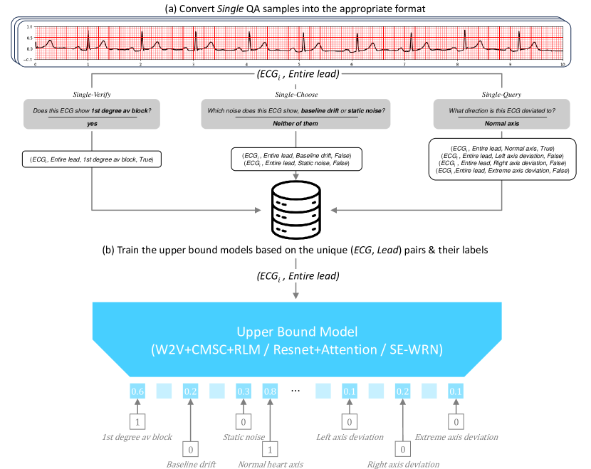
The procedure of training upper bound models with QA samples is illustrated in Figure 3. For a fair comparison with QA models, we need to train the upper bound models using the comparable dataset with the ECG-QA dataset, requiring converting QA samples to the format that the upper bound models can process. Accordingly, we convert each Single QA sample pair (Question, ECG, Answer) in the training set into 4-tuples (ECG, Lead, Attribute, Label), and collect all the corresponding (Attribute, Label) pairs for each unique (ECG, Lead) pair as shown in Figure 3 (a). Then, each (ECG, Lead) pair is fed to the upper bound (ECG classification) model along with its labels, and the model is trained by the binary cross entropy (BCE) losses calculated from the corresponding classification heads (See Figure 3 (b)). When training the upper bound model, although the model outputs the scores (i.e., probabilities) for all the possible attributes from the classification heads, we only calculate BCE losses from the corresponding attributes that have been labeled for each (ECG, Lead) pair so that the classification heads for unlabeled attributes are not trained. In addition, if Lead in each (ECG, Lead) pair indicates the specific lead (e.g., Lead I), not an entire ECG, we forward the ECG to the model after zero-padding the other leads.
The strategies for converting QA samples into the 4-tuple format for different question types are described in the following paragraphs.
| Name | W2V+CMSC+RLM [19] | Resnet+Attention [18] | SE-WRN [6] |
| Training configurations | |||
| Training step | |||
| Batch size | |||
| Learning rate | e- | e- | e- |
| LR scheduler | Tri(, , ) | Tri(, , ) | Tri(, , ) |
| Resources | |||
| GPU device | RTX 3090 | RTX 3090 | RTX 3090 |
| VRAM | 24GB | 24GB | 24GB |
| Training time | hours | hours | hours |
| Tri(x, y, z): warmup ratio (x), hold ratio (y), exponential decay ratio (z), final lr decaying scale= | |||
| RTX 3090: NVIDIA GeForce RTX 3090 | |||
Single-Verify samples
These types of QA samples can be directly convertible to the 4-tuple format according to their answers. For example, if we have a QA sample (“Does this ECG show baseline drift in lead I?”, ECGA, “yes”), then this QA sample is converted to (ECGA, Lead I, Baseline drift, True), which means that "ECGA has Baseline drift in Lead I". On the other hand, if we have another QA sample where the answer is “no”, such as (“Does this ECG show baseline drift in Lead I?”, ECGB, “no”), we can derive (ECGB, Lead I, Baseline drift, False), which means that "ECGB does not have Baseline drift in lead I".
Single-Choose samples
Because there are two relevant attributes in these types of QA samples, we can extract two 4-tuples for each QA sample. For example, if we have a QA sample (“Which noise does this ECG show in Lead I, baseline drift or static noise?”, ECGA, “baseline drift”), then we convert this QA sample into two 4-tuples: 1) (ECGA, Lead I, Baseline drift, True) and 2) (ECGA, Lead I, Static noise, False).
Single-Query samples
Similar to Single-Choose samples, we derive 4-tuples for each QA sample as many as the number of the relevant attributes. For example, if we have a QA sample (“What kind of noises does this ECG show in Lead I?”, ECGA, “baseline drift, burst noise”), then we can derive four 4-tuples: 1) (ECGA, Lead I, Baseline drift, True), 2) (ECGA, Lead I, Static noise, False), 3) (ECGA, Lead I, Burst noise, True), and 4) (ECGA, Lead I, Electrode problems, False).
The detailed upper bound model implementations are described in the following paragraphs. In addition, training configurations for upper bound models including resource information are presented in Table 12.
-
1.
W2V+CMSC+RLM [19]: A Transformer-based model pre-trained with the W2V+CMSC+ RLM [19] method. This model comprises several convolutional layers to extract features from ECGs, followed by Transformer Encoder to contextualize the features. For the model configurations, we follow the original implementation. Specifically, we employ four convolutional layers, each of which has 256 channels with strides of two and kernel lengths of two, and 12 Transformer Encoder layers with 12 self-attention heads and 768 hidden dimensions.
-
2.
Resnet-Attention [18]: This model was introduced in PhysioNet/Computing in Cardiology Challenge 2021 [21] (CinC 2021), which won first place in the challenge. It is composed of several residual blocks followed by a multi-head attention layer, and two output layers: 1) a conventional BCE loss layer and 2) another loss layer specifically designed for optimizing the challenge score. Since we do not need to optimize the model to achieve a high challenge score, we omit the second loss layer and use only BCE loss to train the model. For any other model configurations such as the number of residual blocks or dropout rate, we follow the original implementation.
-
3.
SE-WRN [6]: This model was introduced in the same challenge (CinC 2021), which won second place. It consists of a series of wide residual networks [30] combined with squeeze-and-excitation modules [7]. In the original paper, they additionally use demographic features such as age and sex, but we do not use those features. We follow the original implementation for the model configurations. However, because the original authors did not mention the kernel lengths for each convolutional layer, we selected kernel lengths of 11 after searching for the best options among 3, 5, 7, and 11.
C.1.3 Modeling with LLMs
The procedure of modeling the ECG-QA dataset with LLMs is visualized in Figure 4. To provide the results of the ECG interpretations to the LLMs, we transform the outputs from the best upper bound model (SE-WRN [6]) into the text descriptions that LLMs can interpret. Specifically, we apply a threshold value of 0.5 to each score in the model’s outputs to get only the attributes whose score is more than 0.5. Then, based on the selected attributes with their scores, we forward the following prompts to LLMs and measure the exact match accuracy. To enable the one-to-one comparison between LLM’s answer and GT answer, we induce the LLMs to output the answers only from the valid answer options by giving the candidate options in the prompt for each question.
C.2 Detailed experimental results
Detailed QA results
The QA baseline results for different question types & attribute types are presented in Table 13.
| Question & Attribute Type | per Q-type majority | M3AE† [2] | MedViLL† [17] | Fusion Transf. | |||
| EM Acc. | AUROC | EM Acc. | AUROC | EM Acc. | AUROC | ||
| S-Verify | |||||||
| SCP code | 68.0 | ||||||
| Noise | 67.1 | ||||||
| Stage of infarction | 71.4 | ||||||
| Extra systole | 69.6 | ||||||
| Heart axis | 67.5 | ||||||
| Numeric feature | 66.7 | ||||||
| S-Choose | |||||||
| SCP code | 31.1 | ||||||
| Noise | 31.5 | ||||||
| Stage of infarction | 33.3 | ||||||
| Extra systole | 27.3 | ||||||
| Heart axis | 33.3 | ||||||
| Numeric feature | 34.6 | ||||||
| S-Query | |||||||
| SCP code | 24.5 | ||||||
| Noise | 39.3 | ||||||
| Stage of infarction | 28.1 | ||||||
| Extra systole | 41.5 | ||||||
| Heart axis | 29.9 | ||||||
| Numeric feature | 6.8 | ||||||
| CC-Verify | |||||||
| SCP code | 62.0 | ||||||
| Numeric feature | 66.7 | ||||||
| CC-Query | |||||||
| SCP code | 15.4 | ||||||
| Numeric feature | 18.9 | ||||||
| CI-Verify | |||||||
| SCP code | 66.0 | ||||||
| Numeric feature | 66.7 | ||||||
| CI-Query | |||||||
| SCP code | 0.66 | ||||||
| Numeric feature | 3.08 | ||||||
| S: Single, CC: Comparison-Consecutive, CI: Comparison-Irrelevant | |||||||
Detailed upper bound results
Appendix D Author statement
We bear all responsibility in case of violation of rights, etc. associated with the ECG-QA dataset.
| Attribute name | W2V+CMSC+RLM [19] | Resnet-Attention [18] | SE-WRN [6] | MAX | ||||
| Acc. | AUROC | Acc. | AUROC | Acc. | AUROC | Acc. | AUROC | |
| Macro-average | ||||||||
| Abnormal qrs | ||||||||
| Above the normal range of p duration | ||||||||
| Above the normal range of pr interval | ||||||||
| Above the normal range of qrs duration | ||||||||
| Above the normal range of qt corrected | ||||||||
| Above the normal range of qt interval | ||||||||
| Above the normal range of rr interval | ||||||||
| Any diagnostic symptoms | ||||||||
| Any form-related symptoms | ||||||||
| Any kind of abnormal symptoms | ||||||||
| Any kind of extra systoles | ||||||||
| Any kind of noises | ||||||||
| Any rhythm-related symptoms | ||||||||
| Atrial fibrillation | ||||||||
| Atrial flutter | ||||||||
| Atrial premature complex | ||||||||
| Baseline drift | ||||||||
| Below the normal range of pr interval | ||||||||
| Below the normal range of qrs duration | ||||||||
| Below the normal range of qt corrected | ||||||||
| Below the normal range of qt interval | ||||||||
| Below the normal range of rr interval | ||||||||
| Bigeminal pattern (unknown origin, supraventricular, or ventricular) | ||||||||
| Burst noise | ||||||||
| Complete left bundle branch block | ||||||||
| Complete right bundle branch block | ||||||||
| Conduction disturbance | ||||||||
| Digitalis effect | ||||||||
| Early stage of myocardial infarction | ||||||||
| Electrodes problems | ||||||||
| Extrasystoles | ||||||||
| Extreme axis deviation | ||||||||
| First degree av block | ||||||||
| High qrs voltage | ||||||||
| Hypertrophy | ||||||||
| Incomplete left bundle branch block | ||||||||
| Incomplete right bundle branch block | ||||||||
| Inverted t-waves | ||||||||
| Ischemic | ||||||||
| Late stage of myocardial infarction | ||||||||
| Left anterior fascicular block | ||||||||
| Left atrial overload/enlargement | ||||||||
| Left axis deviation | ||||||||
| Left posterior fascicular block | ||||||||
| Left ventricular hypertrophy | ||||||||
| Long qt-interval | ||||||||
| Low amplitude t-wave | ||||||||
| Low qrs voltages in the frontal and horizontal leads | ||||||||
| Middle stage of myocardial infarction | ||||||||
| Myocardial infarction | ||||||||
| Non-diagnostic t abnormalities | ||||||||
| Non-specific intraventricular conduction disturbance (block) | ||||||||
| Non-specific ischemic | ||||||||
| Non-specific st changes | ||||||||
| Non-specific st depression | ||||||||
| Non-specific st elevation | ||||||||
| Non-specific t-wave changes | ||||||||
| Normal ecg | ||||||||
| Normal functioning artificial pacemaker | ||||||||
| Normal heart axis | ||||||||
| Prolonged pr interval | ||||||||
| Q waves present | ||||||||
| Right atrial overload/enlargement | ||||||||
| Right axis deviation | ||||||||
| Sinus arrhythmia | ||||||||
| Sinus bradycardia | ||||||||
| Sinus rhythm | ||||||||
| Sinus tachycardia | ||||||||
| St/t change | ||||||||
| Static noise | ||||||||
| Subendocardial injury | ||||||||
| Supraventricular extrasystoles | ||||||||
| Supraventricular tachycardia | ||||||||
| T-wave abnormality | ||||||||
| Ventricular extrasystoles | ||||||||
| Ventricular premature complex | ||||||||
| Voltage criteria (qrs) for left ventricular hypertrophy | ||||||||
| Within the normal range of p duration | ||||||||
| Within the normal range of pr interval | ||||||||
| Within the normal range of qrs duration | ||||||||
| Within the normal range of qt corrected | ||||||||
| Within the normal range of qt interval | ||||||||
| Within the normal range of rr interval | ||||||||
| Attribute name | M3AE† [2] | MedViLL† [17] | Fusion Transf. | |||
| Acc. | AUROC | Acc. | AUROC | Acc. | AUROC | |
| Macro-average | ||||||
| Abnormal qrs | ||||||
| Above the normal range of p duration | ||||||
| Above the normal range of pr interval | ||||||
| Above the normal range of qrs duration | ||||||
| Above the normal range of qt corrected | ||||||
| Above the normal range of qt interval | ||||||
| Above the normal range of rr interval | ||||||
| Any diagnostic symptoms | ||||||
| Any form-related symptoms | ||||||
| Any kind of abnormal symptoms | ||||||
| Any kind of extra systoles | ||||||
| Any kind of noises | ||||||
| Any rhythm-related symptoms | ||||||
| Atrial fibrillation | ||||||
| Atrial flutter | ||||||
| Atrial premature complex | ||||||
| Baseline drift | ||||||
| Below the normal range of pr interval | ||||||
| Below the normal range of qrs duration | ||||||
| Below the normal range of qt corrected | ||||||
| Below the normal range of qt interval | ||||||
| Below the normal range of rr interval | ||||||
| Bigeminal pattern (unknown origin, supraventricular, or ventricular) | ||||||
| Burst noise | ||||||
| Complete left bundle branch block | ||||||
| Complete right bundle branch block | ||||||
| Conduction disturbance | ||||||
| Digitalis effect | ||||||
| Early stage of myocardial infarction | ||||||
| Electrodes problems | ||||||
| Extrasystoles | ||||||
| Extreme axis deviation | ||||||
| First degree av block | ||||||
| High qrs voltage | ||||||
| Hypertrophy | ||||||
| Incomplete left bundle branch block | ||||||
| Incomplete right bundle branch block | ||||||
| Inverted t-waves | ||||||
| Ischemic | ||||||
| Late stage of myocardial infarction | ||||||
| Left anterior fascicular block | ||||||
| Left atrial overload/enlargement | ||||||
| Left axis deviation | ||||||
| Left posterior fascicular block | ||||||
| Left ventricular hypertrophy | ||||||
| Long qt-interval | ||||||
| Low amplitude t-wave | ||||||
| Low qrs voltages in the frontal and horizontal leads | ||||||
| Middle stage of myocardial infarction | ||||||
| Myocardial infarction | ||||||
| Non-diagnostic t abnormalities | ||||||
| Non-specific intraventricular conduction disturbance (block) | ||||||
| Non-specific ischemic | ||||||
| Non-specific st changes | ||||||
| Non-specific st depression | ||||||
| Non-specific st elevation | ||||||
| Non-specific t-wave changes | ||||||
| Normal ecg | ||||||
| Normal functioning artificial pacemaker | ||||||
| Normal heart axis | ||||||
| Prolonged pr interval | ||||||
| Q waves present | ||||||
| Right atrial overload/enlargement | ||||||
| Right axis deviation | ||||||
| Sinus arrhythmia | ||||||
| Sinus bradycardia | ||||||
| Sinus rhythm | ||||||
| Sinus tachycardia | ||||||
| St/t change | ||||||
| Static noise | ||||||
| Subendocardial injury | ||||||
| Supraventricular extrasystoles | ||||||
| Supraventricular tachycardia | ||||||
| T-wave abnormality | ||||||
| Ventricular extrasystoles | ||||||
| Ventricular premature complex | ||||||
| Voltage criteria (qrs) for left ventricular hypertrophy | ||||||
| Within the normal range of p duration | ||||||
| Within the normal range of pr interval | ||||||
| Within the normal range of qrs duration | ||||||
| Within the normal range of qt corrected | ||||||
| Within the normal range of qt interval | ||||||
| Within the normal range of rr interval | ||||||
| Placeholder | Regular Expression |
| [**lead_position**] | v leads|chest|limb|anterior|antero\-?\s?lateral|antero\-?\s?septal| inferior|infero\-?\s?lateral|infero\-?\s?septal|lateral|high\-?\s? lateral|precordial|peripheral|standard|lateral\-?\s?chest |
| [**lead**] | (((v?\d)|(lead)?\s?(?<=[^A-Za-z])iii(?=[^A-Za-z])|(lead)?\s?(?<=[^ A-Za-z])ii(?=[^A-Za-z])|(lead)?\s?(?<=[^A-Za-z])i(?=[^A-Za-z])|(lea d)?\s?avr|(lead)?\s?avl|(lead)?\s?avf|v leads|all leads|chest( lead (s)?)?|limb( lead(s)?)?|anterior( lead(s)?)?|antero\-?\s?lateral( l ead(s)?)?|antero\-?\s?septal( lead(s)?)?|inferior( lead(s)?)?|infero \-?\s?lateral( lead(s)?)?|infero\-?\s?septal( lead(s)?)?|lateral( le ad(s)?)?|high\-?\s?lateral( lead(s)?)?|precordial( lead(s)?)?|periph eral( lead(s)?)?|standard( lead(s)?)?|infero\-lateral( lead(s)?)?|la teral\-?\s?chest( lead(s)?)?)\s?([/\),\.\-\&\s])?\s?(and)?\s?)+ |
| Statement | Lead Position |
| chest | V1, V2, V3, V4, V5, V6 |
| v leads | V1, V2, V3, V4, V5, V6 |
| limb | I, II, III, aVR, aVL, aVF |
| anterior | V3, V4 |
| antero-lateral | I, aVL, V3, V4, V5, V6 |
| antero-septal | V1, V2, V3, V4 |
| inferior | II, III, aVF |
| infero-lateral | I, II, III, aVL, aVF, V5, V6 |
| infero-septal | II, III, aVF, V1, V2 |
| lateral | I, aVL, V5, V6 |
| high-lateral | I, aVL |
| precordial | V1, V2, V3, V4, V5, V6 |
| peripheral | I, II, III, aVR, aVL, aVF |
| standard | I, II, III, aVR, aVL, aVF, V1, V2, V3, V4, V5, V6 |
| lateral-chest | I, aVL, V1, V2, V3, V4, V5, V6 |
| SCP Code | Example Target Statements | Regular Expression |
| NDT | t-change neg in [**lead**] t wave flattening in [**lead**] t waves flat in [**lead**] t wave changes in [**lead**] t wave inversion in [**lead**] t waves are low or flat in [**lead**] t waves are low in [**leads**] t waves low in [**lead**] t waves are inverted in [**lead**] t waves inverted in [**lead**] t waves are biphasic in [**lead**] t abnormal in [**lead**] t abnormality in [**lead**] t flat in [**lead**] flat t in [**lead**] | (t wave(s)?|t waves are|t wve|t-change(s)?(:)?| t change(s)?(:)?|flat t|biphasic t|neg t|high t )\s*(are)?\s*(neg in|flattening in|flattening o r slight inversion in|flat in|changes in|invers ion in|low or flat in|generally low and are fla t in|low in|flattened in|now generally flatter and are slightly inverted in |inverted in|sligh tly inverted in|low or flat in|flat or slightly inverted in|now slightly inverted in|now inver ted in|biphasic in|abnormal in|abnormality in|s inus rhythm abnormal in|flat|flach in|in|biphas .|neg\. in|biphasic|negative in|neg in|term. neg in|high in|)\s*([**lead**]) |
| NST_ | st-t wave changes in [**lead**] st-t changes in [**lead**] st-t wave changes are more marked in [**lead**] st-t wave changes persist in [**lead**] t changes in [**lead**] [**lead_position**] st-t changes [**lead_position**] st-t wave changes | ((st-t wave changes|st-t changes|t changes)\s*( are marked in|persist in|in|)\s*([**lead**]))|( ([**lead_position**])\s*(st-t wave changes|st-t changes)) |
| DIG | digitalis t pointed in [**lead**] digitalis change in [**lead**] digitalis changes [**lead**] digitalis change trough [**lead**] digitalis change r trough [**lead**] | (digitalis)\s*(t pointed in|change in|changes|c hange trough|change r trough|in|)\s*([**lead**]) |
| STD_ | st segment depression in [**lead**] st segments are depressed in [**lead**] st depression in [**lead**] st depression [**lead**] st depression above [**lead**] st lowering in [**lead**] st-lowering [**lead**] st reduction in [**lead**] st reduction discrete in [**lead**] st-senkung in [**lead**] st-thinking in [**lead**] | (st segment(s)?|st(\-)?st-senkung|st-thinking)\s *(are)?\s*(depression in|depression and t wave f lattening in|depressed in|depressed and t wave.{ 0,15} in|depression|depression above|lowering in |lowering|reduction in|reduction discrete in|in) \s*([**lead**]) |
| VCLVH | voltages are high in [**lead**] suggesting lvh [**lead**] voltages suggest possible lv hypertrop hy left ventricular hypertrophy are satisfied in [**lea d**] voltages in [**lead**] are at upper limit voltages in [**lead**] of left ventricular hypertro phy | (voltages are high in ([**lead**]) suggesting lv h)|(([**lead**]) voltages suggest possible lv hy pertrophy)|(left ventricular hypertrophy are sat isfied in ([**lead**]))|(voltages in ([**lead**] ) are at upper limit)|(voltages in ([**lead**]) of left ventricular hypertrophy)|(r wave height in ([**lead**]) suggests the possibility of left ventricular hypertrophy) |
| QWAVE | q waves in [**lead**] q wave in [**lead**] q wave present in [**lead**] q in [**lead**] q wave and small r wave in [**lead**] q waves and t wave inversion in [**lead**] [**lead_position**] q waves noted | ((q wave(s)?|q)\s*(are)?\s*(present in|in|(and|, ).{0,55} in)\s*([**lead**]))|(([**lead_position* *]) q waves noted) |
| LOWT | t waves are low in [**lead**] t waves are low or flat in [**lead**] | (t waves are)\s*(low or flat in|low in)\s*([**le ad**]) |
| NT_ | t wave flattening in [**lead**] t wave flattening persists in [**lead**] t wave flattening or inversion in [**lead**] t wave changes in [**lead**] | (t wave)\s*(flattening in|flattening persists in |flattening or slight inversion in|flattening or inversion in|changes in)\s*([**lead**]) |
| INVT | t waves are inverted in [**lead**] t wave inversion in [**lead**] t waves inverted in [**lead**] | (t wave(s)?)\s*(are)?\s*(inverted in|inversion i n|flattening in.{0,30} and inverted in)\s*([**le ad**] |
| LVOLT | low limb lead voltage peripheral low voltage low voltage in [**lead**] | (low limb lead voltage|peripheral low voltage|pe ripheral low-voltage|peripheral low tension|low voltage in ([**lead**])) |
| HVOLT | high v lead voltages voltages in [**lead**] are at upper limit voltages are high in [**lead**] | (high v lead voltages)|(voltages in ([**lead**]) \s*are at upper limit)|(voltages are high in ([* *lead**])) |
| Continued on next page. | ||
| SCP Code | Example Target Statements | Regular Expression |
| TAB_ | t changes negative in [**lead**] t-changes in [**lead**] t-changes [**lead**] t-changes neg in [**lead**] t-changes neg [**lead**] t-changes neg. t in [**lead**] t changes high in [**lead**] t changes biphas. in [**lead**] t abnormal in [**lead**] | (t(\-)?\s?change(s)?(:)?|t abnormal)\s*(negativ e in|neg in|neg t|neg\. t in|neg\. in|high in|hi gh t in|flat in|biphas|biphas\.|biphas\. in|exc essive|in|)\s*([**lead**]) |
| STE_ | st elevation in [**lead**] st elevation discrete in [**lead**] st elevation over [**lead**] st elevation [**lead**] st elevation discrete [**lead**] st-hebung in [**lead**] | (st\-?\s?elevation|st-hebung)\s*(in|discrete in |over|discrete|)\s*([**lead**]) |
| ID | Question Type | Attribute Type | Template |
| 1 | S-Verify | SCP code | Does this ECG show symptoms of ${scp_code}? |
| 2 | S-Verify | SCP code | Is this a normal ECG? |
| 3 | S-Choose | SCP code | Which symptom does this ECG show, ${scp_code1} or ${scp_code2}? |
| 4 | S-Choose | SCP code | Which diagnostic symptom does this ECG show, ${scp_code1} or ${scp_code2}, including uncertain symptoms? |
| 5 | S-Choose | SCP code | Which diagnostic symptom does this ECG show, ${scp_code1} or ${scp_code2}, excluding uncertain symptoms? |
| 6 | S-Choose | SCP code | Which form-related symptom does this ECG show, ${scp_code1} or ${scp_code2}? |
| 7 | S-Choose | SCP code | Which rhythm-related symptom does this ECG show, ${scp_code1} or ${scp_code2}? |
| 8 | S-Query | SCP code | What diagnostic symptoms does this ECG show, including uncertain symptoms? |
| 9 | S-Query | SCP code | What diagnostic symptoms does this ECG show, excluding uncertain symptoms? |
| 10 | S-Query | SCP code | What rhythm-related symptoms does this ECG show? |
| 11 | S-Query | SCP code | What form-related symptoms does this ECG show? |
| 12 | S-Verify | SCP code | Does this ECG show any kind of abnormal symptoms? |
| 13 | S-Verify | SCP code | Does this ECG show any diagnostic symptoms, including uncertain symptoms? |
| 14 | S-Verify | SCP code | Does this ECG show any diagnostic symptoms, excluding uncertain symptoms? |
| 15 | S-Verify | SCP code | Does this ECG show any form-related symptoms? |
| 16 | S-Verify | SCP code | Does this ECG show any rhythm-related symptoms? |
| 17 | S-Verify | SCP code | Does this ECG show symptoms of ${scp_code} in ${lead}? |
| 18 | S-Query | SCP code | What form-related symptoms does this ECG show in ${lead}? |
| 19 | S-Query | SCP code | What leads are showing symptoms of ${scp_code} in this ECG? |
| 20 | S-Verify | Heart axis | Does this ECG show ${heart_axis}? |
| 21 | S-Choose | Heart axis | Which cardiac axis does this ECG show, ${heart_axis1} or ${heart_axis2}? |
| 22 | S-Query | Heart axis | What direction is this ECG deviated to? |
| 23 | S-Verify | Stage of infarction | Does this ECG show ${stage_of_infarction}? |
| 24 | S-Choose | Stage of infarction | Which stage of infarction is this ECG at, ${stage_of_infarction1} or ${stage_of_infarction2}? |
| 25 | S-Query | Stage of infarction | What stage of infarction is this ECG at? |
| 26 | S-Verify | Noise | Does this ECG show ${noise} in ${lead}? |
| 27 | S-Verify | Noise | Does this ECG show ${noise}? |
| 28 | S-Verify | Noise | Does th is ECG show any kind of noises in ${lead}? |
| 29 | S-Verify | Noise | Does this ECG show any kind of noises? |
| 30 | S-Choose | Noise | Which noise does this ECG show, ${noise1} or ${noise2}? |
| 31 | S-Choose | Noise | Which noise does this ECG show in ${lead}, ${noise1} or ${noise2}? |
| 32 | S-Query | Noise | What kind of noises does this ECG show? |
| 33 | S-Query | Noise | What kind of noises does this ECG show in ${lead}? |
| 34 | S-Query | Noise | What leads are showing ${noise} in this ECG? |
| 35 | S-Verify | Extra systole | Does this ECG show any kind of extra systoles? |
| 36 | S-Verify | Extra systole | Does this ECG show ${extra_systole}? |
| 37 | S-Choose | Extra systole | Which kind of extra systoles does this ECG show, ${extra_systole1} or ${extra_systole2}? |
| 38 | S-Query | Extra systole | What kind of extra systole does this ECG show? |
| 39 | S-Verify | Numeric feature | Does the ${numeric_feature} of this ECG fall ${numeric_range}? |
| 40 | S-Choose | Numeric feature | Which range does the ${numeric_feature} of this ECG fall in, ${numeric_range1} or ${numeric_range2}? |
| 41 | S-Query | Numeric feature | What range does the ${numeric_feature} of this ECG fall in? |
| 42 | S-Query | Numeric feature | What numeric features of this ECG fall ${numeric_range}? |
| 43 | CC-Verify | SCP code | Compared to the previous tracing, has ${scp_code} been resolved in the recent tracing? |
| 44 | CC-Verify | SCP code | Compared to the previous tracing, has ${scp_code} been newly detected in the recent tracing? |
| 45 | CC-Verify | SCP code | Compared to the previous tracing, does ${scp_code} still remain in the recent tracing? |
| 46 | CC-Verify | SCP code | Compared to the previous tracing, is ${scp_code} still not found in the recent tracing? |
| 47 | CC-Query | SCP code | What symptoms have been resolved in the recent tracing as compared to the previous one? |
| 48 | CC-Query | SCP code | What symptoms are newly detected in the recent tracing as compared to the previous one? |
| Continued on next page. | |||
| ID | Question Type | Attribute Type | Template |
| 49 | CC-Query | SCP code | What symptoms still remain in the recent tracing as compared to the previous one? |
| 50 | CC-Verify | Numeric feature | Compared to the previous tracing, has the ${numeric_feature} of the recent tracing become normal? |
| 51 | CC-Verify | Numeric feature | Compared to the previous tracing, has the ${numeric_feature} of the recent tracing changed to an abnormal value? |
| 52 | CC-Verify | Numeric feature | Compared to the previous tracing, does the ${numeric_feature} still show an abnormal value in the recent tracing? |
| 53 | CC-Verify | Numeric feature | Compared to the previous tracing, is the ${numeric_feature} still considered normal in the recent tracing? |
| 54 | CC-Query | Numeric feature | What numeric features of the recent tracing now have become normal compared to the previous one? |
| 55 | CC-Query | Numeric feature | What numeric features of the recent tracing are now considered abnormal values compared to the previous one? |
| 56 | CC-Query | Numeric feature | What numeric features of the recent tracing are still abnormal compared to the previous one? |
| 57 | CI-Verify | SCP code | Compared to the first ECG, has ${scp_code} been resolved in the second ECG? |
| 58 | CI-Verify | SCP code | Compared to the first ECG, has ${scp_code} been newly detected in the second ECG? |
| 59 | CI-Verify | SCP code | Compared to the first ECG, does ${scp_code} still remain in the second ECG? |
| 60 | CI-Verify | SCP code | Compared to the first ECG, is ${scp_code} still not found in the second ECG? |
| 61 | CI-Query | SCP code | What symptoms have been resolved in the second ECG as compared to the first ECG? |
| 62 | CI-Query | SCP code | What symptoms are newly detected in the second ECG as compared to the first ECG? |
| 63 | CI-Query | SCP code | What symptoms still remain in the second ECG as compared to the first ECG? |
| 64 | CI-Verify | Numeric feature | Compared to the first ECG, has the ${numeric_feature} of the second ECG become normal? |
| 65 | CI-Verify | Numeric feature | Compared to the first ECG, has the ${numeirc_feature} of the second ECG changed to an abnormal value? |
| 66 | CI-Verify | Numeric feature | Compared to the first ECG, does the ${numeric_feature} still show an abnormal value in the second ECG? |
| 67 | CI-Verify | Numeric feature | Compared to the first ECG, is the ${numeric_feature} still normal in the second ECG? |
| 68 | CI-Query | Numeric feature | What numeric features of the second ECG now have become normal as compared to the first ECG? |
| 69 | CI-Query | Numeric feature | What numeric features of the second ECG are now considered abnormal values as compared to the first ECG? |
| 70 | CI-Query | Numeric feature | What numeric features of the second ECG are still abnormal as compared to the first ECG? |
| ID | Train & validation paraphrases | Test paraphrases |
| 1 | Is ${scp_code} present in this ECG? Does this ECG reveal any signs of ${scp_code}? Is there evidence of ${scp_code} on this ECG? Are there any manifestations of ${scp_code} on this ECG? Can the presence of ${scp_code} be identified from this ECG? Is ${scp_code} detectable from this ECG? Is ${scp_code} indicated by this ECG? | Are there any indications of ${scp_code} in this ECG? Can ${scp_code} be detected from this ECG? Does this ECG display any features of ${scp_code}? |
| 2 | Does this ECG display normal results? Is the ECG result within the standard range of a healthy person? Is this ECG pattern indicative of normal cardiac function? Is the ECG tracing normal? Does the ECG result suggest a healthy heart function? Is this ECG tracing considered normal in medical terms? Does the ECG report indicate normal cardiac activity? | Is this electrocardiogram (ECG) reading within normal parameters? Is the ECG reading typical for a healthy individual? Are the results of this ECG normal? |
| 3 | What are the symptoms suggested by this ECG, ${scp_code1} or ${scp_code2}? Which symptom does this ECG illustrate, ${scp_code1} or ${scp_code2}? What is the symptom displayed in this ECG, ${scp_code1} or ${scp_code2}? Which one does this ECG point to, ${scp_code1} or ${scp_code2} as a symptom? Which of the following symptoms is displayed by this ECG, ${scp_code1} or ${scp_code2}? What symptoms are evident from this ECG, ${scp_code1} or ${scp_code2}? Which one can this ECG be used to diagnose, ${scp_code1} or ${scp_code2}? | Which one is this ECG indicating, ${scp_code1} or ${scp_code2} symptoms? What are the symptoms on this ECG, ${scp_code1} or ${scp_code2}? Which symptom does this ECG show, ${scp_code1} or ${scp_code2}? |
| 4 | Can you identify which diagnostic symptom is indicated in this ECG, whether it is ${scp_code1} or ${scp_code2}, even if there are symptoms that are not entirely clear? Regarding this ECG, which diagnostic symptom is being exhibited, specifically ${scp_code1} or ${scp_code2}, including any ambiguous symptoms? What is the diagnostic symptom displayed in this ECG, ${scp_code1} or ${scp_code1}, even if there are uncertain symptoms? Could you identify the diagnostic symptom depicted in this ECG, specifically ${scp_code1} or ${scp_code2}, including any indeterminate symptoms? What diagnostic symptom is being manifested in this ECG, ${scp_code1} or ${scp_code2}, even though there are unclear symptoms? Regarding this ECG, can you identify which diagnostic symptom is being shown, whether it is ${scp_code1} or ${scp_code2}, including symptoms that are uncertain? What is the diagnostic symptom indicated by this ECG, specifically ${scp_code1} or ${scp_code2}, including any uncertain symptoms? | In reference to this ECG, can you determine which diagnostic symptom is being shown, ${scp_code1} or ${scp_code2}, even if they are not entirely clear? Which diagnostic symptom is being demonstrated by this ECG, ${scp_code1} or ${scp_code2}, even if there are symptoms that are uncertain? Can you indicate which diagnostic symptom is being demonstrated in this ECG, ${scp_code1} or ${scp_code2}, including unclear symptoms? |
| 5 | Which diagnostic symptom does this ECG show, ${scp_code1} or ${scp_code2}, excluding uncertain symptoms? What is the diagnostic symptom that can be identified from this ECG, excluding any symptoms that are unclear, ${scp_code1} or ${scp_code2}? By excluding uncertain symptoms, which diagnostic symptom is apparent in this ECG, ${scp_code1} or ${scp_code2}? Excluding any uncertain symptoms, what is the diagnostic symptom that this ECG displays, ${scp_code1} or ${scp_code2}? Which diagnostic symptom is clearly indicated in this ECG, ${scp_code1} or ${scp_code2}, with ambiguous symptoms removed from consideration? What is the diagnostic symptom that can be identified from this ECG, ${scp_code1} or ${scp_code2}, when we ignore any symptoms that are uncertain? Excluding ambiguous symptoms, which diagnostic symptom can be identified from this ECG, ${scp_code1} or ${scp_code2}? | Can you determine from this ECG which diagnostic symptom is present, ${scp_code1} or ${scp_code2}, without including ambiguous symptoms? Which diagnostic symptom is evident in this ECG, ${scp_code1} or ${scp_code2}, but only if we discount ambiguous symptoms? Can you determine which diagnostic symptom is present in this ECG, ${scp_code1} or ${scp_code2}, by excluding any symptoms that are unclear? |
| 6 | Which one is the ECG indicating, ${scp_code1} or ${scp_code2} as a form-related symptom? Which type of form-related symptom is illustrated on this ECG, ${scp_code1} or ${scp_code2}? Which one of ${scp_code1} or ${scp_code2} is indicated as a form-related symptom on this ECG? What is the form-related symptom displayed on this ECG, ${scp_code1} or ${scp_code2} What form-related symptom is shown on this ECG, ${scp_code1} or ${scp_code2}? What is the form-related symptom present on this ECG, specifically ${scp_code1} or ${scp_code2}? What form-related symptom is being presented on this ECG, ${scp_code1} or ${scp_code2}? | Which one does this ECG display, ${scp_code1} or ${scp_code2} in terms of form-related symptoms? Which of the form-related symptoms, ${scp_code1} or ${scp_code2} is evident on this ECG? What symptoms does this ECG show, ${scp_code1} or ${scp_code2} as a symptom related to form? |
| Continued on next page. | ||
| ID | Train & validation paraphrases | Test paraphrases |
| 7 | Which one is the ECG indicating, ${scp_code1} or ${scp_code2} as a rhythm-related symptom? What does the ECG demonstrate, ${scp_code1} or ${scp_code2} as a rhythm-related feature? What is the rhythm-related symptom displayed by the ECG, ${scp_code1} or ${scp_code2}? Which rhythm-related symptom is highlighted in this ECG, ${scp_code1} or ${scp_code2}? Which rhythm-related element is being presented on this ECG, ${scp_code1} or ${scp_code2}? Which one of ${scp_code1} or ${scp_code2} is being indicated as a rhythm-related issue in this ECG? What rhythm-related symptom can be inferred from this ECG, ${scp_code1} or ${scp_code2}? | Which one does this ECG display, ${scp_code1} or ${scp_code2} as a rhythm-based manifestation? What is the rhythm-related issue displayed on this ECG, ${scp_code1} or ${scp_code2}? Which rhythm-related characteristic is shown by the ECG, ${scp_code1} or ${scp_code2}? |
| 8 | Can you point out the diagnostic symptoms shown in this ECG, including those that are uncertain? Could you explain the diagnostic symptoms in this ECG, both certain and uncertain? Can you highlight the diagnostic indications seen in this ECG, including those that are not conclusive? Can you specify the diagnostic symptoms shown in this ECG, including those that are inconclusive? What are the diagnostic features of this ECG, including the uncertain ones? Could you elaborate on the diagnostic symptoms present in this ECG, including those that are uncertain? What does this ECG reveal in terms of diagnostic symptoms, both certain and questionable? | Could you identify the diagnostic symptoms of this ECG, including those that are unclear? What are the diagnostic indications that this ECG presents, including those that are not definitive? What are the symptoms of diagnosis present in this ECG, including those that are uncertain? |
| 9 | Can you identify the diagnostic symptoms present in this ECG, disregarding any uncertain symptoms? Which symptoms in this ECG are considered diagnostic, leaving out any symptoms that are uncertain? What are the distinguishable diagnostic symptoms displayed in this ECG, leaving aside any uncertain symptoms? Could you point out the certain diagnostic symptoms seen in this ECG, omitting any uncertain symptoms? Excluding any uncertain symptoms, what are the diagnostic features evident in this ECG? Can you identify the diagnostic indicators present in this ECG, leaving out any uncertain symptoms? What are the evident diagnostic features in this ECG, disregarding any uncertain symptoms? | What are the specific diagnostic symptoms shown in this ECG, excluding any uncertain symptoms? What are the conclusive diagnostic symptoms exhibited in this ECG, excluding any uncertain symptoms? Excluding any uncertain symptoms, what are the definitive diagnostic findings in this ECG? |
| 10 | Which rhythm-based indications are evident in this ECG? What are the rhythmical indications that can be observed on this ECG? What signs of a rhythm-related disorder can be found in this ECG recording? What rhythmical symptoms are displayed on this ECG? What indications of a rhythm-related issue can be found in this ECG? What rhythm-based signs are present in this ECG? What are the rhythm-related indications that can be seen on this ECG? | What are the rhythm-based symptoms displayed in this ECG? What are the rhythm-related indications exhibited by this ECG? Which rhythm-based symptoms are evident on this ECG reading? |
| 11 | Can you identify the form-related symptoms on this ECG? What are the form-related indicators displayed on this ECG? Could you point out the ECG symptoms that are related to form? What are the form-related features displayed on this ECG? Which form-related abnormalities can be observed in this ECG? Which symptoms on this ECG are associated with form-related problems? Which form-related characteristics are visible in this ECG? | What form-related symptoms are evident in this ECG? What does the ECG reveal in terms of form-related symptoms? What are the form-related manifestations apparent in this ECG? |
| 12 | Is there anything unusual in this ECG? Are there any abnormalities evident in the ECG? Are there any abnormal findings on the ECG results? Are there any indications of irregular cardiac activity on the ECG? Are there any signs of abnormalities in the ECG tracing? Are there any atypical features in the ECG? Does this ECG show any indications of abnormal cardiac activity? | Are there any signs of abnormalities on this ECG? Is there any indication of irregularities on this ECG? Does the ECG display any atypical patterns or symptoms? |
| 13 | Are there any discernible diagnostic indications, including ambiguous ones, in this ECG? Does this ECG exhibit any indicative symptoms, whether or not they are ambiguous? Are there any signs or symptoms that may aid in the diagnosis, even if they are unclear, in this ECG? Does this ECG show any potential diagnostic symptoms, including uncertain ones? Are there any identifiable diagnostic signs, even if they are uncertain, in this ECG? Are there any diagnostic symptoms, including those that are indeterminate, in this ECG? Are there any observable diagnostic indications or symptoms, even if they are uncertain, in this ECG? | Does this ECG display any diagnostic clues, including uncertain ones? Are there any observable diagnostic features, even if they are uncertain, in this ECG? Are there any signs or symptoms that may point to a diagnosis, even if they are unclear, in this ECG? |
| Continued on next page. | ||
| ID | Train & validation paraphrases | Test paraphrases |
| 14 | Can this ECG provide any conclusive evidence of diagnosis, except for symptoms that are uncertain? Is there any evident diagnostic manifestation in this ECG, with uncertain symptoms being excluded? Can this ECG exhibit any decisive diagnostic symptoms, with ambiguous symptoms being ruled out? Is there any conclusive diagnostic evidence in this ECG, with uncertain symptoms being eliminated? Does this ECG present any unmistakable diagnostic signs, leaving out any uncertain symptoms? Is there any irrefutable diagnostic indication in this ECG, without including any unclear symptoms? Are there any conspicuous diagnostic features displayed in this ECG, disregarding any uncertain symptoms? | Are there any clear diagnostic symptoms present in this ECG, with uncertain symptoms being excluded? Are there any unmistakable diagnostic symptoms visible in this ECG, disregarding any unclear symptoms? Are there any noticeable diagnostic characteristics in this ECG, with ambiguous symptoms being excluded? |
| 15 | Can any form-related symptoms be detected in this ECG? Are there any observable form-related symptoms in this ECG? Is there evidence of any form-related symptoms in this ECG? Does this ECG exhibit any form-related symptoms? Are there any apparent form-related symptoms in this ECG? Are there any observable form-related issues in this ECG result? Do the results of this ECG suggest any form-related symptoms? | Are there any signs of form-related symptoms in this ECG? Does this ECG reveal any form-related symptoms? Is there any indication of form-related issues in this ECG reading? |
| 16 | Are there any signs of rhythm-related symptoms on this ECG? Is there evidence of a rhythm-related condition in this ECG? Are there any rhythm-related symptoms displayed on this ECG? Does the ECG suggest any rhythm-related symptoms? Are there any rhythm-related signs apparent on this ECG? Can any rhythm-related symptoms be identified on this ECG? Does this ECG point towards any rhythm-related symptoms? | Is there any indication of a rhythm-related condition on this ECG? Are there any signs of a rhythm-related ailment in this ECG? Is there any evidence of rhythm-related symptoms shown in this ECG? |
| 17 | Is ${lead} showing indications of ${scp_code} in this ECG? Are there any signs of ${scp_code} present in $lead} on this ECG? Can ${lead} provide evidence of ${scp_code} in this ECG? Are there any indications of ${scp_code} in ${lead} on this ECG reading? Is there any suggestion of ${scp_code} in ${lead} on this ECG? Is there any evidence of ${scp_code} in ${lead} on this ECG recording? Is there any indication of ${scp_code} in ${lead} on this ECG tracing? | Is ${scp_code} identifiable in ${lead} on this ECG? Is there evidence of ${scp_code} in ${lead} on this ECG? Does this ECG reveal symptoms of ${scp_code} in ${lead}? |
| 18 | What are the signs related to the form that this ECG shows in ${lead}? What form-related traits are exhibited by this ECG in ${lead}? In ${lead}, what signs of form does this ECG indicate? What are the form-related indicators that are manifested in ${lead} in this ECG? What are the ECG form-related indications that are noticeable in ${lead}? What form-related manifestations are apparent in ${lead} in this ECG? What are the ECG form-related symptoms displayed in ${lead}? | In ${lead}, what are the symptoms related to the form that is shown by this ECG? In ${lead}, what form-related features does this ECG display? Could you identify the form-related features shown by this ECG in ${lead}? |
| 19 | What specific leads are showing any indications of ${scp_code} on this ECG? Which leads on this ECG are exhibiting symptoms of ${scp_code}? Which leads in this ECG display characteristics of ${scp_code} symptoms? In what leads of this ECG do we see signs of ${scp_code}? What are the leads that demonstrate symptoms of ${scp_code} in this ECG? Which leads of this ECG exhibit ${scp_code} symptoms? Can you tell me which leads in this ECG show features of ${scp_code} symptoms? | Can you identify which leads in this ECG display signs of ${scp_code} symptoms? Can you point out the leads in this ECG that show manifestations of ${scp_code}? In which leads of this ECG can we identify characteristics of ${scp_code}? |
| 20 | Is the ${heart_axis} discernible in this ECG? Is there evidence of ${heart_axis} in this ECG? Does the ECG tracing suggest ${heart_axis}? Can you identify the ${heart_axis} from the ECG tracing? Does this ECG show ${heart_axis}? Is there any indication of ${heart_axis} in this ECG? Is there any suggestion of ${heart_axis} in this ECG? | From the ECG, can we infer the ${heart_axis}? Does the ECG exhibit any indications of ${heart_axis}? Does the ECG display any patterns indicative of ${heart_axis}? |
| 21 | What is the cardiac axis depicted in this ECG pointing towards, ${heart_axis1} or ${heart_axis2}? Which cardiac axis does this ECG illustrate, ${heart_axis1} or ${heart_axis2}? Which direction is the cardiac axis pointing in this ECG, ${heart_axis1} or ${heart_axis2}? What is the cardiac axis indicated in this ECG directed towards, ${heart_axis1} or ${heart_axis2}? Which of the two directions, ${heart_axis1} or ${heart_axis2}, does the cardiac axis on this ECG represent? What does the cardiac axis displayed in this ECG correspond to, ${heart_axis1} or ${heart_axis2}? On this ECG, which direction is the cardiac axis positioned in, ${heart_axis1} or ${heart_axis2}? | What is the cardiac axis orientation consistent with this ECG, ${heart_axis1} or ${heart_axis2}? Which direction of the cardiac axis does this ECG tracing exhibit, ${heart_axis1} or ${heart_axis2}? In this ECG, is the cardiac axis directed towards ${heart_axis1} or ${heart_axis2}? |
| Continued on next page. | ||
| ID | Train & validation paraphrases | Test paraphrases |
| 22 | To what direction is this ECG showing a deviation? What is the deviation direction of this ECG? Which direction is indicated by the deviation in this ECG? To which side is this ECG deviating? What is the direction of the deviation illustrated in this ECG? What is the direction of the deviation in the ECG tracing? In which direction is the ECG recording deviating from the norm? | What is the direction of deviation shown by this ECG? What direction is the deviation in this ECG taking place? What direction is this ECG recording deviating towards? |
| 23 | Is ${stage_of_infarction} present based on this ECG result? Can the presence of ${stage_of_infarction} be confirmed through this ECG? Is there any indication of ${stage_of_infarction} from this ECG recording? Does this ECG suggest the occurrence of ${stage_of_infarction}? Is ${stage_of_infarction} identifiable by this ECG analysis? Can this ECG be used to diagnose ${stage_of_infarction}? Is there any possibility of ${stage_of_infarction} based on this ECG reading? | Can ${stage_of_infarction} be identified through this ECG reading? Does this ECG reveal signs of ${stage_of_infarction}? Does this ECG indicate the presence of ${stage_of_infarction}? |
| 24 | At what stage of infarction is this ECG, ${stage_of_infarction1} or ${stage_of_infarction2}? Which infarction stage is this ECG displaying, ${stage_of_infarction1} or ${stage_of_infarction2}? At which stage of infarction is this ECG at, ${stage_of_infarction1} or ${stage_of_infarction2}? Is this ECG at ${stage_of_infarction1} or ${stage_of_infarction2}? Which stage of myocardial infarction is this ECG at, ${stage_of_infarction1} or ${stage_of_infarction2}? Which stage of infarction is represented by this ECG, ${stage_of_infarction1} or ${stage_of_infarction2}? Is this ECG showing ${stage_of_infarction1} or ${stage_of_infarction2}? | Does this ECG indicate ${stage_of_infarction1} or ${stage_of_infarction2}? At what stage of myocardial infarction is this ECG, ${stage_of_infarction1} or ${stage_of_infarction2}? Which one does this ECG correspond to, ${stage_of_infarction1} or ${stage_of_infarction2}? |
| 25 | Can you determine the infarction stage from this ECG? Which stage of myocardial infarction is indicated by this ECG? What is the infarction stage depicted in this ECG? Which phase of infarction is this ECG reflecting? At what point of infarction is this ECG showing? From this ECG, what can you infer about the stage of infarction? What is the infarction phase that this ECG corresponds to? | What is the phase of infarction represented in this ECG? Can you recognize the stage of infarction from this ECG tracing? What is the stage of myocardial infarction that can be deduced from this ECG? |
| 26 | Does ${lead} show any ${noise} on this ECG? Is there ${noise} in ${lead} as displayed in this ECG? Is there any ${noise} in ${lead} according to this ECG? Does the ECG reveal any ${noise} in ${lead}? Is ${noise} present in ${lead} based on the ECG? Can ${noise} be detected in ${lead} based no the ECG results? Does the ECG demonstrate any ${noise} in this ${lead}? | Does ${lead} exhibit any ${noise} on this ECG? Is ${lead} showing any ${noise} on this ECG? Can ${noise} be observed in ${lead} on this ECG? |
| 27 | Is this ECG reading being affected by any ${noise}? Is ${noise} visible in this ECG? Is there any indication of ${noise} on this ECG? Is there any ${noise} present on this ECG? Is the ECG recording showing any signs of ${noise} interference? Is ${noise} disrupting the ECG reading? Is there any interference from ${noise} in this ECG recording? | Are there any signs of ${noise} in this ECG reading? Is this ECG affected by ${noise}? Does this ECG exhibit any ${noise}? |
| 28 | Is there any interference present in ${lead} on this ECG? Is there any interference or distortion present in ${lead} on this ECG? Does ${lead} on this ECG have any noises? Is ${lead} on this ECG showing any signs of interference or distortion? Does ${lead} on this ECG have any noises that can be detected? Does this ECG show any kind of signal distortions in ${lead}? Are there any noises detected in ${lead} on this ECG? | Are there any abnormal noises detected in ${lead} on this ECG? Are there any disruptive noises present in ${lead} on this ECG? Is ${lead} on this ECG showing any indication of interference or noises? |
| 29 | Are there any disturbances present in this ECG reading? Is there any interference on this ECG? Can you detect any noise in this ECG? Does the ECG display any noise or disturbance? Are there any distortions visible on this ECG? Is the ECG recording affected by any kind of interference or noise? Does the ECG signal contain any kind of disturbances? | Is there any noise in this ECG reading? Is there any kind of distortion or interference present in this ECG? Does the ECG exhibit any signs of interference or noise? |
| 30 | Which type of noise is present in this ECG, ${noise1} or ${noise2}? Which one of the ${noise1} or ${noise2} is evident in this ECG? What is causing disturbance in this ECG, ${noise1} or ${noise2}? What does this ECG contain as a signal disturbance, ${noise1} or ${noise2}? What type of interference is visible in this ECG, ${noise1} or ${noise2}? Which noise source is present in this ECG, ${noise1} or ${noise2}? Which type of noise is affecting the ECG signal, ${noise1} or ${noise2}? | Can you identify whether the ECG displays ${noise1} or ${noise2} interference? Which noise is causing a disruption in this ECG, ${noise1} or ${noise2}? What type of noise is present in this ECG, ${noise1} or ${noise2}? |
| 31 | What does ${lead} of the ECG display, ${noise1} or ${noise2}? What does ${lead} of the ECG exhibit, ${noise1} or ${noise2}? Which type of noise, ${noise1} or ${noise2}, is present in ${lead} on this ECG? Is ${lead} of the ECG affected by ${noise1} or ${noise2}? Which interferences does ${lead} of the ECG contain, ${noise1} or ${noise2}? Which one of ${noise1} or ${noise2} is detected in ${lead} on the ECG? Is ${noise1} or ${noise2} present in ${lead} of the ECG? | Is ${lead} of the ECG showing ${noise1} or ${noise2}? What noises can be observed in ${lead} of the ECG, ${noise1} or ${noise2}? Which type of noise, ${noise1} or ${noise2}, is visible in ${lead} of the ECG? |
| 32 | What are the noises represented in this ECG? What noises are being displayed in this ECG? What noises are being registered by this ECG? What noises are visible in this ECG? What kind of noise is being captured by this ECG reading? What kind of noise can be seen in this ECG reading? What is the type of noise that is shown on this ECG? | What types of noise are indicated by this ECG reading? What kind of noise can be observed in this ECG? What kind of noise is visible in this ECG? |
| 33 | Which noises are evident in ${lead} on this ECG recording? What type of noises are displayed in ${lead} in this ECG waveform? What kind of noises are present in ${lead} on this ECG tracing? What kind of noises does this ECG show in ${lead}? What type of noise can be seen on this ECG in ${lead}? What types of noise can be observed in ${lead} on this ECG? What noise is present in ${lead} on this ECG? | In ${lead}, what are the noises that can be identified in this ECG? Which noises are shown in ${lead} on this ECG recording? What is the noise pattern that is visible in ${lead} on this ECG? |
| Continued on next page. | ||
| ID | Train & validation paraphrases | Test paraphrases |
| 34 | Which ECG leads are affected by ${noise}? Which ECG leads are demonstrating ${noise} on the tracing? What are the leads on the ECG that are manifesting ${noise}? Which ECG leads are exhibiting ${noise} that is noticeable? What are the leads on the ECG where ${noise} is present? Which leads on the ECG tracing are showing ${noise} that needs to be addressed? What specific leads on the ECG are affected by ${noise}? | In this ECG, which leads are exhibiting ${noise}? What leads are causing ${noise} in this ECG? Which leads on the ECG are producing ${noise}? |
| 35 | Are there any extra systoles present on this ECG? Is there any indication of extra systoles on this ECG recording? Does this ECG display evidence of any extra systoles? Can any extra systoles be observed from this ECG result? Does this ECG show any evidence of extra heart rhythms? Are there any indications of extra beats on this ECG? Does this ECG reveal any evidence of extra contractions? | Is there any indication of extra systoles on this ECG? Is there any indication of extra heartbeats on this ECG? Can any extra heart contractions be detected from this ECG? |
| 36 | Does this ECG display any signs of ${extra_systole}? Is there evidence of ${extra_systole} in this ECG? Is ${extra_systole} detectable from this ECG trace? Does this ECG indicate the presence of ${extra_systole}? Is there any indication of ${extra_systole} in this ECG? Can ${extra_systole} be detected in this ECG? Is there any sign of ${extra_systole} in this ECG record? | Can ${extra_systole} be observed from this ECG result? Does this ECG suggest the existence of ${extra_systole}? Does this ECG reveal any evidence of ${extra_systole}? |
| 37 | Can you identify whether the ECG shows ${extra_systole1} or ${extra_systole2}? Could you determine whether the ECG reveals ${extra_systole1} or ${extra_systole2}? Can you differentiate if the ECG displays ${extra_systole1} or ${extra_systole2}? Which extra systoles are visible on this ECG, ${extra_systole1} or ${extra_systole2}? Could you specify if the ECG presents ${extra_systole1} or ${extra_systole2}? Which type of extra systoles does this ECG indicate, ${extra_systole1} or ${extra_systole2}? What type of extra systoles are identifiable on this ECG, specifically ${extra_systole1} or ${extra_systole2}? | What type of extra systoles is evident on this ECG, ${extra_systole1} or ${extra_systole2}? Which specific type of extra systoles is exhibited on this ECG, ${extra_systole1} or ${extra_systole2}? Can you determine the type of extra systoles displayed on this ECG, specifically ${extra_systole1} or ${extra_systole2}? |
| 38 | What is the specific extra systole pattern displayed on this ECG? Could you identify the extra systole type from this ECG reading? What type of extra systole is observed in this ECG? Which type of extra systole is evident from this ECG? What extra systole is present in this ECG recording? What is the particular type of irregular heartbeat shown in this ECG reading? What extra systole is indicated on this ECG? | What kind of extra systole is visible in this ECG? Could you specify the extra heartbeat pattern shown in this ECG? What type of extra heartbeat is evident in this ECG tracing? |
| 39 | Does the ${numeric_feature} of this ECG fall ${numeric_range}? Does the ${numeric_feature} displayed on this ECG fall ${numeric_range}? Does the ${numeric_feature} shown on this ECG fall ${numeric_range}? Is the ${numeric_feature} seen on this ECG ${numeric_range}? Is the ${numeric_feature} in this ECG ${numeric_range}? Is the ${numeric_feature} presented in this ECG ${numeric_range}? Does the ${numeric_feature} of this ECG lie ${numeric_range}? | Is the ${numeric_feature} exhibited on this ECG ${numeric_range}? Does the ${numeric_feature} in this ECG fall ${numeric_range}? Does the ${numeric_feature} of this ECG lie ${numeric_range}? |
| 40 | Within which numeric range does the ${numeric_feature} of this ECG fall, ${numeric_range1} or ${numeric_range2}? Can you tell me if the ${numeric_feature} of this ECG falls ${numeric_range1} or ${numeric_range2}? Where does the ${numeric_feature} of this ECG fall, ${numeric_range1} or ${numeric_range2}? Is the ${numeric_feature} of this ECG situated ${numeric_range1} or ${numeric_range2}? What is the range in which the ${numeric_feature} of this ECG is situated, ${numeric_range1} or ${numeric_range2}? Which one is the ${numeric_feature} on the ECG be considered, ${numeric_range1} or ${numeric_range2}? In what range does the ECG’s ${numeric_feature} fall, ${numeric_range1} or ${numeric_range2}? | In which range does the ${numeric_feature} of this ECG lie, ${numeric_range1} or ${numeric_range2}? Where is the ${numeric_feature} of this ECG situated, ${numeric_range1} or ${numeric_range2}? Can you specify whether the ${numeric_feature} of this ECG falls ${numeric_range1} or ${numeric_range2}? |
| 41 | Within what range does the ECG’s ${numeric_feature} occur? What is the range that the ${numeric_feature} on this ECG falls into? At what range is the ${numeric_feature} of this ECG located? What is the range of the ${numeric_feature} seen in this ECG? What is the ${numeric_feature} range detected in this ECG? What range encompasses the ${numeric_feature} shown on this ECG? Within what range does the ${numeric_feature} of this ECG lie? | What is the ${numeric_feature} range observed on this ECG? What range does the ECG’s ${numeric_feature} fall into? In what interval range does the ${numeric_feature} of this ECG exist? |
| 42 | Which numerical values of this ECG fall ${numeric_range}? What are the numeric attributes of this ECG that are ${numeric_range}? What are the numerical measurements of this ECG that fall ${numeric_range}? What ECG values are present ${numeric_range}? Which ECG measurements fall ${numeric_range}? Which numerical features of this ECG are found ${numeric_range}? What are the numerical attributes of this ECG that lie ${numeric_range}? | Which numeric features of this ECG are ${numeric_range}? What numerical values on this ECG fall ${numeric_range}? Which ECG measurements have values ${numeric_range}? |
| Continued on next page. | ||
| ID | Train & validation paraphrases | Test paraphrases |
| 43 | Compared to the previous tracing, has ${scp_code} been resolved in the recent tracing? When comparing the recent tracing to the previous one, has the ${scp_code} been resolved? Has ${scp_code} been resolved in the recent tracing compared to the previous one? Has ${scp_code} been eliminated in the recent tracing in comparison to the previous one? Has ${scp_code} been removed from the recent tracing in contrast to the previous one? Does the recent tracing no longer indicate ${scp_code} when compared to the previous one? Does the recent tracing show no evidence of ${scp_code} when compared to the previous tracing? | Has ${scp_code} been eliminated in the recent tracing in comparison to the previous one? Is ${scp_code} no longer visible in the recent tracing in comparison to the previous one? Does the recent tracing show the absence of ${scp_code} compared to the previous one? |
| 44 | Has ${scp_code} been identified in the recent tracing, as opposed to the previous one? Has the recent tracing revealed any new instances of ${scp_code} in comparison to the previous tracing? Has ${scp_code} been found in the recent tracing, which was not identified in the previous tracing? Has the recent tracing detected any new cases of ${scp_code}, as compared to the previous one? Are there any instances of ${scp_code} that have been newly detected in the recent tracing, as opposed to the previous tracing? Has any new presence of ${scp_code} been observed in the recent tracing, as opposed to the previous one? Are there any new instances of ${scp_code} identified in the recent tracing, as compared to the previous one? | Are there any new occurrences of ${scp_code} in the recent tracing compared to the previous one? Does the recent tracing show any newly detected ${scp_code}, unlike the previous tracing? Has there been any newly detected presence of ${scp_code} in the recent tracing, which was not found in the previous one? |
| 45 | Is ${scp_code} still included in the recent tracing as compared to the previous one? Has ${scp_code} been retained in the recent tracing as compared to the previous tracing? Compared to the previous tracing, does ${scp_code} still remain in the recent tracing? Is ${scp_code} still accounted for in the recent tracing, in comparison to the previous one? Are ${scp_code} still being tracked in the recent tracing, as compared to the previous tracing? Does ${scp_code} continue to be part of the recent tracing as compared to the previous one? Are ${scp_code} still detected in the recent tracing as compared to the previous one? | Does the recent tracing still contain ${scp_code} as compared to the previous one? Are ${scp_code} still present in the recent tracing, as compared to the previous tracing? Does the recent tracing still involve ${scp_code} as compared to the previous one? |
| 46 | Compared to the previous tracing, is ${scp_code} still not found in the recent tracing? When compared to the previous tracing, is ${scp_code} still not located in the recent tracing? Is ${scp_code} still absent in the recent tracing when compared to the previous one? When compared to the previous tracing, is ${scp_code} still undiscovered in the recent tracing? Has ${scp_code} been undiscovered in the recent tracing when compared to the previous tracing? Is ${scp_code} still unlocated in the recent tracing when compared to the previous tracing? In the recent tracing, is ${scp_code} still not present when compared to the previous tracing? | When compared to the previous tracing, is ${scp_code} still not discovered in the recent tracing? Is ${scp_code} still not detected in the recent tracing when compared to the previous one? Has ${scp_code} still not been identified in the recent tracing when compared to the previous tracing? |
| 47 | What symptoms have been resolved in the recent tracing as compared to the previous one? Which symptoms have been removed during the recent tracing compared to the previous one? What symptoms have been eliminated in the recent tracing that were present in the previous one? In comparison to the previous tracing, what symptoms have been resolved in the recent tracing? What are the symptoms that were present in the previous tracing that have been resolved in the previous one? What symptoms were present in the previous tracing that are no longer present in the recent one? Which symptoms have been cured during the recent tracing in contrast to the previous one? | Which symptoms have been alleviated in the recent tracing as opposed to the previous one? What are the resolved symptoms during the recent tracing in contrast to the previous one? What symptoms have disappeared in the recent tracing as compared to the previous one? |
| 48 | What symptoms are newly detected in the recent tracing as compared to the previous one? In contrast to the previous tracing, what symptoms have been newly detected in the recent one? What new symptoms have been detected in the recent tracing in comparison with the previous one? What are the newly identified symptoms in the recent tracing compared to the previous one? What are the newly discovered symptoms in the recent tracing as compared to the previous one? What are the symptoms that are newly discovered in the recent tracing that were not detected in the previous one? What new symptoms have emerged in the recent tracing that were not present in the previous one? | In the recent tracing, what new symptoms have been identified in contrast to the previous tracing? What are the new symptoms detected in the recent tracing in comparison to the previous one? As compared to the previous tracing, what new symptoms have been detected in the recent one? |
| Continued on next page. | ||
| ID | Train & validation paraphrases | Test paraphrases |
| 49 | What symptoms still remain in the recent tracing as compared to the previous one? Which symptoms remain present in the latest tracing in contrast to the previous one? What symptoms are still observable in the recent tracing, as compared to the earlier one? In the recent tracing, what symptoms are still present in contrast to the preceding one? Which symptoms are still evident in the recent tracing, in comparison to the preceding one? What symptoms continue to be present in the recent tracing, relative to the earlier one? In comparison to the previous tracing, which symptoms still remain in the recent tracing? | In the recent tracing, what symptoms persist in comparison to the previous tracing? What symptoms continue to exist in the recent tracing, relative to the prior one? What symptoms remain in the recent tracing as compared to the prior one? |
| 50 | Has the recent tracing’s ${numeric_feature} become normal when compared to the previous tracing? When compared to the previous tracing, has the ${numeric_feature} of the recent tracing returned to its normal state? In contrast to the previous tracing, is the ${numeric_feature} of the recent tracing now normal? Is the ${numeric_feature} of the recent tracing back to normal when compared to the previous tracing? Compared to the previous tracing, has the ${numeric_feature} in the recent tracing become typical? Has the ${numeric_feature} of the recent tracing normalized compared to the previous tracing? Is the ${numeric_feature} of the recent tracing now typical, in comparison to the previous tracing? | Has the ${numeric_feature} of the recent tracing returned to normal after being compared to the previous tracing? Is the ${numeric_feature} of the recent tracing now normal, as opposed to the previous tracing? Has the ${numeric_feature} of the recent tracing returned to typical levels compared to the previous tracing? |
| 51 | Is the ${numeric_feature} in the recent tracing considered abnormal when compared to the previous tracing? Is the ${numeric_feature} of the recent tracing deemed abnormal, while it was not in the previous tracing? Does the recent tracing indicate an abnormal ${numeric_feature}, unlike the previous tracing? Has an abnormal ${numeric_feature} been detected in the recent tracing but not in the previous tracing? Does the recent tracing display an abnormal ${numeric_feature} when compared to the previous tracing? In comparison to the previous tracing, is the ${numeric_feature} in the recent tracing considered abnormal? Does the recent tracing exhibit an abnormal ${numeric_feature} in contrast to the previous tracing? | Has the ${numeric_feature} in the recent tracing been identified as abnormal, whereas it was not in the previous tracing? Is the ${numeric_feature} in the recent tracing abnormal compared to the previous tracing? Is the ${numeric_feature} in the recent tracing regarded as abnormal, unlike the previous tracing? |
| 52 | Has the value of ${numeric_feature} continued to be abnormal in the current tracing, when compared to the previous one? Does the recent tracing still exhibit an unusual value for ${numeric_feature} in comparison to the previous tracing? Is ${numeric_feature} still displaying an unusual value in the recent tracing compared to the previous one? Has the abnormality of ${numeric_feature} persisted in the current tracing as compared to the previous tracing? Is the current tracing still showing an abnormal value for ${numeric_feature} when compared to the previous tracing? Has the abnormal value of ${numeric_feature} persisted in the current tracing, in comparison to the previous one? Does the recent tracing still indicate an abnormal value for ${numeric_feature} in comparison to the previous tracing? | Is the ${numeric_feature} still showing an abnormal value in the recent tracing, when compared to the previous one? Is ${numeric_feature} still displaying an abnormal value in the current tracing, in comparison to the previous one? Does the recent tracing show an abnormal value for ${numeric_feature} similar to the previous tracing? |
| 53 | Compared to the previous tracing, is the ${numeric_feature} still considered normal in the recent tracing? Does the recent tracing confirm the normalcy of the ${numeric_feature} as shown in the previous tracing? Is the ${numeric_feature} still in the normal range on the recent tracing, as it was in the previous one? Does the ${numeric_feature} value remain within normal limits on the recent tracing, similar to the previous tracing? Is the ${numeric_feature} still considered to be within the normal limits on the recent tracing, as it was on the previous tracing? Is the ${numeric_feature} value on the recent tracing consistent with the previous tracing, indicating normality? | Is the ${numeric_feature} still within the normal range on the recent tracing when compared to the previous tracing? Has the ${numeric_feature} remained normal in the recent tracing compared to the previous tracing? Is the ${numeric_feature} still considered normal in the latest tracing when compared to the previous tracing? |
| 54 | Which numeric attributes in the recent tracing have returned to the normal range from the previous one? What numeric features in the recent trace are now considered normal, relative to the previous one? What numeric factors in the recent tracing have now fallen within the normal range when compared to the prior one? What numerical elements of the latest tracing have become normal compared to the previous one? Which numeric values in the recent tracing are now in the standard range, in contrast to the prior one? Which numeric attributes in the recent tracing are now considered typical when compared to the previous one? What numeric indicators in the recent trace have returned to the standard range compared to the previous one? | What numeric features in the recent trace have returned to the normal range compared to the previous one? What numerical values in the recent tracing have returned to the standard range from the previous one? In comparison to the previous one, which numeric features in the recent tracing have become normal? |
| Continued on next page. | ||
| ID | Train & validation paraphrases | Test paraphrases |
| 55 | What numeric attributes in the latest tracing are currently identified as abnormal when compared to the previous one? Which numerical features of the latest tracing are deemed abnormal in contrast to the previous one? What numeric values in the recent tracing are now considered abnormal compared to the previous one? Which numerical features of the recent tracing are now abnormal values when compared to the previous one? What numerical features of the recent tracing are currently abnormal compared to the previous one? Which numeric values in the latest tracing are now considered abnormal in comparison to the previous one? Which numerical features of the latest tracing are regarded as unusual values compared to the previous one? | Which numerical features in the recent tracing are considered abnormal values in comparison to the previous one? What are the newly abnormal numeric features of the recent tracing when compared to the previous one? What numeric attributes of the recent tracing are deemed abnormal values in contrast to the previous one? |
| 56 | What numerical attributes of the recent tracing remain atypical when compared to the previous one? Which numerical values of the recent tracing remain abnormal relative to the previous one? What numeric indicators in the recent tracing still stand out as abnormal compared to the previous one? What kinds of numeric features in the recent tracing are still considered abnormal when compared to the previous one? Which specific numerical values in the recent tracing are still considered abnormal when compared to the previous one? Which numerical factors of the recent tracing remain abnormal relative to the previous one? What are the abnormal numerical measurements that still exist in the recent tracing compared to the previous one? | What numeric features of the recent tracing are still abnormal compared to the previous one? What numerical attributes of the recent tracing remain abnormal as compared to the previous tracing? What numerical values of the recent tracing are still atypical compared to the previous tracing? |
| 57 | Has ${scp_code} been eliminated in the second ECG as compared to the first ECG? Is ${scp_code} absent in the second ECG as opposed to the first ECG? Does the second ECG demonstrate the removal of ${scp_code} in contrast to the first ECG? Has the second ECG shown the removal of ${scp_code} in relation to the first ECG? Does the second ECG show the elimination of ${scp_code}, in relation to the first ECG? Has the second ECG shown that there is no ${scp_code} in comparison to the first ECG? Has ${scp_code} been ruled out in the second ECG, compared to the first ECG? | Has the second ECG shown the absence of ${scp_code} in comparison to the first one? Does the second ECG show the removal of ${scp_code} in comparison to the first ECG? Has ${scp_code} been removed in the second ECG, when compared to the first ECG? |
| 58 | Is ${scp_code} a newly observed finding in the second ECG when compared to the first one? Is ${scp_code} a newly discovered feature in the second ECG compared to the first one? Has the second ECG indicated the presence of ${scp_code} that was not previously detected in the first ECG? Has ${scp_code} been newly identified in the second ECG when compared to the initial ECG? Does the second ECG reveal the presence of ${scp_code} that was not previously detected in the first ECG? Has ${scp_code} been newly detected in the second ECG in comparison to the initial ECG? Does the second ECG show the presence of ${scp_code} that was not previously detected in the first ECG? | Have we discovered an occurrence of ${scp_code} on the second ECG that was not apparent on the first? Has ${scp_code} been newly diagnoses on the second ECG compared to the first? Is there any evidence of a newly detected ${scp_code} in the second ECG that was not present in the first? |
| 59 | Is ${scp_code} still present in the second ECG as compared to the first one? Is ${scp_code} still detectable in the second ECG in comparison to the first one? Does the second ECG still show the presence of ${scp_code} like the first ECG? Is ${scp_code} still observable in the second ECG in comparison to the first ECG? Has the presence of ${scp_code} remained consistent in the second ECG when compared to the first ECG? Does ${scp_code} still show up in the second ECG like it did in the first ECG? Has the presence of ${scp_code} been sustained in the second ECG when compared to the first ECG? | Does the second ECG still indicate the presence of ${scp_code} as compared to the first ECG? Has the presence of ${scp_code} persisted in the second ECG as compared to the first ECG? Does the second ECG still reflect the presence of ${scp_code} that was observed in the first ECG? |
| 60 | Has the absence of ${scp_code} persisted in the second ECG when compared to the first ECG? Does the second ECG still indicate the absence of ${scp_code} as compared to the first ECG? Is ${scp_code} still not observable in the second ECG when compared to the first ECG? Does the second ECG still exhibit the absence of ${scp_code} in comparison to the first ECG? Is ${scp_code} still not apparent in the second ECG as compared to the first ECG? Does the second ECG still demonstrate the absence of ${scp_code} in relation to the first ECG? Is ${scp_code} still not detected in the second ECG when compared to the first ECG? | Does the second ECG still show the absence of ${scp_code} when compared to the first ECG? Does the second ECG continue to show the absence of ${scp_code} in comparison to the first ECG? Is ${scp_code} still not present in the second ECG when compared to the first ECG? |
| 61 | Which diagnoses are no longer evident in the second ECG as compared to the first ECG? Which diagnoses are no longer visible in the second ECG compared to the first ECG? What symptoms are present in the first ECG but absent in the second ECG? What symptoms have been resolved in the second ECG as compared to the first ECG? Can you identify any diagnoses that were resolved in the second ECG as compared to the first ECG? What are the symptoms that are present in the first ECG but absent in the second ECG? Which symptoms have been alleviated in the second ECG that were present in the first ECG? | Can you identify which symptoms were present in the first ECG but absent in the second ECG? What diagnoses were ruled out in the second ECG compared to the first ECG? What diagnoses were removed or resolved in the second ECG as opposed to the first ECG? |
| Continued on next page. | ||
| ID | Train & validation paraphrases | Test paraphrases |
| 62 | What new symptoms are visible in the second ECG that were not present in the first one? What symptoms have been detected in the second ECG that were not present in the first? What symptoms have emerged on the second ECG as opposed to the first? Which symptoms have been newly identified in the second ECG in comparison to the first? Which new symptoms have been detected in the second ECG that were not present in the first? What symptoms are new on the second ECG that were not present in the first ECG? Can you describe any new symptoms that have appeared on the second ECG but not on the first? | Can you identify any new symptoms in the second ECG compared to the first one? What new symptoms can be seen on the second ECG compared to the first one? What are the newly discovered symptoms on the second ECG when compared to the first? |
| 63 | What are the symptoms that are still evident in the second ECG when compared to the first ECG? Which symptoms remain unchanged in the second ECG as compared to the first ECG? Which symptoms have persisted in the second ECG as compared to the first ECG? Which symptoms persist in the second ECG compared to the first ECG? What are the symptoms that are still present in the second ECG that were also present in the first ECG? Which symptoms continue to exist in the second ECG when compared to the first ECG? Which symptoms are still evident in the second ECG that were present in the first ECG? | What are the symptoms that have not resolved in the second ECG compared to the first ECG? Which symptoms are still detectable in the second ECG as compared to the first ECG? Which symptoms remain unchanged between the first and second ECG? |
| 64 | Has the ${numeric_feature} in the second ECG returned to a normal state compared to the first ECG? Is the ${numeric_feature} value on the second ECG now within the normal range when compared to the first ECG? Has the ${numeric_feature} returned to a normal range on the second ECG compared to the first ECG? Is the ${numeric_feature} measurement on the second ECG now within the normal range compared to the first ECG? Does the ${numeric_feature} value on the second ECG return to a normal state compared to the first ECG? Is the ${numeric_feature} reading on the second ECG indicative of a normal state as opposed to the first ECG? Is the ${numeric_feature} on the second ECG within the normal range as opposed to the first ECG? | Is the ${numeric_feature} reading on the second ECG now considered normal compared to the first ECG? Has the ${numeric_feature} reached a normal level on the second ECG as opposed to the first ECG? Has the ${numeric_feature} returned to normal levels on the second ECG in comparison to the first ECG? |
| 65 | Has the ${numeric_feature} value on the second ECG shifted from the normal range in comparison to the first ECG? Is the ${numeric_feature} on the second ECG now considered abnormal when compared to the first ECG? Does the ${numeric_feature} value on the second ECG fall outside the normal range as opposed to the first ECG? Is the ${numeric_feature} on the second ECG indicating abnormality as opposed to the first ECG result? Does the ${numeric_feature} value on the second ECG indicate abnormality compared to the first ECG result? Has the ${numeric_feature} result on the second ECG moved away from the normal range relative to the first ECG result? Is the ${numeric_feature} result on the second ECG now indicative of abnormality when compared to the first ECG reading? | Is the ${numeric_feature} value on the second ECG indicating abnormality as opposed to the first ECG? Is the ${numeric_feature} result on the second ECG indicating deviation from normal as opposed to the first ECG? Has the ${numeric_feature} on the second ECG moved out of the normal range compared to the first ECG? |
| 66 | Does the ${numeric_feature} on the second ECG still demonstrate an abnormal value compared to the first one? Is the ${numeric_feature} still abnormal on the second ECG as compared to the first one? Does the second ECG exhibit an abnormal value for the ${numeric_feature}, similar to the first ECG? Is the ${numeric_feature} in the second ECG still outside the normal range seen in the first ECG? Does the second ECG still show the same abnormality in the ${numeric_feature} as the first ECG? Is there still an abnormality in the second ECG with respect to the ${numeric_feature}? Does the ${numeric_feature} on the second ECG still indicate an abnormality similar to the first ECG? | Does the ${numeric_feature} of the second ECG still indicate an abnormal value compared to the first one? Is there still an abnormality in the ${numeric_feature} on the second ECG when compared to the first one? Has the abnormal value in the ${numeric_feature} persisted in the second ECG? |
| 67 | Does the second ECG still show a normal reading for the ${numeric_feature} compared to the first ECG? Does the second ECG continue to show a normal ${numeric_feature} reading when compared to the first ECG? Is the ${numeric_feature} reading still normal on the second ECG as compared to the first ECG? Does the second ECG still demonstrate a normal ${numeric_feature} reading in comparison to the first ECG? Is the ${numeric_feature} reading still normal on the second ECG in comparison to the first ECG? Is the ${numeric_feature} reading on the second ECG still normal as compared to the first ECG? Does the second ECG still indicate a normal reading for the ${numeric_feature} as compared to the first ECG? | Is the ${numeric_feature} still in the normal range on the second ECG as compared to the first ECG? Is the ${numeric_feature} still within the normal range on the second ECG when compared with the first ECG? Does the second ECG still present a normal ${numeric_feature} reading as compared to the first ECG? |
| 68 | Which numerical features in the second ECG have returned to a normal state in comparison to the first ECG? What are the numeric features that are now considered normal in the second ECG, as opposed to the first one? What numerical values in the second ECG have returned to a standard state in comparison to the first ECG? Which numeric features of the second ECG have now become typical in contrast to the first ECG? What are the numerical features of the second ECG that are now within a normal range in contrast to the first ECG? What numeric measurements of the second ECG have changed from abnormal to normal compared to the first ECG? Which numeric measurements in the second ECG are now considered stable compared to the first ECG? | What are the numerical features of the second ECG that have reverted to normal compared to the first ECG? Which numeric features in the second ECG are now within the normal range in contrast to the first ECG? Which numeric measurements in the second ECG have become normal as compared to the first ECG? |
| Continued on next page. | ||
| ID | Train & validation paraphrases | Test paraphrases |
| 69 | Which numerical features of the second ECG are considered unusual in contrast to the first ECG? Which numeric values in the second ECG are newly considered abnormal when compared to the first ECG? Which numerical attributes of the second ECG are now abnormal values when compared to the first ECG? What numeric factors in the second ECG are now considered irregular values compared to the first ECG? Which numeric measurements in the second ECG are newly considered abnormal values compared to the first ECG? What are the numeric values of the second ECG that are regarded as abnormal in contrast to the first ECG? What are the numerical measurements in the second ECG that deviate from the normal range established by the first ECG? | Which numeric features in the second ECG are now considered abnormal in relation to the first ECG? Which numerical values of the second ECG are considered irregular values as opposed to the first ECG? What are the numeric measurements in the second ECG that are newly considered abnormal compared to the first ECG? |
| 70 | Which numerical features of the second ECG remain irregular compared to the first ECG? Which numerical features of the second ECG are not still normal when compared to the first ECG? Which numeric features of the second ECG are still irregular compared to the first ECG? What are the numeric measurements of the second ECG that remain atypical compared to the first ECG? Which numeric attributes of the second ECG are still irregular relative to the first ECG? What are the numerical measurements of the second ECG that are still not normal compared to the first ECG? What numeric features of the second ECG are still abnormal as compared to the first ECG? | What are the numerical attributes of the second ECG that are still abnormal relative to the first ECG? Which numerical factors of the second ECG continue to be abnormal as compared to the first ECG? What are the numeric factors of the second ECG that are still abnormal as compared to the first ECG? |