Theory of active self-organization of dense nematic structures in the actin cytoskeleton
Abstract
The actin cytoskeleton is remarkably adaptable and multifunctional. It often organizes into nematic bundles such as contractile rings or stress fibers. However, how a uniform and isotropic actin gel self-organizes into dense nematic bundles is not fully understood. Here, using an active gel model accounting for nematic order and density variations, we identify an active patterning mechanism leading to localized dense nematic structures. Linear stability analysis and nonlinear finite element simulations establish the conditions for nematic bundle self-assembly and how active gel parameters control the architecture, orientation, connectivity and dynamics of self-organized patterns. Finally, we substantiate with discrete network simulations the main requirements for nematic bundle formation according to our theory, namely increased active tension perpendicular to the nematic direction and generalized active forces conjugate to nematic order. Our work portrays actin gels a reconfigurable active materials with a spontaneous tendency to develop patterns of dense nematic bundles.
Introduction
Actin networks are remarkably dynamic and versatile and organize in a variety of architectures to accomplish crucial cellular functions [1]. For instance, isotropic thin actin gels form the cell cortex, which largely determines cell shape [2] and motility in confined non-adherent environments [3, 4]. Polar structures at the edge of adherent cells, either forming filaments as in filopodia or sheets as in lamellipodia [5], enable cells to probe their environment and crawl on substrates. Nematic actin bundles conform a variety of contractile structures [6], including sub-cellular rings during cytokinesis [7] and cortical repair [8], supra-cellular rings during wound healing [9] or development [10], bundle networks during cellularization [11], or stress fibers in adherent cells [12, 13, 14]. Nematic bundles consist of highly aligned and densely packed actin filaments of mixed polarity connected by a diversity of crosslinkers. In vivo and in vitro observations show the key role of actin nucleators and regulators, of myosin activity and of crosslinkers in the assembly and maintenance of actin bundles [13, 5, 6, 1, 15, 16, 17, 18, 19, 2, 20, 21].
Various studies have emphasized the morphological, dynamical, molecular and functional specificities of different types of actin bundles such as dorsal, transverse, and ventral stress fibers or contractile rings [22, 23, 13, 14, 24]. Here, we ask the question of whether, despite this diversity, the ubiquity of actin bundles in different contexts can be explained by the intrinsic ability of the active actomyosin gel to self-organize into patterns of dense nematic structures. Suggestive of such active self-organization, stress fibers often form dynamic highly organized patterns, e.g. involving families of fibers along orthogonal directions [25, 14, 19, 26, 27, 12, 22]. Other kinds of actin bundles also develop patterns of remarkable regularity. For instance, parallel arrangements of -spaced actin bundles serve as templates for extra-cellular matrix deposition during butterfly wing morphogenesis and determine their iridescence [28]. Similarly, the morphogenesis of the striated tracheal cuticle in Drosophila is pre-patterned by a parallel arrangement of actin bundles spaced by , spanning from sub-cellular to supra-cellular and organ scales [21, 29]. Muscle-like actin bundles form regular parallel patterns spanning organs as in C elegans [19] or the entire organism of hydra [30]. Furthermore, dense nematic bundles have been shown to assemble de novo from the sparse isotropic cortex in a process controlled by myosin activity [20], and to form a mechanically integrated network with the cortex [31].
To understand the mechanisms underlying the self-assembly of dense actin bundles from a low-density isotropic cortex, we develop a theory for the self-organization of dense nematic structures in the actomyosin cytoskeleton based on a nematic active gel theory [32, 7, 33] but accounting for density variations and compressibility [34, 35]. In our theory, nematic patterning is driven by activity rather than by a more conventional crowding mechanism [36, 37]. Linear stability analysis and fully nonlinear simulations show that, when coupled to nematodynamics, the well-known patterning mechanism based on self-reinforcing flows [38, 39, 29] leads to a rich diversity of patterns combining density and nematic order. See [34] for a related study, further discussed later. The geometry and dynamics of the emergent patterns are very similar to those observed in diverse cellular contexts. Finally, we test the key requirements on phenomenological activity parameters for such self-organization according to our theory using discrete network simulations.
Theoretical model
We describe a thin layer actomyosin cytoskeleton with a 2D nematic active gel theory [35]. A number of active cellular and cytoskeletal systems have been modeled using nematic active liquid-crystal theory, including dense colonies of elongated cells or dense confined cytoskeletal gels [40, 41, 42]. In such systems, crowding drives strong nematic order everywhere except at defects, which are generated by activity or required by topology [37]. We argue that the porous actin cytoskeleton is not a nematic liquid-crystal because it can adopt extended isotropic/low-order phases, and hence defects are not topologically required. Furthermore, the main mechanism driving nematic order in actomyosin gels is active contractility [22, 19, 20]. Finally, liquid crystals are nearly incompressible, whereas actin gels develop large density variations. We thus consider an active gel model capable of accommodating large density variations and in which nematic ordering is actively driven.
At time and position , the local state of the system is described by the areal density of cytoskeletal material and by the network architecture given by the symmetric and traceless nematic tensor , see Fig. 1a. Assuming a fixed volumetric density , can be mapped to cytoskeletal thickness . The nematic tensor can be expressed as , where is the average molecular alignment, the degree of local alignment about and is the Kronecker delta; here and in the following we use Einstein summation convention for repeated indices. We denote by the velocity field of the gel and by and the rate-of-deformation and spin tensors. The rate of change of relative to a frame that translates and locally rotates with the flow generated by is given by the Jaumann derivative [43].
A systematic derivation of the governing equations summarized next is given elsewhere [35], based on Onsager’s variational formalism of irreversible thermodynamics. Because we consider a bi-periodic domain, we ignore other boundary conditions. Transport and turnover of the cortical material is described by
| (1) |
where is an effective diffusivity, is the steady-state areal density and is the depolymerization rate. Balance of linear momentum takes the form , where models friction with a background medium and is the Cauchy stress tensor, which in 2D has units of tension. This tensor is not symmetric and reads
| (2) |
where is the shear viscosity, measures the dissipative coupling between nematic order and strain rate [7], which here induces a stress proportional to changes in nematic tensor, is the active tension resulting from mechanical transduction of of chemical power in the gel, and is the Frank constant. To model the dependence of contractility on network architecture, we assume
| (3) |
where measures the sign and strength of active tension anisotropy. When order is low (), active tension is isotropic, Fig. 1b, whereas when order is high, active tension becomes anisotropic, Fig. 1c, with active tension along the nematic direction reflecting the sliding of antiparallel fibers driven by myosin motors, and perpendicular to it reflecting the the out-of-equilibrium binding of bundling proteins or myosins [44, 45, 5, 46, 47, 48, 49, 50].
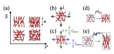
Balance of the generalized forces power-conjugate to also includes viscous, elastic-nematic and active contributions, and takes the form
| (4) |
where is a nematic viscous coefficient, the second term models alignment induced by strain rate (Fig. 1e) with the deviatoric part of the rate-of-deformation tensor given by , and and are susceptibility coefficients. The last term is an active generalized force controlled by activity parameter tending to further align filaments (Fig. 1d) [7]. This term is linear in because in the expansion the constant contribution can be subsumed by the susceptibility parameter . Thus, the active term acts as a negative density-dependent susceptibility. When , the system can sustain a uniform quiescent state with , and a non-zero nematic tensor satisfying . Even if and hence the uniform quiescent state is devoid of order, pattern formation can induce density variations such that becomes locally negative and actively favors local nematic order. The entropy production inequality requires that [35].
By freezing an isotropic state, , our model reduces to an orientation-independent active gel model, which develops periodic patterns driven by self-reinforcing active flows sustained by diffusion and turnover [29]. In the present model, however, translational, orientational and density dynamics are intimately coupled through the terms involving , , and .
We readily identify the hydrodynamic length , above which friction dominates over viscosity, the Damkölher length above which reactions dominate over diffusion, and the nematic length . Non-dimensional analysis reveals a set of non-dimensional groups that control the system behavior, namely the non-dimensional turnover rate , the Frank constant , the susceptibility parameters and , the drag coefficients and , the nematic activity coefficient , and the active tension parameters and (Section II of SI). The full list of material parameters for each figure given in Supplementary Tables (I,II) and justified in Section IV of SI.
Results
Onset and nature of pattern formation. To examine the role of nematic order in the emergence of various actin architectures, we performed linear stability analysis of our model particularized to 1D, whose dynamical variables are velocity, density and nematic order, , , and , along , where corresponds to a nematic orientation parallel (perpendicular) to the axis (Section I of SI). We first focused on the case to examine the loss of stability of a uniform, isotropic, and quiescent steady state (, , ) by increasing the master activity parameter and identifying the most unstable modes. This allowed us to determine a threshold activity for pattern formation and the wavelength of the emerging pattern. Since the exact evaluation of such quantities requires solving nonlinear equations, we derived explicit expansions in the limit of small for the critical contractile activity
| (5) |
where and , and for the the corresponding wavenumber
| (6) |
where .
The active origin of pattern formation in our model is evident from these expressions. When or , and hence , we recover the predictions of an active gel model not accounting for network architecture [29], and . However, active tension anisotropy () and flow-induced alignment () fundamentally change the nature of pattern formation. Nematic order introduces quantitative changes in critical tension and wavenumber, which can be very significant depending on the ratio of hydrodynamic and Damkölher lengths and on the strength and sign of nematic coupling. The nematic corrections increase as , close to the point where the uniform quiescent state develops spontaneous order. We thus studied separately the regime (Section III of SI), finding analogous expansions for the critical tension and wavenumber in terms of . Interestingly, Eq. (5) shows that the activity threshold is reduced, and hence pattern formation facilitated, when , i.e. when active tension is larger perpendicular to the nematic direction. Besides modifying critical tension and wavenumber, the present model predicts that the dynamical modes with self-reinforcing flows generate patterns where high density co-localizes with high nematic order.
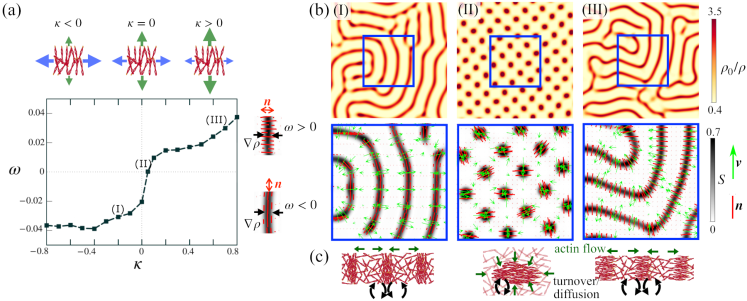
To test the validity of this analysis and further understand the system beyond the onset of pattern formation, we performed fully non-linear finite element simulations in a periodic 2D domain [35]. In these simulations, we increased the activity parameter beyond the instability starting from a quiescent uniform state. We found that the linear stability analysis very accurately predicts the activity thresholds and pattern wave-numbers within two percent across a wide range of parameters. In the nonlinear regime, the exponentially-growing instabilities eventually reach out-of-equilibrium quasi-steady-state patterns involving self-reinforcing flows towards regularly-spaced regions of high density surrounded by a low density matrix. In the absence of nematic coupling (), these high density domains are droplets arranged in a regular hexagonal lattice with throughout the domain, Movie 1. In contrast, for a generic parameter set with finite , and , high density domains adopt elongated configurations or bands where order is high, surrounded by a low density and low order matrix, Fig. 2b and Movie 2. Thus, the nematic active gel develops out-of-equilibrium localized states through a chemomechanical mechanism [51], which unlike those in [34], exhibit localization of both density and nematic order, and hence resemble dense nematic structures embedded in a low-density isotropic actin cortex. Furthermore, the shape and internal architecture of dense and nematic phases are qualitatively modified by the nematic coupling.
Our simulations show that self-reinforcing flows develop along the direction of largest active tension, and consequently the pattern architecture depends on the sign of , Fig. 2c. For , the system self-organizes into high-density and high-order bands, where nematic direction is parallel to their axis, in what we call fibrillar pattern, Fig. 2b(I). Instead, for nematic order is perpendicular to the axis of the bands, in what we call sarcomeric pattern, Fig. 2b(III). To systematically study the effect of active tension anisotropy, we varied between -0.8 and 0.8 while keeping all other non-dimensional groups fixed and setting to be 1.3 times the critical activity. We defined the order parameter , Fig. 2a, allowing us to distinguish between sarcomeric () and fibrillar () organizations. We found a sharp transition between fibrillar and sarcomeric regimes around , during which elongated high-density and high-order domains fragment into nematic droplets or tactoids [52, 53], Fig. 2b(II).
Our results for , leading to self-organized dense nematic fibrillar patterns from an isotropic low-density network, are in agreement with evidence suggesting that stress fibers can assemble from the actin cortex without the involvement of stress fiber precursors or actin polymerization at focal adhesions [20]. They also agree with observations showing that actin bundles form a mechanical continuum with the surrounding sparse and isotropic cortex [31]. Their morphology and patterning dynamics is strikingly reminiscent of actin microridges, formed at the apical surfaces of mucosal epithelial cells [55, 56]. Finally, we also note the similarity in terms of density and nematic architecture between our fibrillar patterns and those emerging in other active systems through different mechanisms of self-organization, including polar motile filaments [57, 58] or mean-field models of dry mixtures of microtubules and motors [59].
Requirements for fibrillar and sarcomeric patterns. At linear order, our theory shows that the distinctly nematic self-organization requires both flow-induced alignment () and active tension anisotropy (), whereas no condition is required on nematic activity (). We performed further simulations to establish the requirements for fibrillar and sarcomeric active patterning in the nonlinear regime. We found that both sarcomeric and fibrillar patterns readily form for and finite , yet a finite value of enhances fibrillar formation, leading to longer and more stable dense bands, and hinders sarcomeric organization, Movie 3. This behavior is expected since the velocity gradients of the self-reinforcing flows tend to align filaments parallel to high-density bands due to in Eq. (Theoretical model).
The active nematic coefficient modifies the onset of pattern formation through according to linear stability analysis. Apart from this linear effect, it should also contribute to condensation in high-density regions since it appears multiplied by in Eq. (Theoretical model). To examine this nonlinear effect, we perform simulations with but keeping fixed. This leads to very different patterns without clear elongated structures and very mild nematic patterning, Supplementary Figure 1(a). Enhancing nematic patterning by considering the largest thermodynamically allowed value of leads to elongated structures for , but rather than high-order co-localizing with high density, the nematic field develops domains with 90∘ angles between high- and low-density regions as in [34], Supplementary Figure 1(b), in an architecture enhanced by higher friction , Supplementary Figure 1(c). Thus, the architectures found for and are distinct from the fibrillar pattern described previously. Similarly, rather than sarcomeres, for , and high we found patterns of nematic asters (high-density droplets with radial nematic organization around them), Supplementary Figure 1(b,c).
Together, these results show that active tension anisotropy () and nematic activity () are necessary and sufficient for nematic self-organization into fibrillar or sarcomeric patterns, with flow-induced alignment () favoring fibrillar organization.
Morphological and dynamical diversity of self-organized fibrillar patterns. Given the morphological and dynamical diversity of nematic bundles in actin gels across cell types, geometric confinement, mechanical environment, or biological and pharmacological treatments [26, 27, 11, 60, 61, 62], we varied model parameters to examine the architectures predicted by our active gel model, focusing on . Significant changes in the effective parameters of our active gel model are reasonable since the active mechanical properties of actomyosin gels strongly depend on micro-architecture both in reconstituted systems and in cells [49, 63].
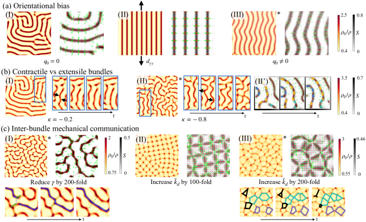
With our default parameter set, bundle junctions and free ends are unfavorable and reorganize during pattern formation to annihilate as much as possible, Movie 4(I). However, because the initial state of the system is isotropic but fibrillar patterns are not, this coarsening process leads to frustrated labyrinth patterns with domains and defects, which depending on parameters can remain frozen in quasi-steady states as in Fig. 3a(I). We then asked the question of whether an orientational bias, which physically may be caused by cytoskeletal flows, boundaries or directed polymerization [22], could direct the pattern and result in fewer defects. We first slightly modified the system by including a small anisotropic strain-rate, according to which friction is computed relative to an elongating background. This directional bias is sufficient to produce well-oriented defect-free patterns aligned with the direction of elongation, Fig. 3a(II) and Movie 4(II). Alternatively, we considered to be slightly negative, leading to the uniform and nematic steady state , and . The linear stability analysis around this state and further nonlinear simulations show that the essential phenomenology of Eqs. (5,6) and Fig. 2 is not altered by the slight pre-existing order (Section III of SI). Again, the weak pre-existing order provides sufficient bias to direct pattern orientation, Fig. 3a(III). Thus, our model indicates that an anisotropic bias can guide and anneal nematic fibrillar patterns, in agreement with remarkably oriented patterns of actin bundles in elongated cells [28], as a result of uniaxial cellular stretch [56, 19], or on anisotropically curved surfaces [21, 29].
At longer times, the nematic bundles in Fig. 3a(III) develop secondary active instabilities leading to coordinated bending, curvature amplification, defect nucleation and annihilation, Movie 4(III), in a behavior reminiscent of active extensile systems. This possibility is puzzling because our active gel is contractile. To systematically examine it, we performed simulations at higher active tension anisotropies , which we compared with our reference , Fig. 3b. While in our reference system bundles behave like contractile objects tending to straighten, for they behave like extensile objects enhancing curvature, which results in continuous defect nucleation as highly bent bundles destabilize and fragment, as well as defect annihilation as pairs of free ends merge to reorganize the network, in a behavior akin to active turbulence [64]. See Movie 5 for an illustration and for the slightly extensile case . To understand the origin of this behavior, we examined the individual components of the total tension, Eq. (2), along and perpendicular to the fibrillar pattern, Supplementary Fig. 2. Because in all of these simulations active contraction is larger perpendicular to nematic bundles () as required for their self-assembly, bundles are effectively extensile with regards to the active component of the total tension. Competing with active tension, however, viscous tension is negative and larger perpendicular to the bundles. Hence, depending on their relative magnitude, the total tension can be larger along or perpendicular to the bundles. We found that for contractile bundles leading to stable fibrillar patterns (), total tension is larger along bundles, indicating that a large fraction of the active power perpendicular to the bundles is dissipated in the self-reinforcing flows, and hence is not available to perform power at a mesoscale. Instead, for stronger active anisotropy (), total tension along nematic bundles is smaller than perpendicular to them, Supplementary Fig. 2(c), confirming the extensile nature of the self-organized bundles leading to the secondary wrinkling instability. In summary, our theory predicts that a contractile nematic active gel can self-organize into fibrillar patterns with mesoscale bundles that are either contractile or extensile depending on the parameter regime.
Focusing on contractile bundles, we then examined a different parameter regime known to trigger sustained pattern dynamics. For isotropic gels, previous work has shown that reducing friction triggers chaotic dynamics as the distance between high-density regions, , becomes comparable or smaller than the hydrodynamic length scale [29], thus enabling their mechanical interaction. In a model devoid of orientational order, reducing friction is equivalent to increasing . In our model, however, we can either reduce , which in non-dimensional terms means increasing , , and in concert, or increase while leaving all other non-dimensional parameters fixed. The first of these choices leads to dynamic and hierarchical networks with very dense and highly-aligned bundles, which coexist with perpendicular weak bundles with much lower density enrichment and ordering. These two families of bundles enclose cells of isotropic and sparse gel, Fig. 3c(I). Junctions where two or more dense bundles meet are very unfavorable and short-lived, but junctions of two dense and a weak bundle are much more stable. Because bundles are mechanically coupled, the networks actively reorganizes in events where dense bundles become weak bundles and vice-versa, inset and Movie 6(I). We note that here total tension along bundles is much larger than perpendicular to them, Supplementary Fig. 2(d), and hence the persistent dynamics are unrelated to the previously described behavior of extensile bundles.
The second choice to favor mechanical interaction of bundles, increasing only, leads to very different networks with high-density aster-like clusters interconnected by straight actin bundles. Because now is not particularly large, order is low at the core of these clusters, enabling high-valence networks where four bundles often meet at one cluster. For , the network is stable and nearly crystalline, Fig. 3b(II), whereas for , it becomes highly dynamical and pulsatile with frequent collapse of polygonal cells by fusion of neighboring actin clusters and their attached bundles (black polygons) and nucleation of new bundles within large low-density cells (dashed/solid green lines), Fig. 3b(III) and Movie 6(III). This architecture and dynamics resemble those of early C elegans embryos [65], adherent epithelial cells treated with epidermal growth factor [27] and mouse embryonic stem cells [61]. Recent active gel models accounting for RhoA signaling develop similar pulsatile behaviors in 2D, but do not predict the orientational order of the spatio-temporal patterns of the actomyosin cortex [66].
In summary, our theory maps how effective parameters of the actin gel control the active self-organization of a uniform and isotropic gel into a pattern of high-density nematic bundles embedded in a low-density isotropic matrix, including the activity threshold, the bundle spacing, orientation, connectivity and dynamics.

Microscopic origin of and through discrete network simulations. A somewhat counter-intuitive prediction of our model is that the self-organization of nematic bundles, the most prominent emerging organization in actin gels across cell types and length-scales, requires that active tension perpendicular to nematic orientation is larger than along this direction (), at least at the onset of pattern formation. While our continuum hydrodynamical model can address mesoscopic conditions for self-organization, it cannot provide insight about the microscopic origin of effective activity parameters. To examine whether the conditions and for spontaneous formation of fibrillar patterns are plausible from a microscopic point of view, we performed discrete network simulations using Cytosim [67].
Addressing the self-organization of the actin cytoskeleton at mesoscales directly with discrete network simulations is very challenging due to the wide range in time- and length-scales and the need to realistically model diffusion, network renewal, friction and gel hydrodynamics. Instead, we aimed at characterizing the out-of-equilibrium mechanical behavior of a representative volume element of the material with uniform mesoscopic properties, Fig. 4a. We performed 2D simulations in which semi-flexible filaments interact with crosslinkers and myosin motors, all of which undergo turnover and have a stoichiometry previously used to model the actin cytoskeleton [68], Fig. 4b. See Section V of SI for a detailed description of the simulation protocol. Briefly, we modified Cytosim to account for average orientational order in the simulation box, which we evaluated as a sample average of orientations over the ensemble of segments composing the filaments. We further introduced a nematic energy penalty in the network allowing us to restrain average nematic order to a target value .
We first prepared a system consisting only of randomly oriented actin fibers, imposed the desired orientational order using the nematic penalty and equilibrated the system. In a first set of simulations, once was reached, we deactivated the nematic penalty and added crosslinkers and myosins, driving the system out-of-equilibrium. The free contraction of the system was prevented by the addition of anchors at the boundary of the representative volume element, which also allowed us to compute anchor forces and hence estimate the effective active tension along the nematic direction and perpendicular to it, with and , Fig. 4(b).
Addition of crosslinkers and myosins leads to bundling of actin filaments at the microscale, Movie 7, distinct from the mesoscale fibrillar pattern formation emerging from the active gel model. It also leads to out-of-equilibrium tension as measured by the anchors. For isotropic networks (), active tension is isotropic with . For anisotropic networks, however, we found that tension becomes anisotropic with , Fig. 4(c).
We systematically characterized this behavior varying initial orientational order and network density. According to our active gel model, Eqs. (2,3), in the absence of nematic gradients and flow, the tension components and satisfy the following relations in terms of mean and deviatoric tensions
| (7) |
Remarkably, our discrete network simulations closely followed these relations, Fig. 4(d,e), which allowed us to estimate . We robustly found that for perturbations of selected parameters of the discrete network model as long as turnover rates of crosslinkers and myosins were relatively fast.
We then wondered if the discrete network simulations could provide evidence for the orientational activity parameter in our theory, . For a uniform system with nematic order along a given direction and ignoring the susceptibility parameter , Eq. (Theoretical model) becomes
| (8) |
where is the viscous drag of the filaments in the discrete network simulations, the entropic tendency of the model to return to isotropy, the active forcing of nematic order resulting from crosslinkers and motors, and the last term accounts for the effect of the nematic penalty with coefficient . As a first test of this model, we started from an isotropic and periodic network and tracked the athermal dynamics of under the action of the nematic penalty in the absence of anchors, crosslinkers and myosins. For and , Eq. (8) predicts an exponential relaxation given by , which very closely matched the simulation data for different values of and for a single fitting parameter , Supplementary Fig. 3(III) and Movie 8. We then deactivated the nematic penalty and added crosslinkers and motors, but not anchors, to track unconstrained dynamics of nematic order starting from different values of . In agreement with the notion of an active force driving nematic order, we found that monotonically increased, Fig. 4(f). More quantitatively, we tested the short-time prediction of Eq. (8), , by plotting as estimated from our simulations, as a function of , Fig. 4(g). We found a nearly linear relation with positive slope, hence providing evidence for an active generalized force driving order. In agreement with the theory, in the athermal limit, the tendency to actively increase order is faster as the entropic tendency to disorder is absent ().
In summary, discrete network cytoskeletal simulations provide a microscopic justification for two key ingredients of our active gel theory, namely that nematic order elicits (1) anisotropic active tensions, which can be larger perpendicular to the nematic direction (), and (2) active generalized forces driving further ordering.
Conclusions
We have developed a theory for the active self-organization of initially uniform and isotropic actin gels into various localized dense nematic architectures embedded in an isotropic matrix of low density. This model predicts a variety of emergent patterns involving asters, tactoids, and sarcomeric bands. More importantly, it identifies a wide parameter space where the active gel spontaneously develops patterns of dense nematic bundles, the most prominent nematic architecture across scales and cell types. We have characterized how the activity threshold, spacing, geometry, connectivity and dynamics these patterns depends on effective active gel parameters. Because these mesoscale parameters depend on the composition and dynamics of the network at the molecular scale, our results portray actin gels as responsive and reconfigurable active materials that cells can finely regulate. We have further shown that the spontaneous tendency of the gel to assemble bundle patterns can be directed via subtle cues. Thus, a combination of biochemical control of actin dynamics along with geometric, mechanical or biochemical guiding [69, 70] may explain the emergence and context-dependent organization of regular patterns of bundle networks, from sub-cellular to organism scales [25, 14, 19, 26, 27, 12, 22, 28, 21, 30]. Consistently, perturbations of myosin contractility have been shown to alter, disorder, or even prevent the formation patterns of parallel bundles in C elegans [19], whereas perturbations of actin polymerization in drosophila embryos impair the robust organization of actin bundle patterns at the cellular and organ scales by disrupting orientation and spacing, but not the tendency of the actomyosin cytoskeleton to form patterns of parallel bundles [21].
Our theory identifies two key requirements on activity parameters for the self-organization of patterns of nematic bundles, namely active tension anisotropy with larger tension perpendicular to the nematic direction and generalized active forces tending to increase nematic order. Although our discrete network simulations support these two conditions, we expect that in a different regime anisotropic tension may be larger along the nematic direction (). For instance, once bundles are dense and maximally aligned, the ability of the active nematic gel to perform active tension perpendicular to the nematic direction may saturate, while myosin motors may contract the gel along the nematic direction more effectively. The regime studied here explains the initial assembly of dense nematic bundles, but not their maturation to become highly contractile or viscoelastic as demonstrated for stress fibers depending on different isoforms of nonmuscle myosin II or on actin regulators such as zyxin [71, 72]. Our work thus suggests further experimental and computational work to establish a comprehensive mapping between molecular and mesoscale properties of the active gel, and hence a bridge between molecular regulation and emergent cytoskeletal organization.
Author contributions
WM, ATS and MA conceived the study and developed the active gel theory. WM, GV and ATS developed and implemented numerical methods. WM performed and analyzed active gel simulations. WM and MP performed and analyzed discrete network simulations. MP developed the code and simulation protocols for discrete network simulations. MDC contributed to linear stability analysis. WM and MA wrote the manuscript. ATS and MA supervised the study.
Acknowledgements
The authors acknowledge the support of the European Research Council (CoG-681434) and the Spanish Ministry for Science and Innovation (PID2019-110949GB-I00). WM acknowledges the La Caixa Fellowship and the European Union’s Horizon 2020 research and innovation program under the Marie Skłodowska-Curie action (GA 713637). MP acknowledges the support from the Spanish Ministry of Science and Innovation & NextGenerationEU/PRTR (PCI2021-122049-2B). MA acknowledges the Generalitat de Catalunya (ICREA Academia prize for excellence in research). MDC acknowledges funding from the Spanish Ministry for Science and Innovation through the Juan de la Cierva Incorporación fellowship IJC2018-035270-I. IBEC and CIMNE are recipients of a Severo Ochoa Award of Excellence.
References
- Banerjee et al. [2020] S. Banerjee, M. L. Gardel, and U. S. Schwarz, The actin cytoskeleton as an active adaptive material, Annual Review of Condensed Matter Physics 11, 421 (2020).
- Chugh and Paluch [2018] P. Chugh and E. K. Paluch, The actin cortex at a glance, Journal of Cell Science 131, jcs186254 (2018).
- Blaser et al. [2006] H. Blaser, M. Reichman-Fried, I. Castanon, K. Dumstrei, F. L. Marlow, K. Kawakami, L. Solnica-Krezel, C.-P. Heisenberg, and E. Raz, Migration of zebrafish primordial germ cells: a role for myosin contraction and cytoplasmic flow, Developmental Cell 11, 613 (2006).
- Ruprecht et al. [2015] V. Ruprecht, S. Wieser, A. Callan-Jones, M. Smutny, H. Morita, K. Sako, V. Barone, M. Ritsch-Marte, M. Sixt, R. Voituriez, and C.-P. Heisenberg, Cortical contractility triggers a stochastic switch to fast amoeboid cell motility, Cell, Cell 160, 673 (2015).
- Blanchoin et al. [2014] L. Blanchoin, R. Boujemaa-Paterski, C. Sykes, and J. Plastino, Actin dynamics, architecture, and mechanics in cell motility, Physiological Reviews 94, 235 (2014).
- Schwayer et al. [2016] C. Schwayer, M. Sikora, J. Slováková, R. Kardos, and C.-P. Heisenberg, Actin rings of power, Developmental Cell 37, 493 (2016).
- Reymann et al. [2016] A.-C. Reymann, F. Staniscia, A. Erzberger, G. Salbreux, and S. W. Grill, Cortical flow aligns actin filaments to form a furrow, eLife 5, e17807 (2016).
- Mandato and Bement [2001] C. A. Mandato and W. M. Bement, Contraction and polymerization cooperate to assemble and close actomyosin rings around xenopus oocyte wounds, The Journal of cell biology 154, 785 (2001).
- Martin and Lewis [1992] P. Martin and J. Lewis, Actin cables and epidermal movement in embryonic wound healing, Nature 360, 179 (1992).
- Krieg et al. [2008] M. Krieg, Y. Arboleda-Estudillo, P. H. Puech, J. Käfer, F. Graner, D. J. Müller, and C. P. Heisenberg, Tensile forces govern germ-layer organization in zebrafish, Nature Cell Biology 10, 429 (2008).
- Dudin et al. [2019] O. Dudin, A. Ondracka, X. Grau-Bové, A. A. Haraldsen, A. Toyoda, H. Suga, J. Bråte, and I. Ruiz-Trillo, A unicellular relative of animals generates a layer of polarized cells by actomyosin-dependent cellularization, eLife 8, e49801 (2019).
- Senger et al. [2019] F. Senger, A. Pitaval, H. Ennomani, L. Kurzawa, L. Blanchoin, and M. Théry, Spatial integration of mechanical forces by alpha-actinin establishes actin network symmetry, Journal of Cell Science 132, 10.1242/jcs.236604 (2019), jcs236604.
- Tojkander et al. [2012] S. Tojkander, G. Gateva, and P. Lappalainen, Actin stress fibers–assembly, dynamics and biological roles, Journal of Cell Science 125, 1855 (2012).
- Tojkander et al. [2015] S. Tojkander, G. Gateva, A. Husain, R. Krishnan, and P. Lappalainen, Generation of contractile actomyosin bundles depends on mechanosensitive actin filament assembly and disassembly, eLife 4, e06126 (2015).
- Chrzanowska-Wodnicka and Burridge [1996] M. Chrzanowska-Wodnicka and K. Burridge, Rho-stimulated contractility drives the formation of stress fibers and focal adhesions., The Journal of Cell Biology 133, 1403 (1996).
- Thoresen et al. [2011] T. Thoresen, M. Lenz, and M. L. Gardel, Reconstitution of contractile actomyosin bundles, Biophysical Journal 100, 2698 (2011).
- Strehle et al. [2011] D. Strehle, S. Jorg, C. Heussinger, J. Alvarado, M. Bathe, J. Kas, and B. Gentry, Transiently crosslinked f-actin bundles, European Biophysics Journal 40, 93 (2011).
- Laporte et al. [2012] D. Laporte, N. Ojkic, D. Vavylonis, and J.-Q. Wu, -actinin and fimbrin cooperate with myosin ii to organize actomyosin bundles during contractile-ring assembly, Molecular Biology of the Cell (MBoC) 23, 3094 (2012).
- Wirshing and Cram [2017] A. C. Wirshing and E. J. Cram, Myosin activity drives actomyosin bundle formation and organization in contractile cells of the caenorhabditis elegans spermatheca, Molecular Biology of the Cell (MBoC) 28, 1937 (2017).
- Lehtimäki et al. [2021] J. I. Lehtimäki, E. K. Rajakylä, S. Tojkander, and P. Lappalainen, Generation of stress fibers through myosin-driven reorganization of the actin cortex, eLife 10, e60710 (2021).
- Öztürk-Çolak et al. [2016] A. Öztürk-Çolak, B. Moussian, S. J. Araújo, and J. Casanova, A feedback mechanism converts individual cell features into a supracellular ecm structure in Drosophila trachea, eLife 5, e09373 (2016).
- Hotulainen and Lappalainen [2006] P. Hotulainen and P. Lappalainen, Stress fibers are generated by two distinct actin assembly mechanisms in motile cells, The Journal of cell biology 173, 383 (2006).
- Naumanen et al. [2008] P. Naumanen, P. Lappalainen, and P. Hotulainen, Mechanisms of actin stress fibre assembly, Journal of Microscopy 231, 446 (2008).
- Lee et al. [2018] S. Lee, E. Kassianidou, and S. Kumar, Actomyosin stress fiber subtypes have unique viscoelastic properties and roles in tension generation, Molecular Biology of the Cell 29, 1992 (2018).
- Tee et al. [2015] Y. H. Tee, T. Shemesh, V. Thiagarajan, R. F. Hariadi, K. L. Anderson, C. Page, N. Volkmann, D. Hanein, S. Sivaramakrishnan, M. M. Kozlov, et al., Cellular chirality arising from the self-organization of the actin cytoskeleton, Nature Cell Biology 17, 445 (2015).
- Yolland et al. [2019] L. Yolland, M. Burki, S. Marcotti, A. Luchici, F. N. Kenny, J. R. Davis, E. Serna-Morales, J. Müller, M. Sixt, A. Davidson, et al., Persistent and polarized global actin flow is essential for directionality during cell migration, Nature Cell Biology 21, 1370 (2019).
- Jalal et al. [2019] S. Jalal, S. Shi, V. Acharya, R. Y.-J. Huang, V. Viasnoff, A. D. Bershadsky, and Y. H. Tee, Actin cytoskeleton self-organization in single epithelial cells and fibroblasts under isotropic confinement, Journal of Cell Science 132, jcs220780 (2019).
- Dinwiddie et al. [2014] A. Dinwiddie, R. Null, M. Pizzano, L. Chuong, A. Leigh Krup, H. Ee Tan, and N. H. Patel, Dynamics of f-actin prefigure the structure of butterfly wing scales, Developmental Biology 392, 404 (2014).
- Hannezo et al. [2015] E. Hannezo, B. Dong, P. Recho, J.-F. Joanny, and S. Hayashi, Cortical instability drives periodic supracellular actin pattern formation in epithelial tubes, Proceedings of the National Academy of Sciences 112, 8620 (2015).
- Maroudas-Sacks et al. [2021] Y. Maroudas-Sacks, L. Garion, L. Shani-Zerbib, A. Livshits, E. Braun, and K. Keren, Topological defects in the nematic order of actin fibres as organization centres of hydra morphogenesis, Nature Physics 17, 251 (2021).
- Vignaud et al. [2021] T. Vignaud, C. Copos, C. Leterrier, M. Toro-Nahuelpan, Q. Tseng, J. Mahamid, L. Blanchoin, A. Mogilner, M. Théry, and L. Kurzawa, Stress fibres are embedded in a contractile cortical network, Nature materials 20, 410 (2021).
- Salbreux et al. [2009] G. Salbreux, J. Prost, and J.-F. Joanny, Hydrodynamics of cellular cortical flows and the formation of contractile rings, Physical Review Letters 103, 058102 (2009).
- Jülicher et al. [2018] F. Jülicher, S. W. Grill, and G. Salbreux, Hydrodynamic theory of active matter, Reports on Progress in Physics 81, 076601 (2018).
- Zumdieck et al. [2005] A. Zumdieck, M. C. Lagomarsino, C. Tanase, K. Kruse, B. Mulder, M. Dogterom, and F. Jülicher, Continuum description of the cytoskeleton: Ring formation in the cell cortex, Phys. Rev. Lett. 95, 258103 (2005).
- Mirza et al. [2023] W. Mirza, A. Torres-Sánchez, G. Vilanova, and M. Arroyo, Variational formulation of active nematics: theory and simulation, arXiv (2023), 2306.01515 [physics.bio-ph] .
- e Silva et al. [2011] M. S. e Silva, J. Alvarado, J. Nguyen, N. Georgoulia, B. M. Mulder, and G. H. Koenderink, Self-organized patterns of actin filaments in cell-sized confinement, Soft Matter 7, 10631 (2011).
- Doostmohammadi et al. [2018] A. Doostmohammadi, J. Ignés-Mullol, J. M. Yeomans, and F. Sagués, Active nematics, Nature Communications 9, 1 (2018).
- Bois et al. [2011] J. S. Bois, F. Jülicher, and S. W. Grill, Pattern formation in active fluids, Physical Review Letters 106, 028103 (2011).
- Callan-Jones and Voituriez [2013] A. Callan-Jones and R. Voituriez, Active gel model of amoeboid cell motility, New Journal of Physics 15, 025022 (2013).
- Giomi et al. [2014] L. Giomi, M. J. Bowick, P. Mishra, R. Sknepnek, and M. Cristina Marchetti, Defect dynamics in active nematics, Philosophical Transactions of the Royal Society A: Mathematical, Physical and Engineering Sciences 372, 20130365 (2014).
- Duclos et al. [2017] G. Duclos, C. Erlenkämper, J.-F. Joanny, and P. Silberzan, Topological defects in confined populations of spindle-shaped cells, Nature Physics 13, 58 (2017).
- Kumar et al. [2018] N. Kumar, R. Zhang, J. J. de Pablo, and M. L. Gardel, Tunable structure and dynamics of active liquid crystals, Science Advances 4, eaat7779 (2018).
- de Gennes and Prost [1993] P. de Gennes and J. Prost, The Physics of Liquid Crystals, International Series of Monogr (Clarendon Press, 1993).
- Harris et al. [2006] E. S. Harris, I. Rouiller, D. Hanein, and H. N. Higgs, Mechanistic differences in actin bundling activity of two mammalian formins, frl1 and mdia2, Journal of Biological Chemistry 281, 14383 (2006).
- Courson and Rock [2010] D. S. Courson and R. S. Rock, Actin cross-link assembly and disassembly mechanics for -actinin and fascin, Journal of Biological Chemistry 285, 26350 (2010).
- Schuppler et al. [2016] M. Schuppler, F. C. Keber, M. Kröger, and A. R. Bausch, Boundaries steer the contraction of active gels, Nature Communications 7, 1 (2016).
- Li et al. [2017] J. Li, T. Biel, P. Lomada, Q. Yu, and T. Kim, Buckling-induced f-actin fragmentation modulates the contraction of active cytoskeletal networks, Soft Matter 13, 3213 (2017).
- Nandi [2018] S. K. Nandi, Activity-dependent self-regulation of viscous length scales in biological systems, Physical Review E 97, 052404 (2018).
- Ennomani et al. [2016] H. Ennomani, G. Letort, C. Guérin, J.-L. Martiel, W. Cao, F. Nédélec, E. M. D. L. Cruz, M. Théry, and L. Blanchoin, Architecture and connectivity govern actin network contractility, Current Biology 26, 616 (2016).
- Chen et al. [2020] S. Chen, T. Markovich, and F. C. MacKintosh, Motor-free contractility in active gels, Phys. Rev. Lett. 125, 208101 (2020).
- Barberi and Kruse [2023] L. Barberi and K. Kruse, Localized states in active fluids (2023), arXiv:2209.02581 [physics.bio-ph] .
- Weirich et al. [2017] K. L. Weirich, S. Banerjee, K. Dasbiswas, T. A. Witten, S. Vaikuntanathan, and M. L. Gardel, Liquid behavior of cross-linked actin bundles, Proceedings of the National Academy of Sciences 114, 2131 (2017).
- Weirich et al. [2019] K. L. Weirich, K. Dasbiswas, T. A. Witten, S. Vaikuntanathan, and M. L. Gardel, Self-organizing motors divide active liquid droplets, Proceedings of the National Academy of Sciences 116, 11125 (2019).
- Deshpande and Pfohl [2015] S. Deshpande and T. Pfohl, Real-time dynamics of emerging actin networks in cell-mimicking compartments, PloS one 10, e0116521 (2015).
- Depasquale [2018] J. A. Depasquale, Actin microridges, The Anatomical Record 301, 2037 (2018).
- van Loon et al. [2020] A. P. van Loon, I. S. Erofeev, I. V. Maryshev, A. B. Goryachev, and A. Sagasti, Cortical contraction drives the 3D patterning of epithelial cell surfaces, Journal of Cell Biology 219, 10.1083/jcb.201904144 (2020), e201904144.
- Huber et al. [2018] L. Huber, R. Suzuki, T. Krüger, E. Frey, and A. R. Bausch, Emergence of coexisting ordered states in active matter systems, Science 361, 255 (2018).
- Denk and Frey [2020] J. Denk and E. Frey, Pattern-induced local symmetry breaking in active-matter systems., Proc Natl Acad Sci U S A 117, 31623 (2020).
- Maryshev et al. [2019] I. Maryshev, A. B. Goryachev, D. Marenduzzo, and A. Morozov, Dry active turbulence in a model for microtubule–motor mixtures, Soft Matter 15, 6038 (2019).
- Gupta et al. [2015] M. Gupta, B. R. Sarangi, J. Deschamps, Y. Nematbakhsh, A. Callan-Jones, F. Margadant, R.-M. Mège, C. T. Lim, R. Voituriez, and B. Ladoux, Adaptive rheology and ordering of cell cytoskeleton govern matrix rigidity sensing, Nature Communications 6, 1 (2015).
- Xia et al. [2019] S. Xia, Y. B. Lim, Z. Zhang, Y. Wang, S. Zhang, C. T. Lim, E. K. Yim, and P. Kanchanawong, Nanoscale architecture of the cortical actin cytoskeleton in embryonic stem cells, Cell Reports 28, 1251 (2019).
- Verkhovsky et al. [1997] A. Verkhovsky, T. Svitkina, and G. Borisy, Polarity sorting of actin filaments in cytochalasin-treated fibroblasts, Journal of Cell Science 110, 1693 (1997).
- Chugh et al. [2017] P. Chugh, A. G. Clark, M. B. Smith, D. A. Cassani, K. Dierkes, A. Ragab, P. P. Roux, G. Charras, G. Salbreux, and E. K. Paluch, Actin cortex architecture regulates cell surface tension, Nature cell biology 19, 689 (2017).
- Alert et al. [2022] R. Alert, J. Casademunt, and J.-F. Joanny, Active turbulence, Annual Review of Condensed Matter Physics 13, 143 (2022).
- Munro et al. [2004] E. Munro, J. Nance, and J. R. Priess, Cortical flows powered by asymmetrical contraction transport par proteins to establish and maintain anterior-posterior polarity in the early c. elegans embryo, Developmental Cell 7, 413 (2004).
- Staddon et al. [2022] M. F. Staddon, E. M. Munro, and S. Banerjee, Pulsatile contractions and pattern formation in excitable actomyosin cortex, PLOS Computational Biology 18, 1 (2022).
- Nedelec and Foethke [2007] F. Nedelec and D. Foethke, Collective langevin dynamics of flexible cytoskeletal fibers, New Journal of Physics 9, 427 (2007).
- Cortes et al. [2020] D. B. Cortes, M. Gordon, F. Nédélec, and A. S. Maddox, Bond type and discretization of nonmuscle myosin ii are critical for simulated contractile dynamics, Biophysical Journal 118, 2703 (2020).
- Gross et al. [2017] P. Gross, K. V. Kumar, and S. W. Grill, How active mechanics and regulatory biochemistry combine to form patterns in development, Annual Review of Biophysics 46, 337 (2017), pMID: 28532214.
- Burkart et al. [2022] T. Burkart, M. C. Wigbers, L. Würthner, and E. Frey, Control of protein-based pattern formation via guiding cues, Nature Reviews Physics 4, 511 (2022).
- Weißenbruch et al. [2021] K. Weißenbruch, J. Grewe, M. Hippler, M. Fladung, M. Tremmel, K. Stricker, U. S. Schwarz, and M. Bastmeyer, Distinct roles of nonmuscle myosin ii isoforms for establishing tension and elasticity during cell morphodynamics, eLife 10, e71888 (2021).
- Oakes et al. [2017] P. W. Oakes, E. Wagner, C. A. Brand, D. Probst, M. Linke, U. S. Schwarz, M. Glotzer, and M. L. Gardel, Optogenetic control of rhoa reveals zyxin-mediated elasticity of stress fibres, Nature Communications 8, 15817 (2017).