Large saturation moment and high ferromagnetic transition temperature in a structurally disordered inverse Heusler alloy Fe2RuGe
Abstract
We report the successful synthesis of a new 4 based polycrystalline inverse Heusler alloy Fe2RuGe by an arc melting process and have studied in detail its structural, magnetic and transport properties complemented with first principle calculations. X-ray and neutron diffraction, Extended X-ray Absorption Fine Structure and 57Fe Mössbauer spectroscopic studies confirm the single phase nature of the system where the Fe and Ru atoms are randomly distributed in the 4 and 4 Wyckoff positions in a ratio close to 50:50. The formation of the disordered structure is also confirmed by the theoretical energy minimization calculation. Despite the random cross-site disorder of Fe and Ru atoms, magnetic measurements suggest not only a high Curie temperature of 860 K, but also a large saturation magnetic moment 4.9 per formula unit at 5 K, considerably exceeding the theoretical limit (4 per formula unit) predicted by the Slater-Pauling rule. Only a few Fe-based inverse Heusler alloys are known to exhibit such high Curie temperatures. Neutron diffraction analysis coupled with the isothermal magnetization value indicates that the magnetic moments in Fe2RuGe are associated with Fe-atoms only, which is also confirmed by Mössbauer spectrometry. Interestingly, in comparison to the cubic or hexagonal phase of the parent compound, Fe3Ge, the Curie temperature of Fe2RuGe has increased significantly despite the substitution of the nonmagnetic, yet isoelectronic element Ru in this structurally disordered compound. Our theoretical calculation reveals that the large Fe moment (/Fe) on the 4 site can be attributed to a charge transfer from this Fe site towards its Ru neighbours. Such a substantial increase in magnetic moment due to electron charge transfer has not previously been reported in a Heusler alloy system.
I Introduction
Heusler alloys (HA) are known to be a series of enigmatic compounds that are often known to exhibit high ferromagnetic transition temperatures, even when all individual constituents are nonmagnetic [1, 2]. HAs have a general chemical formula of , where and are transition metal atoms and is generally a main group element atom. They crystallize in the Cu2MnAl-type or -type structure (). Most HAs are highly ordered cubic intermetallic compounds with three crystallographic positions in their crystal lattice, viz. 4 (0,0,0), 4 (,,) and 8 (,,) [1]. It is generally seen that the occupancy of an atom at a particular site is determined by its number of valence electrons. Those with more valence electrons () tend to occupy the 4 and 4 positions, while atoms with fewer valence electrons () tend to occupy the 4 position, which is generally known as the valence electron number rule [3]. However, there is also a subclass of HAs, where and one of the atoms interchange their occupancy, and such systems are known as inverse HA, forming in the XA-type structure, also known as the Hg2CuTi structure [4, 5]. This latter crystallises in space group with a splitting of sites from into two individual and sites. Another fascinating feature of HA system is the structural tunability, as many different combinations of 3, 4 and 5 transition metal elements can be used in the and positions. Interestingly, one may realize that the choice of constituent elements and their occupancies determines of the electronic band structure, which in turn controls most of the physical properties of these materials. As a result, HAs remain a very attractive system for appropriate crystal engineering [6, 7, 8, 9].
The high Curie temperature and large saturation magnetic moment displayed by Heusler alloys recently got renewed attention due to their multi-functional properties viz. Half metallic ferromagnetism (HMF) [10, 11, 12], semi-metallic behavior [3], magnetic shape memories [13, 14], magnetic spin valve effects [15], topological insulators [16, 17], magnetic skyrmions [18, 19], spin-gapless semiconductors [20, 21], etc., as all these properties could be used in many practical applications developed recently. For example, spintronics devices are considered to have huge advantages over conventional electronic devices due to their high speed for data processing, non-volatile nature and reduced power consumption rate [22]. It is generally seen that HA with the combination of both Co and Fe is particularly good for obtaining a high magnetic moment and elevated Curie temperature. A substantial number of Co-based systems are particularly known to exhibit HMF behaviour and could be used for spintronic-related applications [11] [23]. On the other hand, although Fe-based HAs are rarely known to show such features, they have an enormous potential to be used as economically viable rare-earth free permanent magnet. In the quest for such new permanent magnetic material, the iron-rich compound, Fe3Ge turns out to be one of the most remarkable systems [24]. The compound could be considered as the binary equivalent of Heusler alloy, where both the and atoms are identical. Very interestingly, the compound is found to be stable at room temperature in two polymorphic crystal structures, viz., a cubic phase (Cu3Au type, , space group no. 221) formed by annealing at low temperatures ( 1073 K) and a hexagonal phase (Mg3Cd type, , space group no. 194) stabilized when the system is annealed at higher temperatures (1073 K) [24]. Although both phases exhibit high Curie temperatures (655 K for the hexagonal phase and 755 K for the cubic phase) and large magnetic moments (/f.u.) at room temperature, the anisotropic magnetic properties of the hexagonal phase have generated considerable interest in its use as a permanent magnet. A lot of attempts have been made to modify the magnetic anisotropy by partially replacing Fe atoms with other 3 elements, viz., V, Cr, Mn, Co and Ni [25, 26, 27, 4]. It has been found that depending on the use of heavier or lighter 3 element substitution, the structural as well as magnetic properties can vary widely. Both Fe2CoGe and Fe2NiGe form in cubic inverse Heusler alloy structure [4], while Fe3-xCrxGe ( 0.70) and Fe2MnGe retain the hexagonal crystal structure [26] [27]. Fe3-xVxGe remains hexagonal for 0.125, but changes to cubic for 0.125 0.75 [25]. The ferromagnetic Curie temperature for Fe2CoGe ( = 925 K) and Fe2NiGe ( = 760 K) is also higher than that of both the hexagonal as well as cubic phases of the parent compound, Fe3Ge. In addition, although Co and Ni are considered magnetic elements, the magnetic moment is confined only to Fe in Fe2NiGe, while both Fe and Co carry magnetic moment in Fe2CoGe. On the other hand, the Curie temperature decreases in case of substitution by 3 elements lighter than Fe [25, 26, 27, 4].
In light of such contrasting magnetic properties of FeGe depending on the electronic configuration of 3 -element, it would be interesting to understand the role of the valence electron and the electronic band structure in this system. The substitution of isoelectronic elements belonging to the 4 (Ru) or 5 (Os) series would be highly helpful, despite the fact that both are non-magnetic and isoelectronic to Fe, and are also susceptible to structural disorder. In this work, we report the successful synthesis of a new inverse Heusler alloy Fe2RuGe and explored its structural and physical properties through different experimental and theoretical techniques. Two different spectroscopic techniques, viz., 57Fe Mössbauer spectrometry and Extended X-ray Absorption Fine Structure (EXAFS) have been adopted to investigate the local structural disorders, While neutron diffraction studies have been carried out to investigate the magnetic spin arrangements. The electronic band structure calculation is also presented to complement the experimental results.
II Methods
II.1 Experimental details
The Fe2RuGe polycrystalline ingot was prepared by the standard arc melting method in argon atmosphere on water-cooled Cu hearth. A stoichiometric amount of Fe chunk (99.9%), Ru powder (99.9%) and Ge lump (99.999%) were used as starting materials. All the elements were melted and re-melted together for 5 times after flipping each time to improve homogeneity. A negligible amount (0.6%) of weight loss was recorded after the melting process. X-ray diffraction (XRD) measurement at room temperature was carried out using a commercial diffractometer (rotating Cu anode, 9 kW, Model: TTRAX-III, Rigaku Corp., Japan). The single-phase nature of the as-cast sample was confirmed by Rietveld refinement using FULLPROF software [28]. Microstructural analysis and compositional homogeneity were further investigated using a wavelength dispersive electron probe microanalysis (EPMA) spectrometer [Model: SX 100, Cameca, France].
Extended X-ray Absorption Fine Structure (EXAFS) experiments on Fe2RuGe were carried out at the Indus-2 Synchrotron Source (2.5 GeV, 100 mA) at the Energy-Scanning EXAFS beamline (BL-9) at Raja Ramanna Centre for Advanced Technology (RRCAT), Indore, India which operates in the energy range of 4 - 25 keV. Both transmission and fluorescent modes can be used to perform EXAFS measurements in this beamline. A Rh/Pt coated collimating meridional cylindrical mirror is used and the collimated beam is reflected by a mirror that is monochromatized by a double crystal monochromator (DCM) of Si(111) (2d = 6.2709 Å). For horizontal focusing, a sagittal cylinder (the second crystal of DCM) is used, and for vertical focusing of the beam, a Rh/Pt coated bendable post mirror facing down at the sample position is employed. The higher harmonics content of the X-ray beam is rejected by detuning the second crystal of DCM. For EXAFS measurements, we have used the fluorescence mode. In this method, the incident X-ray beam angle is 45 degrees to the sample position, and a fluorescence detector, positioned at 90 degrees to the incident X-ray beam, is used to collect the signal. To measure the incident flux (I0), an ionization chamber detector is placed before the sample, and fluorescence intensity (If) is measured by a fluorescence detector. The relation, = (/), is used to calculate the X-ray absorption coefficient of the sample. The spectrum (as a function of energy) was acquired by scanning the monochromator over a particular range. In order to extract qualitative information about the local structure, the oscillations of the normalized absorption spectra () were transformed to absorption function using the relation:
| (1) |
here, is the absorption edge energy, is the background of exposed atom and is the step in value at the absorption edge. The absorption coefficient as a function of energy , was changed to the absorption coefficient as a function of wave number by the following equation,
| (2) |
here, is the mass of the electron. To intensify the oscillation at high , it is multiplied by and then to obtain the plots, a Fourier transformation of functions into space, regarding to the real distances calculated from the centre of the absorbing atom is done. The set of EXAFS data was analyzed by the program available in the Demeter software package. Background reduction, Fourier transformation of the absorption spectra to derive plots (using ATHENA code) [29], simulation of the theoretical plots from the assumed crystallographic structure, and finally the fitting of experimental data using the theoretically generated spectra (by ARTEMIS code) were also included in this package [29].
Isothermal magnetisation measurements at low temperatures were performed in commercially available SQUID VSM (Quantum Design Inc., USA). Magnetic measurements in the temperature range 300 - 900 K were done in a high-temperature VSM equipped with an electromagnet (Model EV9, MicroSense, LLC Corp., USA).
Room temperature, as well as high-temperature neutron diffraction experiments ( = 2.4395 Å), were performed at ECHIDNA beam line in ANSTO, Sydney, Australia [30]. The details of the crystal structure as well as the analysis of the magnetic structure were performed by Rietveld refinement of neutron diffraction data using the FULLPROF software.
57Fe transmission Mössbauer spectrometry was used to study the hyperfine structure at Fe sites. The spectra were obtained at 300 K and 77 K in a bath cryostat and using an electromagnetic transducer with a triangular velocity nature and a 57Co source in a Rh matrix. The sample consists of a thin powdered and homogeneous layer containing about 5 mg-Fe/cm2. The least-squares fitting method, which assumes quadrupolar doublets and/or magnetic sextets involving Lorentzian lines, has been applied to model the hyperfine structures using the MOSFIT program. The refined isomer shift values are corrected to that of -Fe at 300 K, and an -Fe standard was used to calibrate the velocity.
Resistivity experiments using a standard four-probe method were performed in the temperature range 2 - 300 K using a PPMS Evercool-II instrument, (Quantum Design Inc., USA).
II.2 Computational methods
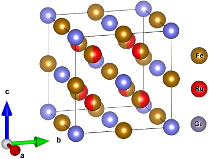
For the modelling part, the , inverse XA and disordered structures of Heusler alloys have been described in the same space group having both 4c and 4d positions (splitting of site in ), for a fuller discussion of site comparison.
The calculations of enthalpy of formation, electronic structure and spin polarisation at 0 K were carried out using density functional theory (DFT). They were conducted using the projector augmented wave (PAW) method [31] implemented in the Vienna ab initio simulation package (VASP) [32, 33]. The exchange-correlation was described by the generalized gradient approximation modified by Perdew, Burke and Ernzerhof (GGA-PBE) [34]. Energy bands up to a cutoff of = 600 eV were used in all calculations and the convergence tolerance for the calculations was selected as a difference on the total energy within 1 10-6 eV/atom. For each structure, volume and ionic (for disordered compounds) relaxation steps were performed and the tetrahedron method with Blöchl correction [35] was applied. Spin-polarization calculations were considered for all the structures. In order to statistically simulate the chemical disorder in Fe2RuGe, unit cells based on the concept of special quasirandom structure (SQS) [36] were generated for different possible disorder schemes. To generate the SQS, the cluster expansion formalism for multicomponent and multisublattice systems [37] was used as implemented in the Monte-Carlo (MCSQS) code contained in the Alloy-Theoretic Automated Toolkit (ATAT) [38, 39]. Subsequent DFT calculations were performed in order to test the quality of the SQS and to see how reliable the DFT results are. The root mean square (rms) error on correlation functions was used as another quality criterion in addition to the calculations including a different order of interactions. The error describes the deviation of the correlation function of the SQS () from the correlation function of a fully random structure () for all clusters .
| (3) |
Several tests on the dependence of the type and number of clusters were performed to generate the disordered structure: Ge at 4 (0,0,0), Fe at 4 (,,) and Fe = 0.5/Ru = 0.5 at 4 (,,) and Fe = 0.5 & Ru = 0.5 at 4 (,,). Finally, 7 pairs, 5 triplets and 11 quadruplets interactions were considered to obtain reliable results. Using these parameters, a disordered quaternary SQS cell of 28 atoms was generated.
III Results and Discussions
III.1 Structural Characterization
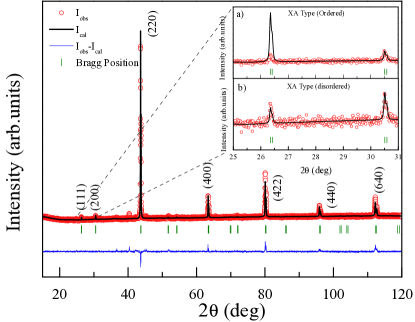
The powder XRD data taken at room temperature is presented in Fig. 1. With the exception of very few small peaks (2% of main peak intensity), all the peaks in the XRD pattern can be well indexed with a Hg2CuTi-type (space group , No - 216) inverse Heusler (XA-type) crystal structure with lattice parameter = 5.871(4) Å. It may be noted that Heusler alloys are prone to structural disorder, and despite the formation in cubic structure, such atomic disorders are known to affect the intensity of the (111) and (200) superlattice Bragg peaks in the XRD patterns. For example, in -type disorder (all atoms mixed on a single site), both (111) and (200) peaks are absent, while in -type disorder (atoms mixed on 2 sites), only the (200) peak is present [3] [12]. The occurrence of both (111) and (200) lines in Fig. 2 allows discarding or type of disorder. Our attempt to describe the crystal structure with an ordered variant (Ge occupy 4 position, Fe at 4, Fe at 4 and Ru in 4 site) strongly undermined the experimentally observed (111) peak intensity (Fig. 1, Inset), suggesting a disordered structure of different variant. Accordingly, we tried various combinations of site-disorders and obtained the best result by considering a 50% permutation between Fe and Ru in the tetrahedral 4 and 4 sites (Fig. 2).
The Rietveld analysis, considering the above model, can fit the XRD pattern quite well (Fig. 1). The trace amount of the minority phase turns out to be that of FeGe2 of less than 2% of the weight fraction of the primary phase. The structural disorder is further confirmed by the EXAFS and neutron diffraction data analysis which will be discussed in later sections. This result is also in conformity with the theoretical calculations establishing that the disordered XA structure is more stable than the ordered one.
III.2 EXAFS analysis
| Fe edge | Ru edge | ||||||||||
| Path | R (Å) | N | Path | R (Å) | N | ||||||
| Fe-Ru | 2.53 ±0.01 | 4 |
|
Ru-Ge | 2.53 ±0.01 | 4 |
|
||||
| Fe-Fe | 2.48±0.01 | 4 |
|
Ru-Fe | 2.53 ±0.01 | 4 |
|
||||
| Fe-Ge | 2.48±0.01 | 3 |
|
Ru-Ru | 2.88 ±0.01 | 2.3 |
|
||||
| Fe-Ru | 2.89±0.01 | 1.7 |
|
Ru-Fe | 2.88 ±0.01 | 3.7 |
|
||||
| Fe-Fe | 2.88±0.01 | 4.3 |
|
Ru-Ru | 4.07±0.03 | 5.1 |
|
||||
| Fe-Ge | 3.11±0.01 | 6 |
|
Ru-Fe | 4.07±0.03 | 6.9 |
|
||||
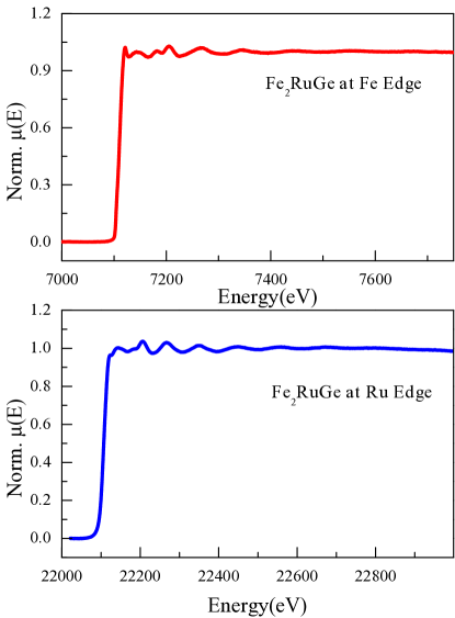
The normalized EXAFS () spectra of Fe2RuGe at the Fe and Ru edges, following the treatment presented in Eq. 1 and Eq. 7, are shown in Fig. 3. The plots (Fourier transformed EXAFS spectra) generated by the crystallographic information and the EXAFS equation of the Fe and Ru edge is shown in Fig. 4. Simultaneous fitting with several edges of various data sets is used in this approach. The plots calculated at the Ru and Fe edges are fitted at the same time with common fitting parameters. The statistical significance of the fitting is improved by reducing the number of independent parameters below the Nyquist criterion. The value of was used to determine the goodness of fit, which is calculated by:
| (4) |
where and are the theoretical and experimental values, respectively. and are the real and imaginary components of the respective quantities. To theoretically simulate the EXAFS spectra of Fe2RuGe, the structural information (lattice parameters and nearest neighbour numbers) have been taken from the XRD results. The atomic distances (R), coordination numbers (N), and disorder (Debye-Waller) factors (), which give the mean square variations of the distances, were used as fitting parameters. The fitting was done up to 4 Å. A simultaneous fitting of the Ru and Fe edges could be obtained for the disordered structure only. The refined values of the parameters are listed in Table 1.
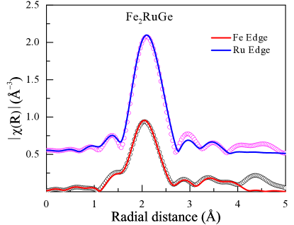
From the fitting of Fe edge of the Fourier transformed spectra, we found that the main peak near 2 Å has a contribution from all Fe-Fe (2.48 Å), Fe-Ru (2.53 Å) and Fe-Ge (2.48 Å) distances. The second intense peak near 3 Å arises due to the contribution of Fe-Ru (2.89 Å) and Fe-Fe (2.89 Å) and the peak near 4 Å consists of the contribution of Fe-Fe (4.07 Å) and Fe-Ru (4.17 Å). From the coordination number and bond length analysis, we found occupancy of the , and sites between Fe and Ru is nearly 71%:29%, close to 66%:33%. Similarly, we analysed the Ru-edge Fourier transformed spectra, where the main peak near 2 Å arises due to the contribution of scattering paths of Ru-Ge (2.53 Å) and Ru-Ru (2.53 Å). The second peak consists of the scattering paths of Ru-Ru (2.88 Å) and Ru-Fe (2.88 Å) and the third peak near 4 Å consists of Ru-Fe (4.07 Å) and Ru-Ru (4.07 Å). From these data we also calculated the intermixing between 4c and 4d sites to be 58:42. This data is consistent with the Fe edge data also. From the XRD analysis in Sec. III.1 we found that the disorder between Fe and Ru in 4c and 4d sites is about 50–50. Our EXAFS analysis is also in agreement with this.
III.3 DC Magnetization Study
To measure the magnetic ordering temperature and magnetic moment of Fe2RuGe, the magnetization as a function of temperature as well as magnetic field was measured.
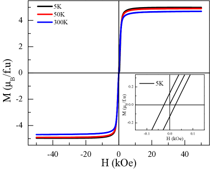
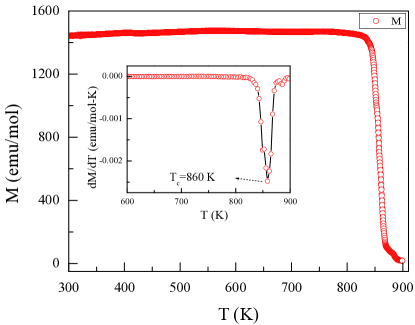
The isothermal magnetic measurements at room temperature show a soft ferromagnetic character with a rather large magnetic moment (4.71 /f.u.) (Fig. 5), suggesting ferromagnetic ordering at a temperature above 300 K. Accordingly, we performed magnetic susceptibility measurements in a VSM with electromagnet in the temperature range 300 - 900 K under a magnetic field of 100 Oe. Very interestingly, the system exhibits ferromagnetic ordering at 860 K (estimated from curve) (Fig. 6, Inset), much higher than its binary parent compound, Fe3Ge ( = 755 K) [24]. It may be noted here that the minor phase of FeGe2 detected from our XRD analysis are known to exhibit a ferromagnetic ordering at a much lower temperature, K [40], and hence the observed ordering at K must arise from the main phase. The large magnetic moment in the isothermal magnetic measurement at 300 K also confirms the ferromagnetic ordering of the primary phase, Fe2RuGe. Substituting a magnetic Fe atom with a non-magnetic Ru atom, having a random mixed site occupancy increases TC even in comparison to the case where 3 atoms like V, Cr, Mn, and Ni are substituted [25, 26, 27, 4]. The isothermal magnetic measurement was also extended to lower temperatures to compare it to the theoretically estimated moments. The data taken at 5, 50 and 300 K are shown in Fig. 5. Even at the lowest measured temperature, 5 K, the coercive field retains its soft magnetic character ( = 130 Oe), and the saturation moment is only slightly enhanced to 4.92 /f.u.. It may be pointed out here that the well-known Slater-Pauling (SP) rule provides a theoretical limit of the magnetic moments in Heusler alloy systems. According to the SP rule, the saturation magnetic moment () can be expressed as (NV -24)/f.u. where NV is the valence electron number [1]. The atomic disorder present in the system is known to reduce the experimental value in comparison to the SP-rule prediction [12]. Since Fe and Ru are randomly distributed in 4 and 4 positions in Fe2RuGe, one thus expects a considerably diminished value of magnetic moment. Surprisingly, the experimentally observed saturation magnetic moment (4.92 /f.u.) for Fe2RuGe is found to be much higher than that theoretically predicted (4 /f.u.). Although a few Fe-based HA are already known to exhibit saturation moment higher than that predicted by the SP-rule [4], Fe2RuGe occupies a special place because it exhibits such a large value despite its inherent atomic disorder involving the moment carrying Fe-atoms. As up to now all studied HMF systems obey the SP rule [12], the specific behaviour of Fe2RuGe needs other experimental measurements (neutron diffraction and Mössbauer spectrometry) as well as theoretical calculations to be more clearly understood.
III.4 Neutron Diffraction
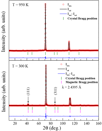
Neutron powder diffraction measurements have been carried out on Fe2RuGe at 300 K and 950 K, respectively, below and above its Curie temperature ( K) to analyze the nuclear structure at 950 K and the long-range magnetic order at 300 K (Fig. 7). The diffraction pattern at 950 K, i.e., in the paramagnetic region, can be well refined in the cubic space group with lattice parameter as = 5.936(2) Å, while the Fe and Ru occupy the 4 and 4 positions with occupation rates of 55% and 45%, respectively. This agrees with the XRD data analysis described earlier in Sec. III.1 and confirms the larger stability of the disordered XA structure compared to Heusler or ordered XA structure. We may, however, note that the intensity of (111) and (200) Bragg peaks, which are generally considered as a signature of perfect crystalline ordering, have much-reduced intensity in the ND spectra in comparison to XRD data, which can be explained due to the different atomic scattering factors of neutron and X-ray. The small peak at 2 = 33.9° can be refined with 0.6% of FeGe2 impurity. The NPD pattern measured at 300 K, in the magnetically ordered state, appears to be nearly like that at 950 K, except for a moderate increase in the intensity of the (111) and (311) crystal Bragg peaks. Assuming the structural model refined in the paramagnetic region (950 K) remains similar at 300 K, the absence of additional peaks beyond the allowed crystal Bragg positions ruled out the antiferromagnetic type of order in this system. Rietveld refinement of the 300 K data yields a refined cell parameter = 5.8787(2) Å close to the XRD value. A good refinement was obtained assuming a ferromagnetic alignment of the Fe sublattice in agreement with the magnetization data. In a powder pattern, all the axes are equivalent to a cubic structure, and it is not possible to specify a preferred orientation of the magnetic moments. Since the intensity of only a few Bragg peaks is related to magnetic ordering, with a small intensity compared to the nuclear contribution, it was not possible to refine independently each Fe moment without a divergence of the fit. A unique Fe moment was therefore assumed for all sites and the refinement of the Fe moment intensity lead to 2.49(4) /Fe or 4.98 /f.u ( = 0.65 % and =7.78%). This value agrees quite well with the isothermal magnetization measurements (Sec. III.3) with = 4.69 /f.u at 300 K and = 4.92 /f.u at 5 K. In order to check if a Ru magnetic contribution had to be taken into account, a fit was performed by fixing the values of Fe and Ru moment to the values calculated by DFT: (Fe-) = 2.74 /Fe, (Fe-) = 1.92 /Fe and (Ru) = 0.33 /Ru (See Sec. III.6). This lead to higher values of the refinement’s factors (RBnucl = 1.25% and RMag =15%). To conclude this part, we consider that the larger total Fe moment in the XA ordered or disordered structure is related to the Fe:Ru charge transfer.
III.5 57Fe Mössbauer Spectrometry
| T (K) | Site |
|
|
|
|
|||||||||||
| 300 K | Fe- | 0.17 | 0.01 | 306 | 55 | |||||||||||
| Fe- | 0.39 | -0.01 | 233 | 23 | ||||||||||||
| Fe- | 0.41 | 0.02 | 218 | 22 | ||||||||||||
| <0.28> | <0.01> | <269> | ||||||||||||||
| 77 K | Fe- | 0.28 | -0.01 | 330 | 52 | |||||||||||
| Fe- | 0.50 | 0.01 | 252 | 24 | ||||||||||||
| Fe- | 0.53 | 0.01 | 234 | 24 | ||||||||||||
| <0.40> | <0.01> | <288> |
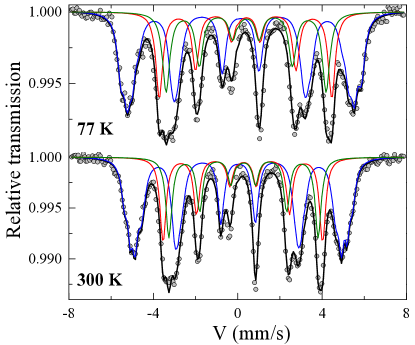
As the analysis of the Fe2RuGe ND spectra does not allow the determination of site-specific Fe moments, 57Fe Mössbauer spectrometry was used to check the relative population of Fe atoms and their respective magnetic moments (determined appropriately from the magnetic hyperfine fields of the individual components) in the three crystallographic positions, namely 4, 4 and 4. The Mössbauer spectra taken at 300 K and 77 K (shown in Fig. 8) are quite similar and show complex magnetic structures consisting of well-resolved magnetic hyperfine structures with broadened and asymmetrical lines. Several models can be proposed to describe each spectrum: the first solution consists of two magnetic components with broad and Lorentzian lines characterized by two different values of isomer shift attributed to the two Fe 4 and 4 sites. The Fe, Ru and Ge disorder explains why only two components are not able to describe perfectly the hyperfine structures observed at 300 and 77 K. Indeed, the best fitting model for both spectra at once requires three magnetic components, each resulting from the superposition of several magnetic sextets to describe the asymmetry and width of the lines, in order to take into account the disordered atomic environments. The corresponding refined mean values of the hyperfine parameters are listed in Table 2. The relative absorption area is estimated at 55%, 23% and 22% for the three Fe sites. Assuming the same values of their respective Lamb-Mössbauer factors, they directly give rise to the proportions of Fe atoms that match quite well with our analysis of ND and XRD patterns (Sec. III.1, Sec. III.4). Considering the fact the larger hyperfine field value corresponds to the 4 site in inverse Heusler alloys [4], the mean hyperfine field values at 300 K for the three magnetic components as 306 kOe, 233 kOe and 218 kOe are assigned to 4, 4 and 4 sites, respectively. The individual Fe moments corresponding to the Wyckoff positions 4, 4 and 4 in Fe2RuGe can be estimated from the three different hyperfine field components, provided that the magnetic hyperfine field values can be properly scaled with the respective magnetic moments. Usually, -Fe with a magnetic moment of 2.2 yields a hyperfine field of 330 kOe, and most often this reference is used as the calibration factor. Using this approach, it was concluded in a previous study on the structurally similar compound Fe3-xRuxSi that Ru atoms contains a magnetic moment of about 1.3 for = 0.75, while the proposed moment in Ru is slightly reduced to 1 in Fe2RuSi [41]. This result is really surprising, as only a handful of materials are known where Ru carries magnetic moments [42]. If we implement the same calibration factor in Fe2RuGe as well, and compare the result with isothermal magnetisation data, we also would have to assign a magnetic moment of 0.7 on Ru atoms in our sample of Fe2RuGe. However, the neutron diffraction experiment presented above ruled out the presence of any significant magnetic moment at the Ru site. In this context, we may like to point out that the relationship between the magnetic hyperfine field and the magnetic moment are not so straight-forward as elaborated earlier in literature for many different Fe-containing DO3-type alloys [43], [44] as well as for other binary systems [45]. In some other Fe-based Heusler compounds too, it has also been suggested that a magnetic moment of 2 2 /Fe would correspond to 255 kOe [4]. Such modification is generally caused by the presence of a large anisotropic hyperfine field involving a dipolar field, non-cubic distribution of spin density in the iron atom, an anisotropic g-factor, etc [44]. For our calculation, we have thus utilized the reference of Fe3Ge, which can be regarded as the parent compound of our studied Heusler system, Fe2RuGe, where a magnetic hyperfine field value of 220 kOe had been associated with 2 /Fe atom [4]. If we use this calibration factor for Fe2RuGe, we find the Fe magnetic moment values at 4, 4 and 4 sites as 2.78(1), 2.12(1) and 1.98(1) , respectively, giving a resulting magnetic moment of 4.83(2) , that matches quite well with the isothermal magnetisation measurement.
III.6 Theoretical analysis
In order to theoretically establish the most stable crystal structure, several crystal configurations were studied by DFT calculations. We considered the ordered Cu2MnAl-type corresponding to the full Heusler structure () and the Hg2CuTi-type known as the XA structure. For both structures, the optimized crystal structure was obtained from DFT calculations and the enthalpies of formation are summarized in Table 3. Our calculations show that the Cu2MnAl-type structure is less stable than the XA structure which is in good agreement with the structural analysis (Sec. III.A.).
| 4a | 4b | 4c | 4d | (kJ/mol | Magnetic Moment (/f.u.) | |
| Heusler (Cu2MnAl) | Ge | Ru | Fe | Fe | 2.79 | 5.0 |
| XA structure (Hg2CuTi) | Ge | Fe | Ru | Fe | -18.94 | 5.0 |
| Disordered XA structure | Ge | Fe | Fe:Ru | Fe:Ru | -20.24 | 4.9 |
Moreover, a structural disorder of Fe and Ru on the 4 and 4 positions, in the XA structure, leads to a more stable compound, thus confirming the XRD, ND and EXAFS data.
| 4a (/f.u.) | 4b (/f.u.) | 4c (/f.u.) | 4d (/f.u.) | (%) | |
|---|---|---|---|---|---|
| (Cu2MnAl) | Ge = -0.05 | Ru = 1.3 | Fe = 1.94 | Fe = 1.94 | -33.3 % |
| XA (Hg2CuTi) | Ge = -0.04 | Fe = 2.76 | Ru = 0.33 | Fe = 1.92 | -29.9 % |
| Disordered XA | Ge = -0.04 | Fe = 2.74 | Fe:Ru with Ru = 0.16 | Fe:Ru with Fe =1.98 | -2.7 % |
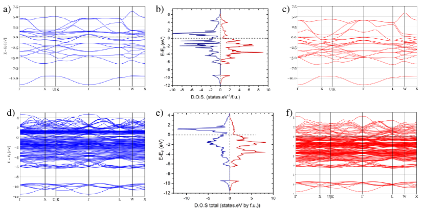
Fig. 9(a-c) shows the calculated spin-polarized band structure and the calculated density of states (DOS) of the ordered XA structure. As it can be seen, the DOS shows no band gap at the Fermi level () for either the majority or minority spin population. This explains a rather low value of the spin polarization, with =29.9%, and (Heusler structure)=33.3%. As in many other Heusler compounds, the spin polarization drastically decreases when the disorder increases, drops to -2.7% in the disordered XA inverse Fe2RuGe too. As in Fig. 9(a-c), Fig. 9(d-f) shows the calculated spin-polarized band structure and the associated DOS of the disordered XA structures where no semiconducting behaviour can be noticed because both spin directions correspond to a metallic ground state. The calculated total magnetic moment, for all configurations, is in good agreement with the experimental magnetic moment, always around 5 /f.u.. Although all calculated total magnetic moments are almost the same for the three configurations, only the ordered and disordered XA types show significant differences in atomic moment of Fe compared to the full Heusler configuration. It may be noted that the magnetic moment is shared between the Fe and Ru elements for the Heusler type. In contrast to this, in the XA structure type, the magnetic moment of the Ru element is considerably decreased whereas the magnetic moment carried by the Fe atom in 4 positions is strongly increased (Table 4).
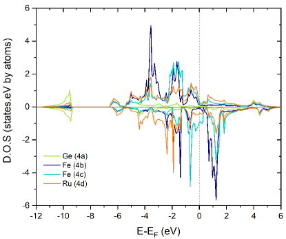
In the full Heusler configuration, the Fe- site is surrounded by 4 Ge and 4 Ru atoms, while the nearest Fe neighbours are at a large distance and the mean Fe moment is 1.98 /Fe. In the ordered and disordered XA structures, it can be observed that Fe- atoms surrounded by 4 Fe and 4 Ru atoms have a enhanced moment close to 2.8 /Fe while Fe-() atoms surrounded by 4 Fe and 4 Ge atoms retain a moment close to 1.9 /Fe. The spin-polarized DOS projected on each site for the XA ordered structure clearly shows a different contribution of the Fe site to the SP-DOS (Fig.10), with a stronger interaction of Ru orbitals with Fe- than with Fe-. The calculation of the Bader charges, as shown in Table 5, reveals a larger electronic charge transfer from Fe- (-0.35) to Ru- (+0.47) in the disordered XA structure compared to the direct structure, -0.13 to +0.26, for both Fe and Ru sites, respectively. In fact, the charge transfer from Fe- is less important in XA structure since its atomic environment is surrounded by Fe and Ge. Due to the charge transfer from Fe to Ru, the number of unpaired electrons for the Fe- atom increases, explaining the enhancement of its magnetic moment in comparison with Fe- and the low value for the Ru moment. Such charge transfer is also responsible for the enhancement of the magnetic moment of the Fe at site, compared to the average moment on Fe in the parent compound Fe3Ge. To our knowledge, such a large enhancement of magnetic moment has not been reported earlier in any Heusler alloy system.
| 4a (electron) | 4b (electron) | 4c (electron) | 4d (electron) | |
|---|---|---|---|---|
| Heusler (Cu2MnAl) | Ge: +0.00 | Ru: +0.26 | Fe: -0.13 | Fe: -0.13 |
| XA structure (Hg2CuTi) | Ge: -0.11 | Fe: -0.35 | Ru: +0.53 | Fe: -0.06 |
| Disordered XA structure | Ge: -0.07 | Fe: -0.35 | Ru: +0.47 | Fe: -0.06 |
III.7 Resistivity study
In sec. III.6, it is predicted that Fe2RuGe has density of states in both up and down spin configuration at the Fermi level. To check its validity, we have performed resistivity experiments. Fig. 11 represents the temperature-dependent electrical resistivity () behaviour of Fe2RuGe measured in the absence of magnetic field ( = 0 kOe) in both heating and cooling cycles. We could not observe any irreversibility between these data: thus we ruled out any possibility of a structural transition in the system. In the case of a ferromagnetic material, the temperature dependence of electrical resistivity can be attributed to three different types of scattering factors. According to Matthiessen rule, all these contributions arise due to different types of factors such as, a) contribution due to scattering of conduction electrons by the lattice defects, , b) phonon scattering, , c) magnon scattering and electron–electron interaction, , are additive in nature. From Fig. 11 we can see that the residual resistivity ratio (RRR) of Fe2RuGe (/) is around 1.625, which clearly indicates the existence of substantial structural disorder in the sample [46, 47]. We can say that resistivity of any ferromagnetic material is
| (5) |
The phonon scattering term can be written as [12] [48]
| (6) |
and the magnon and electron contribution as . Here is the Debye temperature and is scattering constant [12]. As the is quite high, we have fitted the data using Eq. 5
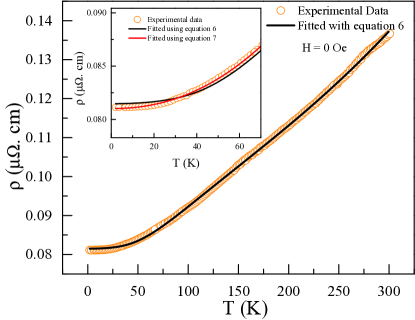
From the fitted parameters, phonon contribution was found much higher compared to the magnon contribution. However, Eq. 5 fits the data well within the region 60 T 300 K only, but is unable to fit the low temperature region properly. From the fitted results we get = 360 K. Generally, electron-electron (e-e) and electron-magnon (e-m) lead to a quadratic temperature dependence of resistivity. However, between these two, the dominant contribution can be inferred from the magnitude of the coefficient, . Normally, is of the order of 10-2 ncm.K2 for e-e interaction while it is normally two orders higher for e-m interaction. From the fitting, we got the value of (e-e interaction coefficient) to be 4.57 cm.K2 which falls within the accepted range for electron-electron scattering, indicating the very weak presence of a single-magnon-scattering mechanism. We have tried to fit the low temperature resistivity data by using the equation
| (7) |
From the fitting, we have found the value of , from which we can confirm that at low temperatures also there is a dominant magnon contribution, which is quite surprising and calls for further investigations of magnetic dynamics in this material. Previously, several authors have obtained different power () values depending on the temperature range considered. The value of above 2 gives some signs of the weak-metallic character even at higher temperatures [12, 47].
IV Concluding Remarks
To summarize, we have synthesized a new Fe-based Heusler alloy Fe2RuGe through the arc melting technique. The combined XRD, ND and EXAFS study reveals that the material crystallizes in a disordered structure, which is also supported by the lower formation ground state energy of the disordered structure over the ordered one, calculated by SQS+DFT. 57Fe Mössbauer spectrometry confirms the inverse Heusler XA disorder with different sextets, strongly suggesting different Fe positions. From neutron diffraction, we confirm that the disorder between Fe and Ru in the and sites is 50:50 in nature. This leads to the mixing of Fe and Ru in the same plane. All these results validate that there are two different Fe environments compared to the Heusler structure. The compound exhibits a ferromagnetic to paramagnetic transition at a significantly high Curie temperature of 860 K and the saturation moment at 5 K is estimated to be 4.90 /f.u., which is higher than the expected SP value of 4 /f.u.. Such enhancement of the magnetic moment above the predicted SP value is observed for the first time. The neutron diffraction experiments reveal that only Fe, distributed over different sites, contributes to the net magnetic moment while no moment could be detected on Ru site. Both theoretical calculation and the Mössbauer experiment support that there is a larger moment on Fe- site, in comparison with the other disordered Fe- sites. This enhanced magnetic moment is explained by a large electronic charge transfer from Fe- to Ru, as calculated by DFT. The resistivity experiment confirms the presence of electron–electron behaviour at the high-temperature range and a weak phonon contribution at low temperature compared to the magnon contribution.
Acknowledgements.
S.C. and S.G. would like to sincerely acknowledge UGC, India, and SINP, respectively, for their fellowship. DFT calculations were performed using HPC resources from GENCI-CINES (Grant No. 2021-A0100906175). Work at the Ames National Laboratory (in part) was supported by the Department of Energy–Basic Energy Sciences, Materials Sciences and Engineering Division, under Contract No. DEAC02- 07CH11358.References
- Graf et al. [2011] T. Graf, C. Felser, and S. S. Parkin, Progress in Solid State Chemistry 39, 1 (2011).
- Felser and Hirohata [2015] C. Felser and A. Hirohata, Heusler alloys (Springer, 2015).
- Mondal et al. [2018] S. Mondal, C. Mazumdar, R. Ranganathan, E. Alleno, P. Sreeparvathy, V. Kanchana, and G. Vaitheeswaran, Physical Review B 98, 205130 (2018).
- Gasi et al. [2013] T. Gasi, V. Ksenofontov, J. Kiss, S. Chadov, A. Nayak, M. Nicklas, J. Winterlik, M. Schwall, P. Klaer, P. Adler, et al., Physical Review B 87, 064411 (2013).
- Du et al. [2013] Y. Du, G. Xu, X. Zhang, Z. Liu, S. Yu, E. Liu, W. Wang, and G. Wu, EPL (Europhysics Letters) 103, 37011 (2013).
- Fu et al. [2015] C. Fu, T. Zhu, Y. Liu, H. Xie, and X. Zhao, Energy & Environmental Science 8, 216 (2015).
- Chauhan et al. [2020] N. S. Chauhan, P. R. Raghuvanshi, K. Tyagi, K. K. Johari, L. Tyagi, B. Gahtori, S. Bathula, A. Bhattacharya, S. D. Mahanti, V. N. Singh, et al., The Journal of Physical Chemistry C 124, 8584 (2020).
- Chauhan et al. [2019] N. S. Chauhan, S. Bathula, B. Gahtori, S. D. Mahanti, A. Bhattacharya, A. Vishwakarma, R. Bhardwaj, V. N. Singh, and A. Dhar, ACS Applied Materials & Interfaces 11, 47830 (2019).
- Özdoğan et al. [2008] K. Özdoğan, E. Şaşıoğlu, and I. Galanakis, Journal of Applied Physics 103, 023503 (2008).
- De Groot et al. [1983] R. De Groot, F. Mueller, P. Van Engen, and K. Buschow, Physical Review Letters 50, 2024 (1983).
- Wurmehl et al. [2005] S. Wurmehl, G. H. Fecher, H. C. Kandpal, V. Ksenofontov, C. Felser, H.-J. Lin, and J. Morais, Physical Review B 72, 184434 (2005).
- Gupta et al. [2022] S. Gupta, S. Chakraborty, S. Pakhira, C. Barreteau, J.-C. Crivello, B. Bandyopadhyay, J.-M. Greneche, E. Alleno, and C. Mazumdar, Physical Review B 106, 115148 (2022).
- Siewert et al. [2012] M. Siewert, M. E. Gruner, A. Hucht, H. C. Herper, A. Dannenberg, A. Chakrabarti, N. Singh, R. Arróyave, and P. Entel, Advanced Engineering Materials 14, 530 (2012).
- Chopra et al. [2000] H. D. Chopra, C. Ji, and V. Kokorin, Physical Review B 61, R14913 (2000).
- Huang et al. [2013] H. Huang, X. Ma, Z. Liu, C. Zhao, S.-Q. Shi, and L. Chen, Applied Physics Letters 102, 042405 (2013).
- Lin et al. [2010] H. Lin, L. A. Wray, Y. Xia, S. Xu, S. Jia, R. J. Cava, A. Bansil, and M. Z. Hasan, Nature Materials 9, 546 (2010).
- Chadov et al. [2010] S. Chadov, X. Qi, J. Kübler, G. H. Fecher, C. Felser, and S. C. Zhang, Nature Materials 9, 541 (2010).
- Zuo et al. [2018] S. Zuo, F. Liang, Y. Zhang, L. Peng, J. Xiong, Y. Liu, R. Li, T. Zhao, J. Sun, F. Hu, et al., Physical Review Materials 2, 104408 (2018).
- Giri et al. [2020] B. Giri, A. I. Mallick, C. Singh, P. P. Madduri, F. Damay, A. Alam, and A. K. Nayak, Physical Review B 102, 014449 (2020).
- Ouardi et al. [2013] S. Ouardi, G. H. Fecher, C. Felser, and J. Kübler, Physical Review Letters 110, 100401 (2013).
- Bainsla et al. [2015] L. Bainsla, A. Mallick, M. M. Raja, A. Nigam, B. C. S. Varaprasad, Y. Takahashi, A. Alam, K. Suresh, and K. Hono, Physical Review B 91, 104408 (2015).
- Žutić et al. [2004] I. Žutić, J. Fabian, and S. D. Sarma, Reviews of Modern Physics 76, 323 (2004).
- Telling et al. [2006] N. Telling, P. S. Keatley, G. van der Laan, R. Hicken, E. Arenholz, Y. Sakuraba, M. Oogane, Y. Ando, and T. Miyazaki, Physical Review B 74, 224439 (2006).
- McGuire et al. [2018] M. A. McGuire, K. Shanavas, M. S. Kesler, and D. S. Parker, Scientific Reports 8, 14206 (2018).
- Mahat et al. [2020] R. Mahat, K. Shambhu, D. Wines, F. Ersan, S. Regmi, U. Karki, R. White, C. Ataca, P. Padhan, A. Gupta, et al., Journal of Alloys and Compounds 830, 154403 (2020).
- Mahat et al. [2021] R. Mahat, K. Shambhu, D. Wines, S. Regmi, U. Karki, Z. Li, F. Ersan, J. Law, C. Ataca, V. Franco, A. Gupta, and P. LeClair, Journal of Magnetism and Magnetic Materials 521, 167398 (2021).
- Keshavarz et al. [2019] S. Keshavarz, N. Naghibolashrafi, M. E. Jamer, K. Vinson, D. Mazumdar, C. L. Dennis, W. Ratcliff II, J. A. Borchers, A. Gupta, and P. LeClair, Journal of Alloys and Compounds 771, 793 (2019).
- Rodríguez-Carvajal [1993] J. Rodríguez-Carvajal, Physica B: Condensed Matter 192, 55 (1993).
- Ravel and Newville [2005] B. Ravel and M. Newville, Journal of Synchrotron Radiation 12, 537 (2005).
- Avdeev and Hester [2018] M. Avdeev and J. R. Hester, Journal of Applied Crystallography 51, 1597 (2018).
- Blöchl [1994] P. E. Blöchl, Physical Review B 50, 17953 (1994).
- Kresse and Hafner [1994a] G. Kresse and J. Hafner, Physical Review B 49, 14251 (1994a).
- Kresse and Hafner [1994b] G. Kresse and J. Hafner, Journal of Physics: Condensed Matter 6, 8245 (1994b).
- Perdew et al. [1996] J. P. Perdew, K. Burke, and M. Ernzerhof, Physical Review Letters 77, 3865 (1996).
- Blöchl et al. [1994] P. E. Blöchl, O. Jepsen, and O. K. Andersen, Physical Review B 49, 16223 (1994).
- Zunger et al. [1990] A. Zunger, S.-H. Wei, L. Ferreira, and J. E. Bernard, Physical Review Letters 65, 353 (1990).
- Sanchez et al. [1984] J. M. Sanchez, F. Ducastelle, and D. Gratias, Physica A: Statistical Mechanics and its Applications 128, 334 (1984).
- Van de Walle et al. [2013] A. Van de Walle, P. Tiwary, M. De Jong, D. Olmsted, M. Asta, A. Dick, D. Shin, Y. Wang, L.-Q. Chen, and Z.-K. Liu, Calphad 42, 13 (2013).
- Van De Walle [2009] A. Van De Walle, Calphad 33, 266 (2009).
- Jeong [2007] T. Jeong, Solid State Communications 141, 329 (2007).
- Mishra et al. [1985] S. Mishra, D. Rambabu, A. Grover, R. Pillay, P. Tandon, H. Devare, and R. Vijayaraghavan, Solid State Communications 53, 321 (1985).
- Cao et al. [1997] G. Cao, S. McCall, M. Shepard, J. Crow, and R. Guertin, Physical Review B 56, 321 (1997).
- Dubiel [2009] S. Dubiel, Journal of Alloys and Compounds 488, 18 (2009).
- Drijver et al. [1976] J. Drijver, S. Sinnema, and F. Van der Woude, Journal of Physics F: Metal Physics 6, 2165 (1976).
- Arzhnikov et al. [2001] A. Arzhnikov, L. Dobysheva, G. Konygin, E. Voronina, and E. Yelsukov, Physical Review B 65, 024419 (2001).
- Bœuf et al. [2006] J. Bœuf, C. Pfleiderer, and A. Faißt, Physical Review B 74, 024428 (2006).
- Nag et al. [2022] J. Nag, D. Rani, D. Singh, R. Venkatesh, B. Sahni, A. Yadav, S. Jha, D. Bhattacharyya, P. Babu, K. Suresh, et al., Physical Review B 105, 144409 (2022).
- Rossiter [1991] P. L. Rossiter, The electrical resistivity of metals and alloys, Vol. 6 (Cambridge university press, 1991).