Feature Imitating Networks Enhance the Performance, Reliability and Speed of Deep Learning on Biomedical Image Processing Tasks
Abstract
Feature-Imitating-Networks (FINs) are neural networks with weights that are initialized to approximate closed-form statistical features. In this work, we perform the first-ever evaluation of FINs for biomedical image processing tasks. We begin by training a set of FINs to imitate six common radiomics features, and then compare the performance of networks with and without the FINs for three experimental tasks: COVID-19 detection from CT scans, brain tumor classification from MRI scans, and brain-tumor segmentation from MRI scans; we find that FINs provide best-in-class performance for all three tasks, while converging faster and more consistently when compared to networks with similar or greater representational power. The results of our experiments provide evidence that FINs may provide state-of-the-art performance for a variety of other biomedical image processing tasks.
1 Introduction
1.1 Motivation
Medical imaging has benefited greatly from the rise of deep-learning techniques for tasks including: image segmentation, classification, detection, retrieval, reconstruction, filtering, denoising, and super resolution [1]. Indeed, there are countless studies that have demonstrated the superiority of deep learning approaches compared to classical machine learning algorithms (e.g. SVM) using expert-features [2]. Importantly however, there remain domains where deep learning approaches have yet to provide breakthrough performance: when data is too small, computational resources needed to train models are too large, or where model interpretation is a strict requirement. This is often the case in medical imaging tasks; a recent review article on biomedical image classification techniques by Tchapga et al. reported that Neural Networks were less accurate, less tolerant to redundant attributes, slower to train, and more likely to over-fit compared to SVM models with expert features on a variety of biomedical image classification tasks when data was scarce [3].
Feature Imitating Networks (FIN) refers to a recently developed deep learning paradigm that solves the aforementioned challenges of standard deep learning in data-scarce environments [4]. A FIN is a small network pre-trained to simulate one or more closed-form statistical features which are thought to be relevant for a given task; for example, Site Zone Uniformity is a Radiomics feature [5] that is used to measure the variability of size zone volumes in a radiological image. FINs can be trained to emulate one-or-more features, and may then be integrated within a larger, more complex network architecture that obtains the power of the feature, without the strict limitations that would result from including the feature as an input to the model directly. That is, as part of network fine-tuning, the representation captured by the FIN evolves from the static feature representation it was first trained to emulate into an instantiation that is most effective for the task at hand; For instance, a FIN that is designed to emulate Shannon’s entropy, may evolve into a Tsalis entropy representation during fine-tuning.
1.2 Our contributions
Previously, FINs have been shown to provide state-of-the-art performance on several biomedical signal processing tasks using EEG [4]. However, there has been no work to evaluate the potential of FINs for biomedical imaging tasks. In this work, we extend the FIN paradigm beyond the dense implementations described in the original work [4] to enable their emulation of common radiomics features and apply them on three biomedical imaging tasks: COVID-19 detection from CT scans, brain tumor classification from MRI, and brain-tumor segmentation from MRI. We demonstrate that FINs provided best-in-class performance for all three tasks, while converging faster and more consistently when compared to networks with similar or greater representational power.
Our results are important because they demonstrate the ability of FINs to provide state-of-the-art performance without demanding the collection of ever-larger data-sets. This is particularly important for domains like medical imaging, where data is expensive to collect, and there are numerous forms of downstream error that can be caused by merging many small datasets into one large sample [6]. That is, our work provides evidence that FIN-embedded networks can learn robust representations from a patchwork of small noisy datasets collected in a variety of clinical contexts.
1.3 Related work
Conventional neural networks (CNN) have been used with great success for a variety of biomedical tasks including classification, segmentation, object detection, etc. [7]. Below we provide an overview of specific works that are relevant for this study. More specifically, we provide a brief discussion of the state-of-the-art approaches that will serve as baselines for the experimental results discussed later in this paper.
1.3.1 Image classification
The state-of-the-art approach for most radiomics classification tasks use either conventional neural networks (CNN) or SVMs with expert features. There has been an increase in accuracy with deep learning methods in radiology, and deep learning is likely to continue to advance in medical imaging sciences in the future as dataset grow larger, and are better integrated [8]. As such, for the purposes of this study, we compared FINs against several CNN architectures with similar representational power.
1.3.2 Segmentation
U-nets are neural networks designed for image segmentation that contains two parts: (1) a CNN architecture and (2) an up-sampling path. Using highly limited trading samples, U-nets can create highly detailed segmentation maps of images. Based on its context-based learning, U-nets can also be trained much faster than most segmentation models. [9] As a result, we will use the U-net architecture as a baseline for comparison against the FINs for the segmentation task.
2 METHODS
In this section, we provide a discussion of our proposed approach. More specifically, we discuss the procedure used to select and train the Feature Imitating Networks which were later applied to the three experimental tasks outlined in section 3. For our purposes herein, the goal of the FINs is to imitate common radiomics features used in medical imaging tasks, and to embed these FINs within larger network architectures to observe changes in performance and convergence.
Radiomics Features Selected
There are three categories of features that are often used for radiomics tasks: first-order features (e.g. entropy, skewness), shape-based features (e.g mesh surface), and texture-based features (e.g. autocorrelation) [6]. For the purposes of this study, we trained six FINs to emulate common radiomics features. Five FINs were each independently trained to emulate texture based features: Autocorrelation, Gray Level Variance, Cluster Shade, Difference Entropy, and Size Zone Non-uniformity. The sixth FIN was trained to imitate a first-order feature: Skewness. We intentionally excluded shape-based features because the extraction of shape features requires significantly more computational power (over 10x) than texture or first-order features, without significant downstream benefits for the classification and segmentation tasks at hand.
Data and Training Approach
The FINs were trained with a feed-forward neural network (DFNN) structure [10] with random topology using an open source imaging dataset. For each of the images, we extracted radiomics features using PyRadiomics [11]. Next, for each feature, a separate neural network was trained to imitate the feature, given the image. Several network topological configurations were explored at random, and the best configuration was retained. For each FIN, we used a as the activation function for feature approximation. The training was conducted on a NVIDIA A100 GPU. Following training, the FINs were integrated within a baseline network for the experimental task at hand (see following section).
3 Experiments
In this section we describe three radiomics tasks (two classification, and one image segmentation) where we compare the performance of FIN-embedded models against baseline approaches. Within the description of each experiment, we elaborate on the data used for the task, as well as any fine-tuning considerations for integration of the FINs within the baseline approaches for the task. All experiments described here used publicly available data.
3.1 Experiment I
The task for the first experiment was the binary classification of COVID-19 detection using Lung CT images.
Data and Prepossessing
The data for this experiment consisted of Lung CT scans from the COVID-19 Lung CT dataset [12]. The lung scans consisted of non-COVID images and COVID images. The images were portioned into 10-folds; in each fold, 90% of the data was used for model training and validation, and the remaining 10% of the data was used for model testing. All models were assessed according to the mean and standard deviation of their Area Under the Receiver Operator Characteristic Curve (AUROC) across the ten folds, as well as the number of epochs required for convergence of the validation loss.
| AUROC () | AUROC () |
|
|||
|---|---|---|---|---|---|
| DFNN | 0.667 | 0.0629 | 6.9 | ||
| CNN | 0.995 | 0.0050 | 7.2 | ||
| FINs | 0.998 | 0.0029 | 5.7 |
| F-1 Score () | F-1 score () | Accuracy () | Accuracy () | Training epochs () | |
|---|---|---|---|---|---|
| RGB CNN | 0.617 | 0.0177 | 0.677 | 0.0177 | 4.25 |
| Grey-scaled CNN | 0.629 | 0.0137 | 0.684 | 0.0089 | 4.38 |
| FINs | 0.643 | 0.0084 | 0.697 | 0.0059 | 4.25 |
Models
We trained three binary classification models using the collected data: (1) a Deep Forward Neural Network (DFNN) using the radiomics features as inputs, (2) a CNN model that utilized the raw images as inputs, with a final DFNN layer, and (3) the same CNN model, now with an embedded FIN ensemble that imitated the six radiomics features (described in section 2) that also had a final DFNN layer.
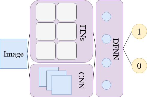
The FINs were trained to imitate the radiomics features of the entire image using a separate open-source imaging dataset from Lung CT Segmentation Challenge [13] provided by TCIA [14]. The challenge dataset consisted of CT images from thoracic patients undergoing radiation treatment. To ensure a fair comparison between the models we explored twenty more topological configurations of each model; the best performing topological configuration was retained. See Figure 1 for an illustration for the FIN embedding approach. Importantly, we explored CNN and DFNN configurations with the same (or greater) number of parameters as the FIN ensemble model; this ensured that the raw representational power of all three models was comparable. The number of parameters for the final CNN, DFNN, and FIN ensemble models was 149M, 150M and 147M respectively.
Results
In Table 1 we compare the performance of the three models with respect to the mean AUROC across the ten testing folds, the standard deviation of the test set performance, and the number of epochs required for the models to converge. As seen in the table, the DFNN using the raw radiomics features had the lowest overall AUROC (0.667) of the three models. The mean AUROC of the FIN-embedded model was only slightly better than the CNN model (0.998 and 0.995 respectively); however, the standard deviation of the FIN-embedded model was 42% lower than the CNN approach, and 95% lower than the DFNN. Importantly, the FIN-embedded model required the fewest number of epochs before convergence; the FIN-embedded model converged 20% faster than the CNN, and faster 17% than the DFNN. These results imply FIN-embedded models provide enhanced classification performance, that is more robust, and faster to train than the alternative approaches.
3.2 Experiment II
The task for the second experiment was a multi-class, brain tumor classification task. Our second experiment builds on the encouraging results seen in Experiment I (Section 3.1) by using the same FINs and baseline CNN model structure.
Data and Prepossessing
The data for this experiment consisted of brain MRI scans from a brain tumor classification dataset [15]. The training data consisted of scans, describing four outcome classes: glioma tumors (), meningioma tumors (), pituitary tumors (), and non-tumors (). Within the test set, there were scans for glioma tumors, scans for meningioma tumors, scans for pituitary tumors, and scans for non-tumors. Prior to modeling, all images were converted to dimensions of using SimpleITK [16], and greyscaled using the following forumla: .
Models
We trained three multi-class classification models with the same structure as the baseline CNN and FINs in experiment I (Section 3.1) using the collected data: (1) a CNN model with an embedded FIN ensemble that imitated the six radiomics features described in section 2, (2) a baseline CNN model with RGB image inputs, and (3) a baseline CNN model with grey-scaled image inputs.
We trained two CNN models, one with grey-scaled inputs and the other one with RGB inputs to ensure there was no color sensitivity when modeling with CNNs. The number of parameters for the two CNNs and FIN ensemble was 149M and 147M respectively. We shuffled and regrouped 90% of the data for model training, and the remaining 10% of the data was used for validation to perform a model training. All models were assessed according to the mean and standard deviation of their F-1 score, accuracy, and the number of epochs until convergence of the validation loss.
Result
In Table 2, we compare the performance of our proposed approach with the baseline models. We observed that the CNN model was more sensitive to grey-scaled inputs as demonstrated by a higher average accuracy and F-1 score, and lower standard deviation, relative to the RGB model. Importantly, the FINs ensemble outperformed both the RGB and grey-scaled CNN models: the FINs ensemble had a higher F-1 score and accuracy, with a 39% lower standard deviation for the F-1 score and 33% lower standard deviation for accuracy than the best performing baseline model (grey-scaled CNN). The three models all converged at similar epochs in term of the validation loss.
3.3 Experiment III
The task for the third experiment was a segmentation task using brain MRI scans from TCIA and TCGA111We thank the data generated by the TCGA Research Network.
Data and Prepossessing
The data for this experiment consisted of brain MRI scans with corresponding segmentation masks [17] . The images were divided into three categories: 70% for training, 15% for validation, and 15% for testing. All the images we normalized from the value range of 0-255 to 0-1.
Models
We trained two models using the training data: (1) A standard U-net approach with filters that increased quadratically to filters, followed by up-sampling back [9], (2) the standard U-net approach with FINs imitating the six features described in section 2. The FINs in this task were trained using the training data from this task to imitate the radiomics features in sub-segments of the image. The outputs of the FINs were inserted after the U-net’s first max-pooling layer. All models were assessed according to their intersection over union (IoU) and dice similarity coefficient; standard measures of image segmentation accuracy [18].
Result
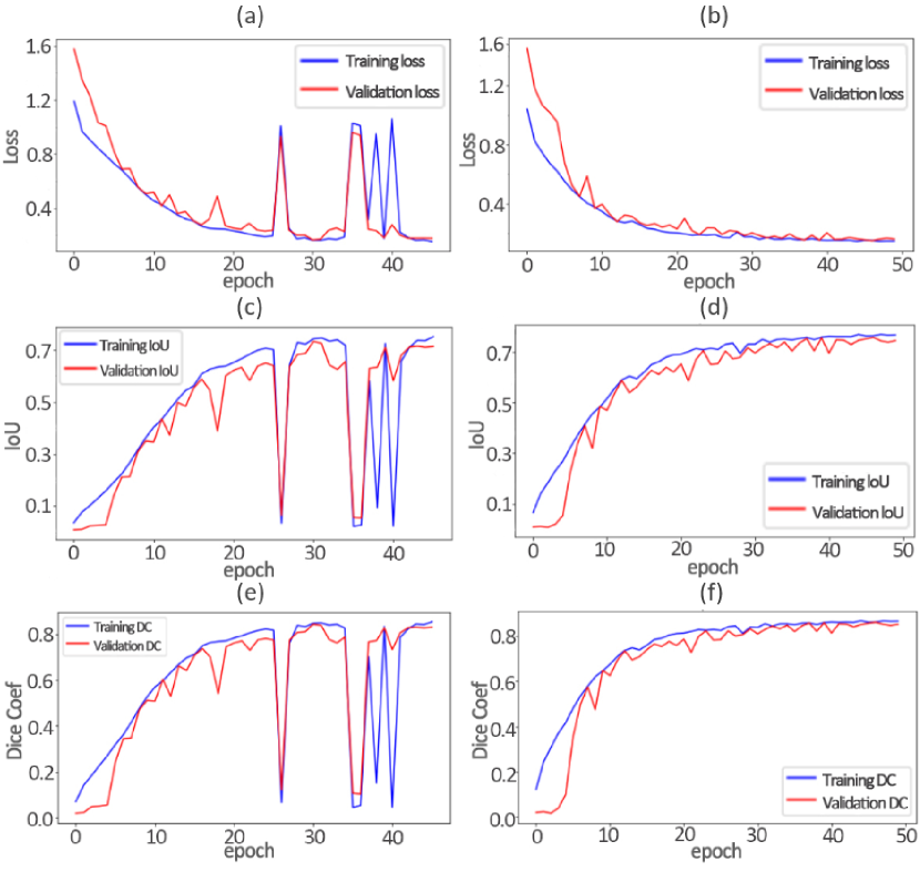
Figure 2 compares the loss, IoU, and dice similarity coefficient of the two models (U-net, U-net + FINs) during the training and validation phases. As seen in the figure, the baseline model of the U-net approach more frequently experienced problems with loss instability and poor validation set performance. In contrast, the FIN approach reduced the sudden irregularities in loss, and the corresponding effects on the IoU and dice coefficient measures. When evaluating on the test set, the performance of the U-net combined with FINs was higher than the U-net model alone, as demonstrated by an increase in both IoU (from to ), and dice coefficient (from to ).
4 Discussion
In the section below, we provide an interpretation of the key results of this work, design nuances that were important for successful use of the FINs, the limitations of the current approach and how these may be overcome by us (or others) in future research.
Key Results
The results of all three experiments provide evidence that embedding FINs within conventional neural networks enhances their ability to learn faster and more reliably without additional parameters. More specifically, the results of experiment I (see Table 1), demonstrate that FINs can provide enhanced performance on binary classification tasks; the results of experiment II (see Table 2 demonstrate that FINs provide enhanced performance on multinomial classification tasks; the results of experiment III (see Figure 2) demonstrate that FINs provide enhanced performance on segmentation tasks. Critically, in all three experiments, FINs required less training epochs for validation loss convergence and exhibited lower variance over multiple folds compared to conventional architectures (CNN or DFNN) with similar representational power (i.e. number of parameters).
Critically, the enhanced performance of the FINs can not be achieved by simply making the raw feature values available to the networks. In Experiment I, we found that the DFNN model, with access to the raw features that the FINs were trained to imitate, performed more poorly than a basic CNN model for radiomics tasks. This highlights FINs are able to adapt the features to be best suited for the task at hand; that is, using a FIN is not equivalent to simply passing in feature values at the input of the network.
An important attribute of the FIN models is not only their higher performance, but also the speed of model training (less epochs), and stability of the model performance (lower variance in test performance across folds); these properties of FINs may be explained by the fact that they are imitating features which are known to be important for the task at hand. That is, the network is initiated in a parameter space which we have reason to believe is better than a random initialization.
Future Directions
Contemporary radiomics tasks utilize large-scale batch processing via high-throughput computational pipelines that integrate image pre-processing, segmentation, feature extraction, feature selection, predictive modeling, and model validation [19]. In this work, we tackled the first step in bringing deep-learning to the radiomics workflow by generating six radiomics FINs to improve the performance of deep learning approaches on three tasks. An obvious direction for future research is to generate a larger variety of radiomics FINs beyond the six used in this study. Furthermore, it would be interesting to experiment with the cross-functionality of FINs for tasks in different domains. For example, it would be interesting to explore if radiomics FINs are useful for tasks not directly related to biomedical image processing.
While radiomics analysis is complex, determining the optimal analysis parameters is also challenging. The results indicate that overall the repeatability is highly sensitive to the processing parameters [20]. In the future, the selection of the analysis parameters and features may also be accomplished using FINs.
Limitations
It remains challenging to interpret the features and calculations used by deep learning models to make a classification. As such, it is therefore difficult to resolve a discrepancy between physicians / radiologists and trained models through discussion if their judgments differ from those of trained models [21]. However, when external performance and interpretability improve, machine learning algorithms, such as FINs, may contribute to a gradual shift in clinical practice by improving radiologists’ performance, increasing inter-observer reliability, and improving workflow for more timely recommendations [21].
References
- [1] Mathieu Hatt, Chintan Parmar, Jinyi Qi, and Issam El Naqa, “Machine (deep) learning methods for image processing and radiomics,” IEEE Transactions on Radiation and Plasma Medical Sciences, vol. 3, no. 2, pp. 104–108, 2019.
- [2] Justin Ker, Lipo Wang, Jai Rao, and Tchoyoson Lim, “Deep learning applications in medical image analysis,” IEEE Access, vol. 6, pp. 9375–9389, 2018.
- [3] Christian Tchito Tchapga, Thomas Attia Mih, Aurelle Tchagna Kouanou, Theophile Fozin Fonzin, Platini Kuetche Fogang, Brice Anicet Mezatio, and Daniel Tchiotsop, “Biomedical image classification in a big data architecture using machine learning algorithms,” Journal of Healthcare Engineering, vol. 2021, 2021.
- [4] Sari Saba-Sadiya, Tuka Alhanai, and Mohammad M Ghassemi, “Feature imitating networks,” in ICASSP 2022-2022 IEEE International Conference on Acoustics, Speech and Signal Processing (ICASSP). IEEE, 2022, pp. 4128–4132.
- [5] Philippe Lambin, Emmanuel Rios-Velazquez, Ralph Leijenaar, Sara Carvalho, Ruud GPM Van Stiphout, Patrick Granton, Catharina ML Zegers, Robert Gillies, Ronald Boellard, André Dekker, et al., “Radiomics: extracting more information from medical images using advanced feature analysis,” European journal of cancer, vol. 48, no. 4, pp. 441–446, 2012.
- [6] Virendra Kumar, Yuhua Gu, Satrajit Basu, Anders Berglund, Steven A. Eschrich, Matthew B. Schabath, Kenneth Forster, Hugo J.W.L. Aerts, Andre Dekker, David Fenstermacher, Dmitry B. Goldgof, Lawrence O. Hall, Philippe Lambin, Yoganand Balagurunathan, Robert A. Gatenby, and Robert J. Gillies, “Radiomics: the process and the challenges,” Magnetic Resonance Imaging, vol. 30, no. 9, pp. 1234–1248, 2012, Quantitative Imaging in Cancer.
- [7] Muralikrishna Puttagunta and S Ravi, “Medical image analysis based on deep learning approach,” Multimedia Tools and Applications, vol. 80, no. 16, pp. 24365–24398, 2021.
- [8] Luca Saba, Mainak Biswas, Venkatanareshbabu Kuppili, Elisa Cuadrado Godia, Harman S. Suri, Damodar Reddy Edla, Tomaž Omerzu, John R. Laird, Narendra N. Khanna, Sophie Mavrogeni, Athanasios Protogerou, Petros P. Sfikakis, Vijay Viswanathan, George D. Kitas, Andrew Nicolaides, Ajay Gupta, and Jasjit S. Suri, “The present and future of deep learning in radiology,” European Journal of Radiology, vol. 114, pp. 14–24, 2019.
- [9] Nahian Siddique, Sidike Paheding, Colin P. Elkin, and Vijay Devabhaktuni, “U-net and its variants for medical image segmentation: A review of theory and applications,” IEEE Access, vol. 9, pp. 82031–82057, 2021.
- [10] Daniel Svozil, Vladimir Kvasnicka, and Jiri Pospichal, “Introduction to multi-layer feed-forward neural networks,” Chemometrics and intelligent laboratory systems, vol. 39, no. 1, pp. 43–62, 1997.
- [11] Joost JM Van Griethuysen, Andriy Fedorov, Chintan Parmar, Ahmed Hosny, Nicole Aucoin, Vivek Narayan, Regina GH Beets-Tan, Jean-Christophe Fillion-Robin, Steve Pieper, and Hugo JWL Aerts, “Computational radiomics system to decode the radiographic phenotype,” Cancer research, vol. 77, no. 21, pp. e104–e107, 2017.
- [12] Mehrad Aria, Mustafa Ghaderzadeh, Farkhondeh Asadi, and Ramezan Jafari, “Covid-19 lung ct scans,” 2021.
- [13] Yang J., Sharp G., Veeraraghavan H., Van Elmpt W., Dekker A., Lustberg T., and Gooding M., “Data from lung ct segmentation challenge (version 3) [data set],” 2017.
- [14] Kenneth Clark, Bruce Vendt, Kirk Smith, John Freymann, Justin Kirby, Paul Koppel, Stephen Moore, Stanley Phillips, David Maffitt, Michael Pringle, et al., “The cancer imaging archive (tcia): maintaining and operating a public information repository,” Journal of digital imaging, vol. 26, no. 6, pp. 1045–1057, 2013.
- [15] Sartaj Bhuvaji, Ankita Kadam, Prajakta Bhumkar, Sameer Dedge, and Swati Kanchan, “Brain tumor classification (mri),” 2020.
- [16] Ziv Yaniv, Bradley C Lowekamp, Hans J Johnson, and Richard Beare, “Simpleitk image-analysis notebooks: a collaborative environment for education and reproducible research,” Journal of digital imaging, vol. 31, no. 3, pp. 290–303, 2018.
- [17] Pedano N., Flanders A. E., Scarpace L., T. Mikkelsen, Eschbacher J. M., Hermes B., Sisneros V., Barnholtz-Sloan J., and Ostrom Q., “The cancer genome atlas low grade glioma collection (tcga-lgg) (version 3) [data set],” 2016.
- [18] Debesh Jha, Pia H Smedsrud, Michael A Riegler, Pål Halvorsen, Thomas de Lange, Dag Johansen, and Håvard D Johansen, “Kvasir-seg: A segmented polyp dataset,” in International Conference on Multimedia Modeling. Springer, 2020, pp. 451–462.
- [19] Alberto Traverso, Leonard Wee, Andre Dekker, and Robert Gillies, “Repeatability and reproducibility of radiomic features: A systematic review,” International Journal of Radiation Oncology*Biology*Physics, vol. 102, no. 4, pp. 1143–1158, 2018, Imaging in Radiation Oncology.
- [20] Michael Schwier, Joost van Griethuysen, Mark G Vangel, Steve Pieper, Sharon Peled, Clare Tempany, Hugo JWL Aerts, Ron Kikinis, Fiona M Fennessy, and Andriy Fedorov, “Repeatability of multiparametric prostate mri radiomics features,” Scientific reports, vol. 9, no. 1, pp. 1–16, 2019.
- [21] Koichiro Yasaka and Osamu Abe, “Deep learning and artificial intelligence in radiology: Current applications and future directions,” PLoS medicine, vol. 15, no. 11, pp. e1002707, 2018.