Trapping and Scattering of a Multiflagellated Bacterium by a Hard Surface
Abstract
Thiovulum majus, which is one of the fastest known bacteria, swims using hundreds of flagella. Unlike typical pusher cells, which swim in circular paths over hard surfaces, a T. majus cell turns its flagella normal to the surface. To probe the torques that stabilize this unusual fixed point, the trajectories of several thousand collisions between a T. majus cell and a wall of a quasi-two-dimensional microfluidic chamber are analyzed. Measuring the fraction of cells escaping the wall either to the left or to the right of the point of contact—and how this probability varies with incident angle and time spent in contact with the surface—maps the scattering dynamics onto a first passage problem. These measurements are compared to the prediction of a Fokker-Planck equation to fit the angular velocity of a cell in contact with a hard surface. This analysis reveals a bound state with a narrow basin of attraction in which cells orient their flagella normal to the surface. The escape angle predicted by matching these near field dynamics with the far field hydrodynamics is consistent with observation. We discuss the significance of these results for the ecology of T. majus and their self organization into active chiral crystals.
I Introduction
Microbes in their natural environments often live in close proximity to surfaces such as the air-water interface, sediment grains in water-saturated soils, and sinking detritus fletcher2013bacterial ; harshey2003bacterial ; azam2001sea ; datta2016microbial . When a cell swims near a surface, the flow it generates is perturbed by the presence of the boundary. The perturbed flow turns and advects the swimming cell lauga2009hydrodynamics ; lopez2014dynamics . Microbes have evolved to exploit these flows to better colonize new environments durham2012division , improve foraging and mating strategies drescher2009dancing , and navigate heterogeneous environments muller2020compass ; waisbord2021fluidic ; petroff2022phases .
Past work on the hydrodynamic coupling between cells and surfaces has focused on a relatively small number of domesticated species lauga2009hydrodynamics , which poorly represent the true diversity of microbial form and locomotion observed in nature zinder2006morphological . To better understand how more complex flagellation patterns allow microbes to exploit new hydrodynamic effects, we study the sediment microbe Thiovulum majus de1961observations ; lariviere1963cultivation ; wirsen1978physiological ; marshall2012single , which is one the the fastest-known bacteria schulz2001big . T. majus uses hundreds of flagella to swim at a speed up to m s-1 garcia1989rapid and turn smoothly in chemical gradients thar2003bacteria . T. majus cells attach to surfaces by means of a m-long mucus tether that is extruded from the cell posterior de1961observations . Once attached, the cell rotates its flagella to generate a flow that efficiently stirs its chemical environment fenchel1998veil ; thar2002conspicuous ; petroff2015biophysical . When many free-swimming cells collide with a flat surface, they self-organize into two-dimensional active crystals petroff2015fast ; petroff2018nucleation . The fluid mechanics that underlie the tethering of cells to surfaces and the formation of active crystals are not well understood.
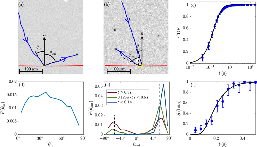
Here we experimentally investigate these dynamics by tracking the motion of several thousand T. majus cells as they collide with the wall of a quasi-two-dimensional microfluidic chamber. After a collision, a cell remains in contact with the surface for an average trapping time s. However trapping times are widely distributed. In approximately of collisions, the cell is only momentarily (s) in contact with the surface before escaping back into the bulk fluid. Fig. 1(a) and supplemental video SV1.mp4 show a one such collision. A similar fraction of collisions result in the trapping of the cell (Fig. 1[b] and supplemental video SV2.mp4) near the surface for more than s and as long as s. These trapped bacteria do not swim along the circular paths that are typical of pusher cells lauga2009hydrodynamics . Rather, a cell orients its flagella normal to the surface das2019transition ; ishimoto2019bacterial ; petroff2015fast and lateral motion is purely diffusive petroff2018nucleation . The distribution of escape times for all collision is shown in Fig. 1(c).
Understanding the dynamics that lead a T. majus cell to either scatter quickly from a surface or become trapped for extended periods is challenging from both a theoretical and experimental stand point. It is difficult to gauge the relative importance of contact forces and hydrodynamic forces even for well characterized cells with few flagella lauga2009hydrodynamics ; drescher2011fluid ; lushi2017scattering . T. majus is far more complex than these model organisms. It is covered with several hundred flagella and it remains unknown how these flagella interact with one another (e.g., by forming bundles). It similarly difficult to probe these dynamics experimentally. Because T. majus cannot be grown in pure culture, the standard experimental techniques (e.g., genetic manipulation of strains) that have proven vital to the study of model organisms are unavailable. Extending our understanding of cell-surface interactions from model organisms to the broader field of environmental microbiology requires experimental techniques and analysis to overcome these challenges.
We probe the dynamics of scattering and trapping indirectly by analyzing the statistics of 3661 collisions between a swimming cell and a wall. We observe striking simplicity in the trajectories of escaping cells. Although the distribution of incident angles is widely distributed (Fig. 1[d]), escape angles are narrowly distributed (Fig. 1[e]). Cells that are in fleeting (s) contact with the surface tend to escape at an average angle of Any such cell that approaches the wall from the left escapes to the right and vice versa. In our coordinate system, these cells escape at an angle . By contrast, cells that remain in contact with the surface for at least s are bimodally distributed, being equally likely to escape at either positive or negative angles with mean .
The bimodality of escape directions develops gradually as a cell remains in contact with surface. We measure the probability that a cell escapes the surface at an angle and calculate the the associated binomial entropy of the escape direction. As shown in Fig. 1(f), the information of a cell’s trajectory before the collision is gradually lost over the course of half a second.
In this article, we show how this erasure of information can be mapped onto a first passage problem berg1993random ; moen2022trapping to infer the torques acting on a multiflagellated bacterium that is in contact with a wall. Matching the inferred near field dynamics with the far field hydrodynamics of a pusher cell predicts the angle at which cells escape.
II Materials and Methods
II.1 Enrichment of Bacteria
Because there are no known techniques to grow Thiovulum majus in pure culture, cells must be enriched from environmental samples using well established methods de1961observations ; marshall2012single ; petroff2015fast ; petroff2018nucleation . We collect sediment from a shallow tide pool in Little Sippewissett Marsh (N, W), which is near Woods Hole, Massachusettes. This sediment is stored in the lab in a container covered in cm of natural sea water. After three to five days in the container, the sediment-water interface becomes euxinic and a T. majus veil forms a few millimeters above the sediment jorgensen1983colorless ; fenchel1998veil , often between strands of eel grass. Cells are collected from a fresh veil with a ml pipette and lightly mixed. After collection the veil reforms, typically within a day.
II.2 Microfluidic Device
Microfluidic chambers—which are produced using standard soft photolithography techniques whitesides2001soft —are composed from Polydimethylsiloxane (PDMS) and sealed on one side by a glass slide. Chambers are quasi-two dimensional, with a height of both m and centimeter-scale lateral dimensions. Two similarly designed microfluidic chambers are used in these experiment. In the first set of experiments the shape of the chamber, when viewed from above, is a square (). In the second set of experiments (as in Fig. 1[a,b]), the chamber is circular with a radius of cm. We initially suspected that the slight curvature of the wall could lead to measurable differences in the scattering dynamics, however the statistics presented here were found to be indistinguishable between the two chamber designs. Because we limit our analysis to the trajectories of cells within m of a wall, the curvature of a walls is small, being within of flat. Consequently, these measurements are combined in the analysis presented here.
II.3 Tracking of Cells
T. majus cells are inoculated into the microfluidic chambers describe above. The motion of cells as they swim near the outer wall of the chamber is observed using the Zeiss objective, which is focused to a plane m between to top and bottom of the chamber to limit hydrodynamic interactions between the swimming cell and the chamber. In each experiment, the motion of cells near one wall of the chamber is recorded at fps using a Nikon D7000 camera.
We track the motion of cells swimming within m of the wall of the chamber. Swimming cells appear as dark spots under transmitted light. To identify cells, we first average all frames over the course of a five minute experiment. This background image is subtracted from each frame of the video to highlight motion. Swimming cells are identified using an intensity threshold. These instantaneous measurements of cell position are connected into trajectories by applying Munkres’ Assignment Algorithm. Two representative trajectories are shown as solid blue lines in Fig 1(a,b). A small fraction of the trajectories corresponded to cells that are close to division. These cells are atypically large and swim in slightly helical trajectories. To better distinguish torques arising from interactions with the chamber walls from those that are due to the particular flagellation pattern of the cell these helical trajectories are discarded.
From each trajectory, we calculate the velocity of each cell that collides with the wall of the chamber. Cells are identified as in contact with the surface if the distance between the center of the cell and the chamber wall is less than or equal to the measured radius of the cell. To find the incident angle , we measure the incident velocity from the frames immediately before the cell contacts the surface and measure angle between and the local normal at the point of first contact. Similarly, the asymptotic escape angle is measured relative to the local surface normal at the point where the cell escapes. The velocity of the escaping cell is calculated when the cell is two body lengths () from the surface, at which point hydrodynamic torques are negligible (see Fig 2). The slight curvature of the circular chamber wall creates an ambiguity in of less than , which we ignore.
III Results
III.1 Motion of a cell far from a wall
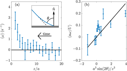
We begin by analyzing a cell’s approach to a surface. Fig. 2(a) shows that the average angular velocity of cells decreases with the distance between the cell and the wall. Cells initially approach the wall at a constant angle, . However, when the cell arrives to within a distance of , it begins to turn parallel to the surface.
These dynamics are explained by the far-field hydrodynamics of a pusher cell near a wall lauga2009hydrodynamics ; drescher2011fluid . At distances , the cell can be approximated as a force dipole. A swimming cell moves as it pushes on the fluid and is advected and rotated by its hydrodynamic image blake1971note ; lauga2009hydrodynamics . As T. majus is well approximated as a sphere schulz2001big , the predicted angular velocity is drescher2011fluid
| (1) |
where is ratio of the dipole length of the cell to its radius. Fig 2(b) shows fair agreement between the theory and observation with a single fit parameter .
III.2 Motion of a cell near a wall
The observation that cells align their flagella with the surface normal when they are in contact with a boundary (see Fig 1[b], supplemental video S2, and Refs. petroff2018nucleation ; das2019transition ; ishimoto2019bacterial ) but not as they approach the boundary (see Fig. 2) strongly constrains the functional form of the torque acting on a cell. As the fixed point changes its stability, this bifurcation must be either a transcritical bifurcation or a pitchfork bifurcation strogatz2018nonlinear . The former of these options is excluded by the symmetry of the system, which requires that the angular velocity be an odd function of .
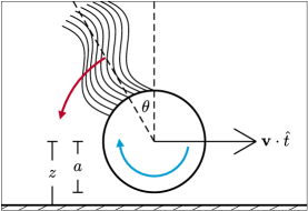
As illustrated in Fig 3, physical reasoning suggests that the orientation undergoes a subcritical pitchfork bifurcation as the cell approaches the wall. Consider the torques acting on a cell in contact with a wall. If the flagella are slightly misaligned with the local normal, the cell moves laterally over the surface. As the resulting velocity gradients are greater in the narrow gap between cell and the surface than between the top of the cell and the bulk fluid, there is a net torque on the cell that turns the flagella normal to the surface petroff2018nucleation ; das2019transition . This torque is countered by drag on the flagella, however because the flagella of T. majus are relatively short compared to the cell size, viscous dissipation between the cell and the wall dominates, and is stable das2019transition . As the angle between the flagella and the surface normal grows, the flagella are rotated closer to the surface and hydrodynamic attraction das2019transition between the flagella and the surface becomes dominant. These competing torques suggest there is an unstable orientation at which the torque due to the flow between the cell body and the surface balance hydrodynamic attraction between the flagella and the surface. Thus, we expect that as the cell approaches the wall, the fixed point undergoes a bifurcation that stabilizes this orientation while generating unstable fixed points at . This process is typical of a subcritical pitchfork bifurcation.
This reasoning gives little insight into torques acting on cell that is turned to large angle, where contact forces between hundreds of rotating flagella and the surface are likely important. In response to this uncertainty, we make the simple assumption that the angular velocity saturates to some maximum value as .
To analyze the motion of cells in contact with a surface, we consider a minimal model that includes the normal form a subcritical pitchfork bifurcation predicts a bounded angular velocity. We propose
| (2) |
where the angle defines the edge of the basin of attraction of the bound orientation , is a rate coefficient, and is chosen such that is continues.
To test this hypothesis, we map the escape of a cell from the wall onto a first passage problem. Our measurements provide the probability that a cell that strikes the surface at a particular angle escapes at an angle within a time . We recapitulate these measurements in our model. For a given choice of , , , and rotational diffusion coefficient , we calculate the probability that a cell is rotated to an orientation (at which it escapes in the positive sense) before it is turned to an orientation . We calculate how this probability varies with the incident angle and find the distribution of first passage times.
We solve this first passage problem by way of a Fokker-Planck equation. Let be the probability density that the flagella of cell exert a net force oriented at an angle off of the surface normal at time . The probability distribution evolves in time as
| (3) |
For a given choice of model parameters, we discretize the interval into 300 elements and numerically integrate this equation using standard finite element methods for linear equations reddy2005introduction . We then calculate the probability flux . The probability that a cell escapes at a positive angle (as in Fig. 1[a]) is , where is the probability flux evaluated at the absorbing boundaries .
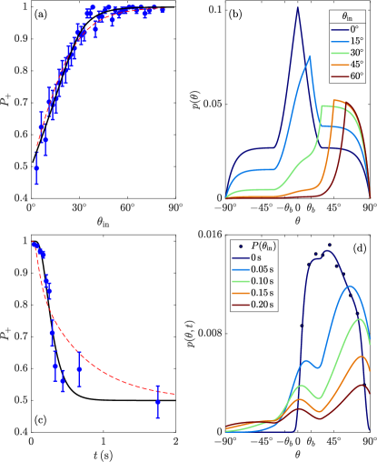
We first consider the fate of a cell that collides with the surface at an angle (see Fig 4[a]). To find the fraction of cells that eventually escape in the positive sense, we solve for the Greens functions of the steady-state Fokker-Planck equation with a source of cells at . Figure 4(b) shows solution for five values of . Note that as increases, the fraction of cells that accumulate at decreases.
We fit three parameters to match these solutions to the measured variation of (see Fig 4[a]). The first parameter is the width of the basin of attraction of trapped orientation . We find that . The second fit parameter represents the typical fluctuations about the stable orientation . Because these fluctuations are somewhat larger than the basin of attraction of this fixed point, we conclude that cells in this experiment are only momentarily bound. The final fit parameter is the rotational Peclet number, which represents the ratio of the rates at which a cell is turned by hydrodynamic and Brownian torques. Its reasonably large value indicates that cells that collide with the surface outside the basin of attraction of rarely become trapped.
Next, we consider how the probability that a cell escapes in the positive sense decays as it remains in contact with the surface, as shown in Fig. 4(c). We solve Eq. (3) using the measured distribution of as the initial distribution of orientations and the parameter values fit to Fig. 4(a). Figure 4[d] shows the evolution of distribution of orientations as cells escape from the surface and become trapped at . These dynamics uniquely define the functional form of , where the diffusive timescale is the only unknown parameter. We fit by rescaling the measured decay of to the predicted functional form. We find rad2/s, which implies s-1 and rad/s. Fig. 1(c) compares the measured cumulative distribution function of escape times to the probability that a cell escapes before time . Similarly, the escape entropy corresponding to the best fit of is shown in Fig. 1(f).
The measured variations of with incident angle and time are inconsistent with the physical null model that the far field hydrodynamic effect of a boundary can be extended to model the motion of a cell in contact with a surface. We repeat the procedure described above to fit to the Fokker-Planck equation where the drift velocity is described by Eq. 1 and the prefactor on the sinusoidal term is fit. As shown in Fig. 3(a–b), while the variation with incident angle is equally well described by either model, the first passage times differ markedly.
III.3 Matching near and far field dynamics
The agreement between theory and observation shown in Fig. 3 leads us to conclude that Eq. (2) captures the essential qualities of the near field dynamic coupling between a T. majus cell and a wall. Two questions remain. First, these results do not constrain the distance between a cell and the wall at which a cell may become trapped. Additionally, it is not clear why all cells escape the surface at narrowly distributed angles (see Fig. 1[e]).
To answer these questions we match the short-range angular velocity (Eq. [2]) with the far field (Eq. [1]). To match the cubic decay of torques on the cell predicted by the far field dynamics, we propose that at short distances from the wall and small angles ()
| (4) |
where is the distance from the wall at which the orientation becomes stable. In the limit and , this expansion matches Eq. (1) where . We conclude that cells may become bound when the gap between the cell and the wall becomes similar to the cell radius. This result is intuitively consistent with the physical model summarized in Fig. 3, in which the bound state is stabilized because velocity gradients in the gap between cell and surface are sharper than those between the cell and the bulk fluid.
Finally, we consider the escape of a cell from the surface. All cells escape by first swimming tangent to the wall. We consider the simplest function that interpolates between the constant angular velocity found when the cell is in contact with the surface the cubic decay predicted by the far field. We expect the angular velocity of an escaping cell to decay as
| (5) |
Cells escape with speed , where is a correction to the swimming speed due to advection by the cell’s hydrodynamic image. Its functional form is provided in Ref. drescher2011fluid .
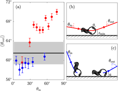
We numerically integrate Eq. (5) to find the asymptotic orientation of a cell’s motion that is initially swimming tangent to the surface. Taking the initial distance between the cell and the wall to be one cell radius, we find—with no fit parameters—a predicted escape angle of . As shown in Fig. 4, this value is consistent with the measured escape angle found for cells that remain in contact with the surface for at least s. Cells that are identified by the tracking algorithm to be in contact with the surface for shorter durations escape at slightly greater angles, which increase with the incident angle. We believe that these cells are turned away from the surface by hydrodynamic torques before they come in physical contact with the wall, similar to the scattering of Chlamydomonas lushi2017scattering . Taking the minimum distance between the cell and wall to be , the observed increase in is consistent with a value of that increases monotonically with .
IV Conclusion
In conclusion, we have used the scattering statistics of collisions between T. majus and a hard wall to probe the near-field dynamics between a fast-swimming multiflagellated cell and a surface. A simple physical picture emerges. As a cell approaches a wall, dipole-dipole interactions with its hydrodynamic image turn the cell to swim parallel to the surface. When the distance between the cell and the surface decreases to a value of cell radii the torques acting on the cell change qualitatively. Shear forces in the gap between the cell and the surface orient the cell to exert a force normal to the surface. The cell becomes trapped. We find that the basin of attraction of the fixed point is narrow. Consequently, most cells collide with the surface outside of this basin and rapidly escape while maintaining their direction of motion tangent to the surface. When a cell is captured by the stable fixed point, its eventual escape is symmetric and all information of its approach to the wall is erased. Because all cells that come in contact with the surface escape by first swimming tangent to the wall at a distance of one cell radius, all cells escape at similar angles. A minority of cell are turned from the wall before coming in physical contact with the wall and escape at slightly greater angles.
Because the near field dynamics described in Eq. (2) are phenomenological, it remains unclear whether the stability of trapped cells is primarily due to hydrodynamic of contact forces. We suspect hydrodynamic torques dominate at small and collisions become increasingly important as the flagella are turned toward the wall. Indeed, Das and Lauga das2019transition analyzed a simplified model of T. majus with a single short flagellum to show that hydrodynamic torques are sufficient to stabilize the bound state. However, this analytic model—which was used primarily to provide intuition for an aspect of their more detailed numeric results—neglected hydrodynamic attraction of the flagella to the surface. As the simplified cell approaches the surface, the orientation becomes stable through a supercritical pitchfork bifurcation, rather than the subcritical bifurcation found here. We encourage computational fluid mechanicians to simulate the fluid flow around a spherical cell with a single flagellum near a wall and compare the angular velocity of a cell to Eqs. (2) and its matching to the far field.
The trapping of T. majus by a hard surface is the first step in the nucleation of active chiral crystalspetroff2015fast . In our previous study, we found that isolated cells remain trapped by the surface for tens of seconds petroff2018nucleation , much longer than average trapping time s found here. We ascribe this difference to the roughness of the walls made of PDMS to those made of polished glass. As the gap between the cell and the surface is presumably much smaller if the surface is smooth, the velocity gradient between the cell and the wall—which stabilize the trapped cell—is much sharper. Consequently, we expect cells by be more strongly trapped by cover slips than microfluidic walls. The escape of cells could be further enhanced by the sporadic binding of quickly-rotating flagella to the PDMS, which may increase the effective rotational diffusion of cells, causing cells to escape more quickly from PDMS than from glass. This result highlights the importance of an unnaturally smooth surface for the formation of active chiral crystals. We are consequently doubtful of the biological significance of this form of collective motion.
Nonetheless, the dynamics by which cells scatter from rough surfaces are likely quite important for the ecology of T. majus. These bacteria, which live in the pore space of water saturated sand, exude mucus tethers from their posteriors to attach to sand grains schulz2001big . It is not understood how cells attach this mucus thread to a surface. Our results show that when a cell collides with a surface outside of a narrow basin of attraction, it rapidly escapes rather than swimming parallel to the surface. As free-swimming T. majus frequently drag short tethers as they swim fenchel1998veil and collisions quickly turn the cell posterior toward the wall, it is plausible that these collisions facilitate attachment of the tether to the surface. Given a swimming speed of m/s and a pore size of several tens of micron, these collision dynamics provide several opportunities a second for a dragged tether to stick to a surface.
Acknowledgements.
We thank Dabasish Das and Arshad Kudrolli for their insightful comments and discussions. This work was supported by NSF Grant no. PHY-2042150.Appendix A Appendix: Supplemental Videos
Two videos of collisions between a cell and a surface are provided.
-
1.
Video SV1.mp4 shows a representative collision of a T. majus with the wall of a microfluidic chamber in which the cell is only momentarily in contact with the surface. The trajectory is similar to that shown in Fig. 1(a).
-
2.
SV2.mp4 shows a collision between the cell that strikes the surface close to the local normal and becomes trapped. This collision is also shown in Fig 1(b).
References
- [1] Madilyn Fletcher and Dwayne C Savage. Bacterial adhesion: mechanisms and physiological significance. Springer Science & Business Media, 2013.
- [2] Rasika M Harshey. Bacterial motility on a surface: many ways to a common goal. Annual Reviews in Microbiology, 57(1):249–273, 2003.
- [3] Farooq Azam and Richard A Long. Sea snow microcosms. Nature, 414(6863):495–498, 2001.
- [4] Manoshi S Datta, Elzbieta Sliwerska, Jeff Gore, Martin F Polz, and Otto X Cordero. Microbial interactions lead to rapid micro-scale successions on model marine particles. Nature communications, 7(1):11965, 2016.
- [5] E. Lauga and T.R. Powers. The hydrodynamics of swimming microorganisms. Reports on Progress in Physics, 72(9):096601, 2009.
- [6] Diego Lopez and Eric Lauga. Dynamics of swimming bacteria at complex interfaces. Physics of Fluids, 26(7):400–412, 2014.
- [7] William M Durham, Olivier Tranzer, Alberto Leombruni, and Roman Stocker. Division by fluid incision: Biofilm patch development in porous media. Physics of Fluids, 24(9):091107, 2012.
- [8] Knut Drescher, Kyriacos C Leptos, Idan Tuval, Takuji Ishikawa, Timothy J Pedley, and Raymond E Goldstein. Dancing volvox: Hydrodynamic bound states of swimming algae. Physical Review Letters, 102(16):168101, 2009.
- [9] Frank D Müller, Dirk Schüler, and Daniel Pfeiffer. A compass to boost navigation: cell biology of bacterial magnetotaxis. Journal of bacteriology, 202(21):e00398–20, 2020.
- [10] Nicolas Waisbord, Amin Dehkharghani, and Jeffrey S Guasto. Fluidic bacterial diodes rectify magnetotactic cell motility in porous environments. Nature Communications, 12(1):5949, 2021.
- [11] Alexander Petroff, Alejandra Rosselli-Calderon, Ben Roque, and Pradeep Kumar. Phases of active matter composed of multicellular magnetotactic bacteria near a hard surface. Physical Review Fluids, 7(5):053102, 2022.
- [12] Stephen H Zinder and Martin Dworkin. Morphological and physiological diversity. The prokaryotes, pages 185–220, 2006.
- [13] W.E. De Boer, J.W.M. La Rivière, and AL Houwink. Observations on the morphology of thiovulum majus hinze. Antonie van Leeuwenhoek, 27(1):447–456, 1961.
- [14] JWM LaRiviere. Cultivation and properties of thiovulum majus hinze. In Symposium on marine microbiology. Charles C Thomas, Publisher, Springfield, Ill, pages 61–72, 1963.
- [15] CO Wirsen and HW Jannasch. Physiological and morphological observations on thiovulum sp. Journal of bacteriology, 136(2):765–774, 1978.
- [16] Ian PG Marshall, Paul C Blainey, Alfred M Spormann, and Stephen R Quake. A single-cell genome for thiovulum sp. Applied and environmental microbiology, 78(24):8555–8563, 2012.
- [17] H.N. Schulz and B.B. Jørgensen. Big bacteria. Annual Reviews in Microbiology, 55(1):105–137, 2001.
- [18] F. Garcia-Pichel. Rapid bacterial swimming measured in swarming cells of thiovulum majus. Journal of bacteriology, 171(6):3560–3563, 1989.
- [19] Roland Thar and Michael Kühl. Bacteria are not too small for spatial sensing of chemical gradients: an experimental evidence. Proceedings of the National Academy of Sciences, 100(10):5748–5753, 2003.
- [20] T. Fenchel and R.N. Glud. Veil architecture in a sulphide-oxidizing bacterium enhances countercurrent flux. Nature, 394(6691):367–369, 1998.
- [21] R. Thar and M. Kühl. Conspicuous veils formed by vibrioid bacteria on sulfidic marine sediment. Applied and environmental microbiology, 68(12):6310–6320, 2002.
- [22] Alexander P Petroff, Alexis L Pasulka, Nadine Soplop, Xiao-Lun Wu, and Albert Libchaber. Biophysical basis for convergent evolution of two veil-forming microbes. Royal Society open science, 2(11):150437, 2015.
- [23] Alexander P Petroff, Xiao-Lun Wu, and Albert Libchaber. Fast-moving bacteria self-organize into active two-dimensional crystals of rotating cells. Physical review letters, 114(15):158102, 2015.
- [24] AP Petroff and A Libchaber. Nucleation of rotating crystals by thiovulum majus bacteria. New Journal of Physics, 20(1):015007, 2018.
- [25] Debasish Das and Eric Lauga. Transition to bound states for bacteria swimming near surfaces. Physical Review E, 100(4):043117, 2019.
- [26] Kenta Ishimoto. Bacterial spinning top. Journal of Fluid Mechanics, 880:620–652, 2019.
- [27] Knut Drescher, Jörn Dunkel, Luis H Cisneros, Sujoy Ganguly, and Raymond E Goldstein. Fluid dynamics and noise in bacterial cell–cell and cell–surface scattering. Proceedings of the National Academy of Sciences, 108(27):10940–10945, 2011.
- [28] Enkeleida Lushi, Vasily Kantsler, and Raymond E Goldstein. Scattering of biflagellate microswimmers from surfaces. Physical Review E, 96(2):023102, 2017.
- [29] Howard C Berg. Random walks in biology. Princeton University Press, 1993.
- [30] Emily Qing Zang Moen, Kristian Stølevik Olsen, Jonas Rønning, and Luiza Angheluta. Trapping of active brownian and run-and-tumble particles: A first-passage time approach. Physical Review Research, 4(4):043012, 2022.
- [31] B.B. Jørgensen and N.P. Revsbech. Colorless sulfur bacteria, beggiatoa spp. and thiovulum spp., in o2 and h2s microgradients. Applied and Environmental Microbiology, 45(4):1261–1270, 1983.
- [32] George M Whitesides, Emanuele Ostuni, Shuichi Takayama, Xingyu Jiang, and Donald E Ingber. Soft lithography in biology and biochemistry. Annual review of biomedical engineering, 3(1):335–373, 2001.
- [33] JR Blake. A note on the image system for a stokeslet in a no-slip boundary. In Proc. Camb. Phil. Soc, volume 70, pages 303–310. Cambridge Univ Press, 1971.
- [34] Steven H Strogatz. Nonlinear dynamics and chaos with student solutions manual: With applications to physics, biology, chemistry, and engineering. CRC press, 2018.
- [35] Junuthula Narasimha Reddy. An introduction to the finite element method, volume 3. McGraw-Hill New York, 2005.