Federated Learning for Medical Image Analysis: A Survey
Abstract
Machine learning in medical imaging faces a fundamental dilemma, namely the small sample size problem. Recent studies suggest using images pooled from different acquisition sites to improve statistical power. However, medical images cannot be easily shared for model training due to privacy protection reasons. As a promising solution, federated learninghas attracted considerable attention recently. In this paper, we conduct a comprehensive survey of the recent development of federated learning in medical image analysis. We first introduce the background knowledge of federated learning. Then we present a comprehensive review of federated learning methods for medical image analysis. Specifically, existing methods are categorized based on three critical aspects of a federated learning system, including client end, server end, and communication techniques. In each category, we summarize the existing federated learning methods according to specific research problems in medical image analysis. In addition, we provide a review of existing benchmark medical imaging datasets and software platforms for current federated learning research. We also conduct an experimental study to empirically evaluate typical federated learning methods for medical image analysis. This survey can help to better understand the current research status, challenges, and potential research opportunities in this promising research field.
Index Terms:
Federated learning, machine learning, medical image analysis, data privacyI Introduction
Medical image analysis has been greatly pushed forward by computer vision and machine learning [1, 2, 3, 4]. The remarkable success of modern machine learning methods, e.g., deep learning [5], can be attributed to the building and release of grand-scale natural image databases, such as ImageNet [6] and Microsoft Common Objects in Context (MS COCO) [7]. Unlike natural image analysis, the field of medical image analysis still faces the fundamental challenge of the “small-sample-size” problem [8, 9]. Based on small sample data, it is difficult for us to estimate real data distributions, greatly hindering the building of robust and reliable learning models for medical image analysis. An intuitive and direct solution to this small sample size problem is to pool images from multiple sites together and build larger datasets to train high-quality machine learning models. However, sharing medical imaging data between different sites is intractable due to strict privacy protection policies such as Health Insurance Portability and Accountability Act (HIPAA) [10] and General Data Protection Regulation (GDPR) [11]. For example, the United States HIPAA has rigidly restricted the exchange of personal health data and images [10]. Thus, directly sharing and pooling medical images across different sites/datasets is typically infeasible in real-world practice.
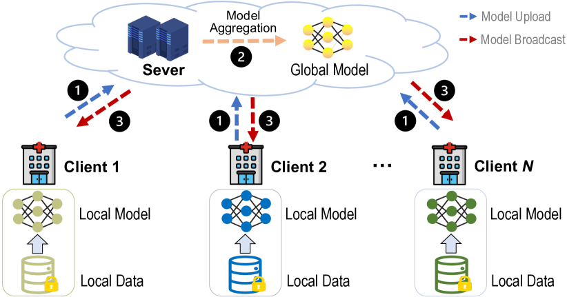
As a promising solution for dealing with the small-sample-size problem and protecting individual privacy, federated learning [12, 13, 14] has become a spotlight research topic in recent years, which aims to train machine learning models in a collaborative manner without exchanging/sharing data among different sites. As an emerging machine learning paradigm, federated learning deliberately avoids demand for all the medical data residing in one single site. Instead, as shown in Fig. 1, it depends on model aggregation/fusion techniques to jointly train a global model which is then sent/broadcast to each site for fine-tuning and deployment.
There have been several survey papers on federated learning [15, 16, 17, 18, 19, 20], but further technical details about facilitating federated learning in medicine and healthcare are not yet covered. Several recent surveys introduce the applications of federated learning in medicine and healthcare areas [21, 22, 23, 24, 25]. However, some of them focus on electronic health records [21, 22] or internet of medical things [26], without paying attention to medical imaging. And some survey papers cover very broad areas [23, 24], without detailed introduction on federated learning in medical image analysis.
To fill this gap, we review and discuss recent advances in federated learning for medical image analysis in this paper. Our survey paper has significant differences from most previous ones in the following aspects. First, we summarize the existing methods from a system perspective. Specifically, we categorize different approaches into three groups: (1) client-end learning methods, (2) server-end learning methods, and (3) server-client communication methods. Different from previous surveys that are based on multiple research issues in federated learning, this categorization can be more intuitive and clear to picture federated learning. Second, when elaborating on the methods in each group, we have designed a novel “question-answer” paradigm to introduce the motivation and mechanism of each method. We deliberately extract the common questions behind different methods and pose them first in each subsection. These questions stem from the characteristics of medical imaging, thus this “question-oriented” approach of introduction is helpful for providing more insights into different methods. Third, we emphasize the implementation of federated learning techniques for medical image analysis. Specifically, we introduce popular software platforms and benchmark medical imaging datasets for federated learning research in medical imaging. In addition, we also conduct an experiment on a benchmark medical image dataset to illustrate the utility and effectiveness of several typical federated learning methods.
The remainder of this paper is organized as follows. In Section II, we introduce the background and motivation of federated learning. We summarize existing federated learning studies for medical image analysis in Section III, IV, V, and VI. In Section VII, software platforms that support federated learning system development are presented. In Section VIII, we introduce medical image datasets that have been widely used in federated learning research. We conduct an experimental study in Section IX to compare several federated learning methods. Challenges and potential research opportunities are discussed in Section X. Finally, we conclude this survey paper in Section XI.
II Background
II-A Motivation
II-A1 Privacy Protection in Medical Image Analysis
Patient data protection has become an important issue in the digital era. Using and selling patient data has many negative implications [27]. First, these data may be used by third parties (e.g., hackers) with unauthorized access. Second, patients may lose control over their health data if the data owners are purchased by third parties who may use the data without the consent of the patients. Third, the anonymized patient data may be reidentified which can lead to the targeting of disadvantaged populations and discrimination. Thus, many governments have introduced tough new laws and regulations on privacy data protection, such as the CCPA in the United States [28] and GDPR in Europe [11]. Collecting, sharing, and processing of personal data are strictly constrained, and violating these laws and regulations may face high-cost penalties [29].
With these strict restrictions from laws, medical images, one of the most important privacy information, cannot be easily shared among different sites/datasets. To this end, federated learning, a distribution-oriented machine learning paradigm without cross-site data sharing, has emerged as a promising technique for developing privacy-preservation machine learning models, thus paving the way for the applications of medical artificial intelligence (AI) in real-world practice.
II-A2 Medical Image Data Limitation and Bias
A traditional way to train machine learning models is to use medical images from a specific site/dataset, with at least two drawbacks.
-
(1)
Due to the cost of imaging and labeling, the amount of images in local datasets is usually small (e.g., tens or hundreds) which is not enough to train a model with good generalizability. This is the well-known “small-sample-size” problem [8, 9]. This problem may lead to sub-par learning performance of a model, and produce results that lack statistical significance.
-
(2)
Data from a specific site/dataset may be biased in distribution and not representative of the true data distribution. It is not unusual that medical sites contain unbalanced data. For example, in some sites, healthy subjects significantly outnumber the patients which may lead to a biased model towards the healthy ones.
Federated learning helps address these limitations, aiming to “pool” medical images together in a distributed way, thereby greatly increasing the sample size. This can effectively take advantage of available data from multiple sites to enhance the statistical power of machine learning models.
II-B Problem Formulation of Federated Learning
Suppose there are independent clients/sites with their own datasets {}, respectively. Each of the clients/sites cannot get access to others’ datasets. Federated learning (FL) aims to collaboratively train a machine learning model by gathering information from those clients /sites without sharing their raw data. The ultimate output of FL is the learned model which is broadcast to each client for deployment, and the generalizability of by FL should outperform each local model (typically with the same model architecture as ) learned through local training.
II-C Typical Process of Federated Learning
In Fig. 1, we illustrate the typical process of federated learning that is embodied in a “client-server” architecture. This process encompasses the Federated Averaging algorithm (FedAvg) proposed by McMahanet al. [12]. It serves as the foundation of most popular algorithms for federated learning. A server triggers and orchestrates the entire training process (without accessing clients’ data) until a certain stop criterion is met, with key components introduced as follows.
-
1)
Client Selection. The server selects a set of clients that meet certain requirements. For example, a medical site/dataset might only check in to the server when it can correctly get access to the intranet of a federation with relatively good bandwidth.
-
2)
Local Training. Every selected client locally trains a machine learning model through optimization methods (e.g., stochastic gradient descent) based on its local data. In the beginning, the model weights can be initialized both on each client or by the server.
-
3)
Model Upload. All the clients that have been selected upload their model (e.g., weights) to the server.
-
4)
Model Aggregation. The server computes/updates a global model by aggregating all client models.
-
5)
Broadcast. The server sends/broadcasts the current shared global model (e.g., weights) to the selected clients. After downloading and deploying the shared model, a client may continue to update/fine-tune it locally using its private data.
III Federated Learning for Medical Image Analysis: A System Perspective
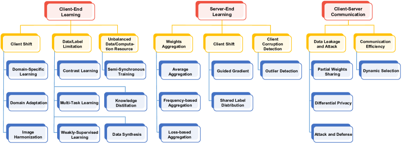
Federated learning (FL) provides a generic framework for distributed learning with privacy preservation. Most existing machine/deep learning methods can be plugged and integrated into an FL framework. For example, a U-Net [30] can be used in each client for medical image segmentation and is trained in a federated manner. Federated learning is concerned with multiple issues such as data, machine learning models, privacy protection mechanisms, and communication architecture. As shown in Fig 2, from a system perspective, we categorize existing FL approaches for medical image analysis into three groups: 1) client-end methods, 2) server-end methods, and 3) communication methods. In each group, different methods are clustered according to the specific research problems they aim to address which will be elaborated in the following sections.
IV Client-End Learning
IV-A Client End: Domain Shift Among Clients
Problem: Different imaging sites often have significant cross-site data distribution variance caused by different scanning settings and/or subject populations, so how to avoid its negative influence on model training?
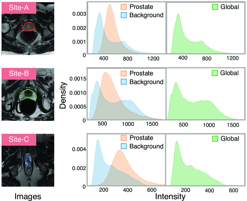
In practice, multi-site medical images may have significantly different data distributions (data heterogeneity), which is the well-known “domain shift” problem [3] (also referred to as “client shift” in an FL system). As shown in Fig. 3, the three imaging sites have significantly different intensity distributions (in terms of both region-wise and global intensity). In an FL system, domain shifts may cause difficult convergence of the global model and performance degradation of some clients. In the following, we present the relevant studies that focus on reducing domain shift among clients for FL research.
(1) Domain-Specific Learning. Federated learning aims to train a global model that fits well with all clients. Due to cross-site data heterogeneity, the global model may not be able to achieve good performance for all clients. One strategy is fine-tuning the global model using domain-specific (local) data to make it more suitable for a specific client. This method is also known as customized/personalized FL [32, 33, 34].
Feng et al. [35] propose an encoder-decoder structure within an FL framework for magnetic resonance (MR) image reconstruction. A globally shared encoder is maintained on the server end to learn domain-invariant representations, while a client-specific decoder is trained with local data to take advantage of domain-specific properties of each client. Similar strategies can also be found in [36, 32]. Chakravarty et al. [37] propose a framework that combines a Convolutional Neural Network (CNN) and a Graph Neural Network (GNN) to tackle the domain shift problem among clients and apply it to chest X-ray image classification. Specifically, model weights of the CNN are shared across clients to learn site-independent features. To address site-specific data variations, a local GNN is built and fine-tuned with local data in each client for disease classification. In this way, both site-independent and site-specific features can be learned. Xu et al. [38] propose an ensemble-based framework to deal with the client shift for medical image segmentation. Their framework is composed of a global model, personalized models, and a model selector. Instead of only using the global model to fit all the client data, they propose to leverage all the produced personalized models to fit different client data distributions through a model selector. Jiang et al. [39] propose to train a locally adapted model that accumulates both global gradients (aggregated from all clients) and local gradients (learned from local data) to optimize the model performance on each client. This helps effectively avoid biased performance of the global model on different clients caused by client domain shift. Ke et al. [40] build an FL framework based on Generative Adversarial Network (GAN) to facilitate harmonization (color normalization) of histopathological images. In this method, each client trains a local discriminator to capture client-specific image style, while the server maintains and updates a global generator model to generate domain-invariant images, thus achieving histopathological image harmonization. Similarly, Wagner et al. [41] propose a GAN model for histopathological image harmonization. In their method, a reference dataset is assumed to be accessible for all clients, which can help the training of all the local GANs at each client.
(2) Domain Adaptation. Domain adaptation is a sound machine learning technique that has been widely used in medical image analysis [3]. It aims to reduce domain shift among different medical image datasets and enhance the generalizability of a learning model. Many medical-related FL studies resort to domain adaptation for improved performance.
Li et al. [42] use domain adaptation to align distribution differences of functional MRI data among clients. In their method, data in each client are added with noise to enhance privacy protection. A domain discriminator/classifier is trained on these data with noises to reduce domain shift. Dinsdale et al. [43] propose a domain adaptation-based FL framework to remove domain shift among clients caused by different scanners. In their framework, medical image features are assumed to follow Gaussian distributions, and the mean and standard deviation of the learned features can be shared among clients. During the training of each client model, a label classifier and a domain discriminator are jointly trained to learn features that are domain-invariant, i.e., removing domain shift. Andreux et al. [44] leverages batch normalization (BN) in a deep neural network to handle client (histopathology datasets) shift. Guo et al. [45] propose a federated learning method for MRI reconstruction, where the learned intermediate latent features among different clients are aligned with the distribution of latent features of a reference site.
(3) Image Harmonization. Qu et al. [46] propose a generative replay strategy to handle data heterogeneity among clients. They first train an auxiliary variational auto-encoder (VAE) to generate medical images which resemble the input images. Then each client can optimize their local classifier using both the real local data and synthesized data with similar data distribution of other clients. In this way, domain shift can be reduced. Yan et al. [47] employ cycleGAN [48] to minimize the variations of medical images among clients. One client/site with low data complexity is selected as a reference, then cycleGAN is used to harmonize medical images from other clients to the reference site. Jiang et al. [49] propose a frequency-based harmonization method to reduce client shift in medical images. Medical images are firstly transformed into frequency domain and phase components are kept locally, while the average amplitudes from each client are shared and then normalized to harmonize all the client medical images.
IV-B Client End: Limited Data and Labels
Problem: Medical imaging datasets are often small-sized and lack label/annotation information, so how to avoid their negative influence on model training (e.g., biased training)?
In real-world practice, there are often limited medical images in one client/site, and labeled medical images are even fewer due to the high cost of image annotation/labeling. A client model may be badly trained with limited labeled data, which can cause negative influences on the entire federation. Therefore, how to alleviate the small-sample-size problem is an important topic of FL in medical image analysis.
(1) Contrast Learning. Contrastive learning [50, 51, 52] is a self-supervised method that can learn useful representations of images by using unlabeled data. A model trained with contrast learning can provide good initialization for further fine-tuning (with a few labeled data) on downstream tasks. Contrast learning has been introduced into federated learning for handling medical data shortage. [53, 54] use contrast learning to pretrain (initialize) the encoder of a U-Net in each client, then the global U-Net is fine-tuned with limited labeled medical images. In this way, the negative influence caused by the shortage of labeled medical images can be largely reduced. Similar strategies can be found in [55].
(2) Multi-Task Learning. Multi-task learning [56] typically solves multiple but related learning tasks at the same time, which can exploit commonalities across tasks. When the training data for each task are small-sized, jointly learning of different tasks can actually share data which is an effective approach for data augmentation. Smith et al. [57] propose a novel optimization framework, i.e., MOCHA, which extends classic multi-task learning in the federated environment. MOCHA is based on a bi-convex alternating method and is guaranteed to converge. Huang et al. [58] build a federated multi-task framework in which several related tasks, i.e., attention-deficit/hyperactivity disorder (ADHD), autism spectrum disorder (ASD), and schizophrenia (SCZ), are jointly trained. In this method, encoders for each task in clients are federated to derive a global encoder that can learn common knowledge among related mental disorders.
(3) Weakly-Supervised Learning. Weakly-supervised learning [59] is an extensive group of methods that train a model under weak supervision. Weak supervision information typically includes three types. 1) Incomplete supervision. Only a small subset of labeled training data is provided while the other data has no labels. Semi-supervised learning [60, 61] is a popular solution for such scenarios. 2) Inexact supervision. Only coarse-grained labels are provided for the training data. Multiple-instance learning [62, 63] is a representative method to handle this problem. 3) Inaccurate supervision. Not all the provided labels are correct. Learning from noisy labels [64, 65] is the corresponding technique.
Yang et al. [66] introduce semi-supervised learning into FL for chest CT segmentation. In their method, unlabeled CT images are leveraged to assist the federated training. For unlabeled CT images in a client, the global model assigns them pseudo labels. Meanwhile, it also outputs predictions on augmented data of the original unlabeled images. A consistency loss is utilized on these predictions to further adjust the global model weights. Lu et al. [67] use multiple-instance learning for local model training on the task of pathology image classification. Whole slide images (WSIs) and weak annotation (e.g., patient or not) are used as the input, with no region-based labels provided. And multiple patches (instances) of a WSI are fed into a network for training. Kassem et al. [68] build a semi-supervised FL system for surgical phase recognition based on laparoscopic cholecystectomy videos. The key idea is to leverage the temporal information in labeled videos to guide unsupervised learning on unlabeled videos.
(4) Knowledge Distillation. Kumar et al. [69] leverage knowledge distillation for COVID-19 detection in chest X-ray images. The network trained on similar data (other chest X-ray image datasets) is used as a “teacher”, while the client model is a “student”. By matching the softmax activation output of the teacher, the student (client model) can learn useful knowledge for the task. In this way, it can help alleviate the demand for large data during the federation learning process.
(5) Data Synthesis. Zhu et al. [70] propose an FL framework with virtual sample synthesis for medical image analysis. Given an image in the client, the authors first use Virtual Adversarial Training [71] to generate synthetic images that are similar to , and then use all the synthesized images for local model training. Peng et al. [72] propose a federated graph learning framework for brain disease prediction, where a Graph Convolutional Network (GCN) is used as the learning model in each client. Considering the missing nodes and edges when separating the global graph into local graphs, the authors leverage network inpainting to predict the missing nodes and their associated edges. This helps complete the graphs for GCN training in each client, with results suggesting its effectiveness in graph data synthesis and augmentation.
IV-C Client End: Unbalanced Data and Computation Resource
Problem: Different medical sites/datasets may have significantly different data scales and computation resources (e.g., number of GPUs) in real-world practice, so how to reduce its influence on federated training?
In the standard FL template (i.e., FegAvg), each client model conducts a predefined number of training epochs (with equal batches and learning rate) before reaching a synchronization time point when it sends its model to the server. However, if the clients have significantly different computation and data resources, this may lead to computational inefficiencies and slow convergence of model optimization. For example, each client is supposed to conduct 50 epochs’ updates before model uploading. In such a setting, a client with advanced GPUs may take 1 second, while a client with weak computation utility may take 100 seconds. Thus, the stronger client will have to spend 99 seconds waiting for weight sharing.
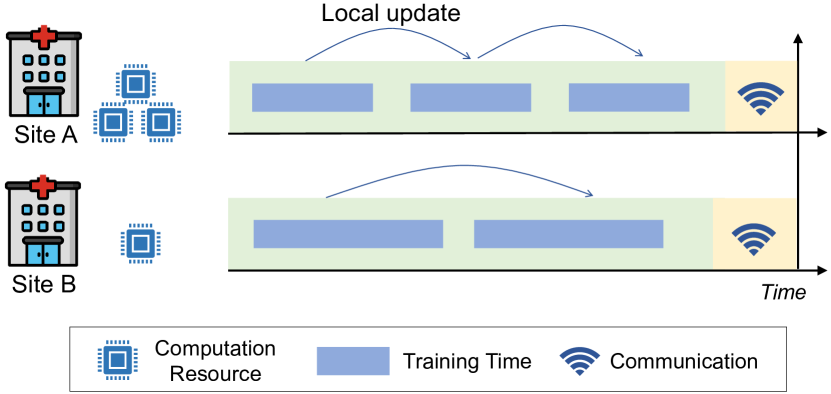
Aiming at handling the computational and data scale heterogeneity among clients, Stripelis et al. [73] propose a Semi-Synchronous Training strategy in federated learning and apply it to brain age prediction. As shown in Fig. 4, each client conducts a variable number of updates (epochs) between synchronization time points which depend on its computational power and data scale. Higher computation power or fewer local data will lead to more local updates (epochs).
V Sever-End Learning
V-A Sever End: Weight Aggregation
Problem: How to aggregate the weights of clients properly to avoid performance degradation after each client-server communication?
Chen et al. [74] propose a Progressive Fourier Aggregation strategy at the server end. Based on previous studies that low-frequency components of parameters form the basis of deep network capability [75], only the low-frequency components are aggregated to share knowledge learned from different clients, while the high-frequency parts are disregarded. Li et al. [76] consider the training loss of each client as the impact factor of the weight aggregation in FL for COVID-19 detection. The client with relatively bad performance caused by uneven image data will get a smaller weight for the global weight aggregation.
V-B Sever End: Domain Shift Among Clients
Problem: The domain shift among clients may cause non-convergence of federated models, so how to avoid this from the server end?
Hosseini et al. [77] argue that the image heterogeneity between different medical centers (clients) may lead to a biased global model, i.e., a model that has good performance for some clients while exhibiting inferior performance for the other clients. Thus, they propose a revised optimization objective (motivated by fair resource allocation approaches in wireless network research), to facilitate uniform model performance in histopathology image analysis across all the clients. In their method, the clients for which the global model has inferior performance will contribute more to the total loss function. [78] leverage the guided-gradient to optimize the global model. After aggregating all the local weights of the clients, only positive values of the aggregated weights are used to update the global. The authors argue that this is helpful for the global gradient descent to go towards the optimal direction, and the guided-gradient can reflect the most influential regions of the medical images.
Luo et al. [79] propose a method called federated learning with shared label distribution (FedSLD) for medical image classification by mitigating label distribution differences among clients. In their method, it is assumed that the amount of samples of each category (label distribution) is known for the entire federation. During local model training in each client, a weighted cross-entropy loss is designed as the batch loss. The weight is computed based on the label distributions in each batch, with respect to their label distributions across the entire federation.
V-C Sever End: Client Corruption/Anomaly Detection
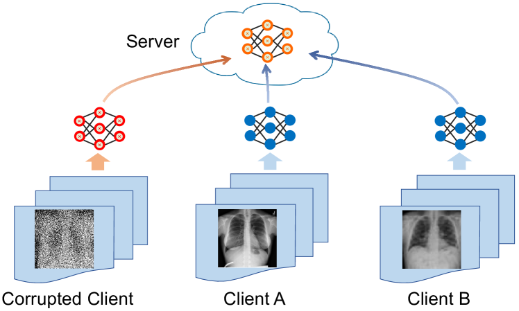
Problem: If one or more clients are corrupted by very noisy image labels or malicious attacks, how to avoid its negative influence on the entire federation?
Classic FL framework holds the assumption that all the clients work normally. In this context, the term “normal” means that a client is trained with correctly labeled images or the client is honest without malicious attack. In real-world practice (as shown in Fig. 5), however, a client may be trained with “dirty” medical images that have noisy labels, poor scanning qualities, or suffer from poisoning attacks from malicious parties. How to deal with this issue is critical for ensuring the safety of a medical federated learning system. Alkhunaizi et al. [80] propose a sever-end outlier detection method for medical images, called Distance-based Outlier Suppression (DOS), which is robust to client corruption/failure. In this method, the weight of each client is calculated based on an anomaly score for the client using Copula-based outlier detection. A client with a high outlier score will get a tiny weight during model aggregation, thus reducing the negative influence of corrupted clients. Experimental results on clients with noisy labels demonstrate the effectiveness of this method.
VI Client-Server Communication
VI-A Data Leakage and Attack
Problem: How to avoid medical image data leakage and privacy violation during the interaction/communication between the server and clients during federated learning?
Protection of privacy, i.e., ensuring the medical image data of each client are not seen and accessed by other clients/sever, is the main concern of FL systems. Prior studies have shown that, even without inter-site data sharing, pixel-level images can be reconstructed or recovered by the leaked gradients of a machine learning model [81, 82, 83]. Therefore, it is critical to study advanced techniques to proactively avoid data leakage during communication between the server and multiple clients. Many studies focus on this topic in recent years.
(1) Partial Weights Sharing. Yang et al. [84] argue that sharing an entire model (network) may not fully protect privacy, and thus propose sharing a partial model for federated learning on medical datasets. Specifically, clients only share the feature-learning part of a model for aggregation on the server while keeping the last several layers private. Similar strategies can also be found in [85].
(2) Differential Privacy. Gradient information of a deep neural work may contain individual privacy that can be reconstructed by malicious parties. Differential privacy [86] could limit the certainty in inferring an individual’s presence in the training dataset. And several recent studies [42, 67, 87] propose to add Gaussian random noise to the computed gradients on the patients’ imaging data in each client/site, thus protecting privacy from the server and other clients.
(3) Attack and Defense. Kaissis et al. [88] apply gradients attack [81] to a medical image classification system, and conduct an empirical study on its capability of reconstructing training images from clients in an FL system. Hatamizadeh et al. [89] design a gradient inversion algorithm to estimate the running statistics (i.e., mean and variance) of batch normalization layers to match the gradients from real images and the synthesized ones, thus generating synthesized images that are very similar to the original ones. They further propose a method to measure and visualize the potential data leakage.
VI-B Communication Efficiency
To improve communication efficiency, Zhang et al. [90] propose a dynamic fusion-based federated learning approach for COVID-19 diagnosis. Their framework dynamically selects the participating clients for weight fusion according to the performance of local client models, and then performs model aggregation based on participating clients’ training time. If a client does not upload its updated model within a certain waiting time, it will be excluded by the central server for this aggregation round.
| Reference | Task | Dataset | Modality | Model |
| Brain | ||||
| Peng et al. (2022)[72] | ASD, AD classification | ABIDE [91], ADNI [92] | fMRI | GCN |
| Gürler et al. (2022)[93] | Brain connectivity prediction | OASIS [94] | MRI | GNN |
| Islam et al. (2022)[95] | Brain tumor classification | UK Data Service [96] | MRI | CNN |
| Dinsdale et al. (2022)[43] | Age prediction | ABIDE [91] | MRI | CNN (VGG) |
| Jiang et al. (2022)[97] | Intracranial hemorrhage diagnosis | RSNA [98] | CT | CNN (DenseNet) |
| Stripelis et al. (2021)[73] | Brain age prediction | UK Biobank [99] | MRI | CNN |
| Liu et al. (2021)[100] | Intracranial hemorrhage diagnosis | RSNA [98] | CT | CNN (DenseNet) |
| Fan et al. (2021)[78] | ASD classification | ABIDE [91] | MRI | CNN |
| Li et al. (2020)[42] | ASD classification | ABIDE [91] | fMRI | MLP |
| Sheller et al. (2019)[101] | Brain tumor segmentation | BraTS [102] | MRI | U-Net |
| Li et al. (2019)[85] | Brain tumor segmentation | BraTS [102] | MRI | CNN |
| Chest | ||||
| Hatamizadeh et al. (2023)[89] | Image generation (attack) |
COVID CXR [103]
ChestX-ray14 [104] |
Chest X-ray | CNN (ResNet) |
| Yan et al. (2023)[105] | Classification | COVID-FL [105] | Chest X-ray | Transformer |
| Alkhunaizi et al. (2022)[80] | Classification | CheXpert [106] | Chest X-ray | CNN |
| Dong et al. (2022)[107] | Classification | ChestX-ray14 [104] | Chest X-ray | CNN |
| Chakravarty et al. (2021)[37] | Classification | CheXpert [106] | Chest X-ray | CNN, GNN |
| Lung | ||||
| Yang et al. (2021)[84] | COVID-19 diagnosis | COVIDx [108] | Chest X-ray | CNN |
| Feki et al. (2021)[109] | COVID-19 diagnosis | Local dataset | Chest X-ray | CNN |
| Kumar et al. (2021)[69] | COVID-19 diagnosis | COVID-19 CXR[103] | Chest X-ray | CNN |
| Dong et al. (2021)[55] | COVID-19 diagnosis | COVID-19 CXR[103] | Chest X-ray | CNN |
| Yang et al. (2021)[66] | Segmentation | Local dataset | CT | CNN |
| Heart | ||||
| Linardos et al. (2022)[110] | Cardiac diagnosis | ACDC[111], M&M[112] | MRI | CNN |
| Qi et al. (2022)[97] | Cardiac segmentation | M&M[112], Emidec[113] | MRI | U-Net |
| Li et al. (2021)[114] | Cardiac image synthesis | Local dataset | CT | GAN |
| Wu et al. (2021)[53] | Cardiac segmentation | ACDC[111] | MRI | U-Net |
| Breast | ||||
| Agbley et al. (2023)[115] | Breast tumor classification | BreakHis [116] | Pathology | CNN |
| Wicaksana et al. (2022)[117] | Breast tumor segmentation | BUS [118], BUSIS [119], UDIAT [120] | Ultrasound | U-Net |
| Skin | ||||
| Yan et al. (2023)[105] | Skin lesion classification | ISIC [121] | Dermoscopy | Transformer |
| Wicaksana et al. (2022)[32] | Skin lesion classification | HAM10000 [122] | Dermoscopy | CNN |
| Alkhunaizi et al. (2022)[80] | Skin lesion classification | HAM10000 [122] | Dermoscopy | CNN |
| Jiang et al. (2022)[97] | Skin lesion classification | HAM10000 [122] | Dermoscopy | CNN (DenseNet) |
| Liu et al. (2021)[100] | Skin lesion classification | HAM10000 [122] | Dermoscopy | CNN (DenseNet) |
| Bdair et al. (2021)[123] | Skin lesion classification |
HAM10000 [122]
ISIC19 [124] Derm7pt [125] PAD-UFES [126] |
Dermoscopy | CNN (EfficientNet) |
| Chen et al. (2021)[127] | Skin lesion classification |
HAM10000 [122]
ISIC17 [128] |
Dermoscopy | CNN (VGG) |
| Eye | ||||
| Yan et al. (2023)[105] | Diabetic classification | Retina[129] | Color retinal image | Transformer |
| Qiu et al. (2023)[130] | Fundus segmentation | RIM-ONE [131] | Color retinal image | CNN (MobileNet) |
| Wang et al. (2023)[132] | Fundus segmentation | RIF [133] | Color retinal image | Transformer |
| Qu et al. (2022)[46] | Diabetic classification | Retina [129] | Color retinal image | VAE, CNN |
| Abdomen | ||||
| Zhu et al. (2023)[134] | Prostate segmentation |
PROMISE12 [135]
NCI-ISBI 2013 [136] |
MRI | U-Net |
| Qiu et al. (2023)[130] | Prostate segmentation | PROMISE12 [135] | MRI | CNN (MobileNet) |
| Xu et al. (2023)[137] | Tumor segmentation | LiTS [138] | CT | U-Net |
| Wicaksana et al. (2022)[32] | Cancer classification | ProstateX [139] | MRI | CNN |
| Luo et al. (2022)[79] | Cancer classification | OrganMNIST [140] | CT | CNN |
| Liu et al. (2022)[141] | Polyp detection | GLRC [142] | Colonoscopy | CNN |
| Yan et al. (2021)[47] | Cancer classification | ProstateX [139] | MRI | GAN |
| Roth et al. (2021)[143] | Prostate segmentation |
MSD-Prostate [144]
PROMISE12 [135] ProstateX [139] NCI-ISBI 2013 [136] |
MRI | U-Net |
| Histology | ||||
| Hosseini et al. (2023)[77] | Cancer classification | TCGA [145] | Pathology | CNN (DenseNet) |
| du Terrail et al. (2023)[146] | Cancer classification | Local dataset | Pathology | CNN |
| Lu et al. (2022)[67] | Cancer classification | TCGA [145] | Pathology | CNN |
| Adnan et al. (2022)[147] | Cancer classification | TCGA [145] | Pathology | CNN (DenseNet) |
| Luo et al. (2022)[79] | Cancer classification | PathMNIST [140] | Pathology | CNN |
| Wagner et al. (2022)[41] | Image harmonization | PESO [148] | Pathology | GAN |
| Ke et al. (2021)[40] | Image harmonization | TCGA [145] | Pathology | GAN |
| Others | ||||
| Feng et al. (2022)[35] | MRI reconstruction | fastMRI [149], BraTS [102] | MRI | U-Net |
| Elmas et al. (2022)[150] | MRI reconstruction | fastMRI, BraTS, IXI | MRI | GAN |
| Guo et al. (2021)[45] | MRI reconstruction | fastMRI, BraTS, IXI | MRI | U-Net |
VII Software Platforms and Tools
In this section, we review several popular and influential FL platforms. These software platforms provide application interfaces (APIs) for the development of FL algorithms, which can boost the efficiency and robustness of building practical FL systems.
VII-A PySyft
PySyft [151]111https://github.com/OpenMined/PySyft is an open-source FL library enabling secure and private machine learning by wrapping popular deep learning frameworks. It is implemented by Python and can run on Linux, MacOS, and Windows systems. PySyft has attracted more than 8,000 stars and 1,900 forks on GitHub222https://github.com, which shows its popularity. Budrionis et al. [152] conduct an empirical study using PySyft on a medical dataset. Their experimental results demonstrate that the performance of machine learning models trained with federated learning is comparable to those trained on centralized data.
VII-B OpenFL
The Open Federated Learning (OpenFL)333https://github.com/securefederatedai/openfl is an open-source FL framework initially developed for use in medical imaging. OpenFL is built through a collaboration between Intel and the University of Pennsylvania (UPenn) to develop the Federated Tumor Segmentation (FeTS) platform444https://www.fets.ai. OpenFL supports model training with PyTorch and TensorFlow. Foley et al. [153] provide several use cases of OpenFL in medical image/data analysis, such as tumor segmentation and respiratory distress syndrome prediction.
VII-C PriMIA
The Privacy-preserving Medical Image Analysis (PriMIA) [88] is an open-source framework for privacy-preserving decentralized deep learning with medical images. PriMIA is built upon the PySyft ecosystem which supports Python and PyTorch for deep learning development. It is compatible with a wide range of medical imaging data formats. The source code, documentation as well as publicly available data can be found online (https://zenodo.org/record/4545599). For example, Kaissis et al. [88] use PriMIA to perform classification on pediatric chest X-rays and achieve good results.
VII-D Fed-BioMed
Fed-BioMed555https://fedbiomed.gitlabpages.inria.fr is an open-source federated learning software for real-world medical applications. It is developed by Python and supports multiple machine learning toolkits such as PyTorch, Scikit-Learn, and NumPy. It can also be used in cooperation with PySyft. Silva et al. [154] use Fed-BioMed as the toolkit to conduct multi-center analysis for structural brain imaging data (MRI) across different medical sites and verify its effectiveness.
VII-E TFF
The TensorFlow Federated (TFF)666https://github.com/tensorflow/federated is an open-source framework for general-purpose federated learning developed by Google. TFF is implemented by Python. Its strength lies in that it can be seamlessly integrated with TensorFlow777https://github.com/tensorflow/tensorflow. Users of TensorFlow and Keras888https://keras.io could easily construct federated learning research using TFF.
VII-F Other
VIII Medical Image Datasets for Federated Learning
In this section, we introduce the benchmark datasets that have been commonly used in federated learning for medical image analysis. For clarity, these datasets are presented in terms of different research objects/organs.
VIII-A Medical Image Data Usage Overview
For most existing FL research in medical image analysis, there are typically two ways of using different imaging datasets for simulation and experiment. The first way is to directly use databases from different medical sites/centers [42, 156]. These databases are typically research projects that are built through multi-center cooperation. Thus, they are ideal choices to set up a FL simulation environment. Another popular way to build an FL experiment platform is to split a very large-scale medical image dataset into several subsets [37, 80], where each subset is treated as a client dataset.
VIII-B Brain Images
VIII-B1 ADNI
The Alzheimer’s Disease Neuroimaging Initiative (ADNI) [92, 157] is the largest and most influential benchmark for the research of Alzheimer’s Disease (AD), including ADNI-1, ADNI-2, ADNI-GO and ADNI-3. Structural brain MRI, functional MRI, and positron emission tomography (PET) from 1,900+ subjects and 59 centers are provided for analysis and research.
VIII-B2 ABIDE
Autism Brain Imaging Data Exchange (ABIDE) initiative [91] is a benchmark database for research on Autism spectrum disorder. ABIDE contains both structural and functional brain images independently collected from more than 24 imaging laboratories/sites around the world.
VIII-B3 BraTS
Multimodal Brain Tumor Image Segmentation Benchmark (BraTS) [102] is a benchmark dataset for brain tumor segmentation. BraTS is updated regularly for the Brain Tumor Segmentation Challenge999https://www.med.upenn.edu/cbica/brats/. It contains brain MRIs acquired by various scanners from around 19 independent institutions.
VIII-B4 RSNA Brain CT
Radiological Society of North America (RSNA) [98] is a large-scale multi-institutional CT dataset for intracranial hemorrhage detection. RSNA contains 874,035 images which are compiled and archived from three different institutions, i.e., Stanford University (Palo Alto, USA), Thomas Jefferson University Hospital (Philadelphia, USA), and Universidade Federal de São Paulo (São Paulo, Brazil).
VIII-B5 UK BioBank
UK Biobank [99] is a large-scale brain imaging dataset that consists of around 100,000 participants with brain imaging in structural, functional, and diffusion modalities.
VIII-B6 IXI
IXI Dataset101010https://brain-development.org/ixi-dataset consists of around 600 MR images from healthy subjects. All the images are acquired from three different hospitals (using different scanners or scanning parameters) in London.
VIII-C Chest/Lung/Heart Images
VIII-C1 CheXpert
CheXpert [106] is a large-scale dataset including 224,316 chest radiographs of 65,240 patients. These images are acquired from Stanford University Medical Center.
VIII-C2 ChestX-ray
The ChestX-ray (also known as ChestX-ray14)111111https://www.kaggle.com/datasets/nih-chest-xrays/data is a large and publicly-available medical image dataset that contains 112,120 X-ray images (in frontal-view) of 30,805 patients with 14 disease labels. It is expanded from the ChestX-ray8 dataset [104] by adding six thorax diseases, including Edema, Emphysema, Fibrosis, Hernia, Pleural, and Thickening.
VIII-C3 COVID-19 Chest X-ray
The COVID-19 Chest X-ray (also known as COVID-19 CXR) [103]121212https://www.kaggle.com/datasets/tawsifurrahman/covid19-radiography-database is a publicly-available database of chest X-ray images, containing 3,616 COVID-19 positive cases, 10,192 normal controls, 6,012 lung opacity (non-COVID infection), and 1,345 viral pneumonia cases.
VIII-C4 COVIDx
The COVIDx dataset [108] is a large-scale and fully accessible database comprising 13,975 chest X-ray images of 13,870 patients. COVIDx includes 358 chest X-ray images from 266 COVID-19 patient cases, 8,066 normal cases, and 5,538 non-COVID-19 pneumonia cases.
VIII-C5 ACDC
Automatic Cardiac Diagnosis Challenge (ACDC) [111] is a large publicly available and fully annotated dataset for cardiac MRI assessment. This dataset consists of 150 patients that are divided into 5 categories in terms of well-defined characteristics based on physiological parameters.
VIII-C6 MM
Multi-Center, Multi-Vendor, and Multi-Disease Cardiac Segmentation (MMs) Challenge [112]131313https://www.ub.edu/mnms is a publicly available cardiac MRI dataset. This dataset contains 375 participants from 6 different hospitals in Spain, Canada, and Germany. All the cardiac MRIs are acquired by 4 different scanners (i.e., GE, Siemens, Philips, and Canon).
VIII-D Skin Images
VIII-D1 HAM10000
The “Human Against Machine with 10000 training images” (HAM10000) [122]141414https://www.kaggle.com/datasets/kmader/skin-cancer-mnist-ham10000 is a popular large-scale dataset for diagnosis of pigmented skin lesions. It consists of 10,015 dermatoscopic images from different sources. Cases in this dataset include a collection of all representative diagnostic categories of pigmented lesions.
VIII-D2 ISIC
The International Skin Imaging Collaboration (ISIC) challenge dataset [121]151515https://challenge.isic-archive.com/data is a large-scale database, containing a series of challenges for skin lesion image analysis. ISIC has become a standard benchmark dataset for dermatoscopic image analysis.
VIII-E Others
VIII-E1 Eye: Kaggle Diabetic Retinopathy (Retina)
The Kaggle Diabetic Retinopathy (Retina)161616https://www.kaggle.com/competitions/diabetic-retinopathy-detection/data is a large-scale dataset of color digital retinal fundus images for diabetic retinopathy detection. It includes 17,563 pairs of color digital retinal fundus images. Each image in this dataset is provided a label (a rated scale from 0 to 4) in terms of the presence of diabetic retinopathy, where 0 to 4 represents no, mild, moderate, severe, and proliferative diabetic retinopathy, respectively.
VIII-E2 Abdomen: PROMISE12
The MICCAI 2012 Prostate MR Image Segmentation challenge dataset (PROMISE12)[135] is a publicly available dataset for the evaluation of prostate MRI segmentation methods. It consists of 100 prostate MRIs acquired by different scanners from 4 independent medical centers, including University College London in the United Kingdom, Haukeland University Hospital in Norway, the Radboud University Nijmegen Medical Centre in the Netherlands, and the Beth Israel Deaconess Medical Center in the USA.
VIII-E3 Histology: TCGA
The Cancer Genome Atlas (TCGA) [145]171717https://www.cancer.gov/ccg/research/genome-sequencing/tcga is a large-scale landmark cancer genomics database. Whole-slide images for normal controls and cancers are provided for histology and microscopy research.
VIII-E4 Knee: fastMRI
VIII-E5 MedMNIST
MedMNIST [140] is a dataset for medical image classification. Similar to the MNIST dataset191919http://yann.lecun.com/exdb/mnist, all the images in the MedMNIST are stored as the size of 28 28. The MedMNIST includes 10 pre-processed subsets, covering primary modalities (e.g., MR, CT, X-ray, Ultrasound, OCT). As a lightweight dataset with diversity, MedMNIST is good for rapid prototyping machine learning algorithms.
IX Experiment
To empirically evaluate the federated learning performance of different approaches for medical image analysis, we conduct an experiment to assess several representative FL methods and some methods with diverse settings on a popular benchmark dataset.
IX-A Dataset
We conduct the experiment on the popular benchmark ADNI dataset [92, 157]. Two studies/phases in ADNI (i.e., ADNI-1 and ADNI-2) with baseline data are used as two client datasets, where subjects that appear in both ADNI-1 and ADNI-2 are removed from ADNI-2 for independent evaluation. Specifically, ADNI-1 consists of 1.5T T1-weighted structural MRIs of 428 subjects (including 199 patients with AD and 229 normal controls (NCs)), while ADNI-2 contains 3.0T T1-weighted structural MRIs of 360 subjects (including 159 AD patients and 201 NC subjects). We use brain regions-of-interest (ROI) as the features to represent each MRI. The ROI features are calculated based on the mean gray matter volumes of 90 brain regions defined in the AAL atlas [159]. In all experiments, for each client, 80% of the dataset is randomly selected to construct the training set, while the remaining 20% samples are used for test. To avoid bias caused by random partition, the random partition process is repeated five times, and we record and report the mean and standard deviation results.
IX-B Experimental Setup
The task here is AD vs. NC classification based on structural MRI data. We use four metrics to evaluate the classification performance, including classification accuracy (ACC), sensitivity (SEN), specificity (SPE), and area under the ROC curve (AUC). Logistic Regression (with model weight ) is used as the machine learning model for each FL setting, which has been widely used in medical imaging analysis [160, 161, 162, 163].
IX-C Federated Learning Settings for Comparison
We compare 3 conventional machine learning and 3 popular FL methods in our study, with details given below.
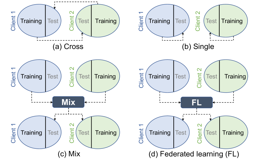
(1) Cross. Training is conducted on one client dataset and then the trained model is directly tested on the data of the other client, as shown in Fig. 6 (a). Specifically, ADNI-1 is used as the training set (denoted as ADNI1-tr), then the trained model is tested on ADNI-2. ADNI-2 is used as the training set (denoted as ADNI2-tr), then the trained model is evaluated on ADNI-1.
(2) Single. Training and testing are conducted within each client dataset separately, as shown in Fig. 6 (b). In each client, 80% of the data is used for training while the other is used for testing.
(3) Mix. All the training data in each client are pooled together for training a model, then the trained model is evaluated on the test data of all the clients, as shown in Fig. 6 (c). Note this strategy needs to share data, and thus, could not preserve privacy.
(4) FedAVG [12, 164]. Each client trains its own model, then their model weights (e.g., the weight of logistic regression) are aggregated to calculate a global model. The final trained global model is tested on all the test data in each client, as shown in Fig. 6 (d). The number of iterations for local model training is set to 10.
(5) FedSGD [12]. Each client trains a local model, then the gradients from each client are aggregated to calculate a global model. The global model is then applied to all the test data in each client for assessment, as shown in Fig. 6 (d). The number of iterations for local model training is set to 10.
(6) FedProx [165]. Every client trains its own model with an additional proximal term (the coefficient is set to 0.1). Local training is conducted only once. The model weights of each client are aggregated to get a global model. The trained global model is then assessed on the test data in each client, as shown in Fig. 6 (d).
| Client | Method | ACC | SEN | SPE | AUC |
| ADNI-1 | ADNI1-tr | – | – | – | – |
| ADNI2-tr | 0.818 | 0.809 | 0.825 | 0.886 | |
| Single | 0.8440.013 | 0.7860.045 | 0.8950.040 | 0.8890.018 | |
| Mix | 0.8700.017 | 0.8230.045 | 0.9230.050 | 0.9010.018 | |
| FedAvg | 0.8600.028 | 0.7830.025 | 0.9010.049 | 0.8970.013 | |
| FedSGD | 0.8230.030 | 0.7520.047 | 0.8820.011 | 0.8800.034 | |
| FedProx | 0.8580.031 | 0.8150.077 | 0.8960.034 | 0.9000.044 | |
| ADNI-2 | ADNI1-tr | 0.811 | 0.623 | 0.960 | 0.885 |
| ADNI2-tr | – | – | – | – | |
| Single | 0.8280.016 | 0.7500.045 | 0.8900.046 | 0.8630.043 | |
| Mix | 0.8720.012 | 0.8430.048 | 0.8980.038 | 0.9100.013 | |
| FedAvg | 0.8420.021 | 0.8230.018 | 0.8640.038 | 0.9070.017 | |
| FedSGD | 0.8440.039 | 0.8190.066 | 0.8710.056 | 0.9080.040 | |
| FedProx | 0.8560.045 | 0.8450.072 | 0.8610.047 | 0.9080.036 |
IX-D Result and Analysis
The classification result of different methods is shown in Table II. In the “Cross” setting, the client dataset for training is denoted as “client (tr)”. Since there is only one test dataset in this setting, no standard deviation is reported.
From Table II, we can get the following observations. 1) The “mix” strategy has the best performance. This is because it combines all the training data of the clients together and the learning model can get access to the largest amount of data information than the other methods. 2) The “cross” strategy has the worst performance. This should be caused by the well-known “domain shift” problem. Since ADNI-1 and ADNI-2 have different scanning parameters, then directly transferring a model may not achieve good classification results. 3) Federated learning methods achieve satisfactory performance. This can be explained by FL can leverage more data information than the baseline methods (i.e., “cross” and “single”), even without cross-site data sharing. 4) Among the FL methods, we find that aggregation of model weights (i.e., FedAvg, FedProx) can be more advantageous than a fusion of the gradients of each client model (i.e., FedSGD).
In this experiment, we conduct an empirical evaluation of FL algorithms on a benchmark dataset. The result can provide some valuable insights for medical imaging researchers. It should be acknowledged that the results may not generalize to all medical imaging scenarios, as different datasets and clinical settings could yield varying outcomes.
X Discussion
X-A Challenges of FL for Medical Image Analysis
X-A1 Data Heterogeneity Among Clients
Data heterogeneity is widespread in real-world medical image sites. Such heterogeneity can hardly be avoided in practice due to the following factors. 1) Medical images from different sites/datasets are typically acquired by different scanners or scanning protocols. 2) Patients in different sites/hospitals have different distributions. The heterogeneous data distribution, i.e., “domain shift” or “client shift”, may cause significant degradation or biased performance of a federated learning system. How to alleviate the negative influence of data heterogeneity is one of the most important and challenging research problems for federated learning in medical imaging.
X-A2 Privacy Leakage/Poisoning Attacks
In classic FL, only the model parameters (e.g., weights) are exchanged and updated without data sharing. This is considered an effective way of privacy protection. But further research reveals that FL still faces privacy and security risks, including privacy leakage [81, 82, 83] and poisoning attacks [166, 167]. These issues can happen at both the server end and the client end. Since an FL system contains the communication and interaction of many entities/parties, how to effectively protect individual privacy and data security is a very challenging problem.
X-A3 Technological Limitations
While a majority of research is centered on algorithm design for various medical applications, the practical implementation of FL systems encounters significant technological hurdles. For instance, certain FL algorithms demand substantial computational resources, posing challenges for the underlying hardware infrastructure. Furthermore, addressing communication costs, optimizing network resource allocation, and ensuring synchronization are all formidable obstacles when striving to construct a robust and functional FL system in real life.
X-A4 Long-Term Viability of FL-based Medical Image Analysis
Federated Learning is not just a novel machine learning algorithm; it represents a dynamic and systematic approach to engineering. It is imperative to focus on the long-term viability of FL systems when applied to medical image analysis. This involves addressing critical issues such as scalability, sustainability, and evolving regulations. In real-world scenarios, unforeseen challenges emerge, such as clients leaving or joining the training process, as well as unexpected technical and connectivity issues. Effectively managing an FL system for robust and stable long-term medical image analysis is a complex endeavor.
X-B Future Research Directions
X-B1 Dealing with Client Shift
Domain shift between client datasets (client shift) has become a major concern of federated learning in medical image analysis. To tackle this problem, domain adaptation [3] has attracted extensive interest. Classic domain adaptation methods typically need access to both source and target domains which may violate the privacy protection restraint in FL. Thus, developing more efficient federated domain adaptation methods will be a promising research direction. Another promising solution is personalized FL techniques [33, 34] which utilize local data to further optimize a trained global model.
X-B2 Multi-Modality Fusion for FL
Numerous imaging techniques/tools have been developed to create various visual representations of every subject, such as structural MRI, functional MRI, computed tomography (CT), and positron emission tomography (PET). Most existing FL studies only focus on images of a single modality in each client. How to leverage multi-modal imaging data in an FL system is an interesting problem with practical value. Currently, a few works make early steps on FL with multiple modalities [168]. More research work is expected on this topic.
X-B3 Model Generalizability for Unseen Clients
Most existing FL studies focus on model training and test within a fixed federation system. That is, a global model is trained on and applied to the same client datasets (internal clients). An interesting question is: When facing data from unseen sites that are outside of a federation (outside clients), how to guarantee the generalizability of an FL model? This is typically a domain generalization problem [169, 170] or a test-time adaptation problem (i.e., using inference samples as a clue of the unseen distribution to facilitate adaptation) [171, 172]. Currently, there are a few works that introduce domain generalization into federated learning [39, 133]. In the future, evaluating and enhancing the generalizability of a trained FL model to unseen sites or even unseen classes (i.e., open-set recognition [173, 174]) will be a promising research direction.
X-B4 Weakly-Supervised Learning for FL
Weakly-supervised learning is a promising technique that handles data with incomplete, inexact, and inaccurate labels. These problems are common and widespread in medical imaging data. How to deal with these “imperfect” data (e.g., learning from noisy labels [175]) in an FL system is worthy of further exploration.
X-B5 FL Security: Attack and Defense
Several existing FL systems have been shown to be vulnerable to internal or external attacks, concerning system robustness and data privacy [166]. Further exploration of strong defense strategies in FL is helpful to enhance the security of FL systems. Another interesting question is: if an institution wants to withdraw from a federation, how to guarantee its data has been removed from the trained FL model? One solution is the data auditing technique [176] which can also be used to check if a poisoned/suspicious dataset is used in FL training.
X-B6 Blockchain and Decentralization of FL
Most existing FL methods on medical tasks employ a centralized paradigm which demands a trustworthy central server. This pattern gradually shows many disadvantages such as vulnerability to poisonous attacks and lack of credibility. Recently, blockchain has been identified as a potentially promising solution to this problem [177]. Using blockchain can avoid the dependence on the central server which can be the bottleneck of the whole federation. Some work has made efforts on this point for medical image analysis through leveraging blockchain [178, 179] or other decentralization methods [180]. Currently, very limited work has been performed in this direction for medical image analysis, thus, there is much room for future research.
X-B7 FL for Medical Video Analysis
Most existing FL systems focus on combining cross-site medical images. As an extension of 2D/3D medical images, medical videos have been rarely explored. Some pioneering work has employed FL to effectively take advantage of medical video from multiple sites/datasets for surgical phase recognition [68]. In the future, FL systems consisting of medical videos for surgical or other applications will attract more research attention.
X-B8 Large-Scale Medical Image Benchmark for FL
Most existing medical image databases for FL research only consist of relatively small datasets at each client side. Some work just split a single large dataset (e.g., CheXpert [106]) into different parts which are simulated as different client datasets. There is a lack of large-scale federations which consist of various sites across the world. Only a few works have leveraged real-world datasets from multiple cities or countries. [76] collected chest X-ray images from different cities for the task of COVID-19 detection. [181] leverage seven clinical institutions from across the world to build a federated learning model for breast density classification. [156] builds a large-scale federation through international cooperation. Building large-scale benchmarks (including publicly available medical imaging databases and state-of-the-art FL algorithms) through extensive international cooperation is very beneficial for real-world FL applications.
X-B9 Model Interpretability
In clinical practice, one of the most significant hurdles in adopting machine learning and AI lies in the “black box” nature of certain machine learning systems, such as deep learning [182, 183]. Even as FL emerges as a promising machine learning prototype, it encounters similar challenges. Thus, the issue of model interpretability remains a critical factor to address for the seamless integration of FL into clinical practice. While some researchers have made an early effort towards this topic [184, 185], more explorations are expected in the future.
X-B10 Real-World Implementation and Practical Issues
The majority of research on FL in medical imaging has primarily focused on algorithm development and simulation. FL methods, while promising, can encounter difficulties during real-world implementation, such as compatibility issues with existing hospital systems, integration challenges, and user adoption hurdles. Addressing these practical considerations is crucial for advancing the application of FL in medical image analysis.
XI Conclusion
In this paper, we review the recent advances in federated learning (FL) for medical image analysis. We summarize existing FL methods from a system view and categorize them into client-end, server-end, and client-server communication methods. For each category, we provide a novel “question-answer” paradigm to elaborate on the motivation and mechanism of different FL methods in medical image analysis. We also introduce existing software tools/platforms and benchmark medical image datasets that have been used for federated learning. In addition, we conduct an experiment to empirically compare different FL methods on a popular benchmark imaging database (i.e., ADNI). We further discuss current challenges, potential research opportunities, and future directions of FL-based medical image analysis. We hope that this survey paper could provide researchers with a clear picture of the recent development of FL in medical image analysis and that more research efforts can be inspired and initiated in this exciting research field.
Acknowledgments
This work was supported by NIH grant RF1AG073297. Part of the data used in this work were obtained from Alzheimer’s Disease Neuroimaging Initiative (ADNI). The investigators within ADNI contributed to the design and implementation of ADNI and provided data but did not participate in the analysis or writing of this article.
References
- [1] A. Barragán-Montero, U. Javaid, G. Valdés, D. Nguyen, P. Desbordes, B. Macq, S. Willems, L. Vandewinckele, M. Holmström, F. Löfman et al., “Artificial intelligence and machine learning for medical imaging: A technology review,” Physica Medica, vol. 83, pp. 242–256, 2021.
- [2] V. Cheplygina, M. de Bruijne, and J. P. Pluim, “Not-so-supervised: A survey of semi-supervised, multi-instance, and transfer learning in medical image analysis,” Medical Image Analysis, vol. 54, pp. 280–296, 2019.
- [3] H. Guan and M. Liu, “Domain adaptation for medical image analysis: A survey,” IEEE Transactions on Biomedical Engineering, vol. 69, no. 3, pp. 1173–1185, 2022.
- [4] G. Litjens, T. Kooi, B. E. Bejnordi, A. A. A. Setio, F. Ciompi, M. Ghafoorian, J. A. Van Der Laak, B. Van Ginneken, and C. I. Sánchez, “A survey on deep learning in medical image analysis,” Medical Image Analysis, vol. 42, pp. 60–88, 2017.
- [5] Y. LeCun, Y. Bengio, and G. Hinton, “Deep learning,” Nature, vol. 521, no. 7553, pp. 436–444, 2015.
- [6] J. Deng, W. Dong, R. Socher, L.-J. Li, K. Li, and L. Fei-Fei, “Imagenet: A large-scale hierarchical image database,” in 2009 IEEE Conference on Computer Vision and Pattern Recognition. IEEE, 2009, pp. 248–255.
- [7] T.-Y. Lin, M. Maire, S. Belongie, J. Hays, P. Perona, D. Ramanan, P. Dollár, and C. L. Zitnick, “Microsoft COCO: Common objects in context,” in European Conference on Computer Vision. Springer, 2014, pp. 740–755.
- [8] S. J. Raudys, A. K. Jain et al., “Small sample size effects in statistical pattern recognition: Recommendations for practitioners,” IEEE Transactions on Pattern Analysis and Machine Intelligence, vol. 13, no. 3, pp. 252–264, 1991.
- [9] A. Vabalas, E. Gowen, E. Poliakoff, and A. J. Casson, “Machine learning algorithm validation with a limited sample size,” PLOS ONE, vol. 14, no. 11, pp. 1–20, 2019.
- [10] US Department of Health and Human Services, “HIPAA,” https://www.hhs.gov/hipaa/index.html, 2020.
- [11] General Data Protection Regulation, “GDPR,” https://gdpr-info.eu/, 2019.
- [12] B. McMahan, E. Moore, D. Ramage, S. Hampson, and B. A. y Arcas, “Communication-efficient learning of deep networks from decentralized data,” in Artificial Intelligence and Statistics. PMLR, 2017, pp. 1273–1282.
- [13] K. Bonawitz, H. Eichner, W. Grieskamp, D. Huba, A. Ingerman, V. Ivanov, C. Kiddon, J. Konevcnỳ, S. Mazzocchi, B. McMahan et al., “Towards federated learning at scale: System design,” Proceedings of Machine Learning and Systems, vol. 1, pp. 374–388, 2019.
- [14] P. Kairouz, H. B. McMahan, B. Avent, A. Bellet, M. Bennis, A. N. Bhagoji, K. Bonawitz, Z. Charles, G. Cormode, R. Cummings et al., “Advances and open problems in federated learning,” Foundations and Trends® in Machine Learning, vol. 14, no. 1–2, pp. 1–210, 2021.
- [15] Q. Li, Z. Wen, Z. Wu, S. Hu, N. Wang, Y. Li, X. Liu, and B. He, “A survey on federated learning systems: Vision, hype and reality for data privacy and protection,” IEEE Transactions on Knowledge and Data Engineering, pp. 1–20, 2021.
- [16] T. Li, A. K. Sahu, A. Talwalkar, and V. Smith, “Federated learning: Challenges, methods, and future directions,” IEEE Signal Processing Magazine, vol. 37, no. 3, pp. 50–60, 2020.
- [17] Q. Yang, Y. Liu, T. Chen, and Y. Tong, “Federated machine learning: Concept and applications,” ACM Transactions on Intelligent Systems and Technology (TIST), vol. 10, no. 2, pp. 1–19, 2019.
- [18] K. J. Rahman, F. Ahmed, N. Akhter, M. Hasan, R. Amin, K. E. Aziz, A. M. Islam, M. S. H. Mukta, and A. N. Islam, “Challenges, applications and design aspects of federated learning: A survey,” IEEE Access, vol. 9, pp. 124 682–124 700, 2021.
- [19] C. Zhang, Y. Xie, H. Bai, B. Yu, W. Li, and Y. Gao, “A survey on federated learning,” Knowledge-Based Systems, vol. 216, p. 106775, 2021.
- [20] X. Yin, Y. Zhu, and J. Hu, “A comprehensive survey of privacy-preserving federated learning: A taxonomy, review, and future directions,” ACM Computing Surveys (CSUR), vol. 54, no. 6, pp. 1–36, 2021.
- [21] R. S. Antunes, C. André da Costa, A. Küderle, I. A. Yari, and B. Eskofier, “Federated learning for healthcare: Systematic review and architecture proposal,” ACM Transactions on Intelligent Systems and Technology (TIST), vol. 13, no. 4, pp. 1–23, 2022.
- [22] S. Rajendran, J. S. Obeid, H. Binol, K. Foley, W. Zhang, P. Austin, J. Brakefield, M. N. Gurcan, and U. Topaloglu, “Cloud-based federated learning implementation across medical centers,” JCO Clinical Cancer Informatics, vol. 5, pp. 1–11, 2021.
- [23] D. C. Nguyen, Q.-V. Pham, P. N. Pathirana, M. Ding, A. Seneviratne, Z. Lin, O. Dobre, and W.-J. Hwang, “Federated learning for smart healthcare: A survey,” ACM Computing Surveys (CSUR), vol. 55, no. 3, pp. 1–37, 2022.
- [24] B. Pfitzner, N. Steckhan, and B. Arnrich, “Federated learning in a medical context: A systematic literature review,” ACM Transactions on Internet Technology (TOIT), vol. 21, no. 2, pp. 1–31, 2021.
- [25] N. Rieke, J. Hancox, W. Li, F. Milletari, H. R. Roth, S. Albarqouni, S. Bakas, M. N. Galtier, B. A. Landman, K. Maier-Hein et al., “The future of digital health with federated learning,” NPJ digital medicine, vol. 3, no. 1, p. 119, 2020.
- [26] O. Aouedi, A. Sacco, K. Piamrat, and G. Marchetto, “Handling privacy-sensitive medical data with federated learning: Challenges and future directions,” IEEE Journal of Biomedical and Health Informatics, vol. 27, no. 2, pp. 790–803, 2023.
- [27] V. Chiruvella, A. K. Guddati et al., “Ethical issues in patient data ownership,” Interactive Journal of Medical Research, vol. 10, no. 2, p. e22269, 2021.
- [28] California Consumer Privacy Act (CCPA), “CCPA,” https://oag.ca.gov/privacy/ccpa, 2018.
- [29] A. Satariano, “Google is fined $57 million under Europe’s data privacy law,” The New York Times, vol. 21, 2019.
- [30] O. Ronneberger, P. Fischer, and T. Brox, “U-net: Convolutional networks for biomedical image segmentation,” in 18th International Conference on Medical Image Computing and Computer-Assisted Intervention (MICCAI), Munich, Germany, October 5-9, 2015. Springer, 2015, pp. 234–241.
- [31] J. Xiao, L. Yu, Z. Zhou, Y. Bai, L. Xing, A. Yuille, and Y. Zhou, “Catenorm: Categorical normalization for robust medical image segmentation,” in Domain Adaptation and Representation Transfer: 4th MICCAI Workshop, DART 2022, Held in Conjunction with MICCAI 2022, Singapore, September 22, 2022, Proceedings. Springer, 2022, pp. 129–146.
- [32] J. Wicaksana, Z. Yan, X. Yang, Y. Liu, L. Fan, and K.-T. Cheng, “Customized federated learning for multi-source decentralized medical image classification,” IEEE Journal of Biomedical and Health Informatics, vol. 26, no. 11, pp. 5596–5607, 2022.
- [33] C. T Dinh, N. Tran, and J. Nguyen, “Personalized federated learning with moreau envelopes,” Advances in Neural Information Processing Systems, vol. 33, pp. 21 394–21 405, 2020.
- [34] A. Z. Tan, H. Yu, L. Cui, and Q. Yang, “Towards personalized federated learning,” IEEE Transactions on Neural Networks and Learning Systems, 2022.
- [35] C.-M. Feng, Y. Yan, S. Wang, Y. Xu, L. Shao, and H. Fu, “Specificity-preserving federated learning for MR image reconstruction,” IEEE Transactions on Medical Imaging, 2022.
- [36] M. Zhang, L. Qu, P. Singh, J. Kalpathy-Cramer, and D. L. Rubin, “SplitAVG: A heterogeneity-aware federated deep learning method for medical imaging,” IEEE Journal of Biomedical and Health Informatics, vol. 26, no. 9, pp. 4635–4644, 2022.
- [37] A. Chakravarty, A. Kar, R. Sethuraman, and D. Sheet, “Federated learning for site aware chest radiograph screening,” in 2021 IEEE 18th International Symposium on Biomedical Imaging (ISBI). IEEE, 2021, pp. 1077–1081.
- [38] A. Xu, W. Li, P. Guo, D. Yang, H. R. Roth, A. Hatamizadeh, C. Zhao, D. Xu, H. Huang, and Z. Xu, “Closing the generalization gap of cross-silo federated medical image segmentation,” in Proceedings of the IEEE/CVF Conference on Computer Vision and Pattern Recognition, 2022, pp. 20 866–20 875.
- [39] M. Jiang, H. Yang, C. Cheng, and Q. Dou, “Iop-fl: Inside-outside personalization for federated medical image segmentation,” IEEE Transactions on Medical Imaging, 2023.
- [40] J. Ke, Y. Shen, and Y. Lu, “Style normalization in histology with federated learning,” in 2021 IEEE 18th International Symposium on Biomedical Imaging (ISBI). IEEE, 2021, pp. 953–956.
- [41] N. Wagner, M. Fuchs, Y. Tolkach, and A. Mukhopadhyay, “Federated stain normalization for computational pathology,” in International Conference on Medical Image Computing and Computer-Assisted Intervention. Springer, 2022, pp. 14–23.
- [42] X. Li, Y. Gu, N. Dvornek, L. H. Staib, P. Ventola, and J. S. Duncan, “Multi-site fMRI analysis using privacy-preserving federated learning and domain adaptation: ABIDE results,” Medical Image Analysis, vol. 65, pp. 1–14, 2020.
- [43] N. K. Dinsdale, M. Jenkinson, and A. I. Namburete, “Fedharmony: Unlearning scanner bias with distributed data,” in International Conference on Medical Image Computing and Computer-Assisted Intervention. Springer, 2022, pp. 695––704.
- [44] M. Andreux, J. O. du Terrail, C. Beguier, and E. W. Tramel, “Siloed federated learning for multi-centric histopathology datasets,” in MICCAI Workshop on Domain Adaptation and Representation Transfer, and Distributed and Collaborative Learning (DART), Lima, Peru, October 4–8, 2020. Springer, 2020, pp. 129–139.
- [45] P. Guo, P. Wang, J. Zhou, S. Jiang, and V. M. Patel, “Multi-institutional collaborations for improving deep learning-based magnetic resonance image reconstruction using federated learning,” in Proceedings of the IEEE/CVF Conference on Computer Vision and Pattern Recognition, 2021, pp. 2423–2432.
- [46] L. Qu, N. Balachandar, M. Zhang, and D. Rubin, “Handling data heterogeneity with generative replay in collaborative learning for medical imaging,” Medical Image Analysis, vol. 78, p. 102424, 2022.
- [47] Z. Yan, J. Wicaksana, Z. Wang, X. Yang, and K.-T. Cheng, “Variation-aware federated learning with multi-source decentralized medical image data,” IEEE Journal of Biomedical and Health Informatics, vol. 25, no. 7, pp. 2615–2628, 2020.
- [48] J.-Y. Zhu, T. Park, P. Isola, and A. A. Efros, “Unpaired image-to-image translation using cycle-consistent adversarial networks,” in Proceedings of the IEEE international conference on computer vision, 2017, pp. 2223–2232.
- [49] M. Jiang, Z. Wang, and Q. Dou, “Harmofl: Harmonizing local and global drifts in federated learning on heterogeneous medical images,” in Proceedings of the AAAI Conference on Artificial Intelligence, vol. 36, no. 1, 2022, pp. 1087–1095.
- [50] K. Chaitanya, E. Erdil, N. Karani, and E. Konukoglu, “Contrastive learning of global and local features for medical image segmentation with limited annotations,” Advances in Neural Information Processing Systems, vol. 33, pp. 12 546–12 558, 2020.
- [51] K. He, H. Fan, Y. Wu, S. Xie, and R. Girshick, “Momentum contrast for unsupervised visual representation learning,” in Proceedings of the IEEE/CVF Conference on Computer Vision and Pattern Recognition, 2020, pp. 9729–9738.
- [52] I. Misra and L. v. d. Maaten, “Self-supervised learning of pretext-invariant representations,” in Proceedings of the IEEE/CVF Conference on Computer Vision and Pattern Recognition, 2020, pp. 6707–6717.
- [53] Y. Wu, D. Zeng, Z. Wang, Y. Shi, and J. Hu, “Distributed contrastive learning for medical image segmentation,” Medical Image Analysis, vol. 81, p. 102564, 2022.
- [54] ——, “Federated contrastive learning for volumetric medical image segmentation,” in International Conference on Medical Image Computing and Computer-Assisted Intervention. Springer, 2021, pp. 367–377.
- [55] N. Dong and I. Voiculescu, “Federated contrastive learning for decentralized unlabeled medical images,” in International Conference on Medical Image Computing and Computer-Assisted Intervention. Springer, 2021, pp. 378–387.
- [56] Y. Zhang and Q. Yang, “A survey on multi-task learning,” IEEE Transactions on Knowledge and Data Engineering, vol. 34, no. 12, pp. 5586–5609, 2021.
- [57] V. Smith, C.-K. Chiang, M. Sanjabi, and A. S. Talwalkar, “Federated multi-task learning,” Advances in Neural Information Processing Systems, vol. 30, 2017.
- [58] Z.-A. Huang, Y. Hu, R. Liu, X. Xue, Z. Zhu, L. Song, and K. C. Tan, “Federated multi-task learning for joint diagnosis of multiple mental disorders on MRI scans,” IEEE Transactions on Biomedical Engineering, 2022.
- [59] Z.-H. Zhou, “A brief introduction to weakly supervised learning,” National Science Review, vol. 5, no. 1, pp. 44–53, 2018.
- [60] X. Yang, Z. Song, I. King, and Z. Xu, “A survey on deep semi-supervised learning,” IEEE Transactions on Knowledge and Data Engineering, 2022.
- [61] J. E. Van Engelen and H. H. Hoos, “A survey on semi-supervised learning,” Machine Learning, vol. 109, no. 2, pp. 373–440, 2020.
- [62] G. Quellec, G. Cazuguel, B. Cochener, and M. Lamard, “Multiple-instance learning for medical image and video analysis,” IEEE Reviews in Biomedical Engineering, vol. 10, pp. 213–234, 2017.
- [63] M.-A. Carbonneau, V. Cheplygina, E. Granger, and G. Gagnon, “Multiple instance learning: A survey of problem characteristics and applications,” Pattern Recognition, vol. 77, pp. 329–353, 2018.
- [64] H. Song, M. Kim, D. Park, Y. Shin, and J.-G. Lee, “Learning from noisy labels with deep neural networks: A survey,” IEEE Transactions on Neural Networks and Learning Systems, 2022.
- [65] B. Frénay and M. Verleysen, “Classification in the presence of label noise: A survey,” IEEE Transactions on Neural Networks and Learning Systems, vol. 25, no. 5, pp. 845–869, 2013.
- [66] D. Yang, Z. Xu, W. Li, A. Myronenko, H. R. Roth, S. Harmon, S. Xu, B. Turkbey, E. Turkbey, X. Wang et al., “Federated semi-supervised learning for COVID region segmentation in chest CT using multi-national data from China, Italy, Japan,” Medical Image Analysis, vol. 70, p. 101992, 2021.
- [67] M. Y. Lu, R. J. Chen, D. Kong, J. Lipkova, R. Singh, D. F. Williamson, T. Y. Chen, and F. Mahmood, “Federated learning for computational pathology on gigapixel whole slide images,” Medical Image Analysis, vol. 76, pp. 1–13, 2022.
- [68] H. Kassem, D. Alapatt, P. Mascagni, C. AI4SafeChole, A. Karargyris, and N. Padoy, “Federated cycling (fedcy): Semi-supervised federated learning of surgical phases,” IEEE Transactions on Medical Imaging, 2022.
- [69] A. Kumar, V. Purohit, V. Bharti, R. Singh, and S. K. Singh, “Medisecfed: private and secure medical image classification in the presence of malicious clients,” IEEE Transactions on Industrial Informatics, vol. 18, no. 8, pp. 5648–5657, 2021.
- [70] W. Zhu and J. Luo, “Federated medical image analysis with virtual sample synthesis,” in International Conference on Medical Image Computing and Computer-Assisted Intervention. Springer, 2022, pp. 728–738.
- [71] T. Miyato, S.-i. Maeda, M. Koyama, and S. Ishii, “Virtual adversarial training: a regularization method for supervised and semi-supervised learning,” IEEE Transactions on Pattern Analysis and Machine Intelligence, vol. 41, no. 8, pp. 1979–1993, 2018.
- [72] L. Peng, N. Wang, N. Dvornek, X. Zhu, and X. Li, “Fedni: Federated graph learning with network inpainting for population-based disease prediction,” IEEE Transactions on Medical Imaging, 2022.
- [73] D. Stripelis, J. L. Ambite, P. Lam, and P. Thompson, “Scaling neuroscience research using federated learning,” in 2021 IEEE 18th International Symposium on Biomedical Imaging (ISBI). IEEE, 2021, pp. 1191–1195.
- [74] Z. Chen, C. Yang, M. Zhu, Z. Peng, and Y. Yuan, “Personalized retrogress-resilient federated learning toward imbalanced medical data,” IEEE Transactions on Medical Imaging, vol. 41, no. 12, pp. 3663–3674, 2022.
- [75] Z. Liu, J. Xu, X. Peng, and R. Xiong, “Frequency-domain dynamic pruning for convolutional neural networks,” Advances in Neural Information Processing Systems, vol. 31, 2018.
- [76] Z. Li, X. Xu, X. Cao, W. Liu, Y. Zhang, D. Chen, and H. Dai, “Integrated cnn and federated learning for COVID-19 detection on chest X-ray images,” IEEE/ACM Transactions on Computational Biology and Bioinformatics, 2022.
- [77] S. M. Hosseini, M. Sikaroudi, M. Babaie, and H. Tizhoosh, “Proportionally fair hospital collaborations in federated learning of histopathology images,” IEEE Transactions on Medical Imaging, 2023.
- [78] Z. Fan, J. Su, K. Gao, D. Hu, and L.-L. Zeng, “A federated deep learning framework for 3D brain MRI images,” in 2021 International Joint Conference on Neural Networks (IJCNN). IEEE, 2021, pp. 1–6.
- [79] J. Luo and S. Wu, “Fedsld: Federated learning with shared label distribution for medical image classification,” in 2022 IEEE 19th International Symposium on Biomedical Imaging (ISBI). IEEE, 2022, pp. 1–5.
- [80] N. Alkhunaizi, D. Kamzolov, M. Takávc, and K. Nandakumar, “Suppressing poisoning attacks on federated learning for medical imaging,” in International Conference on Medical Image Computing and Computer-Assisted Intervention. Springer, 2022, pp. 673–683.
- [81] J. Geiping, H. Bauermeister, H. Dröge, and M. Moeller, “Inverting gradients-how easy is it to break privacy in federated learning?” Advances in Neural Information Processing Systems, vol. 33, pp. 16 937–16 947, 2020.
- [82] H. Yin, A. Mallya, A. Vahdat, J. M. Alvarez, J. Kautz, and P. Molchanov, “See through gradients: Image batch recovery via gradinversion,” in Proceedings of the IEEE/CVF Conference on Computer Vision and Pattern Recognition, 2021, pp. 16 337–16 346.
- [83] L. Zhu, Z. Liu, and S. Han, “Deep leakage from gradients,” Advances in Neural Information Processing Systems, vol. 32, 2019.
- [84] Q. Yang, J. Zhang, W. Hao, G. P. Spell, and L. Carin, “Flop: Federated learning on medical datasets using partial networks,” in Proceedings of the 27th ACM SIGKDD Conference on Knowledge Discovery & Data Mining, 2021, pp. 3845–3853.
- [85] W. Li, F. Milletarì, D. Xu, N. Rieke, J. Hancox, W. Zhu, M. Baust, Y. Cheng, S. Ourselin, M. J. Cardoso et al., “Privacy-preserving federated brain tumour segmentation,” in Machine Learning in Medical Imaging: 10th International Workshop, MLMI 2019, Held in Conjunction with MICCAI 2019, Shenzhen, China, October 13, 2019, Proceedings 10. Springer, 2019, pp. 133–141.
- [86] C. Dwork, A. Roth et al., “The algorithmic foundations of differential privacy,” Foundations and Trends® in Theoretical Computer Science, vol. 9, no. 3–4, pp. 211–407, 2014.
- [87] M. Malekzadeh, B. Hasircioglu, N. Mital, K. Katarya, M. E. Ozfatura, and D. Gündüz, “Dopamine: Differentially private federated learning on medical data,” in The Second AAAI Workshop on Privacy-Preserving Artificial Intelligence (PPAI-21), 2021, pp. 1–9.
- [88] G. Kaissis, A. Ziller, J. Passerat-Palmbach, T. Ryffel, D. Usynin, A. Trask, I. Lima Jr, J. Mancuso, F. Jungmann, M.-M. Steinborn et al., “End-to-end privacy preserving deep learning on multi-institutional medical imaging,” Nature Machine Intelligence, vol. 3, no. 6, pp. 473–484, 2021.
- [89] A. Hatamizadeh, H. Yin, P. Molchanov, A. Myronenko, W. Li, P. Dogra, A. Feng, M. G. Flores, J. Kautz, D. Xu et al., “Do gradient inversion attacks make federated learning unsafe?” IEEE Transactions on Medical Imaging, 2023.
- [90] W. Zhang, T. Zhou, Q. Lu, X. Wang, C. Zhu, H. Sun, Z. Wang, S. K. Lo, and F.-Y. Wang, “Dynamic-fusion-based federated learning for COVID-19 detection,” IEEE Internet of Things Journal, vol. 8, no. 21, pp. 15 884–15 891, 2021.
- [91] A. Di Martino et al., “The autism brain imaging data exchange: towards a large-scale evaluation of the intrinsic brain architecture in autism,” Molecular Psychiatry, vol. 19, no. 6, pp. 659–667, 2014.
- [92] S. G. Mueller, M. W. Weiner, L. J. Thal, R. C. Petersen, C. Jack, W. Jagust, J. Q. Trojanowski, A. W. Toga, and L. Beckett, “The Alzheimer’s disease neuroimaging initiative,” Neuroimaging Clinics, vol. 15, no. 4, pp. 869–877, 2005.
- [93] Z. Gürler and I. Rekik, “Federated brain graph evolution prediction using decentralized connectivity datasets with temporally-varying acquisitions,” IEEE Transactions on Medical Imaging, 2022.
- [94] P. J. LaMontagne, T. L. Benzinger, J. C. Morris, S. Keefe, R. Hornbeck, C. Xiong, E. Grant, J. Hassenstab, K. Moulder, A. G. Vlassenko et al., “OASIS-3: longitudinal neuroimaging, clinical, and cognitive dataset for normal aging and Alzheimer disease,” MedRxiv, pp. 1–37, 2019.
- [95] M. Islam, M. T. Reza, M. Kaosar, and M. Z. Parvez, “Effectiveness of federated learning and CNN ensemble architectures for identifying brain tumors using MRI images,” Neural Processing Letters, pp. 1–31, 2022.
- [96] C. R. Pernet, K. J. Gorgolewski, D. Job, D. Rodriguez, I. Whittle, and J. Wardlaw, “A structural and functional magnetic resonance imaging dataset of brain tumour patients,” Scientific data, vol. 3, no. 1, pp. 1–6, 2016.
- [97] X. Qi, G. Yang, Y. He, W. Liu, A. Islam, and S. Li, “Contrastive re-localization and history distillation in federated CMR segmentation,” in International Conference on Medical Image Computing and Computer-Assisted Intervention. Springer, 2022, pp. 256–265.
- [98] A. E. Flanders et al., “Construction of a machine learning dataset through collaboration: the RSNA 2019 brain CT hemorrhage challenge,” Radiology: Artificial Intelligence, vol. 2, no. 3, pp. 1–8, 2020.
- [99] K. L. Miller et al., “Multimodal population brain imaging in the UK Biobank prospective epidemiological study,” Nature Neuroscience, vol. 19, no. 11, pp. 1523–1536, 2016.
- [100] Q. Liu, H. Yang, Q. Dou, and P.-A. Heng, “Federated semi-supervised medical image classification via inter-client relation matching,” in International Conference on Medical Image Computing and Computer-Assisted Intervention. Springer, 2021, pp. 325–335.
- [101] M. J. Sheller, G. A. Reina, B. Edwards, J. Martin, and S. Bakas, “Multi-institutional deep learning modeling without sharing patient data: A feasibility study on brain tumor segmentation,” in Brainlesion: Glioma, Multiple Sclerosis, Stroke and Traumatic Brain Injuries: 4th International Workshop, BrainLes 2018, Held in Conjunction with MICCAI 2018, Granada, Spain, September 16, 2018. Springer, 2019, pp. 92–104.
- [102] B. H. Menze, A. Jakab, S. Bauer, J. Kalpathy-Cramer, K. Farahani, J. Kirby, Y. Burren, N. Porz, J. Slotboom, R. Wiest et al., “The multimodal brain tumor image segmentation benchmark (BRATS),” IEEE Transactions on Medical Imaging, vol. 34, no. 10, pp. 1993–2024, 2014.
- [103] M. E. Chowdhury, T. Rahman, A. Khandakar, R. Mazhar, M. A. Kadir, Z. B. Mahbub, K. R. Islam, M. S. Khan, A. Iqbal, N. Al Emadi et al., “Can AI help in screening viral and COVID-19 pneumonia?” IEEE Access, vol. 8, pp. 132 665–132 676, 2020.
- [104] X. Wang, Y. Peng, L. Lu, Z. Lu, M. Bagheri, and R. M. Summers, “Chestx-ray8: Hospital-scale chest X-ray database and benchmarks on weakly-supervised classification and localization of common thorax diseases,” in Proceedings of the IEEE Conference on Computer Vision and Pattern Recognition, 2017, pp. 2097–2106.
- [105] R. Yan, L. Qu, Q. Wei, S.-C. Huang, L. Shen, D. Rubin, L. Xing, and Y. Zhou, “Label-efficient self-supervised federated learning for tackling data heterogeneity in medical imaging,” IEEE Transactions on Medical Imaging, 2023.
- [106] J. Irvin et al., “Chexpert: A large chest radiograph dataset with uncertainty labels and expert comparison,” in Proceedings of the AAAI Conference on Artificial Intelligence, vol. 33, no. 01, 2019, pp. 590–597.
- [107] N. Dong, M. Kampffmeyer, and I. Voiculescu, “Learning underrepresented classes from decentralized partially labeled medical images,” in International Conference on Medical Image Computing and Computer-Assisted Intervention. Springer, 2022, pp. 67–76.
- [108] L. Wang, Z. Q. Lin, and A. Wong, “Covid-net: A tailored deep convolutional neural network design for detection of COVID-19 cases from chest X-ray images,” Scientific Reports, vol. 10, no. 1, pp. 1–12, 2020.
- [109] I. Feki, S. Ammar, Y. Kessentini, and K. Muhammad, “Federated learning for COVID-19 screening from chest X-ray images,” Applied Soft Computing, vol. 106, p. 107330, 2021.
- [110] A. Linardos, K. Kushibar, S. Walsh, P. Gkontra, and K. Lekadir, “Federated learning for multi-center imaging diagnostics: a simulation study in cardiovascular disease,” Scientific Reports, vol. 12, no. 1, p. 3551, 2022.
- [111] O. Bernard et al., “Deep learning techniques for automatic MRI cardiac multi-structures segmentation and diagnosis: is the problem solved?” IEEE Transactions on Medical Imaging, vol. 37, no. 11, pp. 2514–2525, 2018.
- [112] V. M. Campello et al., “Multi-centre, multi-vendor and multi-disease cardiac segmentation: The M&Ms challenge,” IEEE Transactions on Medical Imaging, vol. 40, no. 12, pp. 3543–3554, 2021.
- [113] A. Lalande et al., “Emidec: a database usable for the automatic evaluation of myocardial infarction from delayed-enhancement cardiac MRI,” Data, vol. 5, no. 4, p. 89, 2020.
- [114] D. Li, A. Kar, N. Ravikumar, A. F. Frangi, and S. Fidler, “Federated simulation for medical imaging,” in International Conference on Medical Image Computing and Computer-Assisted Intervention. Springer, 2020, pp. 159–168.
- [115] B. L. Y. Agbley, J. P. Li, A. U. Haq, E. K. Bankas, C. B. Mawuli, S. Ahmad, S. Khan, and A. R. Khan, “Federated fusion of magnified histopathological images for breast tumor classification in the internet of medical things,” IEEE Journal of Biomedical and Health Informatics, 2023.
- [116] F. A. Spanhol, L. S. Oliveira, C. Petitjean, and L. Heutte, “Breast cancer histopathological image classification using convolutional neural networks,” in 2016 International Joint Conference on Neural Networks (IJCNN). IEEE, 2016, pp. 2560–2567.
- [117] J. Wicaksana, Z. Yan, D. Zhang, X. Huang, H. Wu, X. Yang, and K.-T. Cheng, “Fedmix: Mixed supervised federated learning for medical image segmentation,” IEEE Transactions on Medical Imaging, 2022.
- [118] W. Al-Dhabyani, M. Gomaa, H. Khaled, and A. Fahmy, “Dataset of breast ultrasound images,” Data in Brief, vol. 28, pp. 1–5, 2020.
- [119] Y. Zhang, M. Xian, H.-D. Cheng, B. Shareef, J. Ding, F. Xu, K. Huang, B. Zhang, C. Ning, and Y. Wang, “BUSIS: A benchmark for breast ultrasound image segmentation,” in Healthcare, vol. 10, no. 4. MDPI, 2022, pp. 1–16.
- [120] M. H. Yap, G. Pons, J. Marti, S. Ganau, M. Sentis, R. Zwiggelaar, A. K. Davison, and R. Marti, “Automated breast ultrasound lesions detection using convolutional neural networks,” IEEE Journal of Biomedical and health Informatics, vol. 22, no. 4, pp. 1218–1226, 2017.
- [121] B. Cassidy, C. Kendrick, A. Brodzicki, J. Jaworek-Korjakowska, and M. H. Yap, “Analysis of the ISIC image datasets: Usage, benchmarks and recommendations,” Medical Image Analysis, vol. 75, pp. 1–15, 2022.
- [122] P. Tschandl, C. Rosendahl, and H. Kittler, “The HAM10000 dataset, a large collection of multi-source dermatoscopic images of common pigmented skin lesions,” Scientific Data, vol. 5, no. 1, pp. 1–9, 2018.
- [123] T. Bdair, N. Navab, and S. Albarqouni, “FedPerl: semi-supervised peer learning for skin lesion classification,” in International Conference on Medical Image Computing and Computer-Assisted Intervention. Springer, 2021, pp. 336–346.
- [124] N. Codella, V. Rotemberg, P. Tschandl, M. E. Celebi, S. Dusza, D. Gutman, B. Helba, A. Kalloo, K. Liopyris, M. Marchetti et al., “Skin lesion analysis toward melanoma detection 2018: A challenge hosted by the international skin imaging collaboration (ISIC),” arXiv preprint arXiv:1902.03368, 2019.
- [125] J. Kawahara, S. Daneshvar, G. Argenziano, and G. Hamarneh, “Seven-point checklist and skin lesion classification using multitask multimodal neural nets,” IEEE Journal of Biomedical and Health Informatics, vol. 23, no. 2, pp. 538–546, 2018.
- [126] A. G. Pacheco, G. R. Lima, A. S. Salomão, B. Krohling, I. P. Biral, G. G. de Angelo, F. C. Alves Jr, J. G. Esgario, A. C. Simora, P. B. Castro et al., “PAD-UFES-20: A skin lesion dataset composed of patient data and clinical images collected from smartphones,” Data in Brief, vol. 32, pp. 1–10, 2020.
- [127] Z. Chen, M. Zhu, C. Yang, and Y. Yuan, “Personalized retrogress-resilient framework for real-world medical federated learning,” in International Conference on Medical Image Computing and Computer-Assisted Intervention. Springer, 2021, pp. 347–356.
- [128] N. C. Codella et al., “Skin lesion analysis toward melanoma detection: A challenge at the 2017 International Symposium on Biomedical Imaging (ISBI), hosted by the International Skin Imaging Collaboration (ISIC),” in 2018 IEEE 15th International Symposium on Biomedical Imaging (ISBI 2018). IEEE, 2018, pp. 168–172.
- [129] J. W. C. Emma Dugas, Jared, “Diabetic retinopathy detection,” 2015. [Online]. Available: https://kaggle.com/competitions/diabetic-retinopathy-detection
- [130] L. Qiu, J. Cheng, H. Gao, W. Xiong, and H. Ren, “Federated semi-supervised learning for medical image segmentation via pseudo-label denoising,” IEEE Journal of Biomedical and Health Informatics, 2023.
- [131] F. Fumero, S. Alayón, J. L. Sanchez, J. Sigut, and M. Gonzalez-Hernandez, “Rim-one: An open retinal image database for optic nerve evaluation,” in 24th International Symposium on Computer-Based Medical Systems (CBMS). IEEE, 2011, pp. 1–6.
- [132] J. Wang, Y. Jin, D. Stoyanov, and L. Wang, “Feddp: Dual personalization in federated medical image segmentation,” IEEE Transactions on Medical Imaging, 2023.
- [133] Q. Liu, C. Chen, J. Qin, Q. Dou, and P.-A. Heng, “Feddg: Federated domain generalization on medical image segmentation via episodic learning in continuous frequency space,” in Proceedings of the IEEE/CVF Conference on Computer Vision and Pattern Recognition, 2021, pp. 1013–1023.
- [134] M. Zhu, Z. Chen, and Y. Yuan, “FedDM: Federated weakly supervised segmentation via annotation calibration and gradient de-conflicting,” IEEE Transactions on Medical Imaging, 2023.
- [135] G. Litjens, R. Toth, W. Van De Ven, C. Hoeks, S. Kerkstra, B. van Ginneken, G. Vincent, G. Guillard, N. Birbeck, J. Zhang et al., “Evaluation of prostate segmentation algorithms for MRI: the PROMISE12 challenge,” Medical Image Analysis, vol. 18, no. 2, pp. 359–373, 2014.
- [136] NCI-ISBI 2013 Challenge: Automated Segmentation of Prostate Structures, https://wiki.cancerimagingarchive.net/pages/viewpage.action?pageId=21267207, 2021.
- [137] X. Xu, H. H. Deng, J. Gateno, and P. Yan, “Federated multi-organ segmentation with inconsistent labels,” IEEE Transactions on Medical Imaging, 2023.
- [138] P. Bilic, P. Christ, H. B. Li, E. Vorontsov, A. Ben-Cohen, G. Kaissis, A. Szeskin, C. Jacobs, G. E. H. Mamani, G. Chartrand et al., “The liver tumor segmentation benchmark (lits),” Medical Image Analysis, vol. 84, pp. 1–24, 2023.
- [139] G. Litjens, O. Debats, J. Barentsz, N. Karssemeijer, and H. Huisman, “Computer-aided detection of prostate cancer in MRI,” IEEE Transactions on Medical Imaging, vol. 33, no. 5, pp. 1083–1092, 2014.
- [140] J. Yang, R. Shi, and B. Ni, “MedMNIST classification decathlon: A lightweight automl benchmark for medical image analysis,” in 2021 IEEE 18th International Symposium on Biomedical Imaging (ISBI). IEEE, 2021, pp. 191–195.
- [141] X. Liu, W. Li, and Y. Yuan, “Intervention & interaction federated abnormality detection with noisy clients,” in International Conference on Medical Image Computing and Computer-Assisted Intervention. Springer, 2022, pp. 309–319.
- [142] K. Li et al., “Colonoscopy polyp detection and classification: Dataset creation and comparative evaluations,” PLOS ONE, vol. 16, no. 8, pp. 1–26, 2021.
- [143] H. R. Roth et al., “Federated whole prostate segmentation in MRI with personalized neural architectures,” in International Conference on Medical Image Computing and Computer-Assisted Intervention. Springer, 2021, pp. 357–366.
- [144] A. L. Simpson et al., “A large annotated medical image dataset for the development and evaluation of segmentation algorithms,” arXiv preprint arXiv:1902.09063, 2019.
- [145] Cancer Genome Atlas Research Network, J. Weinstein, E. Collisson, G. Mills, K. Shaw, B. Ozenberger, K. Ellrott, I. Shmulevich, C. Sande, and J. Stuart, “The cancer genome atlas pan-cancer analysis project,” Nature Genetics, vol. 45, no. 10, pp. 1113–1120, 2013.
- [146] J. O. du Terrail, A. Leopold, C. Joly, C. Béguier, M. Andreux, C. Maussion, B. Schmauch, E. W. Tramel, E. Bendjebbar, M. Zaslavskiy et al., “Federated learning for predicting histological response to neoadjuvant chemotherapy in triple-negative breast cancer,” Nature Medicine, vol. 29, no. 1, pp. 135–146, 2023.
- [147] M. Adnan, S. Kalra, J. C. Cresswell, G. W. Taylor, and H. R. Tizhoosh, “Federated learning and differential privacy for medical image analysis,” Scientific Reports, vol. 12, no. 1, p. 1953, 2022.
- [148] W. Bulten et al., “Epithelium segmentation using deep learning in H&E-stained prostate specimens with immunohistochemistry as reference standard,” Scientific Reports, vol. 9, no. 1, pp. 1–10, 2019.
- [149] F. Knoll, J. Zbontar, A. Sriram, M. J. Muckley, M. Bruno, A. Defazio, M. Parente, K. J. Geras, J. Katsnelson, H. Chandarana et al., “fastMRI: A publicly available raw k-space and DICOM dataset of knee images for accelerated MR image reconstruction using machine learning,” Radiology: Artificial Intelligence, vol. 2, no. 1, p. e190007, 2020.
- [150] G. Elmas, S. U. Dar, Y. Korkmaz, E. Ceyani, B. Susam, M. Ozbey, S. Avestimehr, and T. Cukur, “Federated learning of generative image priors for MRI reconstruction,” IEEE Transactions on Medical Imaging, 2022.
- [151] A. Ziller, A. Trask, A. Lopardo, B. Szymkow, B. Wagner, E. Bluemke, J.-M. Nounahon, J. Passerat-Palmbach, K. Prakash, N. Rose et al., “Pysyft: A library for easy federated learning,” Federated Learning Systems: Towards Next-Generation AI, pp. 111–139, 2021.
- [152] A. Budrionis, M. Miara, P. Miara, S. Wilk, and J. G. Bellika, “Benchmarking PySyft federated learning framework on MIMIC-III dataset,” IEEE Access, vol. 9, pp. 116 869–116 878, 2021.
- [153] P. Foley, M. J. Sheller, B. Edwards, S. Pati, W. Riviera, M. Sharma, P. N. Moorthy, S.-h. Wang, J. Martin, P. Mirhaji et al., “OpenFL: The open federated learning library,” Physics in Medicine & Biology, vol. 67, no. 21, p. 214001, 2022.
- [154] S. Silva, A. Altmann, B. Gutman, and M. Lorenzi, “Fed-biomed: A general open-source frontend framework for federated learning in healthcare,” in Domain Adaptation and Representation Transfer, and Distributed and Collaborative Learning: Second MICCAI Workshop, DART 2020, and First MICCAI Workshop, DCL 2020, Held in Conjunction with MICCAI 2020, Lima, Peru, October 4–8, 2020. Springer, 2020, pp. 201–210.
- [155] I. Kholod, E. Yanaki, D. Fomichev, E. Shalugin, E. Novikova, E. Filippov, and M. Nordlund, “Open-source federated learning frameworks for IoT: A comparative review and analysis,” Sensors, vol. 21, no. 1, p. 167, 2020.
- [156] I. Dayan, H. R. Roth, A. Zhong, A. Harouni, A. Gentili, A. Z. Abidin, A. Liu, A. B. Costa, B. J. Wood, C.-S. Tsai et al., “Federated learning for predicting clinical outcomes in patients with COVID-19,” Nature Medicine, vol. 27, no. 10, pp. 1735–1743, 2021.
- [157] C. R. Jack Jr, M. A. Bernstein, N. C. Fox, P. Thompson, G. Alexander, D. Harvey, B. Borowski, P. J. Britson, J. L. Whitwell, C. Ward et al., “The Alzheimer’s disease neuroimaging initiative (ADNI): MRI methods,” Journal of Magnetic Resonance Imaging, vol. 27, no. 4, pp. 685–691, 2008.
- [158] M. J. Muckley, B. Riemenschneider, A. Radmanesh, S. Kim, G. Jeong, J. Ko, Y. Jun, H. Shin, D. Hwang, M. Mostapha et al., “Results of the 2020 fastMRI challenge for machine learning MR image reconstruction,” IEEE Transactions on Medical Imaging, vol. 40, no. 9, pp. 2306–2317, 2021.
- [159] N. Tzourio-Mazoyer, B. Landeau, D. Papathanassiou, F. Crivello, O. Etard, N. Delcroix, B. Mazoyer, and M. Joliot, “Automated anatomical labeling of activations in SPM using a macroscopic anatomical parcellation of the MNI MRI single-subject brain,” NeuroImage, vol. 15, no. 1, pp. 273–289, 2002.
- [160] R. Divya and R. Shantha Selva Kumari, “Genetic algorithm with logistic regression feature selection for Alzheimer’s disease classification,” Neural Computing and Applications, vol. 33, no. 14, pp. 8435–8444, 2021.
- [161] D. Bzdok, M. Eickenberg, O. Grisel, B. Thirion, and G. Varoquaux, “Semi-supervised factored logistic regression for high-dimensional neuroimaging data,” Advances in Neural Information Processing Systems, vol. 28, 2015.
- [162] C. Wachinger et al., “Domain adaptation for Alzheimer’s disease diagnostics,” NeuroImage, vol. 139, pp. 470–479, 2016.
- [163] V. F. van Ravesteijn, C. van Wijk, F. M. Vos, R. Truyen, J. F. Peters, J. Stoker, and L. J. van Vliet, “Computer-aided detection of polyps in CT colonography using logistic regression,” IEEE Transactions on Medical Imaging, vol. 29, no. 1, pp. 120–131, 2009.
- [164] X. Li, K. Huang, W. Yang, S. Wang, and Z. Zhang, “On the convergence of fedavg on non-iid data,” in Proceedings of International Conference on Learning Representations, 2020, pp. 1–12.
- [165] T. Li, A. K. Sahu, M. Zaheer, M. Sanjabi, A. Talwalkar, and V. Smith, “Federated optimization in heterogeneous networks,” Proceedings of Machine learning and systems, vol. 2, pp. 429–450, 2020.
- [166] L. Lyu, H. Yu, X. Ma, C. Chen, L. Sun, J. Zhao, Q. Yang, and S. Y. Philip, “Privacy and robustness in federated learning: Attacks and defenses,” IEEE Transactions on Neural Networks and Learning Systems, 2022.
- [167] G. Xia, J. Chen, C. Yu, and J. Ma, “Poisoning attacks in federated learning: A survey,” IEEE Access, vol. 11, pp. 10 708–10 722, 2023.
- [168] A. Qayyum, K. Ahmad, M. A. Ahsan, A. Al-Fuqaha, and J. Qadir, “Collaborative federated learning for healthcare: Multi-modal COVID-19 diagnosis at the edge,” IEEE Open Journal of the Computer Society, vol. 3, pp. 172–184, 2022.
- [169] K. Zhou, Z. Liu, Y. Qiao, T. Xiang, and C. C. Loy, “Domain generalization: A survey,” IEEE Transactions on Pattern Analysis and Machine Intelligence, vol. 45, no. 4, pp. 4396–4415, 2023.
- [170] J. Wang, C. Lan, C. Liu, Y. Ouyang, T. Qin, W. Lu, Y. Chen, W. Zeng, and P. Yu, “Generalizing to unseen domains: A survey on domain generalization,” IEEE Transactions on Knowledge and Data Engineering, 2022.
- [171] Y. He, A. Carass, L. Zuo, B. E. Dewey, and J. L. Prince, “Autoencoder based self-supervised test-time adaptation for medical image analysis,” Medical Image Analysis, vol. 72, p. 102136, 2021.
- [172] T. Varsavsky, M. Orbes-Arteaga, C. H. Sudre, M. S. Graham, P. Nachev, and M. J. Cardoso, “Test-time unsupervised domain adaptation,” in International Conference on Medical Image Computing and Computer-Assisted Intervention, Lima, Peru, October 4–8, 2020. Springer, 2020, pp. 428–436.
- [173] C. Geng, S.-j. Huang, and S. Chen, “Recent advances in open set recognition: A survey,” IEEE Transactions on Pattern Analysis and Machine Intelligence, vol. 43, no. 10, pp. 3614–3631, 2020.
- [174] Z. Qin, L. Yang, F. Gao, Q. Hu, and C. Shen, “Uncertainty-aware aggregation for federated open set domain adaptation,” IEEE Transactions on Neural Networks and Learning Systems, 2022.
- [175] D. Karimi, H. Dou, S. K. Warfield, and A. Gholipour, “Deep learning with noisy labels: Exploring techniques and remedies in medical image analysis,” Medical Image Analysis, vol. 65, p. 101759, 2020.
- [176] Y. Huang, C.-Y. Huang, X. Li, and K. Li, “A dataset auditing method for collaboratively trained machine learning models,” IEEE Transactions on Medical Imaging, 2022.
- [177] J. Zhu, J. Cao, D. Saxena, S. Jiang, and H. Ferradi, “Blockchain-empowered federated learning: Challenges, solutions, and future directions,” ACM Computing Surveys, vol. 55, no. 11, pp. 1–31, 2023.
- [178] R. Kumar, A. A. Khan, J. Kumar, N. A. Golilarz, S. Zhang, Y. Ting, C. Zheng, W. Wang et al., “Blockchain-federated-learning and deep learning models for COVID-19 detection using CT imaging,” IEEE Sensors Journal, vol. 21, no. 14, pp. 16 301–16 314, 2021.
- [179] A. A. Noman, M. Rahaman, T. H. Pranto, and R. M. Rahman, “Blockchain for medical collaboration: A federated learning-based approach for multi-class respiratory disease classification,” Healthcare Analytics, p. 100135, 2023.
- [180] A. G. Roy, S. Siddiqui, S. Pölsterl, N. Navab, and C. Wachinger, “Braintorrent: A peer-to-peer environment for decentralized federated learning,” arXiv:1905.06731, 2019.
- [181] H. R. Roth, K. Chang, P. Singh, N. Neumark, W. Li, V. Gupta, S. Gupta, L. Qu, A. Ihsani, B. C. Bizzo et al., “Federated learning for breast density classification: A real-world implementation,” in Domain Adaptation and Representation Transfer, and Distributed and Collaborative Learning: Second MICCAI Workshop, DART 2020, and First MICCAI Workshop, DCL 2020, Held in Conjunction with MICCAI 2020, Lima, Peru, October 4–8, 2020. Springer, 2020, pp. 181–191.
- [182] M. A. Ahmad, C. Eckert, and A. Teredesai, “Interpretable machine learning in healthcare,” in Proceedings of the 2018 ACM international conference on bioinformatics, computational biology, and health informatics, 2018, pp. 559–560.
- [183] A. Vellido, “The importance of interpretability and visualization in machine learning for applications in medicine and health care,” Neural computing and applications, vol. 32, no. 24, pp. 18 069–18 083, 2020.
- [184] A. Li, R. Liu, M. Hu, L. A. Tuan, and H. Yu, “Towards interpretable federated learning,” arXiv preprint arXiv:2302.13473, 2023.
- [185] Z. Qin, L. Yang, Q. Wang, Y. Han, and Q. Hu, “Reliable and interpretable personalized federated learning,” in Proceedings of the IEEE/CVF Conference on Computer Vision and Pattern Recognition, 2023, pp. 20 422–20 431.