Polarizability Plays a Decisive Role in Modulating Association Between Molecular Cations and Anions111 Notice: This manuscript has been authored by UT-Battelle, LLC, under contract DE-AC05-00OR22725 with the US Department of Energy (DOE). The US government retains and the publisher, by accepting the article for publication, acknowledges that the US government retains a nonexclusive, paid-up, irrevocable, worldwide license to publish or reproduce the published form of this manuscript, or allow others to do so, for US government purposes. DOE will provide public access to these results of federally sponsored research in accordance with the DOE Public Access Plan (http://energy.gov/downloads/doe-public-access-plan).
Abstract
Electrostatic interactions involving proteins depend not just on the ionic charges involved but also on their chemical identities. Here we examine the origins of incompletely understood differences in the strength of association of different pairs of monovalent molecular ions that are relevant to protein-protein and protein-ligand interactions. Cationic analogues of the basic amino acid side chains are simulated along with oxyanionic analogues of cation-exchange (CEX) ligands and acidic amino acids. Experimentally observed association trends with respect to the cations, but not anions, are captured by a non-polarizable model. A polarizable model proves decisive in capturing experimentally-suggested trends with respect to both cations and anions. Crucially, relative to a non-polarizable model, polarizability changes the free energy surface for ion-pair association, altering configurational sampling itself. An effective continuum correction to account for electronic polarizability can also capture the experimentally-suggested trends, but at the expense of fidelity to the underlying free energy surface.
{tocentry}![[Uncaptioned image]](/html/2306.03851/assets/Figure0.png)
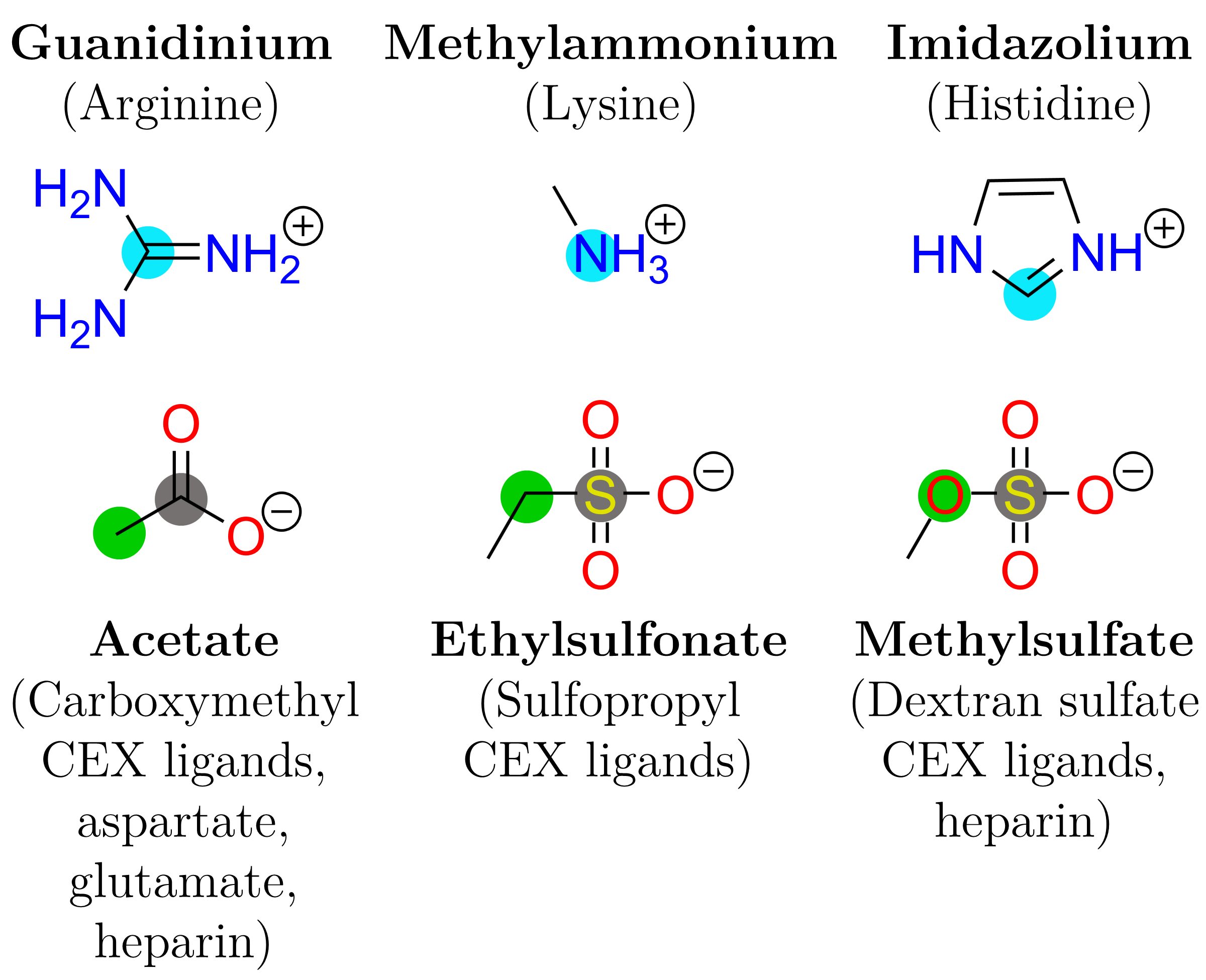
Interactions among charged groups in aqueous solution are widely implicated in protein structural stabilization, biomolecular recognition and surface adsorption.1, 2, 3 Among the specific phenomena of interest to this work is the longstanding puzzle that protein adsorption in cation-exchange (CEX) chromatography is known to be stronger on resins that are decorated with sulfate or sulfopropyl ligands than on resins with carboxymethyl ligands, despite the fact that the ligands bear the same formal charge and may be present on equivalent base matrices at comparable immobilization densities.4, 5 A related puzzle is the stronger binding to a heparin affinity resin of arginine than lysine 7-mers.6 Uncovering the physics that underlie these observations on systems of broad scientific and technological interest still remains largely an open problem.
Given the ionic nature of the constituents noted above, it is a natural first step to model the physics relying solely on electrostatics whilst ignoring the molecular nature of the solvent. Continuum solvent approaches have been enormously influential in guiding our thinking but they treat hydration phenomena only approximately and are unable to resolve puzzles such as those noted above7, 8, 9, 10, 11, 12, 13. It is necessary to account better the underlying physics.
All-atom molecular dynamics (MD) can in principle capture the balance between direct solute-solute and indirect solvent-mediated intermolecular interactions. However, classical non-polarizable force fields (FFs) are generally known to overestimate the strength of ion-pair interactions.14, 15, 16 This may be attributable to the partial inclusion of polarization implicitly in the parameterization of water models but not in ion models.17 To fix this disparity in the treatment of the solvent versus the ion, it has been suggested that ion charges be scaled by a constant factor of , where represents the high-frequency dielectric constant of water.18, 17. Results using this so-called electronic continuum correction (ECC) appear to better capture the strength of ion-pairing in solution. However, relative to the parent FF, the ECC complicates the estimation of solute hydration free energies 17 and may err in describing intermolecular interactions at large separations.
Here we assess the ability of a classical FF (CHARMM3619) with and without the ECC as well as a semiclassical model (AMOEBA20) to explain the two puzzles noted above. (We describe the AMOEBA model as semiclassical because of its rather detailed description of electrostatics and its inclusion of polarizability.) Our previous work treated the solute at a quantum chemical level and the solvent as a continuum;5 that effort captured some of the effects qualitatively, albeit with much uncertainty given the study’s limited configurational exploration. Following that work, here we model analogues of oxyanionic CEX ligands (i.e., acetate, methylsulfate and ethylsulfonate for carboxymethyl, dextran sulfate and sulfopropyl resins, respectively) interacting with comparable analogues of the basic amino acid side chains (i.e., guanidinium, methylammonium and imidazolium for arginine, lysine and histidine residues, respectively) (Fig. 1); acetate is of course also a model for the acidic amino acid side chains. Focusing on ligands alone allows us to study physically critical interactions more thoroughly. The insights thus derived also have broad relevance to modeling protein interactions as well as ionic liquids.
Association constants: Unbiased simulations were performed for each anion-cation pair at a concentration of 0.5 M to compute potential of mean force (PMF) profiles (Supplementary Figure S1); association constants were calculated as21, 22
| (1) |
where and refer to the separation distance and the PMF, respectively, and the ions are considered to be associated for . The location of the first maximum in the PMF was chosen as ,22 which separates the contact ion-pair from the solvent-separated ion pair and occurs at Å (Table S1).
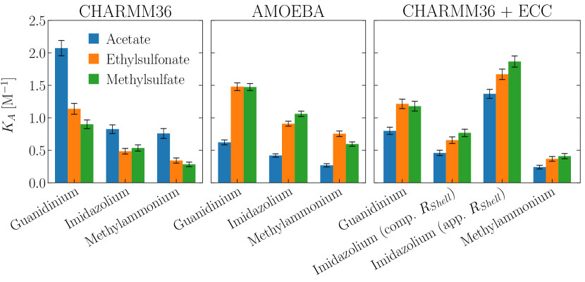
Figure 2 shows that both CHARMM36 and AMOEBA capture the expected rank order in cations (i.e., guanidinium imidazolium methylammonium) based on retention data of individual amino acids on sulfonate resins;23, 24 the order is also consistent with the Lys and Arg oligomer retention in heparin affinity chromatography.6 Although confounded by protein structural details, this order is furthermore consistent with the stronger retention of lysozyme (which is Arg-rich) than cytochrome c (which is Lys-rich) on several CEX resins, which continuum electrostatics models may not be able to capture.4, 25 CHARMM36 with the ECC generally captures the cation order, although the ECC tends to understructure the PMF profiles relative to AMOEBA (Fig. S1). This is especially the case for imidazolium systems, making the definition of somewhat unclear. For this reason, comparable and apparent values are used in Figure 2 that correspond approximately to contact and solvent-separated ion pairs.
The more revealing comparisons are among the anions: CHARMM36 incorrectly predicts stronger cation interactions with acetate than with the sulfur-containing ligands. AMOEBA and the ECC both correct this trend to be consistent with the experimentally observed rank order of CEX resin retentivities. Two competing features stand out. From the perspective of the ions, relative to AMOEBA, CHARMM36 over-stabilizes the interactions with acetate and under-stabilizes the interactions with the sulfur-containing anions (Fig. S1). From the perspective of the solvent, relative to AMOEBA, CHARMM36 predicts weaker acetate-water interactions but stronger water interactions with the sulfur-containing anions (Fig. S3). The ECC brings the classical model predictions closer to those of AMOEBA but sometimes at the expense of fidelity to the underlying PMF for ion-ion and ion-water association (Figs. S1 and S3). However, this observation must be tempered by the recognition that it is likely necessary to update the water models themselves for use in the ECC-ecosystem 17.
There is a paucity of data to support more rigorous quantitative comparisons between these results and experiment, but guanidinium acetate association has been investigated in two independent potentiometric studies.26, 27 Association constants were inferred in those studies from an induced shift in the ionization constant of acetic acid when guanidinium was substituted for another cation that was assumed not to complex with acetate. was independently estimated to be 0.5 M-1 and 0.37 M-1,26, 27 but the ionic strength (IS) was uncontrolled in the first study,26 which obfuscates the result because is expected to decrease with IS due to electrostatic screening. The IS was fixed at 1.02 M in the second study27 and the experimental value of 0.37 M-1 is of comparable magnitude but appropriately lower than the AMOEBA prediction of 0.62 M-1, which was obtained for guanidinium acetate at an IS of 0.5 M. A comparable measurement of the interaction between butylammonium and acetate (0.31 M-1 at 1.02 M IS)27 is also similar to the AMOEBA prediction for the methylammonium acetate system (0.27 M-1 at 0.5 M IS). The data from acetate simulations are similar to previous computational results as well,16, 14, 15 including a difference that was observed in classical simulations of guanidinium acetate using a different FF with and without the ECC (Fig. S4).16
Cross-FF analysis: To assess whether the dissimilarities between the CHARMM36 and AMOEBA results were due to polarizability or simply a difference in the treatment of electrostatics involving permanent charges, a cross-FF analysis was performed in which the in vacuo interaction energies between all ion pairs in AMOEBA simulation trajectories were analyzed retrospectively using both the CHARMM36 and AMOEBA FFs. This permitted an applied comparison of FF parameterization without any differences in configurational sampling. For each FF used in the analysis, the data were ensemble-averaged as a function of the separation distance ; Figures S5-S7 show the decomposition of the in vacuo interaction energy into permanent electrostatic (), van der Waals () and polarization () contributions. Figure S8 shows the magnitude of the difference between the CHARMM36 and AMOEBA profiles.
The permanent electrostatic contributions are nearly identical for the two FFs, with the largest difference observed in methylsulfate systems, where AMOEBA predicts slightly stronger attraction than CHARMM36 (Fig. S5). This similarity of electrostatics is intriguing because AMOEBA uses a sophisticated distributed multipole model whereas CHARMM36 uses only point charges at atom centers.19, 20 AMOEBA predicts generally weaker van der Waals interactions than CHARMM36 but the magnitude of the difference is relatively small (Fig. S6). Polarization contributions represent the largest difference between the two FFs (Fig. S7). Thus while AMOEBA’s more sophisticated multipole treatment of permanent electrostatics does contribute to sampling differences,20 polarizability is the primary factor that is responsible for differences in model performance. Polarization contributions are always observed to stabilize counterion pairs in vacuo.
PMF decomposition: The PMF may be decomposed as , where is the in vacuo interaction energy and is the solvent-mediated contribution. The contributions to were estimated using the trajectory-generating FF and was found from the PMF by difference; the results are shown in Figures S9-S12. Near the PMF minima, the magnitudes of both and are on the order of 90 kcal/mol (Figs. S9 and S10). Thus, it is the fine balance between two large competing contributions that dictates the PMF,22 which has a well depth that is typically two orders of magnitude smaller.
Given the similarities that were observed in the cross-FF analysis, differences between the CHARMM36 and AMOEBA profiles in Figures S9-S12 may be primarily attributed to the sampling differences that polarizability promotes. Permanent electrostatic interactions, which comprise the principal contribution to , are similar for CHARMM36 and AMOEBA in most systems (Fig. S11). However, noticeable discrepancies are apparent in the guanidinium acetate and imidazolium acetate systems, for which CHARMM36 substantially overestimates the magnitude of . The favorable AMOEBA polarization contributions (Fig. S7) decrease this discrepancy in the profiles for imidazolium acetate (Fig. S9) but a substantial difference remains for guanidinium acetate. In general, polarization contributions for the other systems and differences in permanent electrostatics lead to more attractive profiles for AMOEBA than CHARMM36 (Fig. S9). The inferred solvent contribution necessarily follows the opposite trend (Fig. S10).
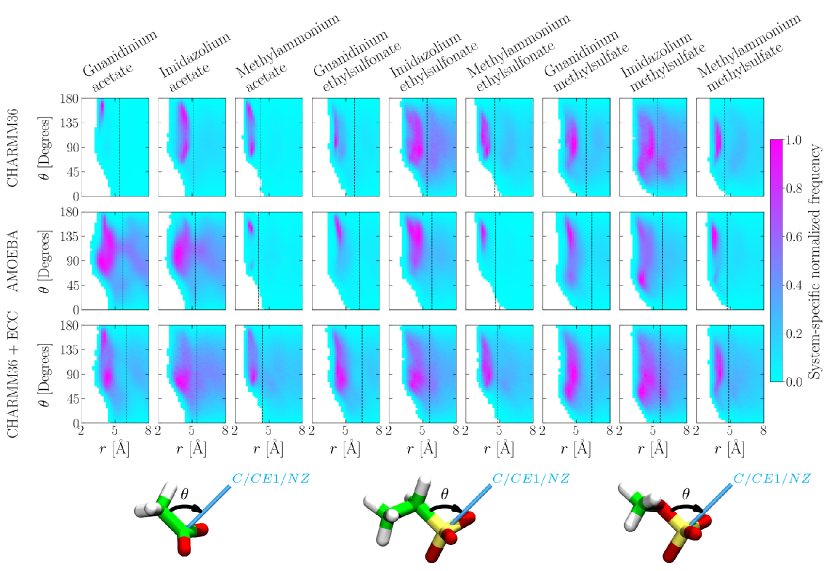
Configurational sampling: Differences in configurational sampling were examined more directly for each system by identifying the 3D spaces of the most frequently observed cation positions relative to anions (Fig. S14) and anion positions relative to cations (Fig. S15). For example, for guanidinium interacting with acetate or ethylsulfonate (Fig. S14), the cation samples a small patch of space around the carboxylate group within the CHARMM36 description but this is relaxed in the AMOEBA description. This trend is approximately reversed for ethylsulfonate. With the ECC correction, no such distinction is evident, suggesting a more promiscuous sampling and hence weaker association. Similar distinctions can be noted from either the anion’s (Fig. S14) or the cation’s perspective (Fig. S15). To quantify better the sampling differences suggested by the 3D plots, we project the data on an map (Fig. 3; is defined in Fig. 1). For between 90° and 180°, the carbonyl or sulfonyl oxygen is directed towards the anion.
Summarizing the observations, in general, for a given cation, CHARMM36 predicts that configurational preferences become less well-defined as the number of oxygen atoms in the anion increases, i.e., as the charge density decreases. This is as expected on the basis of the dominance of electrostatic interactions. With some exceptions, the opposite holds for AMOEBA, suggesting a richer interplay of electrostatic interactions arising from charge-charge interactions and polarization effects. The effect of the ECC is inconsistent.
Hydration free energies: Emergent behavior in ion-pair association is a consequence of the balance between direct interactions and solvent-mediated effects that generally tend to suppress ion-pair formation. The latter arises because there is an energetic penalty for partially dehydrating the interacting ions and reorganizing the nearby solvent structure. Hydration free energies () may therefore provide a useful complement to inform the understanding of ion pairing phenomena. To this end we estimated for individual ions using the molecular quasichemical organization of the potential distribution theorem,28, 12, 29, 30 which allows to be partitioned into physically meaningful contributions as
| (2) |
where the first term represents the free energy required to open a cavity in the solvent to accommodate the ionic solute. Contributions from long-range and short-range solute-solvent interactions are given by the second and third terms, respectively. Each term is a functional of a repulsive potential that is used to condition the solvent up to a maximum range of around the solute, but the sum of the three terms is independent of . We use Å, for which the cavity corresponds roughly to the first hydration shell around the solute.

Experimental estimates of may be obtained using a thermodynamic cycle based on proton dissociation, which requires reference to the free energy of hydrating a proton ().31, 32, 33 However, the value of is the subject of much uncertainty because extrathermodynamic assumptions must be employed to deconvolute experimentally accessible quantities into anion and cation contributions.34, 35, 31 We have taken and its uncertainty from a recent report that summarizes 72 independent estimates of the value.31 Literature data exist to make experimental comparisons for the computed of acetate and the cations,36, 37, 38, 39, 40, 35, 41, 42, 43, 44, 45 which are shown alongside simulation results in the rightmost panel of Figure 4 and are detailed in Table S4. These are comparable to other literature reports that are listed in Table S5.33, 36, 46, 47, 48 Within the appreciable uncertainty, the AMOEBA results agree with experiment. Data for acetate and the cations are also similar to simulation results based on thermodynamic integration.49, 31, 35
We next consider the individual contributions (Eq. 2). Uniformly, the packing contribution, i.e., the primitive hydrophobic contribution, to hydration is somewhat stronger (more positive) in AMOEBA than in CHARMM36. With the exception of guanidinium, the chemistry contribution is also stronger (more negative) in AMOEBA than in CHARMM36, which reflects greater ion-water attraction locally. Thus while packing will tend to favor ion-pair complexation in AMOEBA (over CHARMM36), the local attractive contributions will tend to suppress ion-pair formation in AMOEBA (over CHARMM36).
The long-range contribution to hydration proves surprising. This is more favorable for anions than cations in CHARMM36, which is expected based on the positive potential that exists in the center of a cavity due to the preferential orientation of water protons towards the cavity center 50, 51. However, the trend of long-range interactions found in AMOEBA is nearly reversed. It is well-known that the sign and magnitude of the electric potential that the solvent imposes on the solute charges is sensitive to both the structure of the solvent at the interface and the description of the charge over the solvent molecules 52, 53; for example, ab initio simulations show that the dipole moment of solvent next to a large anion is itself reduced 54. To probe this, in exploratory calculations for a spherical ion in water, we retrospectively included polarizability and multipole electrostatics in analyzing configurations sampled with a non-polarizable model. The shift is similar to the trend seen in Fig. 4 (Long-range). A more thorough investigation is necessarily left for future studies.
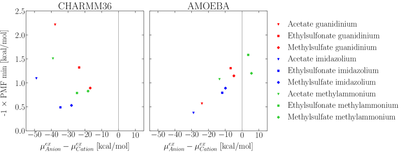
Matching water affinities: To see if we can use the hydration free energies of individual ions to infer ion association, we test the applicability of the empirical law of matching water affinities55, 56. This rule suggests that ions with hydration free energies that are closely matched will tend to associate more readily. Figure 5 shows that when using hydration free energies obtained from AMOEBA (which are closer to experimental estimates), there is a clear correlation between the difference in hydration free energy and the PMF well depth. The greater the difference in hydration free energy, the lower is the ion pair association affinity, which is reminiscent of experimentally observed trends in salt dissolution free energies.55 With CHARMM36 there is considerable scatter and the law of matching water affinities does not seem to hold. Although CHARMM36 predicts correctly that acetate is the most energetically expensive anion to dehydrate, it predicts incorrectly that cation interactions will be strongest with acetate. Overall, the results suggest that the empirical law of matching water affinities could be used in inferring ion-pairing, and hence potential protein adsorption on CEX matrices, provided the hydration of the individual ions is itself well captured (Fig. 4).
In conclusion, the present study suggests that the physics of polarizability is critical in determining why basic amino acid side chains bind more strongly to sulfate and sulfonate moieties than carboxylate groups, despite the fact that carboxylate has a higher (negative) surface charge density and ought to interact more strongly with cations. The carboxylate moiety is more energetically expensive to dehydrate but predicting this is only half of the puzzle. Subtle polarization effects are required to capture experimental trends because it is the fine difference between two large competing potentials (i.e., electrostatic attraction and solvent opposition) that underlies ion complexation in solution. Although the ECC can sometimes improve this balance in classical FFs, it may also promote spuriously promiscuous configurational sampling. Polarizability leads to qualitatively distinct configurational preferences that are expected to be broadly relevant to protein electrostatic interactions.
1 Supporting Information
Supporting information includes methods and supplementary results for (1) unbiased simulations, (2) in vacuo energy analyses (including cross-FF and PMF decomposition analyses), (3) configurational sampling analyses (including 3D maps, coordinate definitions and supplementary heat maps), (4) molecular quasichemical theory simulations and (5) experimental estimates of based on a thermodynamic cycle (PDF)
2 Acknowledgements
We thank Tom Beck (ORNL) for helpful discussions. This research was supported in part through the use of DARWIN computing system: DARWIN – A Resource for Computational and Data-intensive Research at the University of Delaware and in the Delaware Region, which is supported by NSF under Grant Number: 1919839, Rudolf Eigenmann, Benjamin E. Bagozzi, Arthi Jayaraman, William Totten, and Cathy H. Wu, University of Delaware, 2021, URL: https://udspace.udel.edu/handle/19716/29071
This research used resources of the Oak Ridge Leadership Computing Facility at the Oak Ridge National Laboratory, which is supported by the Office of Science of the U.S. Department of Energy under Contract No. DE-AC05-00OR22725.
References
- Haggerty and Lenhoff 1991 Haggerty, L.; Lenhoff, A. M. Relation of Protein Electrostatics Computations to Ion-Exchange and Electrophoretic Behavior. J. Phys. Chem. 1991, 95, 1472–1477
- Roberts and Blanco 2014 Roberts, C. J.; Blanco, M. A. Role of Anisotropic Interactions for Proteins and Patchy Nanoparticles. J. Phys Chem. B 2014, 118, 12599–12611
- Luo et al. 1999 Luo, R.; David, L.; Hung, H.; Devaney, J.; Gilson, M. K. Strength of Solvent-Exposed Salt-Bridges. J. Phys. Chem. B 1999, 103, 727–736
- DePhillips and Lenhoff 2001 DePhillips, P.; Lenhoff, A. M. Determinants of Protein Retention Characteristics on Cation-Exchange Adsorbents. J. Chrom. A 2001, 933, 57–72
- Asthagiri et al. 2000 Asthagiri, D.; Schure, M. R.; Lenhoff, A. M. Calculation of Hydration Effects in the Binding of Anionic Ligands to Basic Proteins. J. Phys. Chem. B 2000, 104, 8753–8761
- Fromm et al. 1995 Fromm, J. R.; Hileman, R. E.; Caldwell, E. E. O.; Weiler, J. M.; Linhardt, R. J. Differences in the Interaction of Heparin with Arginine and Lysine and the Importance of These Basic Amino Acids in the Binding of Heparin to Acidic Fibroblast Growth Factor. Arch. Biochem. Biophys. 1995, 323, 279–287
- Warshel et al. 2006 Warshel, A.; Sharma, P. K.; Kato, M.; Parson, W. W. Modeling Electrostatic Effects in Proteins. Biochim. Biophys. Acta - Proteins Proteom. 2006, 1764, 1647–1676
- Ren et al. 2012 Ren, P.; Chun, J.; Thomas, D. G.; Schnieders, M. J.; Marucho, M.; Zhang, J.; Baker, N. A. Biomolecular Electrostatics and Solvation: A Computational Perspective. Q. Rev. Biophys 2012, 45, 427–491
- Koehl 2006 Koehl, P. Electrostatics Calculations: Latest Methodological Advances. Curr. Opin. Struct. Bio. 2006, 16, 142–151
- Gray and Stiles 2018 Gray, C. G.; Stiles, P. J. Nonlinear Electrostatics: The Poisson-Boltzmann Equation. Eur. J. Phys. 2018, 39, 1–27, 053002
- Sun et al. 2016 Sun, Q.; Klaseboer, E.; Chan, D. Y. C. A Robust and Accurate Formulation of Molecular and Colloidal Electrostatics. J. Chem. Phys. 2016, 145, 1–12, 054106
- Asthagiri et al. 2021 Asthagiri, D. N.; Paulaitis, M. E.; Pratt, L. R. Thermodynamics of Hydration from the Perspective of the Molecular Quasichemical Theory of Solutions. J. Phys. Chem. B 2021, 125, 8294–8304
- van Gunsteren et al. 2006 van Gunsteren, W. F.; Bakowies, D.; Baron, R.; Chandrasekhar, I.; Christen, M.; Daura, X.; Gee, P.; Geerke, D. P.; Glättli, A.; Hünenberger, P. H. et al. Biomolecular Modeling: Goals, Problems, Perspectives. Angew. Chem. Int. Ed. 2006, 45, 4064–4092
- Debiec et al. 2014 Debiec, K. T.; Gronenborn, A. M.; Chong, L. T. Evaluating the Strength of Salt Bridges: A Comparison of Current Biomolecular Force Fields. J. Phys. Chem. B 2014, 118, 6561–6569
- Debiec et al. 2016 Debiec, K. T.; Cerutti, D. S.; Baker, L. R.; Gronenborn, A. M.; Case, D. A.; Chong, L. T. Further along the Road Less Traveled: AMBER ff15ipq, an Original Protein Force Field Built on a Self-Consistent Physical Model. J. Chem. Theory Comput. 2016, 12, 3926–3947
- Mason et al. 2019 Mason, P. E.; Jungwirth, P.; Duboué-Dijon, E. Quantifying the Strength of a Salt Bridge by Neutron Scattering and Molecular Dynamics. J. Phys. Chem. Lett. 2019, 10, 3254–3259
- Duboué-Dijon et al. 2020 Duboué-Dijon, E.; Javanainen, M.; Delcroix, P.; Jungwirth, P.; Martinez-Seara, H. A Practical Guide to Biologically Relevant Molecular Simulations with Charge Scaling for Electronic Polarization. J. Chem. Phys. 2020, 153, 1–15, 050901
- Leontyev and Stuchebrukhov 2011 Leontyev, I.; Stuchebrukhov, A. Accounting for Electronic Polarization in Non-Polarizable Force Fields. Phys. Chem. Chem. Phys. 2011, 13, 2613–2626
- Huang and Mackerell 2013 Huang, J.; Mackerell, A. D., Jr. CHARMM36 All-Atom Additive Protein Force Field: Validation Based on Comparison to NMR Data. J. Comput. Chem. 2013, 34, 2135–2145
- Shi et al. 2013 Shi, Y.; Xia, Z.; Zhang, J.; Best, R.; Wu, C.; Ponder, J. W.; Ren, P. Polarizable Atomic Multipole-Based AMOEBA Force Field for Proteins. J. Chem. Theory Comput. 2013, 9, 4046–4063
- Zhu et al. 2012 Zhu, P.; Pratt, L. R.; Papadopoulos, K. D. Pairing of 1-Hexyl-3-methylimidazolium and Tetrafluoroborate Ions in n-Pentanol. J. Chem. Phys. 2012, 137, 1–4, 174501
- Shi and Beck 2017 Shi, Y.; Beck, T. Deconstructing Free Energies in the Law of Matching Water Affinities. J. Phys. Chem. B 2017, 121, 2189–2201
- Wang et al. 1989 Wang, N.-H. L.; Yu, Q.; Kim, S. U. Cation Exchange Equilibria of Amino Acids. React. Polym. 1989, 11, 261–277
- Moore et al. 1958 Moore, S.; Spackman, D. H.; Stein, W. H. Chromatography of Amino Acids on Sulfonated Polystyrene Resins. An Improved System. Anal. Chem. 1958, 30, 1185–1190
- Yao and Lenhoff 2005 Yao, Y.; Lenhoff, A. M. Electrostatic Contributions to Protein Retention in Ion-Exchange Chromatography. 2. Proteins with Various Degrees of Structural Differences. Anal. Chem. 2005, 77, 2157–2165
- Tanford 1954 Tanford, C. The Association of Acetate with Ammonium and Guanidinium Ions. J. Am. Chem. Soc. 1954, 76, 945–946
- Springs and Haake 1977 Springs, B.; Haake, P. Equilibrium Constants for Association of Guanidinium and Ammonium Ions with Oxyanions: The Effect of Changing Basicity of the Oxyanion. Bioorg. Chem. 1977, 6, 181–190
- Beck et al. 2006 Beck, T. L.; Paulaitis, M. E.; Pratt, L. R. The Potential Distribution Theorem and Models of Molecular Solutions; Cambridge University Press, 2006
- Weber et al. 2011 Weber, V.; Merchant, S.; Asthagiri, D. Communication: Regularizing Binding Energy Distributions and Thermodynamics of Hydration: Theory and Application to Water Modeled with Classical and ab Initio Simulations. J. Chem. Phys. 2011, 135, 1–4, 181101
- Weber and Asthagiri 2012 Weber, V.; Asthagiri, D. Regularizing Binding Energy Distributions and the Hydration Free Energy of Protein Cytochrome C from All-atom Simulations. J. Chem. Theory Comput. 2012, 8, 3409–3415
- Fossat et al. 2021 Fossat, M. J.; Zeng, X.; Pappu, R. V. Uncovering Differences in Hydration Free Energies and Structures for Model Compound Mimics of Charged Side Chains of Amino Acids. J. Phys. Chem. B 2021, 125, 4148–4161
- Lim et al. 1991 Lim, C.; Bashford, D.; Karplus, M. Absolute p Calculations with Continuum Dielectric Methods. J. Phys. Chem. 1991, 95, 5610–5620
- Pearson 1986 Pearson, R. G. Ionization Potentials and Electron Affinities in Aqueous Solution. J. Am. Chem. Soc. 1986, 108, 6109–6114
- Grossfield et al. 2003 Grossfield, A.; Ren, P.; Ponder, J. W. Ion Solvation Thermodynamics from Simulation with a Polarizable Force Field. J. Am. Chem. Soc. 2003, 125, 15671–15682
- Zhang et al. 2017 Zhang, H.; Jiang, Y.; Yan, H.; Yin, C.; Tan, T.; van der Spoel, D. Free-Energy Calculations of Ionic Hydration Consistent with the Experimental Hydration Free Energy of the Proton. J. Phys. Chem. Lett. 2017, 8, 2705–2712
- Cramer and Truhlar 1991 Cramer, C. J.; Truhlar, D. G. General Parameterized SCF Model for Free Energies of Solvation in Aqueous Solution. J. Am. Chem. Soc. 1991, 113, 8305–8311
- Taft and Topsom 1987 Taft, R. W.; Topsom, R. D. In Prog. Phys. Org. Chem., 1st ed.; Taft, R. W., Ed.; John Wiley & Sons, 1987; Vol. 16; pp 1–84
- Cumming and Kebarle 1978 Cumming, J. B.; Kebarle, P. Summary of Gas Phase Acidity Measurements Involving Acids AH. Entropy Changes in Proton Transfer Reactions Involving Negative Ions. Bond Dissociation Energies D(A-H) and Electron Affinities EA(A). Can. J. Chem. 1978, 56, 1–9
- Fujio et al. 1981 Fujio, M.; McIver, R. T., Jr.; Taft, R. W. Effects on the Acidities of Phenols from Specific Substituent-Solvent Interactions. Inherent Substituent Parameters from Gas-Phase Acidities. J. Am. Chem. Soc 1981, 103, 4017–4029
- Settimo et al. 2014 Settimo, L.; Bellman, K.; Knegtel, R. M. A. Comparison of the Accuracy of Experimental and Predicted pKa Values of Basic and Acidic Compounds. Pharm. Res. 2014, 31, 1082–1095
- Wolfenden et al. 1981 Wolfenden, R.; Andersson, L.; Cullis, P. M.; Southgate, C. C. B. Affinities of Amino Acid Side Chains for Solvent Water. Biochem. 1981, 20, 849–855
- Reif et al. 2012 Reif, M. M.; Hünenberger, P. H.; Oostenbrink, C. New Interaction Parameters for Charged Amino Acid Side Chains in the GROMOS Force Field. J. Chem. Theory Comput. 2012, 8, 3705–3723
- Hunter and Lias 1998 Hunter, E. P. L.; Lias, S. G. Evaluated Gas Phase Basicities and Proton Affinities of Molecules: An Update. J. Phys. Chem. Ref. Data 1998, 27, 413–656
- Rizzo et al. 2006 Rizzo, R. C.; Aynechi, T.; Case, D. A.; Kuntz, I. D. Estimation of Absolute Free Energies of Hydration Using Continuum Methods: Accuracy of Partial Charge Models and Optimization of Nonpolar Contributions. J. Chem. Theory Comput. 2006, 2, 128–139
- In et al. 2005 In, Y.; Chai, H. H.; No, K. T. A Partition Coefficient Calculation Method with the SFED Model. J. Chem. Inf. Model. 2005, 45, 254–263
- Gilson and Honig 1988 Gilson, M. K.; Honig, B. Calculation of the Total Electrostatic Energy of a Macromolecular System: Solvation Energies, Binding Energies, and Conformational Analysis. Proteins: Struct. Funct. Bioinform. 1988, 4, 7–18
- Marcus 2013 Marcus, Y. Individual Ionic Surface Tension Increments in Aqueous Solutions. Langmuir 2013, 29, 2881–2888
- Çiftcioǧlu and Trindle 2014 Çiftcioǧlu, G. A.; Trindle, C. Computational Estimates of Thermochemistry and p Values of Cyclopropenyl Imine Superbases. Int. J. Quantum Chem. 2014, 114, 392–399
- Lin et al. 2018 Lin, F.-Y.; Lopes, P. E. M.; Harder, E.; Roux, B.; Mackerell, A. D., Jr. Polarizable Force Field for Molecular Ions Based on the Classical Drude Oscillator. J. Chem. Inf. Model. 2018, 58, 993–1004
- Hummer et al. 1996 Hummer, G.; Pratt, L. R.; Garcia, A. E. Free Energy of Ionic Hydration. J. Phys. Chem. 1996, 100, 1206–1215
- Ashbaugh 2000 Ashbaugh, H. S. Convergence of Molecular and Macroscopic Continuum Descriptions of Ion Hydration. J. Phys. Chem. B 2000, 104, 7235–7238
- Wilson et al. 1988 Wilson, M. A.; Pohorille, A.; Pratt, L. R. Surface potential of the water liquid–vapor interface. J. Chem. Phys. 1988, 88, 3281–3285
- Doyle et al. 2019 Doyle, C. C.; Shi, Y.; Beck, T. L. The Importance of the Water Molecular Quadrupole for Estimating Interfacial Potential Shifts Acting on Ions Near the Liquid–Vapor Interface. J. Phys. Chem. B 2019, 123, 3348–3358
- Guàrdia et al. 2009 Guàrdia, E.; Skarmoutsos, I.; Masia, M. On Ion and Molecular Polarization of Halides in Water. J. Chem. Theory Comput. 2009, 5, 1449–1453
- Collins 1997 Collins, K. D. Charge Density-Dependent Strength of Hydration and Biological Structure. Biophys. J. 1997, 72, 65–76
- Collins 2019 Collins, K. D. The Behavior of Ions in Water Is Controlled by Their Water Affinity. Q. Rev. Biophys. 2019, 52, 1–19, e11