Topotactically induced oxygen vacancy order in nickelate single crystals
Abstract
The strong structure-property coupling in rare-earth nickelates has spurred the realization of new quantum phases in rapid succession. Recently, topotactic transformations have provided a new platform for the controlled creation of oxygen vacancies and, therewith, for the exploitation of such coupling in nickelates. Here, we report the emergence of oxygen vacancy ordering in Pr0.92Ca0.08NiO2.75 single crystals obtained via a topotactic reduction of the perovskite phase Pr0.92Ca0.08NiO3, using CaH2 as the reducing agent. We unveil a brownmillerite-like ordering pattern of the vacancies by high-resolution scanning transmission electron microscopy, with Ni ions in alternating square-pyramidal and octahedral coordination along the pseudocubic [100] direction. Furthermore, we find that the crystal structure acquires a high level of internal strain, where wavelike modulations of polyhedral tilts and rotations accommodate the large distortions around the vacancy sites. Our results suggest that atomic-resolution electron microscopy is a powerful method to locally resolve unconventional crystal structures that result from the topotactic transformation of complex oxide materials.
pacs:
dfI Introduction
In transition metal oxides, strongly correlated valence electrons can couple collectively to the lattice degrees of freedom, which can lead to a variety of emergent ordering phenomena, including exotic magnetism, multiferroicity, orbital order, and superconductivity [1]. In oxides with the perovskite structure, high flexibility and tolerance to structural and compositional changes enable the controlled exploitation and manipulation of the emergent properties [2]. Oxygen vacancies, for example, can radically alter the electronic states in materials, and in turn, suppress or enhance emergent phases via charge compensation and/or structural phase transitions [3, 4, 5]. Understanding the formation of oxygen vacancies and their impact thus provides promising prospects for exploring new physical properties and potential future technological applications.
A prototypical example for correlated transition-metal oxides is the family of perovskite rare-earth nickelates, NiO3 ( = rare-earth ion), exhibiting a rich phase diagram including metal-to-insulator and antiferromagnetic transitions [6, 7, 8]. For = Pr and Nd, these transitions occur concomitantly with a breathing distortion of the NiO6 octahedra and a disproportionation of the Ni-O hybridization [9, 10, 11, 12]. As a consequence, the material family exhibits a pronounced structure-property relationship [13, 14, 15, 16, 17, 18, 19] and a sensitivity to oxygen vacancy formation, which can modify the surrounding Ni-O bonds and the nominal 3 electronic configuration of the Ni3+ ions [20]. Notably, an extensive oxygen reduction of the perovskite phase towards Ni1+ with a cuprate-like 3 electronic configuration was recently realized via topochemical methods in Nd0.8Sr0.2NiO2 thin films, yielding the emergence of superconductivity [21]. Furthermore, superconductivity was also observed in topotactically reduced films with = La and Pr [22, 23, 24], as well as for various Sr-substitution levels [25, 26] and substitution with Ca ions [27]. Since these reduced nickelates with the infinite-layer crystal structure are nominally isoelectronic and isostructural to cuprate superconductors, the degree of the analogy between the two material families is vividly debated [28, 29, 30, 31, 32, 33]. Moreover, vigorous efforts are ongoing to realize superconductivity not only in thin films, but also in polycrystalline powders [34, 35] as well as in single-crystalline samples [36, 37], while also an improved understanding of the topotactic reduction process between the perovskite and infinite-layer phase is highly desirable. In particular, the reduction involves various intermediate (metastable) phases, in which the oxygen vacancy ordering patterns and the nature of the emergent phases have not yet been clarified comprehensively.
For instance, extensive experimental and theoretical studies [38, 39, 40, 41, 42, 43, 44] were performed on oxygen deficient LaNiO3-δ with , suggesting a transition from a paramagnetic metal to a ferromagnetic semiconductor and an antiferromagnetic insulator as a function of increasing vacancy concentration [45, 46, 47]. For , neutron powder diffraction [38] revealed that the parent perovskite crystal structure with uniform NiO6 octahedra changed to a structure with sheets of NiO6 octahedra and square-planar NiO4 units arranged along the pseudocubic [100] direction [38, 39], involving a 2a2a2ap reconstruction of the parent pseudocubic unit cell (ap is the pseudocubic lattice parameter). Yet, the detailed crystal structure for the case is not known, although an electron diffraction study suggested that it involves a 2a2a2ap reconstructed supercell [48]. Moreover, for compounds with = Pr and Nd, even less understanding of the oxygen deficient phases exists. Metastable structures with ferromagnetic order were initially identified for , with x-ray diffraction data indicating a 3apap3ap supercell that possibly comprises two sheets of NiO4 square-planar units connected with one sheet of NiO6 octahedra [49]. In a subsequent neutron powder diffraction study it was suggested, however, that the metastable phase of the Pr compound rather corresponds to with a aaap reconstruction and one sheet of NiO4 square-planar units connected with two sheets of NiO6 octahedra [50].
Here, we use atomic-resolution scanning transmission electron microscopy (STEM) together with electron energy-loss spectroscopy (EELS) to investigate the oxygen vacancy formation occurring in a Pr0.92Ca0.08NiO3-δ single crystal upon topotactic reduction. We resolve the chemical composition and the atomic-scale lattice of the crystal, identifying a 4ap4ap2ap reconstructed superstructure with a highly distorted Pr sublattice. We find that the oxygen vacancy ordering pattern corresponds to a brownmillerite-like structure with a two-layer-repeating stacking sequence of NiO6 octahedra and NiO5 square pyramids, suggesting an oxygen deficiency of . Meanwhile, quantification of the octahedral tilts and Ni-O bond angles reveals distinct periodic wavelike patterns of polyhedra coordination in different layers due to the oxygen vacancies. These results are markedly distinct from previous reports on reduced rare-earth nickelates and provide an atomic-scale understanding of the moderately oxygen deficient structure with , which is one of the metastable phases occurring during the topotactic reduction process towards the infinite-layer phase of the superconducting nickelates with .
II Methods
Single crystals of perovskite Pr1-xCaxNiO3 were synthesized under high pressure and high temperature. Specifically, a 1000 ton press equipped with a Walker module was used to realize a gradient growth under a pressure of 4 GPa, executed in spatial separation of the oxidizing KClO4 and NaCl flux, similarly to the previous synthesis of La1-xCaxNiO3 single crystals [36]. The precursor powders were weighed in according to a desired composition of Pr0.8Ca0.2NiO3, although the incorporated Ca content in the obtained Pr1-xCaxNiO3 was lower and ranged from to 0.1. The Pr1-xCaxNiO3 single crystals were reduced using CaH2 as the reducing agent in spatial separation to the crystals. The duration of the reduction was eight days, using the same procedure and conditions as previously described for the reduction of La1-xCaxNiO3 crystals [36].
Single-crystal x-ray diffraction (XRD) was performed on crystals before and after the reduction. The technical details are given in the Supplemental Material [51].
Electron-transparent TEM specimens of the sample were prepared on a Thermo Fisher Scientific focused ion beam (FIB) using the standard liftout method. Samples with a size of 20 5 m2 were thinned to 30 nm with 2 kV Ga ions, followed by a final polish at 1 kV to reduce effects of surface damage. HAADF, ABF and EELS were recorded by a probe aberration-corrected JEOL JEM-ARM200F scanning transmission electron microscope equipped with a cold-field emission electron source and a probe Cs corrector (DCOR, CEOS GmbH), and a Gatan K2 direct electron detector was used at 200 kV. STEM imaging and EELS analyses were performed at probe semiconvergence angles of 20 and 28 mrad, resulting in probe sizes of 0.8 and 1.0 Å, respectively. Collection angles for STEM-HAADF and ABF images were 75 to 310 and 11 to 23 mrad, respectively. To improve the signal-to-noise ratio of the STEM-HAADF and ABF data while minimizing sample damage, a high-speed time series was recorded (2 s per pixel) and was then aligned and summed. STEM-HAADF and ABF multislice image simulations of the crystal along [100] and [101] zone axis were performed using the QSTEM software [52]. Further details of the parameters used for the simulations are given in the Supplemental Material [51].
III Results
In thin films of infinite-layer nickelates, the highest superconducting transition temperatures are realized through a substitution of approximately 20% of the rare-earth ions by divalent Sr or Ca ions [25, 26, 27]. Accordingly, we have prepared the precursor materials for the synthesis of single crystals with a nominal stoichiometry of Pr0.8Ca0.2NiO3. Using a high-pressure synthesis methods [36], we obtain Pr1-xCaxNiO3 crystals with typical lateral dimensions of 20 – 100 m. Yet, an analysis of the as-grown crystals by scanning electron microscopy (SEM) with energy-dispersive x-ray (EDX) spectroscopy indicates that the incorporated Ca content lies between and 0.1 (see Fig. S1 in the Supplemental Material [51]). This discrepancy to the nominal Ca content suggests that different growth parameters, such as an increased oxygen partial pressure, might be required to achieve stoichiometric Pr0.8Ca0.2NiO3 crystals. By contrast, the employed parameters yielded higher Ca substitutions as high as in the case of La1-xCaxNiO3 as determined on the as-grown crystal surface by EDX [36], which exhibits a less distorted perovskite structure [6].
As a next step, we use single-crystal XRD to investigate a 20 m piece that was broken off from a larger as-grown crystal. The acquired XRD data indicate a high crystalline quality (see Fig. S2 of the Supplemental Material [51]) and can be refined in the orthorhombic space group , which is consistent with PrNiO3 single crystals and polycrystalline powders [53, 54]. The refined Ca content of the crystal is 8.6%. The refined lattice parameters and atomic coordinates are presented in the Supplemental Material [51]. Furthermore, we find that the investigated crystal piece contains three orthorhombic twin domains extracted from the refinement, with volume fractions 0.95/0.04/0.01.
Subsequently, we carry out the topotactic oxygen reduction on a batch of Pr1-xCaxNiO3 single crystals for eight days, using the same conditions as previously described for the reduction of La1-xCaxNiO3 crystals [36]. Single-crystal XRD measurements on a reduced 20 m crystal indicate a significant transformation of the crystal structure after eight days. However, a strong broadening and the resulting overlap of the Bragg reflections in the XRD maps prohibit a structural refinement and determination of the symmetry by this method (see Fig. S2 [51]).
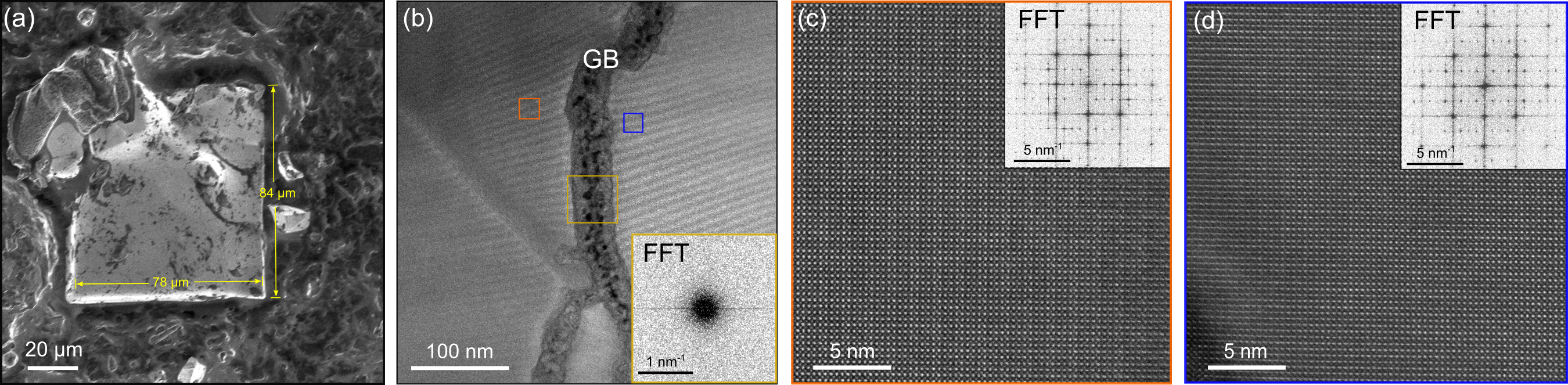
Hence, in order to investigate the topotactic transformation of the crystal lattice on a local scale, we turn to atomic-resolution STEM imaging. We examine a reduced Pr0.92Ca0.08NiO3-δ crystal with lateral dimensions of 80 m. A top-down view of the crystal is shown in Fig. 1(a). Identical TEM specimens were prepared from a region of the crystal without visible surface defects caused by the FIB process. Figure 1(b) shows a low-magnification STEM high-angle annular dark-field (HAADF) image. As a first characteristic of the topotactic-reduced crystal, we note that single-crystalline regions in the specimen are separated by grain boundary (GB) like regions, with a width ranging from a few ten to hundred nanometers and a length ranging from a few nanometers to micrometers. The GBs exhibit mostly an amorphous structure [Fig. 1(b) inset and Fig. S3], exhibiting dark contrast in the images that originate from diffuse scattering [55] (see Fig. S3 for more details [51]). The amorphous character of the GBs is also confirmed in EELS measurements of elemental distribution profiles across a GB, which show a reduced EELS intensity of all cations due to the deteriorating signal in structurally disordered regions (Fig. S4 [51]). The presence of GBs can be a consequence of topotactic reduction. Alternatively, the GBs might have formed already during the high-pressure growth of the perovskite phase.
Zoom-in STEM-HAADF images from areas on either side of a GB [orange and blue squares in Fig. 1(b)] are displayed in Figs. 1(c) and 1(d). The same lattice structure and orientation are observed from the different crystalline domains near the GB. One typical domain size in the crystal is found to be hundreds of nanometers. STEM-HAADF imaging over larger field of view of hundred nanometers does not reveal any regions with defects or impurity phase inside one crystalline domain, suggesting that each domain retains a high crystalline quality and a stable structural phase after the reduction process (see Fig. S3 [51]).
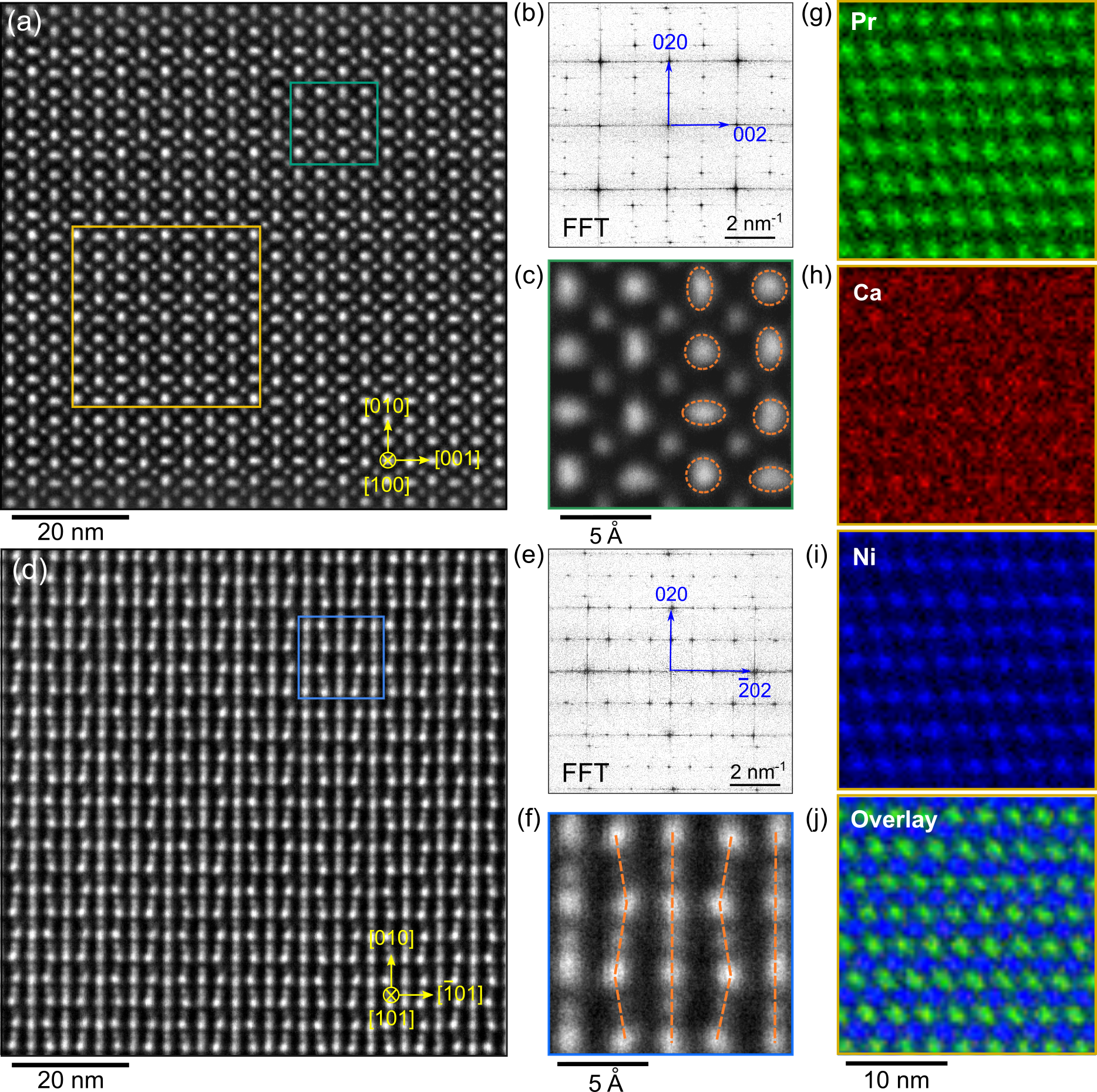
Figure 2(a) and 2(d) show atomic-resolution STEM-HAADF images viewed along the [100] and [101] pseudocubic orientations, respectively. The corresponding fast Fourier transforms (FFT) of the images reveal the crystal symmetry [Figs. 2(b) and 2(e)]. As presented in Fig. 2(b), there is an approximate 8% difference of the distances between the FFT maxima in reciprocal space along the [002] and [020] axis (blue arrows on FFT patterns). Based on the preference of vacancy formation on the apical oxygen sites [56], this indicates a contraction along the [002] axis due to the removal of apical oxygen atoms following the topotactic reduction process. Between the FFT maxima, the satellite spots corresponding to the superstructure periodicity appear. The wave vector of the superstructure reflexes is = 1/4 reciprocal lattice units along [020] axis and = 1/2 along [002] axis, indicating a formation of the 4ap4ap2ap superstructure of perovskite [ = = 16.56(32) Å, = 7.60(14) Å]. In STEM-HAADF images, the contrast is approximately proportional to (where is the atomic number) [57, 58], so Pr columns ( = 59) appear bright and Ni columns ( = 28) exhibit a darker contrast. The fourfold superstructure arises from the periodic changes in the intensity of the STEM image. As shown in the zoomed-in image [Fig. 2(c)], half of the Pr atoms along the [100] projection appear elongated, while the other half remain round and undistorted. Focusing on the first two Pr rows, the atoms exhibit alternating round and vertical oval shapes, while the third and fourth rows exhibit alternating round and horizontal oval shapes.
Considering the projection in our image, the distortions stem from displacements of Pr columns. Figure 2(d) indeed reveals straight and zigzag line patterns of Pr atoms alternating along the [01] direction. A closer look at the atomic arrangement in Fig. 2(f) shows that the zigzag line on the fourth column appears to be a mirror of the second, forming a four-layer repeat sequence (ABAC stacking). EELS elemental maps of Pr, Ca and Ni obtained from the crystal are displayed in Figs. 2(g)-2-(i) using Pr , Ca , and Ni edges, respectively. The maps show that the Pr, Ca and Ni contents are homogeneous over the structure. Integrated concentration profiles of Pr and Ca confirm the A-site cation stoichiometry and uniform distribution (see Fig. S5 [51]). This suggests that strong distortions of the Pr lattice are likely not rooted in an A/B-site deficiency or ordering, but rather originate from other factors such as oxygen vacancy formation.
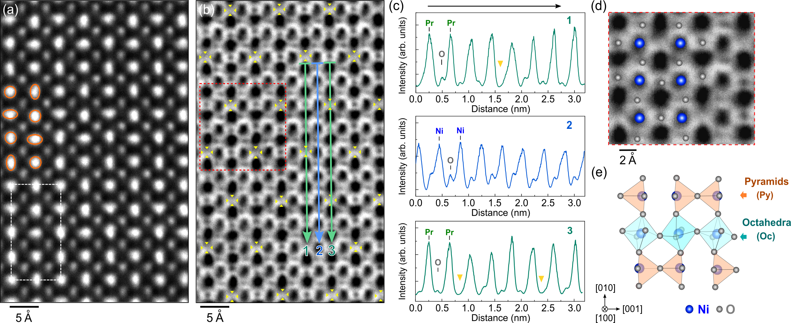
To gain insights into the detailed atomic structure, high-resolution STEM annular bright-field (ABF) images were acquired for imaging lighter elements such as oxygen. Figures 3(a) and 3(b) show the simultaneously acquired HAADF and ABF images of the crystal along the [100] projection. The distribution of oxygen ions including filled and empty apical oxygen sites is clearly visible by ABF imaging. The corresponding inverse intensity profiles extracted from different layers are displayed in Fig. 3(c). The absence of image contrast at every fourth oxygen site of the Pr-O layers confirms the vacancy ordering (profiles 1 and 3), while the oxygen content remains constant in Ni-O layers (profile 2). By overlaying the yellow arrows, the ordering pattern of oxygen vacancies can be clearly visualized. Half of the NiO6 octahedra lose one apical oxygen atom and the remaining five oxygen atoms in a pseudocubic unit cell form a NiO5 pyramid.
Notably, a square pyramidal coordination of the Ni ion is uncommon in nickel-oxide based materials. Few compounds with Ni2+ ions in similar five-fold coordination include KNi4(PO4)3 and BaYb2NiO5 [59]. However, none of these compounds exhibits a perovskite-derived structure. Our investigation therefore reveals the existence of a new structural motif in marked distinction to the NiO4 square-planar coordination in previous reports on oxygen-deficient perovskite nickelates [60, 44].
A magnified view of the ABF image is shown in Fig. 3(d). The apex-linked pyramids as ”bow-tie” dimer units form a one-dimensional chain running along the [001] direction. Such configuration is consistent with the brownmillerite type structure AnBnO3n-1 with = 4 corresponding to the A4B4O11 phase: Layers of apex-linked pyramids are stacked in the sequence …-Oc-Py-Oc-Py-…, where Oc denotes a layer containing only octahedra (cyan), and Py a layer only pyramids (orange). The …-Oc-Py-Oc-Py-… sequence runs parallel to the [010] axis, so that the Py layers are at (001) and (001) planes with a stacking vector [001]. In the pyramidal layers, the remaining oxygen atoms are located at the center of apical sites without causing any tilt of square pyramids. In contrast, the apical oxygen atoms in octahedral layers tend to shift towards the elongated Pr atoms, leading to large octahedral tilts. A corresponding STEM-ABF image simulation was performed based on the predicted atomic model shown in Fig. 3(e) using the multislice method [52] (see Fig. S6 [51]). The simulated image reproduces the vacant sites and distortions well as observed from the STEM measurements, confirming the main crystal structure and alternating-stacking configuration.
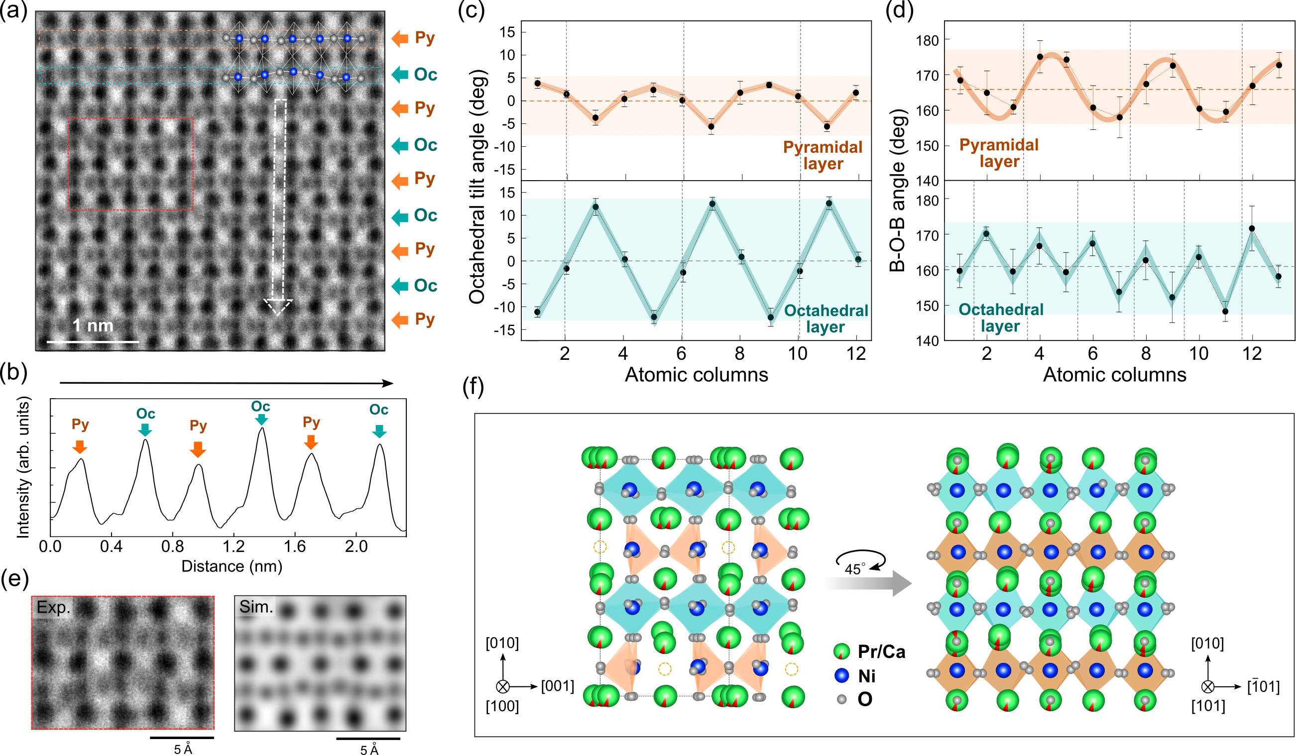
The distribution of oxygen vacancies can lead to modifications in bond angles and tilting of octahedra. Quantitative STEM ABF measurements were used to precisely examine the atomic structure including the oxygen positions along the [101] direction. From the inverse ABF intensity profiles taken in Fig. 4(a), as shown in Fig. 4(b), the oxygen intensities on different Ni-O layers vary as a function of the atomic columns along the [010] direction, caused by different apical oxygen occupancies in octahedral and pyramidal layers. Thus, Ni-O layers are denoted Oc as the oxygen intensity reaches its maximum on the octahedral layer and denoted Py with the lower intensity due to oxygen deficiency on the pyramidal layers. Analyses of the ABF image, quantifying the NiO6 octahedral tilt angles and Ni-O-Ni bond angles, are shown in Figs. 4(c) and 4(d). Notably, we observe two different patterns of octahedral distortions along the [01] direction on the pyramidal and octahedral layers. The pyramidal layer shows a variation of tilt angles, with repeating order -4∘–0∘–4∘–0∘ [Fig. 4(c), upper panel]. Meanwhile, a single wavelike pattern of Ni-O-Ni angles varies from 157∘ to 176∘ with an average of 166.3∘ [Fig. 4(d), upper panel]. Such four perovskite subcell wavelike pattern, as indicated by the dashed lines, can arise from the ordering of oxygen vacancies along the [100] projection on the pyramidal layer. On the octahedral layer, different from the pyramidal layer, the amplitude of the octahedral tilting is larger, varying from -12∘ to 12∘. Ni-O-Ni bond angles on the octahedral layer exhibit a zigzag modulated pattern with two sub-cell repeats and an average of 161.1∘. The overall periodicity of bond angles and tilt modulations is consistent with the alternating zigzag and straight pattern on the Pr columns along the [01] direction. The simulated ABF image for the predicted model along this viewing direction also agrees well with the STEM-ABF image, confirming the polyhedral distortions in the structure [Fig. 4(e)]. The different amplitudes of tilt and bond angles between pyramidal and octahedral layers are the result of the change in oxygen content. This is also revealed in the structure along the [100] projection [Fig. 3(e)], where NiO6 octahedra show highly distorted tilts in the octahedral layer, while less distorted NiO5 pyramids are present in the pyramidal layer for the lattice accommodation due to the removal of oxygen during reduction.
IV Discussion and Conclusion
The obtained insights into the vacancy order in the oxygen sublattice and the distortions of the Pr sublattice are compiled in the two schematics in Fig. 4(f), depicting a brownmillerite-like lattice along the [100] and [101] investigated in this study. Oxygen vacancies at every fourth apical site in every second row lead to a contraction of the out-of-plane lattice parameter and an ordering pattern with alternating NiO6 octahedral and NiO5 pyramidal layers. We note that the STEM-ABF images indicate a nominal structure of Pr0.92Ca0.08NiO2.75 based on the oxygen vacancy ordering in one crystalline domain, and a possible nonuniform oxygen content variation can occur due to the presence of GBs. This can be the subject of future work to explore the mechanism and origin of GBs [61]. Compared to the perovskite structure, the vacancy order reduces the tilt angles and increases the Ni-O bond angles in the pyramidal environment. Also the Pr sublattice is affected by the lack of every fourth apical oxygen ion, and the resulting complex distortion pattern leads to a 4ap4ap2ap reconstructed superstructure. The observed distortion of the Pr cation position and the associated wavelike variation of the surrounding bond angles and polyhedral tilts is highly unusual for perovskite-related materials. Yet, highly distorted A- and B-site cation sublattices were also reported for other topotactically reduced perovskite-related materials, such as CaCoO2 [62], enabling the realization of phases that might be categorically unattainable by direct synthesis methods.
Further studies on Pr0.92Ca0.08NiO2.75 are highly desirable to accurately determine the reconstructed atomic positions and the crystallographic unit cell, which is likely larger than the 4ap4ap2ap supercell. For instance, high-resolution synchrotron XRD might allow to resolve the overlapping structural reflexes in our single-crystal XRD maps (Fig. S2 of the Supplemental Material [51]) and therewith a full structural refinement might be achievable. Moreover, future STEM studies on crystals after prolonged topotactic reduction can reveal whether our alternating pyramidal/octahedral structure eventually transforms into a square-planar/octahedral structure with a aaap supercell, which was proposed in Ref. 50 for PrNiO3-δ with .
Notably, our observed oxygen vacancy ordering with apex-linked pyramidal units is distinct from all previously identified NiO3-δ lattice structures, which contain stacks of alternating square-planar NiO4 and octahedral NiO6 sheets. Moreover, to the best of our knowledge, pyramidal coordination was generally not observed in perovskite-derived Ni compounds to date, whereas it is a common lattice motif in various oxygen-deficient transition metal oxides, including Fe [63, 64], Co [65], and Mn [66, 67] compounds. In particular, SrFeO3-δ hosts a variety of oxygen vacancy ordered phases with distinct spin and charge ordered ground states [68, 69]. Since a closely similar vacancy ordering pattern as in Fig. 4(e) emerges in SrFeO3-δ for (Sr4Fe4O11), an exploration of potentially emerging magnetic order in PrxCa1-xNiO2.75 will be of high interest.
In summary, we examined the topotactic transformation of a Pr0.92Ca0.08NiO3 single crystal to the oxygen-vacancy ordered Pr0.92Ca0.08NiO2.75 phase. The transformed crystal structure contains a 4ap4ap2ap supercell and periodic distortions as well as a zigzag pattern of Pr ions along the [100] and [101] directions, respectively. The ordering of the oxygen vacancies on the apical oxygen sites forms one-dimensional chains of bow-tie dimer units of NiO5 square pyramids. These square pyramidal chains run in parallel to the [001] direction, connecting with flattened NiO6 octahedra. Our atomic-scale observation of the systematic lattice distortions and oxygen vacancies underpins an unexpected pyramidal-type brownmillerite-like phase in the nickelates after a topotactic reduction. Our results are instructive for future efforts to gain a comprehensive understanding of the topotactic reduction of rare-earth nickelates and related materials.
Acknowledgements.
We thank W. Sigle for fruitful discussions and acknowledge T. Heil for valuable technical support and J. Deuschle for TEM specimen preparation. For the crystal growth, the use of the facilities of the Quantum Materials Department of H. Takagi is gratefully acknowledged. This project has received funding in part from the European Union’s Horizon 2020 research and innovation program under Grant Agreement No. 823717–ESTEEM3.References
- Khomskii [2014] D. Khomskii, Transition metal compounds (Cambridge University Press, 2014).
- Torrance et al. [1992] J. B. Torrance, P. Lacorre, A. I. Nazzal, E. J. Ansaldo, and C. Niedermayer, Phys. Rev. B 45, 8209 (1992).
- Zimmermann et al. [2003] M. v. Zimmermann, J. R. Schneider, T. Frello, N. H. Andersen, J. Madsen, M. Kall, H. F. Poulsen, R. Liang, P. Dosanjh, and W. N. Hardy, Phys. Rev. B 68, 104515 (2003).
- Jeong et al. [2013] J. Jeong, N. Aetukuri, T. Graf, T. D. Schladt, M. G. Samant, and S. S. Parkin, Science 339, 1402 (2013).
- Yao et al. [2017] L. Yao, S. Inkinen, and S. Van Dijken, Nat. Commun. 8, 1 (2017).
- Catalan [2008] G. Catalan, Phase Transit. 81, 729 (2008).
- Middey et al. [2016] S. Middey, J. Chakhalian, P. Mahadevan, J. Freeland, A. Millis, and D. Sarma, Ann. Rev. Mater. Res. 46, 305 (2016).
- Catalano et al. [2018] S. Catalano, M. Gibert, J. Fowlie, J. Iniguez, J.-M. Triscone, and J. Kreisel, Rep. Prog. Phys. 81, 046501 (2018).
- Johnston et al. [2014] S. Johnston, A. Mukherjee, I. Elfimov, M. Berciu, and G. A. Sawatzky, Phys. Rev. Lett. 112, 106404 (2014).
- Mercy et al. [2017] A. Mercy, J. Bieder, J. Íñiguez, and P. Ghosez, Nat. Commun. 8, 1 (2017).
- Suyolcu et al. [2021] Y. E. Suyolcu, K. Fursich, M. Hepting, Z. Zhong, Y. Lu, Y. Wang, G. Christiani, G. Logvenov, P. Hansmann, M. Minola, B. Keimer, P. A. van Aken, and E. Benckiser, Phys. Rev. Mater. 5, 045001 (2021).
- Lu et al. [2016] Y. Lu, A. Frano, M. Bluschke, M. Hepting, S. Macke, J. Strempfer, P. Wochner, G. Cristiani, G. Logvenov, H. U. Habermeier, M. W. Haverkort, B. Keimer, and E. Benckiser, Phys. Rev. B 93, 165121 (2016).
- Boris et al. [2011] A. V. Boris, Y. Matiks, E. Benckiser, A. Frano, P. Popovich, V. Hinkov, P. Wochner, M. Castro-Colin, E. Detemple, V. K. Malik, C. Bernhard, T. Prokscha, A. Suter, Z. Salman, E. Morenzoni, G. Cristiani, H.-U. Habermeier, and B. Keimer, Science 332, 937 (2011).
- Chakhalian et al. [2011] J. Chakhalian, J. M. Rondinelli, J. Liu, B. A. Gray, M. Kareev, E. J. Moon, N. Prasai, J. L. Cohn, M. Varela, I. C. Tung, M. J. Bedzyk, S. G. Altendorf, F. Strigari, B. Dabrowski, L. H. Tjeng, P. J. Ryan, and J. W. Freeland, Phys. Rev. Lett. 107, 116805 (2011).
- Hepting et al. [2014] M. Hepting, M. Minola, A. Frano, G. Cristiani, G. Logvenov, E. Schierle, M. Wu, M. Bluschke, E. Weschke, H.-U. Habermeier, E. Benckiser, M. Le Tacon, and B. Keimer, Phys. Rev. Lett. 113, 227206 (2014).
- Disa et al. [2015] A. S. Disa, D. P. Kumah, A. Malashevich, H. Chen, D. A. Arena, E. D. Specht, S. Ismail-Beigi, F. J. Walker, and C. H. Ahn, Phys. Rev. Lett. 114, 026801 (2015).
- Fabbris et al. [2016] G. Fabbris, D. Meyers, J. Okamoto, J. Pelliciari, A. S. Disa, Y. Huang, Z.-Y. Chen, W. B. Wu, C. T. Chen, S. Ismail-Beigi, C. H. Ahn, F. J. Walker, D. J. Huang, T. Schmitt, and M. P. M. Dean, Phys. Rev. Lett. 117, 147401 (2016).
- Hepting et al. [2018] M. Hepting, R. J. Green, Z. Zhong, M. Bluschke, Y. E. Suyolcu, S. Macke, A. Frano, S. Catalano, M. Gibert, R. Sutarto, F. He, G. Cristiani, G. Logvenov, Y. Wang, P. A. van Aken, P. Hansmann, M. Le Tacon, J.-M. Triscone, G. A. Sawatzky, B. Keimer, and E. Benckiser, Nat. Phys. 14, 1097 (2018).
- Fowlie et al. [2019] J. Fowlie, C. Lichtensteiger, M. Gibert, H. Meley, P. Willmott, and J.-M. Triscone, Nano Lett. 19, 4188 (2019).
- Li et al. [2022] J. Li, S. Ramanathan, and R. Comin, Front. Phys. 10:834882 (2022).
- Li et al. [2019] D. Li, K. Lee, B. Y. Wang, M. Osada, S. Crossley, H. R. Lee, Y. Cui, Y. Hikita, and H. Y. Hwang, Nature 572, 624 (2019).
- Osada et al. [2020] M. Osada, B. Y. Wang, B. H. Goodge, K. Lee, H. Yoon, K. Sakuma, D. Li, M. Miura, L. F. Kourkoutis, and H. Y. Hwang, Nano Lett. 20, 5735 (2020).
- Osada et al. [2021] M. Osada, B. Y. Wang, B. H. Goodge, S. P. Harvey, K. Lee, D. Li, L. F. Kourkoutis, and H. Y. Hwang, Adv. Mater. 33, 2104083 (2021).
- Wang et al. [2022] B. Y. Wang, T. C. Wang, Y.-T. Hsu, M. Osada, K. Lee, C. Jia, C. Duffy, D. Li, J. Fowlie, M. R. Beasley, T. P. Devereaux, I. R. Fisher, N. E. Hussey, and H. Y. Hwang, Rare-earth control of the superconducting upper critical field in infinite-layer nickelates (2022), arXiv:2205.15355 [cond-mat.supr-con] .
- Zeng et al. [2020] S. Zeng, C. S. Tang, X. Yin, C. Li, M. Li, Z. Huang, J. Hu, W. Liu, G. J. Omar, H. Jani, et al., Phys. Rev. Lett. 125, 147003 (2020).
- Li et al. [2020a] D. Li, B. Y. Wang, K. Lee, S. P. Harvey, M. Osada, B. H. Goodge, L. F. Kourkoutis, and H. Y. Hwang, Phys. Rev. Lett. 125, 027001 (2020a).
- Zeng et al. [2022] S. Zeng, C. Li, L. E. Chow, Y. Cao, Z. Zhang, C. S. Tang, X. Yin, Z. S. Lim, J. Hu, P. Yang, and A. Ariando, Sci. Adv. 8, eabl9927 (2022).
- Hepting et al. [2021] M. Hepting, M. P. M. Dean, and W.-S. Lee, Front. Phys. 9:808683 (2021).
- Botana and Norman [2020] A. S. Botana and M. R. Norman, Phys. Rev. X 10, 011024 (2020).
- Rossi et al. [2021] M. Rossi, H. Lu, A. Nag, D. Li, M. Osada, K. Lee, B. Y. Wang, S. Agrestini, M. Garcia-Fernandez, J. J. Kas, Y. D. Chuang, Z. X. Shen, H. Y. Hwang, B. Moritz, K. J. Zhou, T. P. Devereaux, and W. S. Lee, Phys. Rev. B 104, L220505 (2021).
- Goodge et al. [2021] B. H. Goodge, D. Li, K. Lee, M. Osada, B. Y. Wang, G. A. Sawatzky, H. Y. Hwang, and L. F. Kourkoutis, Proc. Natl. Acad. Sci. 118, e2007683118 (2021).
- Gu et al. [2020] Q. Gu, Y. Li, S. Wan, H. Li, W. Guo, H. Yang, Q. Li, X. Zhu, X. Pan, Y. Nie, et al., Nat. Commun. 11, 1 (2020).
- Been et al. [2021] E. Been, W.-S. Lee, H. Y. Hwang, Y. Cui, J. Zaanen, T. Devereaux, B. Moritz, and C. Jia, Phys. Rev. X 11, 011050 (2021).
- Li et al. [2020b] Q. Li, C. He, J. Si, X. Zhu, Y. Zhang, and H.-H. Wen, Commun. Mater. 1, 16 (2020b).
- Wang et al. [2020] B.-X. Wang, H. Zheng, E. Krivyakina, O. Chmaissem, P. P. Lopes, J. W. Lynn, L. C. Gallington, Y. Ren, S. Rosenkranz, J. F. Mitchell, and D. Phelan, Phys. Rev. Mater. 4, 084409 (2020).
- Puphal et al. [2021] P. Puphal, Y.-M. Wu, K. Fuersich, H. Lee, M. Pakdaman, J. A. Bruin, J. Nuss, Y. E. Suyolcu, P. A. van Aken, B. Keimer, et al., Sci. Adv. 7, eabl8091 (2021).
- Puphal et al. [2023] P. Puphal, B. Wehinger, J. Nuss, K. Küster, U. Starke, G. Garbarino, B. Keimer, M. Isobe, and M. Hepting, Phys. Rev. Mater. 7, 014804 (2023).
- Alonso et al. [1997] J. A. Alonso, M. J. Martínez-Lope, J. L. García-Muñoz, and M. T. Fernández-Díaz, J. Phys.: Condens. Matter 9, 6417 (1997).
- Sanchez et al. [1996] R. D. Sanchez, M. T. Causa, A. Caneiro, A. Butera, M. Vallet-Regi, M. J. Sayagués, J. González-Calbet, F. Garcia-Sanz, and J. Rivas, Phys. Rev. B 54, 16574 (1996).
- Abbate et al. [2002] M. Abbate, G. Zampieri, F. Prado, A. Caneiro, J. M. Gonzalez-Calbet, and M. Vallet-Regi, Phys. Rev. B 65, 155101 (2002).
- Malashevich and Ismail-Beigi [2015] A. Malashevich and S. Ismail-Beigi, Phys. Rev. B 92, 144102 (2015).
- Misra and Kundu [2016a] D. Misra and T. K. Kundu, Mater. Res. Express 3, 095701 (2016a).
- Nguyen et al. [2020] H. D. Nguyen, C. T. Bach, and Y. Morikawa, Phys. Rev. B 102, 165411 (2020).
- Shin and Rondinelli [2022] Y. Shin and J. M. Rondinelli, Phys. Rev. Research 4, L022069 (2022).
- Wang et al. [2018] B.-X. Wang, S. Rosenkranz, X. Rui, J. Zhang, F. Ye, H. Zheng, R. F. Klie, J. F. Mitchell, and D. Phelan, Phys. Rev. Mater. 2, 064404 (2018).
- Liao et al. [2021] X. Liao, V. Singh, and H. Park, Phys. Rev. B 103, 085110 (2021).
- Misra and Kundu [2016b] D. Misra and T. K. Kundu, Eur. Phys. J. B 89, 1 (2016b).
- Gonzalez-Calbet et al. [1989] J. Gonzalez-Calbet, M. Sayagues, and M. Vallet-Regi, Solid State Ion. 32-33, 721 (1989).
- Moriga et al. [1994] T. Moriga, O. Usaka, I. Nakabayashi, Y. Hirashima, T. Kohno, S. Kikkawa, and F. Kanamaru, Solid State Ion. 74, 211 (1994).
- Moriga et al. [2002] T. Moriga, M. Hayashi, T. Sakamoto, M. Orihara, and I. Nakabayashi, Solid State Ion. 154, 251 (2002).
- [51] See Supplemental Material at http://link.aps.org/supplemental/10.1103/PhysRevMaterials.7.053609 for additional structural characterization.
- Koch [2002] C. T. Koch, Determination of core structure periodicity and point defect density along dislocations, Ph.D. thesis, Arizona State University (2002).
- García-Munoz et al. [1992] J. L. García-Munoz, J. Rodríguez-Carvajal, P. Lacorre, and J. B. Torrance, Phys. Rev. B 46, 4414 (1992).
- Zheng et al. [2019] H. Zheng, J. Zhang, B. Wang, D. Phelan, M. J. Krogstad, Y. Ren, W. A. Phelan, O. Chmaissem, B. Poudel, and J. F. Mitchell, Crystals 9, 324 (2019).
- Wang [1994] Z. Wang, Ultramicroscopy 53, 73 (1994).
- Alonso and Martínez-Lope [1995] J. A. Alonso and M. J. Martínez-Lope, J. Chem. Soc., Dalton Trans. , 2819 (1995).
- Treacy et al. [1978] M. M. J. Treacy, A. Howie, and C. J. Wilson, Philos.Mag. A 38, 569 (1978).
- Pennycook and Boatner [1988] S. J. Pennycook and L. A. Boatner, Nature 336, 565 (1988).
- Waroquiers et al. [2017] D. Waroquiers, X. Gonze, G.-M. Rignanese, C. Welker-Nieuwoudt, F. Rosowski, M. Göbel, S. Schenk, P. Degelmann, R. André, R. Glaum, et al.Chem. Mater. 29, 8346 (2017).
- Manthiram et al. [1999] A. Manthiram, J. P. Tang, and V. Manivannan, J. Solid State Chem. 148, 499 (1999).
- Huang et al. [2021] Q. Huang, Z. Chen, M. J. Cabral, F. Wang, S. Zhang, F. Li, Y. Li, S. P. Ringer, H. Luo, Y.-W. Mai, et al., Nat. Commun. 12, 2095 (2021).
- Kim et al. [2023] W. J. Kim, M. A. Smeaton, C. Jia, B. H. Goodge, B.-G. Cho, K. Lee, M. Osada, D. Jost, A. V. Ievlev, B. Moritz, et al., Nature , 1 (2023).
- Takeda et al. [1986] Y. Takeda, K. Kanno, T. Takada, O. Yamamoto, M. Takano, N. Nakayama, and Y. Bando, J. Solid State Chem. 63, 237 (1986).
- Takano et al. [1988] M. Takano, T. Okita, N. Nakayama, Y. Bando, Y. Takeda, O. Yamamoto, and J. Goodenough, J. Solid State Chem. 73, 140 (1988).
- Vidyasagar et al. [1984] K. Vidyasagar, J. Gopalakrishnan, and C. N. R. Rao, Inorg. Chem. 23, 1206 (1984).
- Poeppelmeier et al. [1982a] K. R. Poeppelmeier, M. E. Leonowicz, J. C. Scanlon, J. M. Longo, and W. B. Yelon, J. Solid State Chem. 45, 71 (1982a).
- Poeppelmeier et al. [1982b] K. R. Poeppelmeier, M. E. Leonowicz, and J. M. Longo, J. Solid State Chem. 44, 89 (1982b).
- Vidya et al. [2006] R. Vidya, P. Ravindran, H. Fjellvåg, and A. Kjekshus, Phys. Rev. B 74, 054422 (2006).
- Reehuis et al. [2012] M. Reehuis, C. Ulrich, A. Maljuk, C. Niedermayer, B. Ouladdiaf, A. Hoser, T. Hofmann, and B. Keimer, Phys. Rev. B 85, 184109 (2012).