TransRUPNet for Improved Out-of-Distribution Generalization in Polyp Segmentation
Abstract
Out-of-distribution (OOD) generalization is a critical challenge in deep learning. It is specifically important when the test samples are drawn from a different distribution than the training data. We develop a novel real-time deep learning based architecture, TransRUPNet that is based on a Transformer and residual upsampling network for colorectal polyp segmentation to improve OOD generalization. The proposed architecture, TransRUPNet, is an encoder-decoder network that consists of three encoder blocks, three decoder blocks, and some additional upsampling blocks at the end of the network. With the image size of , the proposed method achieves an excellent real-time operation speed of 47.07 frames per second with an average mean dice coefficient score of 0.7786 and mean Intersection over Union of 0.7210 on the out-of-distribution polyp datasets. The results on the publicly available PolypGen dataset (OOD dataset in our case) suggest that TransRUPNet can give real-time feedback while retaining high accuracy for in-distribution dataset. Furthermore, we demonstrate the generalizability of the proposed method by showing that it significantly improves performance on OOD datasets compared to the existing methods.
Index Terms:
Computer aided diagnosis, out-of-distribution polyp segmentation, transformer, colonoscopy, residual networkI Introduction
Colonoscopy is widely considered as the gold standard for the diagnosis of colon cancer. Early detection of polyps is important as even a small increase in adenoma detection rate can significantly decrease interval colorectal cancer incidence [1]. Studies suggest a polyps miss rate of around 22-28% [2]. There are several reasons for polyp miss-rate in colonoscopy, for example, the skill of endoscopists, bowel preparation quality, fast withdrawal time, visibility, and differences in polyps characteristics. Deep learning-based algorithms have emerged as a promising approach to improve diagnostic performance by highlighting the presence of precancerous tissue in the colon and reducing the clinical burden.
OOD detection and generalization is essential towards developing computer-aided diagnostic support systems in colonoscopy. It is critical to ensure the reliability and safety of deep learning systems. In this study, we introduce the novel deep learning architecture, TransRUPNet, to address the critical need for clinical integration of polyp segmentation routine, which is real-time time and retains high accuracy. Most existing deep learning models are trained based on closed-world assumption, where the test dataset is assumed to be drawn from the same distribution as the training data, known as in-distribution (ID). However, the models are deployed in an open-world scenario, test samples can be out-of-distribution (OOD) and therefore should be handled in caution. The distributional shifts can be caused by semantic shift (e.g., OOD samples are drawn from different classes), or covariate shift (e.g., OOD samples from a different domain). The detection of semantic distribution shift (e.g., due to the occurrence of new classes) is the focal point of OOD detection tasks, where the label space can be different between ID and OOD data and hence the model should not make any prediction. In addition to OOD detection and generalization, several problems adopt the “open-world” assumption and have a similar goal of identifying OOD examples for developing robust systems. The main contribution are as follows:
-
1.
We propose TransRUPNet, an encoder-decoder architecture specifically designed for accurate, real-time and improved out-of-distribution polyp segmentation.
-
2.
We compared the performance of TransRUPNet with the existing state-of-the-art (SOTA) methods in four different polyp datasets (one in-distribution and three OOD datasets) to show the method’s superiority.
-
3.
Our proposed architecture has strong generalization performance when compared with eight SOTA methods.
II Related Work
Recently, there has been a significant advancement in the development of models for polyp segmentation. While U-Net based architectures have been widely used, several other approaches have also been proposed that focus on capturing boundary details and leveraging the camouflage property of polyps. One such architecture is PraNet [3], which incorporates reverse attention modules to incorporate boundary cues. It combines a global feature map obtained using a parallel partial decoder. Another approach proposed by [4] introduces a boundary constraint network that utilizes a bilateral boundary extraction module to analyze polyp and non-polyp regions. Polyp-PVT [5] takes a different approach by introducing a camouflage identification module with a pyramid vision transformer (PVT) encoder. This module aims to capture polyp cues that are concealed in low-level features. The success of transformer-based approaches in polyp segmentation has led to the development of more similar works in the field. ColonFormer [6] proposes a hierarchical transformer combined with a hierarchical pyramid network, incorporating a residual axial attention module for efficient polyp segmentation. Overall, these research demonstrates a wide range of architectural variations and techniques used for polyp segmentation, inspiring further research for the computer aided diagnosis system for colon polyp segmentation.
III Method
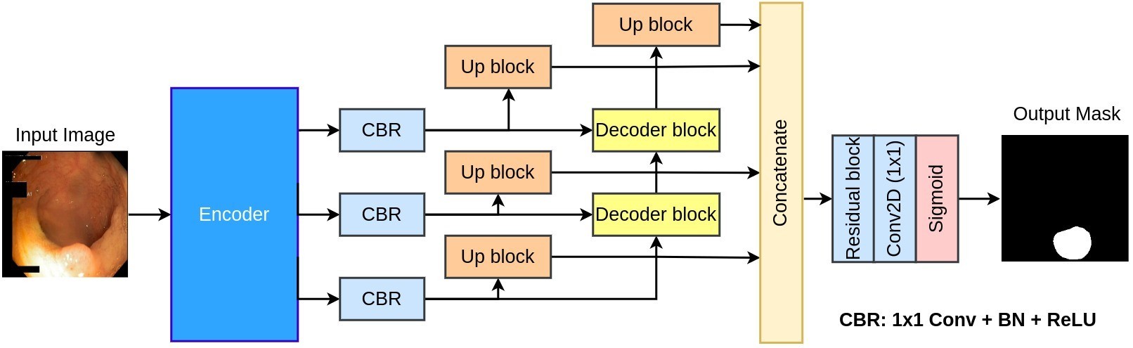
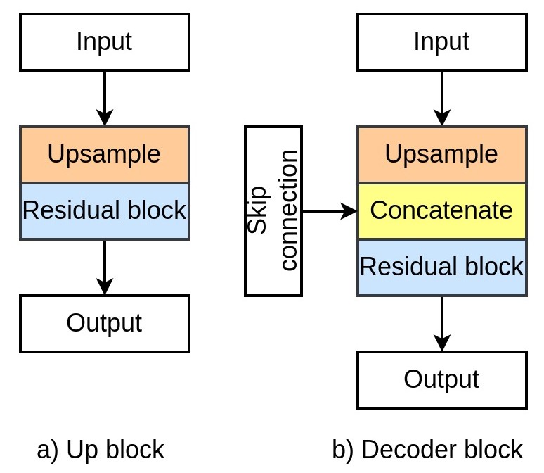
Figure 1 shows the block diagram of the proposed TransRUPNet architecture. It is an encoder-decoder network that begins with a Pyramid Vision Transformer (PVT) [7] as a pre-trained encoder. We extract three different feature maps from the encoder and pass them through a series of Conv, Batch Normalization and ReLU activation for reducing the number of feature channels to . The reduced feature maps are then passed to the up block and the decoder blocks. Within the up block, the input feature map is first passed through a bilinear upsampling to upscale the feature map’s height and width to that of the original input image. Next, the upsampled feature map is passed through a residual block to learn a more robust representation.
In Figure 2, each component of TransRUPNet is illustrated. The decoder block also begins with a bilinear upsampling layer to increase the spatial dimensions by a factor of and then concatenated with the reduced feature from the encoder. Next, the concatenated feature map is passed through a residual block to learn more robust semantic features which help to generate a fine-quality segmentation mask. The output from the first decoder block is passed to the next decoder block, which is further followed by an up block. We concatenate the output from all four up blocks into a single feature representation. After that, the concatenated feature map is followed by a residual block, convolution and a sigmoid activation to generate the final segmentation mask.
| Method | Backbone | mIoU | mDSC | Recall | Precision | F2 | FPS |
|---|---|---|---|---|---|---|---|
| U-Net [8] | - | 0.7472 | 0.8264 | 0.8504 | 0.8703 | 0.8353 | 106.88 |
| U-Net++ [9] | - | 0.7420 | 0.8228 | 0.8437 | 0.8607 | 0.8295 | 81.34 |
| ResU-Net++ [10] | - | 0.5341 | 0.6453 | 0.6964 | 0.7080 | 0.6576 | 43.11 |
| HarDNet-MSEG [11] | HardNet68 | 0.7459 | 0.8260 | 0.8485 | 0.8652 | 0.8358 | 34.80 |
| ColonSegNet [12] | - | 0.6980 | 0.7920 | 0.8193 | 0.8432 | 0.7999 | 73.95 |
| DeepLabV3+ [13] | ResNet50 | 0.8172 | 0.8837 | 0.9014 | 0.9028 | 0.8904 | 67.88 |
| PraNet [3] | Res2Net | 0.8296 | 0.8942 | 0.9060 | 0.9126 | 0.8976 | 31.89 |
| TGANet [14] | ResNet50 | 0.8330 | 0.8982 | 0.9132 | 0.9123 | 0.9029 | 36.58 |
| TransRUPNet (Ours) | PVT | 0.8445 | 0.9005 | 0.9195 | 0.9170 | 0.9048 | 47.07 |
| Method | Backbone | mIoU | mDSC | Recall | Precision | F2 |
|---|---|---|---|---|---|---|
| Training dataset: Kvasir-SEG – Test dataset: PolypGen (C6) | ||||||
| U-Net [8] | - | 0.5384 | 0.6126 | 0.7054 | 0.7508 | 0.6362 |
| U-Net++ [9] | - | 0.5355 | 0.6163 | 0.7340 | 0.7230 | 0.6564 |
| ResU-Net++ [10] | - | 0.2816 | 0.3684 | 0.6220 | 0.3526 | 0.4326 |
| HarDNet-MSEG [11] | HardNet68 | 0.5548 | 0.6341 | 0.7197 | 0.7722 | 0.6487 |
| ColonSegNet [12] | - | 0.4410 | 0.5290 | 0.6199 | 0.6403 | 0.5424 |
| DeepLabV3+ [13] | ResNet50 | 0.7031 | 0.7629 | 0.7773 | 0.8693 | 0.7674 |
| PraNet [3] | Res2Net | 0.6691 | 0.7307 | 0.7612 | 0.8755 | 0.7378 |
| TGANet | ResNet50 | 0.6750 | 0.7382 | 0.7692 | 0.8887 | 0.7391 |
| TransRUPNet (Ours) | PVT | 0.7210 | 0.7786 | 0.8522 | 0.8175 | 0.7929 |
| Training dataset: Kvasir-SEG – Test dataset: CVC-ClinicDB | ||||||
| U-Net [8] | - | 0.5433 | 0.6336 | 0.6982 | 0.7891 | 0.6563 |
| U-Net++ [9] | - | 0.5475 | 0.6350 | 0.6933 | 0.7967 | 0.6556 |
| ResU-Net++ [10] | - | 0.3585 | 0.4642 | 0.5880 | 0.5770 | 0.5084 |
| HarDNet-MSEG [11] | HardNet68 | 0.6058 | 0.6960 | 0.7173 | 0.8528 | 0.7010 |
| ColonSegNet [12] | - | 0.5090 | 0.6126 | 0.6564 | 0.7521 | 0.6246 |
| DeepLabV3+ [13] | ResNet50 | 0.7388 | 0.8142 | 0.8331 | 0.8735 | 0.8198 |
| PraNet [3] | Res2Net | 0.7286 | 0.8046 | 0.8188 | 0.8968 | 0.8077 |
| TGANet | ResNet50 | 0.7444 | 0.8196 | 0.8290 | 0.8879 | 0.8207 |
| TransRUPNet (Ours) | PVT | 0.7765 | 0.8539 | 0.8736 | 0.8870 | 0.8590 |
| Training dataset: Kvasir-SEG – Test dataset: BKAI-IGH | ||||||
| U-Net [8] | - | 0.5686 | 0.6347 | 0.6986 | 0.7882 | 0.6591 |
| U-Net++ [9] | - | 0.5592 | 0.6269 | 0.6900 | 0.7968 | 0.6493 |
| ResU-Net++ [10] | - | 0.3204 | 0.4166 | 0.6979 | 0.3922 | 0.5019 |
| HarDNet-MSEG [11] | HardNet68 | 0.5711 | 0.6502 | 0.7420 | 0.7469 | 0.6830 |
| ColonSegNet [12] | - | 0.4910 | 0.5765 | 0.7191 | 0.6644 | 0.6225 |
| DeepLabV3+ [13] | ResNet50 | 0.6589 | 0.7286 | 0.7919 | 0.8123 | 0.7493 |
| PraNet [3] | Res2Net | 0.6609 | 0.7298 | 0.8007 | 0.8240 | 0.7484 |
| TGANet | ResNet50 | 0.6612 | 0.7289 | 0.7740 | 0.8184 | 0.7412 |
| TransRUPNet (Ours) | PVT | 0.7218 | 0.7945 | 0.8497 | 0.8337 | 0.8072 |
IV Experiment
IV-A Dataset
We use four publicly available colonoscopy polyp segmentation datasets. We consider Kvasir-SEG [15] as the in-distribution dataset and other datasets such as PolypGen [16], BKAI-IGH [17], and CVC-ClinicDB [18] as the OOD dataset. PolypGen dataset is collected from 6 medical centers from Norway, Italy, France, United Kingdom, and Egypt, incorporating more than 300 patients. This dataset is complex as it contains diverse samples from different cohort populations from different countries. Therefore, we use the Kvasir-SEG for in-distribution testing and PolypGen, BKAI-IGH, and CVC-ClinicDB for OOD generalization.
IV-B Experiment setup and configuration
We select Kvasir-SEG [15] dataset for training all the models. It contains 1000 images and mask pair. We use 880 images and masks for training our method and the rest for validation and testing. In addition, we perform extensive data augmentation to increase the size of training samples. All the experiments are implemented using with PyTorch framework. We run all the experiments on an NVIDIA RTX 3090 GPU system. We use Adam optimizer with a learning rate of 1e-4 and a batch size of 8. Additionally, we use a combined binary cross-entropy and dice loss for training our models.
V Result
Comparison with SOTA on in-distrubtion data: Table I shows the result of the TransRUPNet. It obtained an mean dice coefficient of 0.9005, mIoU of 0.8445, recall of 0.9195, precision of 0.9170 and F2-score of 0.9048. With the image resolution of , TransRUPNet obtained a real-time processing speed of 47.07 frames per second (FPS). The most competitive network to TransRUPNet was TGANet, to whom our architecture outperformed by 1.15% in mIoU and 0.23% in DSC. The processing speed of our network is almost 1.5 times TGANet.
Comparison with SOTA on OOD data: We have evaluated the performance of TransRUPNet on three OOD datasets. For this, we train different models on Kvasir-SEG dataset and test it on PolypGen (Center 6). Kindly note that this is the experimental setup for EndoCV 2021 Challenge [19]. We obtained an improvement of 4.6% in mIoU and 4.04% in mDSC. Similarly, we obtained an improvement of 3.21% in mIoU and 3.43% in mDSC on CVC-ClinicDB datasets. Additionally, we obtained an improvement of 6.06% in mIoU and 6.56% in mDSC for the TransRUPNet when tested on BKAI-IGH datasets as compared to the SOTA TGANet [14].
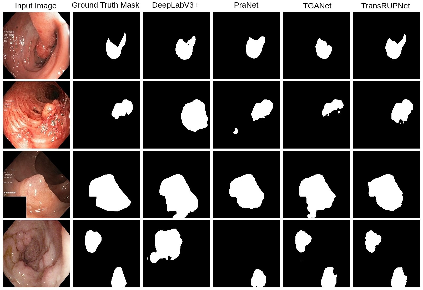
Figure 3 shows the effectiveness of TransRUPNet in qualitative results. As evidenced by the Figure, TransRUPNet avoids the issues such as over-segmentation or under-segmentation, which is observed in the case of SOTA TGANet and PRANet. Additionally, TransRUPNet accurately segments one or more polyps within the frames, even under challenging conditions. This highlights the robustness of TransRUPNet in handling complex scenarios and its ability to delineate the boundaries of polyps correctly. The performance drop of TransRUPNet compared to the in-distribution datasets is observed because there are insufficiently cleaned images in datasets, such as PolypGen (C6), that show elongated black regions on the left side, leading to distorted resizing and decreased OOD performance. Additionally, there are also huge variations among the training dataset and OOD datasets. For instance, BKAI-IGH also contains images from FICE (Flexible spectral Imaging Color Enhancement), BLI (Blue Light Imaging), and LCI (Linked Color Imaging), in addition to WLI (White Light Imaging) which are not present in the training datasets. In case of CVC-ClinicDB, it is a video sequence dataset, whereas our model is trained on still frames which might have affected the performance. However, the performance for all the datasets is satisfactory considering the OOD nature of the experiment.
VI Conclusion
In this study, we proposed TransRUPNet architecture by leveraging a pre-trained Pyramid Vision Transformer (PVT) as an encoder and incorporating a simple residual block for accurate polyp segmentation. The experimental results on various in-distribution and OOD datasets demonstrate that TransRUPNet can provide real-time feedback with high accuracy and perform significantly well on OOD datasets compared to the existing methods. By addressing the challenge of OOD generalization and providing reliable polyp segmentation results, TransRUPNet can be the strong benchmark for developing computer-aided diagnostic support systems in colonoscopy.
References
- [1] G. Urban, P. Tripathi, T. Alkayali, M. Mittal, F. Jalali, W. Karnes, and P. Baldi, “Deep learning localizes and identifies polyps in real time with 96% accuracy in screening colonoscopy,” Gastroenterology, vol. 155, no. 4, pp. 1069–1078, 2018.
- [2] A. Leufkens, M. Van Oijen, F. Vleggaar, and P. Siersema, “Factors influencing the miss rate of polyps in a back-to-back colonoscopy study,” Endoscopy, vol. 44, no. 05, pp. 470–475, 2012.
- [3] D.-P. Fan, G.-P. Ji, T. Zhou, G. Chen, H. Fu, J. Shen, and L. Shao, “Pranet: Parallel reverse attention network for polyp segmentation,” in Proceedings of the International conference on medical image computing and computer-assisted intervention (MICCAI), 2020, pp. 263–273.
- [4] G. Yue, W. Han, B. Jiang, T. Zhou, R. Cong, and T. Wang, “Boundary constraint network with cross layer feature integration for polyp segmentation,” IEEE Journal of Biomedical and Health Informatics, 2022.
- [5] B. Dong, W. Wang, D.-P. Fan, J. Li, H. Fu, and L. Shao, “Polyp-PVT: polyp segmentation with pyramid vision transformers,” arXiv preprint arXiv:2108.06932, 2021.
- [6] N. T. Duc, N. T. Oanh, N. T. Thuy, T. M. Triet, and V. S. Dinh, “Colonformer: An efficient transformer based method for colon polyp segmentation,” IEEE Access, vol. 10, pp. 80 575–80 586, 2022.
- [7] W. Wang, E. Xie, X. Li, D.-P. Fan, K. Song, D. Liang, T. Lu, P. Luo, and L. Shao, “Pyramid vision transformer: A versatile backbone for dense prediction without convolutions,” in Proceedings of the IEEE/CVF international conference on computer vision (ICCV), 2021, pp. 568–578.
- [8] O. Ronneberger, P. Fischer, and T. Brox, “U-Net: Convolutional Networks for Biomedical Image Segmentation,” in Proceedings of the International Conference on Medical image computing and computer-assisted intervention (MICCAI), 2015, pp. 234–241.
- [9] Z. Zhou, M. M. Rahman Siddiquee, N. Tajbakhsh, and J. Liang, “UNet++: a nested u-net architecture for medical image segmentation,” in Deep learning in medical image analysis and multimodal learning for clinical decision support, 2018, pp. 3–11.
- [10] D. Jha, P. H. Smedsrud, M. A. Riegler, D. Johansen, T. De Lange, P. Halvorsen, and H. D. Johansen, “Resunet++: An advanced architecture for medical image segmentation,” in Proceedings of the International Symposium on Multimedia (ISM), 2019, pp. 225–2255.
- [11] C.-H. Huang, H.-Y. Wu, and Y.-L. Lin, “HarDNet-MSEG A Simple Encoder-Decoder Polyp Segmentation Neural Network that Achieves over 0.9 Mean Dice and 86 FPS,” arXiv preprint arXiv:2101.07172, 2021.
- [12] D. Jha, S. Ali, N. K. Tomar, H. D. Johansen, D. Johansen, J. Rittscher, M. A. Riegler, and P. Halvorsen, “Real-time polyp detection, localization and segmentation in colonoscopy using deep learning,” IEEE Access, vol. 9, pp. 40 496–40 510, 2021.
- [13] L.-C. Chen, Y. Zhu, G. Papandreou, F. Schroff, and H. Adam, “Encoder-decoder with atrous separable convolution for semantic image segmentation,” in Proceedings of the European conference on computer vision (ECCV), 2018, pp. 801–818.
- [14] N. K. Tomar, D. Jha, U. Bagci, and S. Ali, “Tganet: text-guided attention for improved polyp segmentation,” in Proceedings of the 25th International Conference on Medical Image Computing and Computer Assisted Intervention (MICCAI 2022), 2022, pp. 151–160.
- [15] D. Jha, P. H. Smedsrud, M. A. Riegler, P. Halvorsen, T. d. Lange, D. Johansen, and H. D. Johansen, “Kvasir-SEG: a segmented polyp dataset,” in Proceedings of the International Conference on Multimedia Modeling (MMM), 2020, pp. 451–462.
- [16] S. Ali, D. Jha, N. Ghatwary, S. Realdon, R. Cannizzaro, O. E. Salem, D. Lamarque, C. Daul, M. A. Riegler, K. V. Anonsen et al., “A multi-centre polyp detection and segmentation dataset for generalisability assessment,” Scientific Data, vol. 10, no. 1, p. 75, 2023.
- [17] P. N. Lan, N. S. An, D. V. Hang, D. Van Long, T. Q. Trung, N. T. Thuy, and D. V. Sang, “NeoUNet: towards accurate colon polyp segmentation and neoplasm detection,” arXiv preprint arXiv:2107.05023, 2021.
- [18] J. Bernal, F. J. Sánchez, G. Fernández-Esparrach, D. Gil, C. Rodríguez, and F. Vilariño, “WM-DOVA maps for accurate polyp highlighting in colonoscopy: Validation vs. saliency maps from physicians,” Computerized Medical Imaging and Graphics, vol. 43, pp. 99–111, 2015.
- [19] S. Ali, N. Ghatwary, D. Jha, E. Isik-Polat, G. Polat, C. Yang, W. Li, A. Galdran, M.-Á. G. Ballester, V. Thambawita et al., “Assessing generalisability of deep learning-based polyp detection and segmentation methods through a computer vision challenge,” arXiv preprint arXiv:2202.12031, 2022.