Review article
Gooshvar, S. et al.
[]Equal contribution
[]Corresponding author: vprakash@miami.edu
Non-bilaterians as Model Systems for Tissue Mechanics
Abstract
In animals, epithelial tissues are barriers against the external environment, providing protection against biological, chemical, and physical damage. Depending on the animal’s physiology and behavior, these tissues encounter different types of mechanical forces and need to provide a suitable adaptive response to ensure success. Therefore, understanding tissue mechanics in different contexts is an important research area. Here, we review recent tissue mechanics discoveries in a few early-divergent non-bilaterian animals – Trichoplax adhaerens, Hydra vulgaris, and Aurelia aurita. We highlight each animal’s simple body plan and biology, and unique, rapid tissue remodeling phenomena that play a crucial role in its physiology. We also discuss the emergent large-scale mechanics that arise from small-scale phenomena. Finally, we emphasize the enormous potential of these non-bilaterian animals to be model systems for further investigation in tissue mechanics.
keywords:
Biomechanics, Biophysics, Tissue Mechanics, Non-bilaterians, Marine Biology1 Introduction
Epithelial tissues in animals are subjected to different types of mechanical forces during their entire life cycle. In order to foster the animal’s survival and success, these tissues must be able to respond, adapt and withstand external forces. Thus, an important goal of tissue mechanics research is to apply the principles of mechanics to characterize and quantify the mechanical properties and understand the response of biological tissues Fung1990 . A key question is to understand how the properties of individual cells and their collective interactions give rise to emergent mechanical properties of the tissue.
Tissue mechanics also plays an important role in biological processes such as development and physiology. Significant advances have already been made in tissue mechanics research focused on human biomedical applications Fung1990 ; Park2015 ; as well as in the context of developmental processes in model animals such as the fruit fly, D. melanogaster, and zebrafish, D. rerio lecuit2013 ; lecuit2007cell ; blankenship2006multicellular ; he2014 ; Mongera2018 . In-vitro cell-culture systems have also been a popular model for tissue mechanics studies Charras2012 ; Latorre2018 ; Xi2019 ; Charras2022 . However, very little is known regarding the tissue mechanics of early divergent, non-bilaterian animals. Here, we highlight recent surprising tissue mechanics discoveries in non-bilaterian animals and their potential in opening up new questions and research directions prakash2021motility ; Carter2016 ; pnas.1502497112 .
Non-bilaterian animals include some of the first multi-cellular animals that evolved from unicellular organisms. These are the most ancestral, earliest diverging animals that do not have a bilateral symmetry axis. The non-bilaterian phyla of animals includes Porifera, Placozoa, Ctenophora, and Cnidaria Dunn2014 . These non-bilaterians have been the focus of several recent biological studies, but have received very little attention from a biophysical perspective. The objective of this review is to attract the attention of biomechanicians and biophysicists and motivate them to work on these important animal systems.
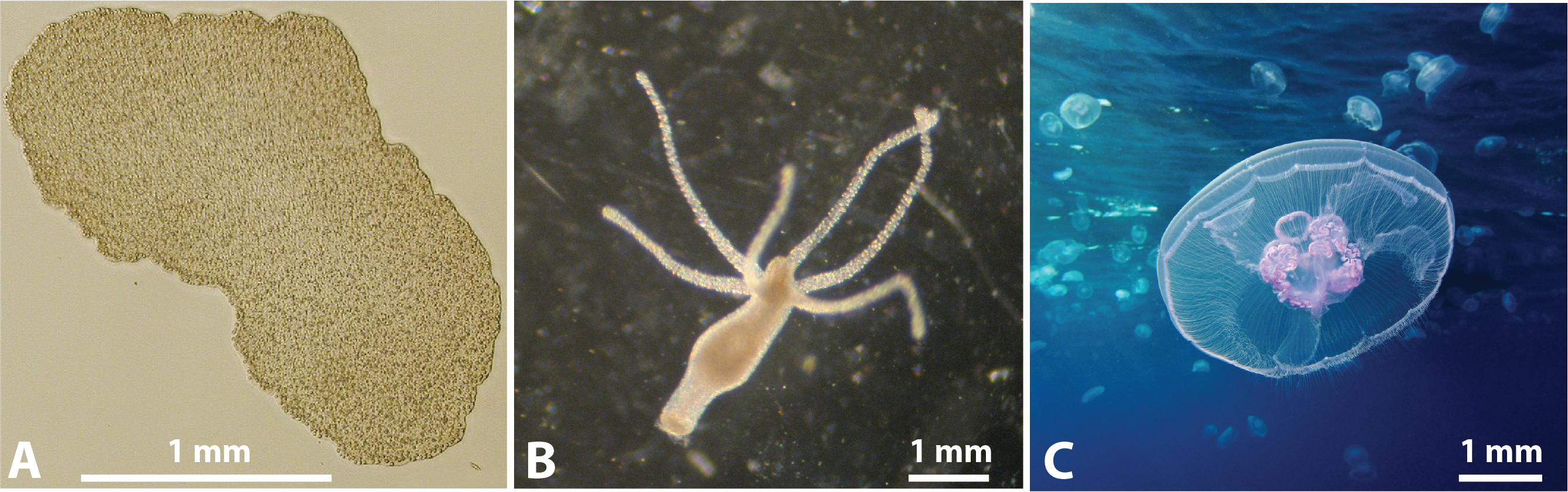
Here, we will focus on three non-bilaterians: the Trichoplax adhaerens of Phylum Placozoa, Hydra vulgaris, and Aurelia aurita of phylum Cnidaria (Figure 1). These non-bilaterian animals provide several key advantages to serve as model research animals for tissue mechanics – they have all been successfully maintained in the lab, are soft and experimentally tractable, have simple body plans, and their tissues are suitable for live imaging. In these animals, the organs and organ systems are not as well developed and specialized as in bilaterians, so organization on the tissue level plays a prominent role in their physiology and behavior. Here, our focus will be on fast-time scale (minutes to hours) tissue remodeling events and their mechanical processes. We will focus on tissue remodeling phenomena such as local cellular rearrangements, tissue fractures and their healing, in the context of physiological processes such as feeding, locomotion, reproduction, and repair. Important aspects such as growth and regeneration that take place over longer time-scales (several days/weeks) are neglected.
This review is organized as follows: In every system, we introduce the animal’s biology and body plan organization. Then, we describe its tissue layers and their role in the animal’s physiology before highlighting recently reported tissue mechanics phenomena in the animal. In the discussion section, we summarize and emphasize how tissue mechanics plays an important role in form-function relationships in these three animal systems. We also reveal how small-scale phenomena give rise to large-scale behaviors. Finally, we conclude by highlighting how non-bilaterians are excellent model systems to investigate the biophysics of tissues.
2 Trichoplax
The early-divergent marine animal Trichoplax adhaerens (Figure 1A) is the most well-studied member of the basal animal phylum of Placozoa Schierwater2018 ; Armon2018a ; Smith2014a ; Srivastava2008 . Trichoplax adhaerens is one of the three species of animals in the entire phylum; and the two other animals were only found recently eitel2018comparative ; osigus2019polyplacotoma . Trichoplax adhaerens has been found in many parts of tropical oceans around the world Pearse2007FieldInteractions ; eitel2013global . Trichoplax adhaerens is considered to be the simplest free-living animal since it consists of less than ten cell types Smith2014a ; romanova2021hidden , and lacks neurons, muscles, extracellular matrix, and a basement membrane Schierwater2018 ; Srivastava2008 . This animal has a simple and flat body plan, with a thickness of only 25 m, but the animals can exhibit a large variation in width, ranging from 50 m to 10 mm (Figure 1A).
2.1 The Role of Tissue Layers in Trichoplax Physiology
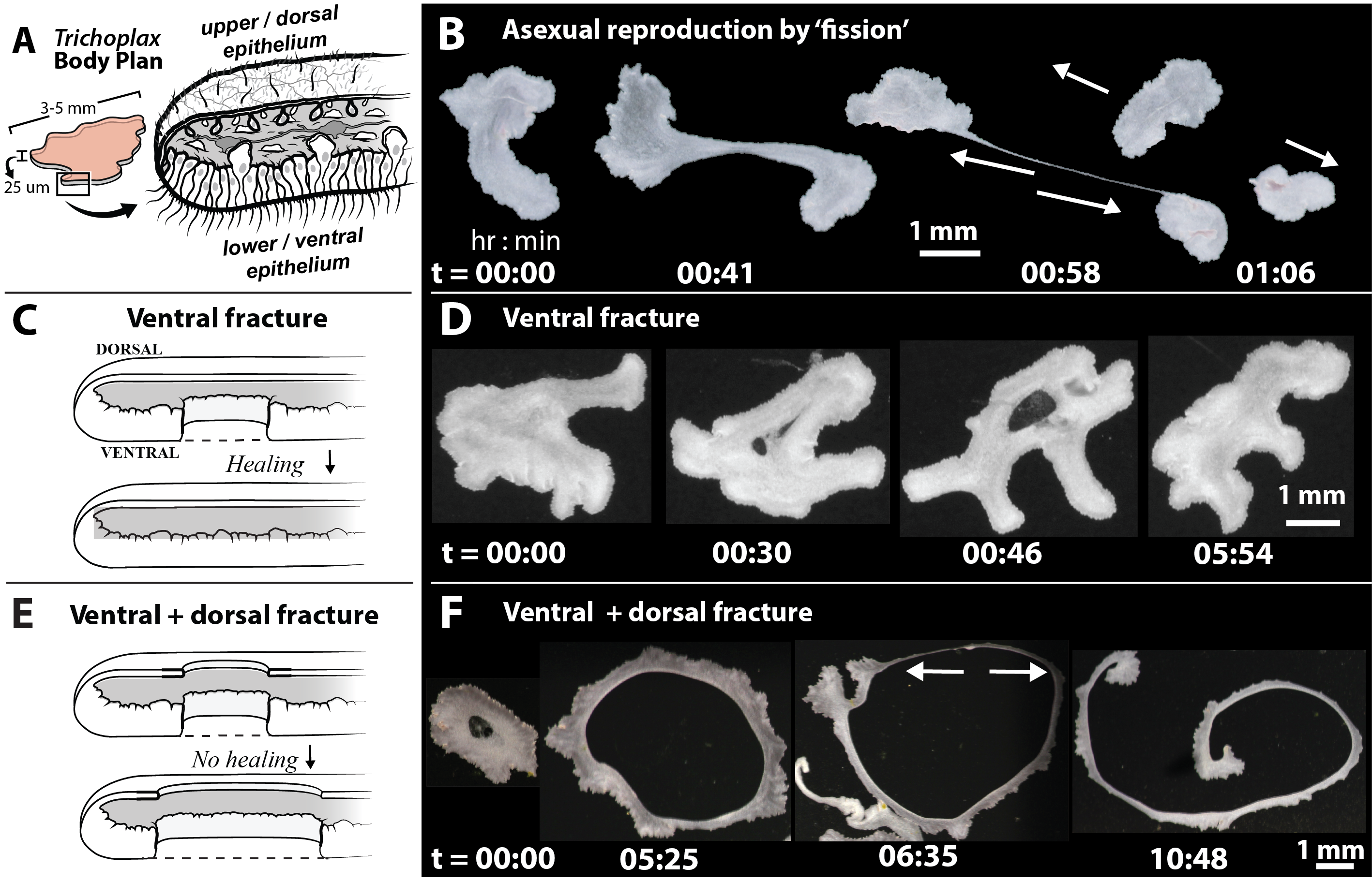
In T. adhaerens, the flat body plan consists of three tissue layers, an upper dorsal epithelium, a central layer of fiber cells, and a lower ventral epithelium. The dorsal epithelial cells have a thin and flat architecture, while the ventral epithelial cells have a columnar structure, and the two tissue layers are coupled at the edge of the animal. The epithelial cells are connected together only by adherens junctions, and no tight junctions have been found Smith2016 . Although both tissue layers have monociliated cells, the cilia of ventral epithelial cells are unique since they can adhere to the bottom substrate, providing the organism sufficient traction forces to be able to walk and generate push/pull forces bull2021a . Trichoplax adhaerens does not have a fixed shape like other animals. Instead, the animal is constantly changing shape in an amorphous manner driven by ciliary traction with the bottom substrate. The animals exhibit an extreme range of shape morphologies ranging from circular-like shapes to long elongated threads prakash2021motility . Trichoplax adhaerens also reproduces by vegetative or asexual fission, resulting in two or more daughter animals prakash2021motility ; Eitel2011 ; Srivastava2008 (Figure 2B). In these animals, epithelial tissue remodeling processes can play an important role in determining the organismal shape change dynamics and asexual reproduction.
2.2 Tissue Remodeling via Cellular Rearrangements
The vegetative (or asexual) reproduction process in Trichoplax adhaerens begins when an individual animal forms two coherent regions that start pulling away from each other (Figure 2B). The tissues in between the two pulling regions are subjected to mechanical forces (tension), and respond by undergoing a rapid thinning deformation to form a thin narrow thread (taking less than 1 hour). From a materials science viewpoint, this rapid thinning process resembles a ‘ductile’ material transformation process, where ‘ductility’ refers to the ability of a material to be drawn into thin wires. This rapid thinning process involves local tissue remodeling via fast time-scale cellular rearrangement mechanisms prakash2021motility . If the two opposing parts of the animal are able to generate sufficient traction forces, eventually the thin thread will break at the length-scale of a single cell and result in the formation of two or more daughter animals, thereby completing the reproduction process prakash2021motility ; Eitel2011 ; Srivastava2008 . The smaller daughter animals will grow in size and again undergo asexual fission when their size reaches 2-3mm prakash2021motility .
2.3 Tissue Remodeling via Fractures
Trichoplax adhaerens can grow to large sizes (2-3mm), and these larger animals are capable of executing extreme shape changes, making them a very interesting model animal for tissue mechanics. Larger animals can generate larger traction forces due to their motility, and this leads to very surprising shape morphologies, such as fracture holes and their healing dynamics, and long string-like animals prakash2021motility (Figure 2C-F). These fracture holes can be rapidly induced in the bulk of their ventral tissues solely due to motility-induced tensile or shear mechanical forces at the organismal scale prakash2021motility (Figure 2C). These fracture holes begin at small scales as micro-fractures and rapidly coalesce (in about half an hour) to form large stable ventral holes that are visible at the organismal scale (Figure 2D). From a materials science viewpoint, this fracture formation and growth process resembles a ‘brittle’ material transformation process, where ‘brittleness’ refers to the tendency of a material to break. In many cases, it was observed these ventral holes can also rapidly heal themselves (in about half an hour) if the hole edges came into contact due to the animal’s motility (Figure 2D).
Sometimes, the ventral holes do not heal and the dorsal epithelium also sustains a fracture hole right above the ventral hole (Figure 2E,F). The animal is now left with a through-hole inside it, and the two tissue layers seal themselves to form an permanent edge inside the animal (preventing any further healing). This animal will now have the geometry of a toroid (like a donut). Over a time-scale of 10 hours, the inside hole diameter keeps increasing until one of the edges becomes thin and eventually breaks, giving rise to long string-like animals (Figure 2F). From an organismal morphology perspective, fractures enable the fastest topological transformations from a circular shape to a long string-like shape, compared to a shape change mechanism that relies on cellular rearrangements.
These tissue remodeling processes in T. adhaerens were further investigated using in-silico tissue models prakash2021motility . The simplified heuristic two-dimensional (sheet) model of ventral epithelium consisted of soft balls (representing cells) that were connected to each other by springs (representing adhesion bonds). The tissue model was subjected to tensile loading (pulling forces) and the resulting tissue response was studied. We emphasize here that the focus is on tissue remodeling processes at fast time-scales (minutes) and the long term effects of growth (several hours) has been neglected. It was found that the two key parameters governing tissue response were the pulling force and the length at which the springs break. Simulations exploring a wide range of these two parameters resulted in a phase diagram that revealed an elastic–ductile–brittle transitions in the material properties. The model captured experimental observations faithfully. Thus confirming that elastic–ductile tissue transitions occur during the local cellular rearrangement process when the animals are dividing by vegetative fission (Figure 2B); and that elastic–brittle transitions indeed occur when the animals sustain tissue fractures (Figure 2D,F) during organismal shape change. Hence, T. adhaerens is an excellent model system for further investigations in tissue mechanics, since solely mechanical forces can give rise to the tissue remodeling processes that play a critical role in their life-cycle prakash2021motility .
3 Hydra
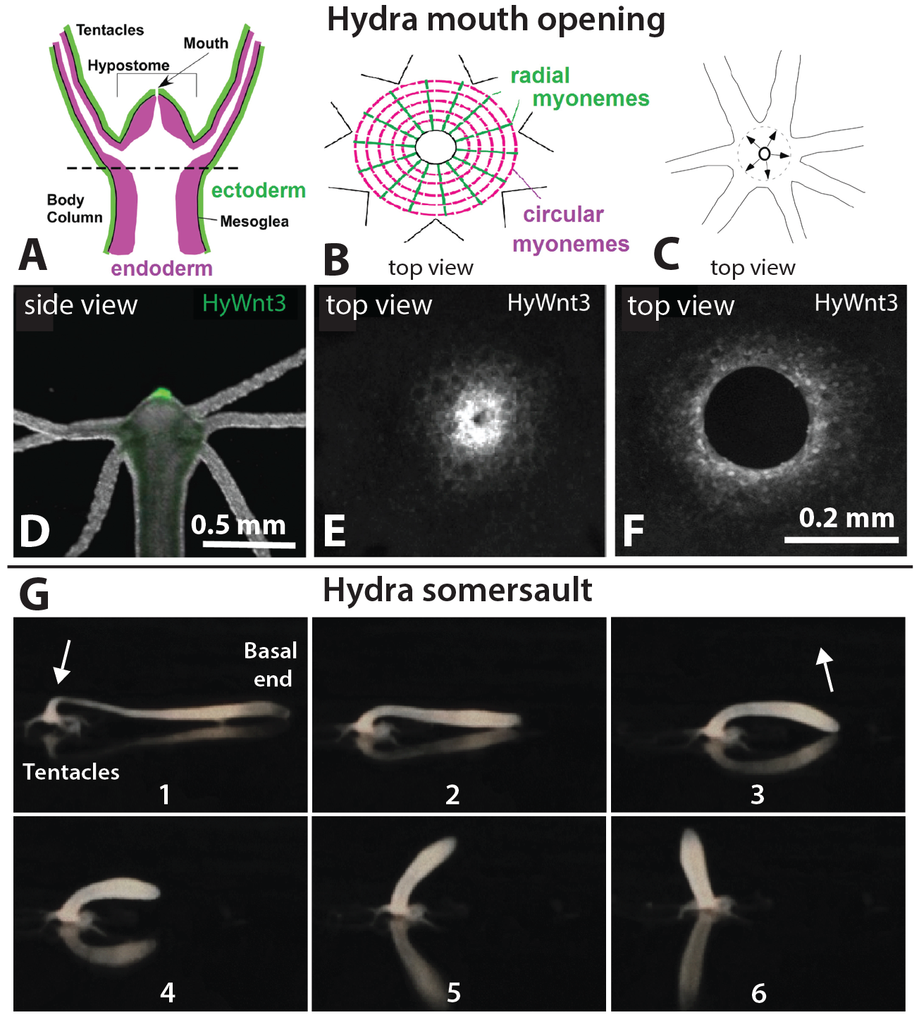
Hydra vulgaris is a freshwater polyp of the Cnidaria phylum which exhibits the characteristic radial symmetry, cnidocytes, and body plan derived from two germ layers McLaughlin2017 . Hydra’s body plan consists of a two-layered tube body and a mouth composed of a ring of tentacles and dome-shaped hypostome (Figure 3 A,D). Although Hydra is an invertebrate composed of two epithelial layers, the endoderm and ectoderm, separated by an extracellular mesoglea. Cells in the tissues of the body column cycle continuously with those of the head and foot regions, maintaining the equilibrium between cell production and loss Wang2023 ; Galliot2006 .
3.1 The Role of Tissue Layers in Hydra Physiology
In addition to the dynamic nature of its tissues, the existence of a differential thickness between the thinner ectodermal layer and the thicker endodermal layer is of particular importance. This difference in thickness is most pertinent when examining the Hydra mouth. When it is closed, the mouth is a continuous epithelial sheet sealed with septate junctions, which were first seen in the model organism Drosophila, and appear as a ladder-like junction between two cells Banerjee2006 ; Izumi2014 . These septate junctions thus act as intercellular connectors functioning to merge adjacent epidermal cells with inner, luminal edges of cells Carter2016 ; Hand1972 .
3.2 Mouth Opening Dynamics
When the Hydra opens its mouth, it must tear a hole through the epithelial tissues at each instance of opening. It does so exclusively through the viscoelastic deformation of cells. This was confirmed via cell shape analysis and by tracking individual cells during mouth openings. It was observed that cells were not undergoing rearrangement and were instead conserving existing cellular contacts Carter2016 . The Hydra must overcome the mouth opening force that exists while the endoderm and ectoderm are sealed. When the mouth is closed, the septate junctions act to connect the cells in both epithelial sheets. In the ectoderm, the sealing force to be overcome is solely that of the septate junctions while the endoderm needs to additionally include myonemes, which are circularly oriented contractile structures (Figure 3 B,E). Both of these closing forces must be exceeded by the opening force to lead to successful mouth opening. These sealing forces have been estimated to be on the order of several nN Carter2016 .
Although the force required must be estimated, the kinematics of the mouth opening can be fully characterized by a logistic equation Carter2016 . Comparing the normalized mouth opening area to the time axis resembles a logistic curve which can then be time-shifted and normalized to the ectoderm and endoderm mouth opening area. This results in a modified logistic equation which accounts for the normalized area of the mouth as a function of time, and various fit parameters to allow for the full capture of the kinematics of mouth opening for both ectoderm and endoderm separately (Figure 3 C,F) Carter2016 .
3.3 The Somersaulting Hydra
Tissue mechanics also plays an important role for Hydra’s locomotion. A differential stiffness within its body enables it to perform the “Hydra somersault” Mackie1974 ; Han2018-om . The Hydra somersault occurs in three general stages: in stage 1, the body column is stretched, and the tentacles hold on to the substrate (Figure 3 G:1,2). In stage 2, the basal end is released (Figure 3 G:3,4). In stage 3, the body column contracts and is lifted, transporting the base to a new location in the direction of the Hydra’s motion (Figure 3 G:5,6).
This type of movement is only possible due to the difference in the local mechanical properties of the Hydra’s body column, specifically that of a 3:1 ratio in the Young’s modulus between the shoulder region and the body column Naik2020 . Thus, when the Hydra moves from one place to another, it utilizes the differential stiffness of the body column to enable efficient transfer of mechanical energy stored in stretching to bending Naik2020 ; Wang2023 .
Hence, Hydra is an interesting animal that employs tissue fracture mechanics for its mouth opening and differential stiffness activity for its locomotion.
4 Jellyfish
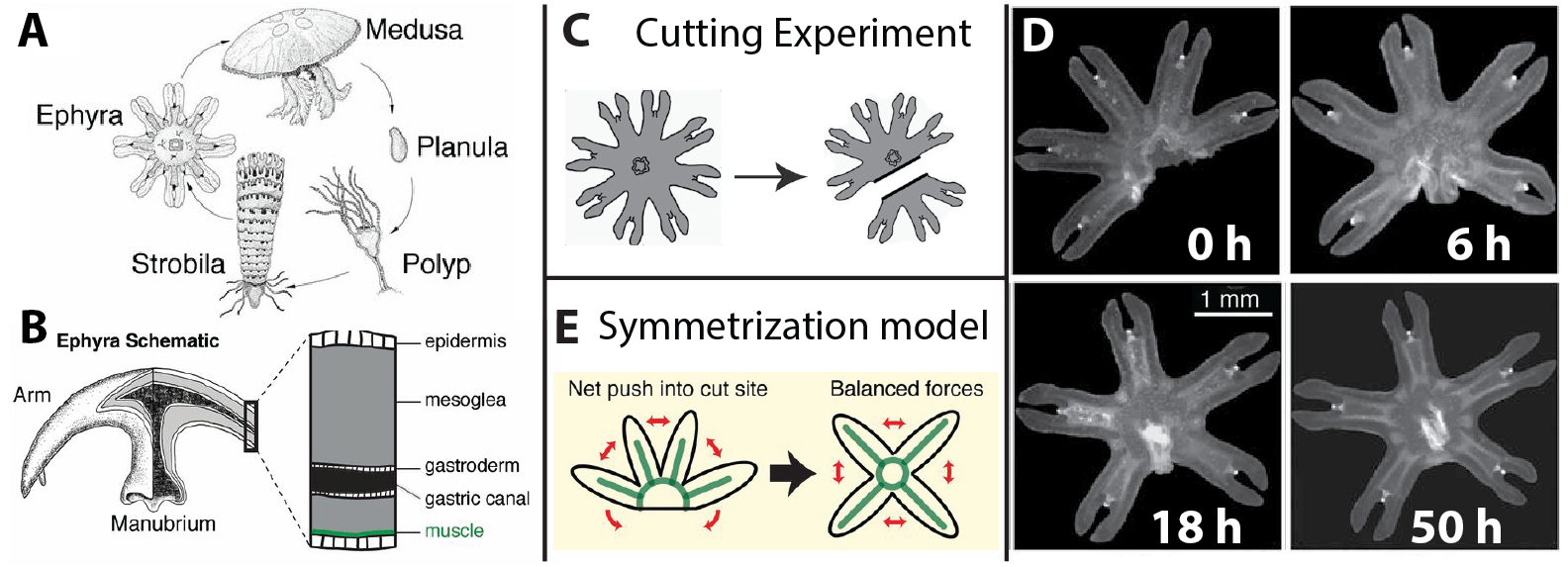
While the previous section discusses tissue fracture mechanics of the Cnidarian Hydra polyp, this section discusses the Cnidarian Medusozoans, commonly known as jellyfish. The Scyphozoan jellyfish have a life cycle with two adult forms, with one being the sexually reproducing fully motile medusae and the other form being the asexually reproducing sessile polyp (Figure 4 A) 10.1007/978-94-010-0722-1_19 .
Jellyfish are morphologically complex invertebrates with striated muscles. Despite lacking a functional brain, they possess a complex radially distributed neural and sensory systems that help them detect light and odour, enabling quick responses to stimuli SATTERLIE2011 . The cross-sectional view of ephyra reveals three layers: the epidermis, mesoglea, and gastrodermis. The epidermis, which is the outer layer, contains the neural net. The endodermis-derived inner layer, the gastrodermis, lines the gastric cavity. The region between the ectoderm and the endoderm is filled by mesoglea, a viscoelastic substance (Figure 4 B) ABRAMS20161 .
The habitat of jellyfish is versatile, ranging across varying temperatures and depths. Though jellyfish can be considered top predators in their ecosystems, many predators, including other jellyfish, attack jellyfish opportunistically predators_jellyfish . Therefore injury via amputation is common among jellyfish, and recovery from such damage could either be from re-organization or regeneration. Jellyfish have been widely studied for their regenerative abilities and symmetrization, specifically the medusae and ephyrae (juvenile jellyfish) stages of their life cycle. The focus here will be on the fast timescale process of tissue symmetrization.
4.1 The Role of Tissue Layers in Aurelia Physiology
A way by which jellyfish can repair sustained damage that is not regenerative is through the restoration of symmetry after tentacle amputation, which occurs purely through responses to mechanical stress. The moon jellyfish (Aurelia aurita) is an excellent example of symmetry restoration after the removal of one of its arms, known as tenticulocytes ABRAMS20161 . An undamaged moon jellyfish has eight tenticulocytes forming its swimming apparatus as the arms symmetrically pulsate. The striated musculature of the arms connected to the jellyfish’s pulsating ”bell” through epithelial tissue drives the recovery and power strokes of the pulsations as the jellyfish swims. The mesoglea’s elastic properties are used in the recovery stroke to restore the bell to its original shape JELLYFISHPROPUSION . Because the architecture of the musculature is so symmetrically interlinked with the umbrella, any loss of symmetry, such as the removal of arms, creates an imbalance in forces during pulsations. Since radial symmetry is essential for propulsion, when any number of these arms are removed in a moon jellyfish ephyra (juvenile jellyfish), a recovery of radial symmetry is expected and observed (Figure 4) in a process termed as ”symmetrization” by Abrams et al. ABRAMS20161 .
4.2 Aurelia aurita-Symmetrization Model
In the symmetrization model, Abrams et al.ABRAMS20161 suggest two repair strategies without increased cell proliferation in Cnidaria, one to restore lost parts and another to restore functional symmetry without restoring lost parts. The latter involves the process of symmetrization, where the arms eventually get rearranged to regain symmetry ABRAMS20161 .
The experiment involving amputation of arms from individual Aurelia ephyrae (as shown in Figure 4 C) resulted in wound closure of the injured site in three hours. Within 18 hours, full symmetrization with the manubrium relocating to the centre was observed (Figure 4 D). The ephyrae could regain radial symmetry with up to six arms removed. Even so, development into the medusae stage, where they fully regain swimming functions, could only be possible with at least four remaining arms. Hence radial symmetry also plays a vital role in facilitating further development of the ephyrae pnas.1502497112 .
The symmetrization process proceeds independently from global factors such as the movement of water, light, or the orientation of the jellyfish in the water column pnas.1502497112 . The process is also suggested to be independent of wound closure, due to the faster time scale of wound closure, and observing wound closure when symmetrization does not occur. It is important to note that neither cell proliferation nor localized cell death played any important role in the process. Concluding that the process is wholly related to symmetry and musculature, each cycle of contraction and repulsion cause the arms to relax to an increasingly stable state until their morphology is geometrically balanced. Asymmetrical contraction might lead to pivoting of the arms towards the injured sight due to lack of bulk tissue at the site (Figure 4 E). Along with the recovery of radial symmetry, this also suggests the existence of a mechanism by which asymmetry is detected pnas.1502497112 .
Abrams et al. derived a mathematical model for the timescale of symmetry recovery. The model is based on the suggestion that muscular contraction forces and the elastic response of mesoglea involved in the propulsion of the uninjured ephyrae can sufficiently explain the recovery of radial symmetry in injured ephyrae. Using the angular movement of each arm with respect to the geometric centre as a parameter, they apply Hooke’s Law to generate a recursive expression for the parameter. The simulation of the mathematical model with parameter values obtained by estimation shows the arms of the ephyrae moving towards the injured site with each contraction and relaxation cycle until symmetry is regained. In other words, symmetry increases with each contraction. Additionally, the model predicts the speed of symmetrization, i.e. how long it takes until the ephyrae regain symmetry, to be dependent on the frequency of muscular contractions. The model suggests that muscular contractions play a dominant role in the mechanism and time required for symmetrization pnas.1502497112 .
The symmetrization process in jellyfish being a fast self-repair strategy, uses existing physiological machinery to drive the mechanical process. These fast processes prioritize the functional recovery of the organism rather than the recovery of the body parts themselves, which could be possible via regeneration at longer time-scales. Hence, the symmetrization process could be an energy conserving process, also increasing the animal’s survivability. This symmetrization process may inspire biomimetic materials and technologies that require conserving functional geometries without the need for regenerating precise shapes and forms JELLYFISHBIOIMETIC .
5 Discussion
5.1 Form-function Relationships
In T. adhaerens, we have discussed the key role of tissue mechanics in its physiological activities such as reproduction by vegetative fission, and continuous organism-scale shape change. Tissue remodeling mechanisms such as ductile transformations for vegetative reproduction, and brittle deformations for extreme morphological shape changes, are unique adaptations found only in this animal. Hence, these ductile-brittle tissue transitions have been optimized for their specific flat body plan. Tissue mechanics therefore acts as an important link between the biological form (flat body plan) and function (reproduction and shape change).
In H. vulgaris, we have described how important physiological activities such as feeding and locomotion are intimately determined by tissue mechanics. The tissue remodeling phenomena involved in the mouth opening for feeding and body column bending for movements are unique phenomena found only in this animal. These tissue remodeling phenomena have been optimized for Hydra’s specific body plan, possibly suggesting that tissue mechanics links their biological form (body plan) and function (feeding and locomotion).
In A. aurita, locomotion in the ephyrae and medusae stage is essential to survival, and we have discussed how there is link between propulsion and maintenance of radial symmetry in these animals. The jellyfish makes use of its radially symmetric musculature to produce power and recovery strokes for propulsion, and these are aided by fast muscle contractions in the arms, and elastic recoil in the viscous mesoglea JELLYFISHPROPUSION . The symmetrization process in jellyfish being a fast self-repair strategy thus adopts its existing physiological machinery to drive the mechanical remodeling process. Symmetrization thus encapsulates the priority afforded to shorter time-scale functional recovery over longer time-scale tissue regeneration. Therefore, propulsion and propulsion-linked recovery from amputation, are both tailored for the radial symmetry, tissue flexibility and musculature of the animal.
5.2 Small-scale Phenomena Lead to Emergent Large-scale Behaviors
In the ventral epithelium of T. adhaerens, micro-scale fractures appear first due to local regions of high tension or shear forces that arise from ciliary-driven traction forces. These micro-fractures subsequently coalesce to form a larger and stable ventral fracture hole. Interestingly, dorsal epithelial fractures also begin with a very small fracture hole that propagates in a different manner and grows in size. Eventually the dorsal hole merges with the ventral hole and an inside edge is created in the animals. Thus, small-scale micro-holes propagate to form larger scale macro-holes in both the ventral and dorsal epithelium. H. vulgaris mouth opening requires the overcoming of dual sealing forces associated with septate junctions at the cellular scale and contractile myonemes, which act together to enable the large scale phenomena of mouth opening with precise control. Also, localized differential stiffness of tissues in the shoulder and body column give rise to the larger-scale mechanics of the organism’s locomotion. Hence, phenomena at the cellular scales determine emergent behavior at the organismal scale.
5.3 Rapid Tissue Remodeling versus Regeneration
This review focuses on rapid (minutes to hours) tissue remodeling phenomena in the three non-bilaterians. However, Cnidarians are also known for their regenerative abilities alvarado2006bridging ; galliot2019 , and in particular H. vulgaris is a popular model system for regeneration. Some Jellyfish species are much more regenerative than the A. aurita discussed above, one such example is Clytia hemisphaerica.Clytia hemisphaerica jellyfish exhibit wound healing and regeneration, and utilize a combination of on tissue re-organization, proliferation of cellular progenitors, and long-range cell recruitment, as shown recently in Ref 10.7554/eLife.54868 . In these animals, it was found that the rapid tissue remodeling is difficult to separate from the regenerative mechanism in terms of recovery even though the time scales differ 10.7554/eLife.54868 .
The conflict between the cost-effectiveness of rapid tissue remodeling in relation to regeneration presents tremendous opportunities for further investigation. Additionally, quantifying the amount and type of mechanical forces that can trigger rapid tissue rearrangement and regeneration could shed light on the cost-effectiveness of both processes. Thus, it will be intriguing to look into the small scale, localized molecular mechanisms that translate forces from muscle contraction into tissue re-organization.
5.4 Tissue Remodeling by Fractures
Hydra vulgaris’ mouth opening mechanism by rupture of epithelial tissue is one among the only two known examples of physiological tissue fractures in animals, the other example being the Trichoplax adhaerens. However, there are several differences in the tissue fracture phenomena between these two animals. An important difference is the location of tissue fractures, in H. vulgaris, this location is always fixed at the mouth, but in T. adhaerens, fractures can arise anywhere in the epithelium – an emergent phenomenon that depends solely on organismal-scale mechanical forces. Physiological tissue fracture and healing dynamics have not yet been observed in any of the Jellyfish so far, and it could be very interesting to look for them in future work.
Hence, H. vulgaris and T. adhaerens are powerful model systems for future cell and molecular biology investigations to determine specific components (e.g. proteins/molecules) that enable these unique tissue fractures and their healing dynamics.
5.5 Tissue Remodeling by Cellular Rearrangements
Tissue remodeling by local cellular rearrangements is a ubiquitous and well-known mechanism during the development of model animals lecuit2013 ; lecuit2007cell ; blankenship2006multicellular ; he2014 ; Mongera2018 . In T. adhaerens, we have observed ductile tissue deformations (rapid thinning of tissues) that suggest local cellular rearrangements. In-silico models have indeed revealed the presence of fast cellular rearrangements in the ventral epithelium prakash2021motility , but this has not yet been observed in experiments.
Unlike T. adhaerens, studies of H. vulgaris have not reported any fast time-scale cellular rearrangements. Given the dynamic movements of H. vulgaris, in future work, it would be interesting to look for localized tissue remodeling events involving cell-cell rearrangements.
The symmetrization model proposed for recovery in A. aurita is based on cellular rearrangements due to forces from contractions that lead to recovery of radial symmetry pnas.1502497112 . It was found that the rearrangement of cells is what drives symmetrization, the process being independent of wound healing at the site of injury, pnas.1502497112 . Despite having different tissue structures and morphologies, other species of jellyfish ABRAMS20161 and the hydromedusae form Hydrmedusae also utilize such a re-organization process in their recovery.
5.6 Tissue Mechanics in Other Non-bilaterian Animals
We have so far described interesting tissue mechanics phenomena in the phyla of Placozoa and Cnidaria, but non-bilaterians also include two other phyla – Porifera and Ctenophora. The animals belonging to these two other phyla also have the potential to be good candidates as tissue mechanics models, given their simple body plans and morphologies dunn2015hidden .
Animals in the phylum Porifera, commonly referred to as sponges, consist of cells in an extracellular matrix, and sometimes also have stiff scaffolding. A recent study dissected their tissues and studied their response to mechanical forces in a rheometer Kraus2022 . It was found that sponge tissues revealed interesting properties, such as anisotropic elasticity, and it was suggested that these properties were linked to the sponge’s flow sensitivity Kraus2022 . The phylum Ctenophora is characterized by several hundred species of ctenophores, which are soft, gelatinous, freely swimming predatory animals pang2008ctenophores . It has been shown that the ctenophore Mnemiopsis leidyi is capable of rapid wound healing when their tissues are cut nikki2019 ; tamm2014cilia .
There is a huge diversity in Poriferans and Ctenophores, and there are hundreds of different species in both these phyla, with unique adaptations. The few studies on them described above give us a glimpse of the promise of these animals to serve as model systems to investigate tissue remodeling phenomena, particulary tissue rheology and rapid wound healing.
6 Conclusion
In this review, we have described interesting tissue mechanics phenomena in two non-bilaterian phyla – Placozoa and Cnidaria. We have shown how these animals have stretched our perspective and understanding of tissue mechanics. We reveal how animals in these phyla, particularly the Placozoan Trichoplax adhaerens, Cnidarians Hydra vulgaris and Aurelia aurita, utilize rapid tissue remodeling for their physiological functioning prakash2021motility ; Carter2016 ; pnas.1502497112 We also illustrate how small-scale cellular reorganization events give rise to phenomena at the larger-scales of tissues. We have shown several examples of how these understudied non-bilaterians present a wealth of opportunities to improve our understanding of rapid tissue remodeling phenomena such as cellular rearrangements, fractures, and wound healing. Hence, these non-bilaterian tissue mechanics model animals complement and contribute to the broader field of biological physics of tissues Park2015 ; Mongera2018 ; Charras2012 ; Latorre2018 ; Xi2019 ; Charras2022 ; bull2021b ; bull2021c ; armon2021 ; Charras2023 ; Kim2021 ; krajnc2020softmatter ; Noll2017 ; Wyatt2016 ; Bi2015 .
We adopt a comparative approach in this review to allow for a rigorous contribution to the search for general biophysical principles. We hope to have made a strong case for studying these non-model organisms to get to the heart of tissue mechanics and appreciate the extreme examples in order to arrive at a comprehensive framework of tissue mechanics in animals.
7 Competing interests
No competing interest is declared.
8 Author contributions statement
S.G., G.M., M.R., and V.N.P. discussed ideas, wrote and reviewed the manuscript. V.N.P. conceived the project.
9 Acknowledgments
The authors thank several colleagues at the University of Miami for useful discussions and suggestions: Ivan Levkovsky and other students of the Fall 2022 PHY 325/625 (Biological Physics - I) course, members of the Prakash Lab, and William Browne. The authors also thank Matthew Storm Bull, Allen Institute and University of Washington, for valuable feeback and suggestions. V.N.P. thanks Manu Prakash, Stanford University, for introduction to Non-bilaterians and for many stimulating and enriching discussions on tissue mechanics over the years. V.N.P. also thanks the University of Miami for startup funding support.
References
- [1] YC Fung. Biomechanics: Motion,Flow,Stress, and Growth. Springer, 1990.
- [2] Jin Ah Park, Jae Hun Kim, Dapeng Bi, Jennifer A. Mitchel, Nader Taheri Qazvini, Kelan Tantisira, Chan Young Park, Maureen McGill, Sae Hoon Kim, Bomi Gweon, Jacob Notbohm, Robert Steward, Stephanie Burger, Scott H. Randell, Alvin T. Kho, Dhananjay T. Tambe, Corey Hardin, Stephanie A. Shore, Elliot Israel, David A. Weitz, Daniel J. Tschumperlin, Elizabeth P. Henske, Scott T. Weiss, M. Lisa Manning, James P. Butler, Jeffrey M. Drazen, and Jeffrey J. Fredberg. Unjamming and cell shape in the asthmatic airway epithelium. Nature Materials, 14(10):1040–1048, 2015.
- [3] Charlène Guillot and Thomas Lecuit. Mechanics of epithelial tissue homeostasis and morphogenesis. Science, 340(6137):1185–1189, 2013.
- [4] Thomas Lecuit and Pierre-Francois Lenne. Cell surface mechanics and the control of cell shape, tissue patterns and morphogenesis. Nature reviews Molecular cell biology, 8(8):633–644, 2007.
- [5] J Todd Blankenship, Stephanie T Backovic, Justina SP Sanny, Ori Weitz, and Jennifer A Zallen. Multicellular rosette formation links planar cell polarity to tissue morphogenesis. Developmental cell, 11(4):459–470, 2006.
- [6] Bing He, Konstantin Doubrovinski, Oleg Polyakov, and Eric Wieschaus. Apical constriction drives tissue-scale hydrodynamic flow to mediate cell elongation. Nature, 508(7496):392–396, 2014.
- [7] Alessandro Mongera, Payam Rowghanian, Hannah J. Gustafson, Elijah Shelton, David A. Kealhofer, Emmet K. Carn, Friedhelm Serwane, Adam A. Lucio, James Giammona, and Otger Campàs. A fluid-to-solid jamming transition underlies vertebrate body axis elongation. Nature, 2018.
- [8] A R Harris, L Peter, J Bellis, B Baum, A J Kabla, and G T Charras. Characterizing the mechanics of cultured cell monolayers. Proceedings of the National Academy of Sciences, 109(41):16449–16454, 2012.
- [9] Ernest Latorre, Sohan Kale, Laura Casares, Manuel Gómez-González, Marina Uroz, Léo Valon, Roshna V. Nair, Elena Garreta, Nuria Montserrat, Aránzazu del Campo, Benoit Ladoux, Marino Arroyo, and Xavier Trepat. Active superelasticity in three-dimensional epithelia of controlled shape. Nature, 563(7730):203–208, 2018.
- [10] Wang Xi, Thuan Beng Saw, Delphine Delacour, Chwee Teck Lim, and Benoit Ladoux. Material approaches to active tissue mechanics. Nature Reviews Materials, 4(1):23–44, 2019.
- [11] Alessandra Bonfanti, Julia Duque, Alexandre Kabla, and Guillaume Charras. Fracture in living tissues. Trends in Cell Biology, 32(6):537–551, June 2022.
- [12] Vivek N Prakash, Matthew S Bull, and Manu Prakash. Motility-induced fracture reveals a ductile-to-brittle crossover in a simple animal’s epithelia. Nature Physics, 17(4):504–511, 2021.
- [13] J. A. Carter, C. Hyland, R. E. Steele, and E. M. Collins. Dynamics of Mouth Opening in Hydra. Biophys J, 110(5):1191–1201, Mar 2016.
- [14] Michael J. Abrams, Ty Basinger, William Yuan, Chin-Lin Guo, and Lea Goentoro. Self-repairing symmetry in jellyfish through mechanically driven reorganization. Proceedings of the National Academy of Sciences, 112(26):E3365–E3373, 2015.
- [15] Casey W. Dunn, Gonzalo Giribet, Gregory D. Edgecombe, and Andreas Hejnol. Animal Phylogeny and Its Evolutionary Implications. Annual Review of Ecology, Evolution, and Systematics, 45(1):371–395, 2014.
- [16] Bernd Schierwater and Rob DeSalle. Placozoa. Current Biology, 28(3):R97–R98, 2018.
- [17] Shahaf Armon, Matthew Storm Bull, Andres Aranda-Diaz, and Manu Prakash. Ultrafast epithelial contractions provide insights into contraction speed limits and tissue integrity. Proceedings of the National Academy of Sciences, 115(44):E10333–E10341, 2018.
- [18] Carolyn L. Smith, Frédérique Varoqueaux, Maike Kittelmann, Rita N. Azzam, Benjamin Cooper, Christine A. Winters, Michael Eitel, Dirk Fasshauer, and Thomas S. Reese. Novel Cell Types, Neurosecretory Cells, and Body Plan of the Early-Diverging Metazoan Trichoplax adhaerens. Current Biology, jun 2014.
- [19] Mansi Srivastava, Emina Begovic, Jarrod Chapman, Nicholas H Putnam, Uffe Hellsten, Takeshi Kawashima, Alan Kuo, Therese Mitros, Asaf Salamov, Meredith L Carpenter, Ana Y Signorovitch, Maria A Moreno, Kai Kamm, Jane Grimwood, Jeremy Schmutz, Harris Shapiro, Igor V Grigoriev, Leo W Buss, Bernd Schierwater, Stephen L Dellaporta, and Daniel S Rokhsar. The Trichoplax genome and the nature of placozoans. Nature, 454(7207):955–60, aug 2008.
- [20] Michael Eitel, Warren R Francis, Frederique Varoqueaux, Jean Daraspe, Hans-Jürgen Osigus, Stefan Krebs, Sergio Vargas, Helmut Blum, Gray A Williams, Bernd Schierwater, et al. Comparative genomics and the nature of placozoan species. PLoS biology, 16(7):e2005359, 2018.
- [21] Hans-Jürgen Osigus, Sarah Rolfes, Rebecca Herzog, Kai Kamm, and Bernd Schierwater. Polyplacotoma mediterranea is a new ramified placozoan species. Current Biology, 29(5):R148–R149, 2019.
- [22] V B Pearse and O Voigt. Field biology of placozoans (Trichoplax): distribution, diversity, biotic interactions. Integrative and Comparative Biology, 47(5):677–692, 2007.
- [23] Michael Eitel, Hans-Jürgen Osigus, Rob DeSalle, and Bernd Schierwater. Global diversity of the placozoa. PloS one, 8(4):e57131, 2013.
- [24] Daria Y Romanova, FrÚdÚrique Varoqueaux, Jean Daraspe, Mikhail A Nikitin, Michael Eitel, Dirk Fasshauer, and Leonid L Moroz. Hidden cell diversity in placozoa: ultrastructural insights from hoilungia hongkongensis. Cell and Tissue Research, 385:623–637, 2021.
- [25] Carolyn L. Smith and Thomas S. Reese. Adherens junctions modulate diffusion between epithelial cells in Trichoplax adhaerens. Biological Bulletin, 231(3):216–224, 2016.
- [26] Matthew S Bull, Laurel A Kroo, and Manu Prakash. Excitable mechanics embodied in a walking cilium. arXiv preprint arXiv:2107.02930, 2021.
- [27] Michael Eitel, Loretta Guidi, Heike Hadrys, Maria Balsamo, and Bernd Schierwater. New insights into placozoan sexual reproduction and development. PloS one, 6(5):e19639, 2011.
- [28] S. Naik, M. Unni, D. Sinha, S. S. Rajput, P. C. Reddy, E. Kartvelishvily, I. Solomonov, I. Sagi, A. Chatterji, S. Patil, and S. Galande. Differential tissue stiffness of body column facilitates locomotion of Hydra on solid substrates. J Exp Biol, 223(Pt 20), Oct 2020.
- [29] Hengji Wang, Joshua Swore, Shashank Sharma, John R Szymanski, Rafael Yuste, Thomas L Daniel, Michael Regnier, Martha M Bosma, and Adrienne L Fairhall. A complete biomechanical model of hydra contractile behaviors, from neural drive to muscle to movement. Proc. Natl. Acad. Sci. U. S. A., 120(11):e2210439120, March 2023.
- [30] Susan McLaughlin. Evidence that polycystins are involved in hydra cnidocyte discharge. Invert. Neurosci., 17(1):1, March 2017.
- [31] Brigitte Galliot, Marijana Miljkovic-Licina, Renaud de Rosa, and Simona Chera. Hydra, a niche for cell and developmental plasticity. Seminars in Cell & Developmental Biology, 17(4):492–502, 2006. Olfaction Animal Stem Cell Types.
- [32] Swati Banerjee, Aurea D. Sousa, and Manzoor A. Bhat. Organization and function of septate junctions: An evolutionary perspective. Cell Biochemistry and Biophysics, 46(1):65–78, 2006.
- [33] Yasushi Izumi and Mikio Furuse. Molecular organization and function of invertebrate occluding junctions. Seminars in Cell & Developmental Biology, 36:186–193, December 2014.
- [34] Arthur R. Hand and Stephen Gobel. THE STRUCTURAL ORGANIZATION OF THE SEPTATE AND GAP JUNCTIONS OF HYDRA. Journal of Cell Biology, 52(2):397–408, February 1972.
- [35] GO Mackie. Coelenterate biology: Reviews and new perspectives, 1974.
- [36] Shuting Han, Ekaterina Taralova, Christophe Dupre, and Rafael Yuste. Comprehensive machine learning analysis of hydra behavior reveals a stable basal behavioral repertoire. Elife, 7, March 2018.
- [37] Cathy H. Lucas. Reproduction and life history strategies of the common jellyfish, aurelia aurita, in relation to its ambient environment. In Jellyfish Blooms: Ecological and Societal Importance, pages 229–246, Dordrecht, 2001. Springer Netherlands.
- [38] Richard A. Satterlie. Do jellyfish have central nervous systems? Journal of Experimental Biology, 214(8):1215–1223, 04 2011.
- [39] Michael J. Abrams and Lea Goentoro. Symmetrization in jellyfish: reorganization to regain function, and not lost parts. Zoology, 119(1):1–3, 2016.
- [40] Jean-Baptiste Thiebot, John PY Arnould, Agustina Gómez-Laich, Kentaro Ito, Akiko Kato, Thomas Mattern, Hiromichi Mitamura, Takuji Noda, Timothée Poupart, Flavio Quintana, Thierry Raclot, Yan Ropert-Coudert, Juan E Sala, Philip J Seddon, Grace J Sutton, Ken Yoda, and Akinori Takahashi. Jellyfish and other gelata as food for four penguin species – insights from predator-borne videos. Frontiers in Ecology and the Environment, 15(8):437–441, 2017.
- [41] John H. Costello, Sean P. Colin, John O. Dabiri, Brad J. Gemmell, Kelsey N. Lucas, and Kelly R. Sutherland. The hydrodynamics of jellyfish swimming. Annual Review of Marine Science, 13(1):375–396, 2021.
- [42] Janna C Nawroth, Hyungsuk Lee, Adam W Feinberg, Crystal M Ripplinger, Megan L McCain, Anna Grosberg, John O Dabiri, and Kevin Kit Parker. A tissue-engineered jellyfish with biomimetic propulsion. Nature Biotechnology, 30(8):792–797, 2012.
- [43] Alejandro Sánchez Alvarado and Panagiotis A Tsonis. Bridging the regeneration gap: genetic insights from diverse animal models. Nature Reviews Genetics, 7(11):873–884, 2006.
- [44] Matthias C Vogg, Brigitte Galliot, and Charisios D Tsiairis. Model systems for regeneration: Hydra. Development, 146(21):dev177212, 2019.
- [45] Chiara Sinigaglia, Sophie Peron, Jeanne Eichelbrenner, Sandra Chevalier, Julia Steger, Carine Barreau, Evelyn Houliston, and Lucas Leclère. Pattern regulation in a regenerating jellyfish. eLife, 9:e54868, sep 2020.
- [46] Charles W. Hargitt. Recent experiments on regeneration. Zoological Bulletin, 1(1):27–34, 1897.
- [47] Casey W Dunn, Sally P Leys, and Steven HD Haddock. The hidden biology of sponges and ctenophores. Trends in ecology & evolution, 30(5):282–291, 2015.
- [48] Emile A. Kraus, Lauren E. Mellenthin, Sara A. Siwiecki, Dawei Song, Jing Yan, Paul A. Janmey, and Alison M. Sweeney. Rheology of marine sponges reveals anisotropic mechanics and tuned dynamics. Journal of The Royal Society Interface, 19(195), October 2022.
- [49] Kevin Pang and Mark Q Martindale. Ctenophores. Current Biology, 18(24):R1119–R1120, 2008.
- [50] Nikki Traylor-Knowles, Lauren E Vandepas, and William E Browne. Still enigmatic: innate immunity in the ctenophore mnemiopsis leidyi. Integrative and comparative biology, 59(4):811–818, 2019.
- [51] Sidney L Tamm. Cilia and the life of ctenophores. Invertebrate Biology, 133(1):1–46, 2014.
- [52] Matthew S Bull, Vivek N Prakash, and Manu Prakash. Ciliary flocking and emergent instabilities enable collective agility in a non-neuromuscular animal. arXiv preprint arXiv:2107.02934, 2021.
- [53] Matthew S Bull and Manu Prakash. Mobile defects born from an energy cascade shape the locomotive behavior of a headless animal. arXiv preprint arXiv:2107.02940, 2021.
- [54] Shahaf Armon, Matthew S Bull, Avraham Moriel, Hillel Aharoni, and Manu Prakash. Modeling epithelial tissues as active-elastic sheets reproduce contraction pulses and predict rip resistance. Communications physics, 4(1):216, 2021.
- [55] Julia Duque, Alessandra Bonfanti, Jonathan Fouchard, Emma Ferber, Andrew Harris, Alexandre Kabla, and Guillaume Charras. Fracture in living cell monolayers. bioRxiv, pages 2023–01, 2023.
- [56] Sangwoo Kim, Marie Pochitaloff, Georgina A Stooke-Vaughan, and Otger Campàs. Embryonic tissues as active foams. Nature physics, 17(7):859–866, 2021.
- [57] Matej Krajnc. Solid–fluid transition and cell sorting in epithelia with junctional tension fluctuations. Soft Matter, 16(13):3209–3215, 2020.
- [58] Nicholas Noll, Madhav Mani, Idse Heemskerk, Sebastian J Streichan, and Boris I Shraiman. Active tension network model suggests an exotic mechanical state realized in epithelial tissues. Nature Physics, 13(August), 2017.
- [59] Tom Wyatt, Buzz Baum, and Guillaume Charras. A question of time: Tissue adaptation to mechanical forces. Current Opinion in Cell Biology, 38:68–73, 2016.
- [60] Dapeng Bi, J. H. Lopez, J. M. Schwarz, and M. Lisa Manning. A density-independent rigidity transition in biological tissues. Nature Physics, 11(12):1074–1079, 2015.