Conditional Diffusion Models for
Semantic 3D Brain MRI Synthesis
Abstract
Artificial intelligence (AI) in healthcare, especially in medical imaging, faces challenges due to data scarcity and privacy concerns. Addressing these, we introduce Med-DDPM, a diffusion model designed for 3D semantic brain MRI synthesis. This model effectively tackles data scarcity and privacy issues by integrating semantic conditioning. This involves the channel-wise concatenation of a conditioning image to the model input, enabling control in image generation. Med-DDPM demonstrates superior stability and performance compared to existing 3D brain imaging synthesis methods. It generates diverse, anatomically coherent images with high visual fidelity. In terms of dice score accuracy in the tumor segmentation task, Med-DDPM achieves 0.6207, close to the 0.6531 accuracy of real images, and outperforms baseline models. Combined with real images, it further increases segmentation accuracy to 0.6675, showing the potential of our proposed method for data augmentation. This model represents the first use of a diffusion model in 3D semantic brain MRI synthesis, producing high-quality images. Its semantic conditioning feature also shows potential for image anonymization in biomedical imaging, addressing data and privacy issues. We provide the code and model weights for Med-DDPM on our GitHub repository (https://github.com/mobaidoctor/med-ddpm/) to support reproducibility.
Conditional diffusion models, semantic image synthesis, generative models, anonymization, data augmentation
1 Introduction
Deep learning has achieved remarkable progress in the medical field [2, 1, 3]. However, it faces challenges due to the scarcity and heterogeneity of medical data, the costs of annotation, and privacy concerns [4, 5, 6, 7]. To overcome these obstacles, generative models have emerged as a promising solution [8], contributing to data augmentation [10], image reconstruction [11], and privacy-preserving data anonymization [12, 13].
In the task of medical image synthesis, Generative Adversarial Networks (GANs) are a widely utilized method [9], but they face issues such as unstable training, mode collapse, and diminished gradients [16]. Moreover, many GAN techniques primarily focus on two-dimensional (2D) images, which falls short of meeting the three-dimensional (3D) data requirements in medical imaging [17, 18]. A common approach generates 2D slices and stacks them to create 3D images, potentially causing spatial inconsistencies and neglecting 3D contextual information [19, 20]. Synthesizing meaningful, high-resolution 3D synthetic medical images, especially for complex organs like the brain, remains challenging.
Several studies have delved into the synthesis of 3D medical images using GANs. G. Kwon et al. [21] initially proposed the generation of 3D brain MRIs using an auto-encoding GAN. Building upon this, L. Sun et al. [22] employed a hierarchical amortized GAN for high-resolution 3D medical image generation, with a focus on 3D thorax CT and brain MRI datasets. Extending this domain further, S. Hong et al. [23] proposed a method to adapt the StyleGAN2 model for 3D image synthesis in medical applications, specifically examining brain MR T1 images. These studies, however, concentrated primarily on unconditional image synthesis.
In contrast, semantic image synthesis offers a more controlled and customizable approach than unconditional generation, proving particularly beneficial in medical imaging for the precise synthesis of pathological images, such as accurately placing abnormal areas. Despite its potential, research in this domain remains limited, especially in the context of brain imaging. Noteworthy studies include those by A.B. Qasim et al. [24], who have employed the SPADE conditional generative network [25] for synthesizing new images from existing masks, focusing on 2D slice-wise brain image synthesis to preserve semantic information for segmentation tasks. Additionally, a significant contribution by H.-C. Shin. [26], utilizing the original pix2pix conditional GAN model [27] for label-to-MRI translation and MRI-to-label segmentation, stands out as a pioneering effort in 3D semantic image synthesis, being the only study to comprehensively address both 2D and 3D semantic image synthesis in the literature. However, training GAN models with the limited datasets, common in the medical domain, often leads to mode collapse, where the model generates similar data points, posing a significant challenge.
Diffusion models have recently emerged as a leading approach in generative modeling, achieving state-of-the-art results in generating high-quality, realistic images[28, 29]. This has led to a growing interest in exploring their potential applications in the field of medical imaging [30].
Several studies have investigated the use of diffusion models for 3D medical image synthesis. Our previous work 3D-DDPM [31] was the first in the literature to apply diffusion models to 3D brain MRI unconditional synthesis, showing their superiority over GAN models. Following this, W. H. L. Pinaya et al. [32] advanced the field by employing Latent Diffusion Models for high-resolution 3D brain MRI synthesis, outperforming existing GAN models. However, despite these advancements, a significant research gap remains in semantic 3D medical image synthesis, with the majority of studies focusing on 2D images [33, 30].
Building on our previous work and aiming to fill the current research gap, this study focuses on improving Denoising Diffusion Probabilistic Models (DDPMs) to address the challenges of limited annotated datasets and privacy concerns in the medical imaging domain. We propose a novel method, Med-DDPM, which incorporates segmentation masks into the diffusion process for pixel-level controllable 3D brain MRI synthesis. Our approach enables the generation of high-resolution, semantically guided 3D brain images and holds potential for extension to diverse image-to-image translation tasks within the medical domain.
To validate our approach, we conducted experiments using raw clinical brain MRI data without skull stripping. Specifically, we examined the impact of the synthesized images on the performance of the tumor segmentation task[35]. The results illustrate the superiority of our approach compared to GAN-based methods, showcasing a wide diversity of generated images and achieving results that closely align with real images in the segmentation task. Our proposed Med-DDPM demonstrates its remarkable effectiveness even with a small number of training images.
Furthermore, we conducted an additional experiment to validate the effectiveness of Med-DDPM, utilizing the brain-extracted MRI dataset from the BraTS2021 challenge111http://braintumorsegmentation.org/. This experiment serves to showcase the remarkable capability of our proposed method in simultaneously generating all four modalities of MRI (T1, T1CE, T2, and Flair) from a segmentation mask.
Our contributions include: (1) Introducing Med-DDPM, a conditional diffusion model that utilizes pixel-level mask images for high-resolution 3D brain MRI synthesis. (2) Demonstrating empirical evidence, Med-DDPM significantly enhances segmentation model performance, bringing them closer to the accuracy achievable with real images. (3) Offering mask conditioning synthesis, enabling the generation of both normal and pathological whole head MRIs of any size based on given masks. Experimental results showcase the generation of diverse and high-quality images, suggesting the potential for Med-DDPM to serve as an advanced data augmentation and anonymization tool with further refinements. (4) Providing a publicly available synthetic dataset comprising brain pathological MR images with corresponding segmentation masks (doi: 10.21227/3ej9-e459), alongside accessible code and model weights on our GitHub repository at https://github.com/mobaidoctor/med-ddpm/.
In summary, this research introduces a novel approach to semantic 3D brain MRI synthesis, emphasizing the potential of diffusion models in addressing challenges related to data scarcity and privacy preservation in the field of medical imaging.
2 Method
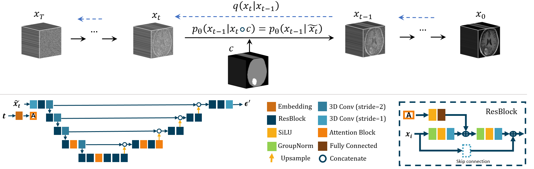
In this study, we extend our previous work on 3D-DDPM [31], which adapted the vanilla DDPM model for generating 3D volumetric images. We further enhance this architecture to facilitate the synthesis of conditional 3D medical images, conditioned on segmentation masks. Initially, our approach was grounded on the original DDPM, as proposed in [34]. The forward diffusion process adds small quantities of Gaussian noise , defined by the variance schedule , to an image sample from the training dataset at each timestep within a given number of timesteps . The noisy sample is defined as follows for :
| (1) |
To prevent sudden noise level fluctuations, we adopt a cosine noise schedule, as defined in [38]:
| (2) |
Here, parameter is a small offset value that prevents the schedule from becoming exceedingly small as the timestep nears zero. As a denoiser model in the reverse diffusion process , we employ a modified 3D U-Net architecture, based on the original work [34] by DDPM authors. Our enhancements involve replacing 2D operations, layers, and noise inputs with 3D counterparts to handle volumetric medical images. We incorporate embedding, ResBlocks, SiLU activation, group normalization, attention and fully connected layers to enhance performance. The detailed architecture is illustrated in Fig. 1.
”Inspired by a super-resolution technique where a lower-resolution image is upsampled and concatenated to the generated image at each iteration, as demonstrated by J. Ho et al. [36], we propose a straightforward, effective method that adapts this approach to modify the input image by channel-wise concatenating the segmentation mask. Contrary to the original DDPM, where is the only input, our model incorporates , which includes an additional channel for the segmentation mask. This mask guides the generation process, enabling the synthesis of meaningful images, such as a pathological MRI of the brain with a tumor precisely positioned. The conditioning process is illustrated in Fig. 1, where the segmentation mask is concatenated with the noisy image at each timestep .
The training images and segmentation masks used in this study are single-channel volumetric images with three dimensions: width (), height (), and depth (). The segmentation mask in the dataset consists of three class labels: 0 represents the background, 1 corresponds to the head area, and 2 indicates the tumor area. To prevent ordinal bias in model training due to the numeric class labels in the segmentation mask [37], we used one-hot encoding on the mask image. This process ignored the irrelevant background class label 0, creating a two-channel mask. In this mask, channel 0 represents the head area, and channel 1 indicates the tumor area. The channel-wise concatenation was then applied to combine the image and the mask, resulting in a concatenated image with three channels denoted as Throughout this paper, we refer to the concatenated image as .
The denoising process, also known as the generative sampling process, , is formulated as follows:
| (3) |
where represents the trained noise predictor U-Net model. We present the complete training and sampling procedures in Algorithms 1 and 2 respectively.
2.1 Loss Function
Pixel-wise losses such as and are commonly used in the literature of DDPM papers [29]. In our study, we observed that the loss (i.e., resulted in noisier images compared to the loss (i.e., The loss function, due to its computation of the squared difference between the estimated value and the target value, is sensitive to outliers. On the other hand, the loss calculates the absolute differences between the estimated value and the target value, making it relatively less sensitive to outliers. Hence, in our main experiments, we utilize the loss:
| (4) |
where and represent the pixels of the original noise added to the input and the predicted noise from the model (i.e., ) respectively, is the total number of pixels ().
3 Experiments and Results
3.1 Datasets and Image Preprocessing
We used unnormalized clinical brain Magnetic Resonance (MR) images without skull stripping. Our evaluation was performed on the clinical stereotactic radiosurgery dataset [39], which included 1688 contrast-enhanced T1-weighted (T1c) whole-head MR images and corresponding segmentation masks for various brain lesions. The dataset was obtained from patients undergoing Cyberknife radiosurgery at the National Taiwan University Hospital (NTUH). For image preprocessing, we employed MRIPreprocessor 222https://github.com/ReubenDo/MRIPreprocessor for image registration and ensured consistent image dimensions by applying cropping and padding. The resulting dimensions were 192x192x192, which were then resized to 128x128x128 with a slice thickness of 1.5x1.5x1.5mm. To enhance the quality of our training data, we removed 188 outlier images that were highly distorted and exhibited strong artifacts. We then utilized only 1,500 images for our experiments. Following this, we performed intensity rescaling and normalized the image intensities to a range of [-1, 1]. The segmentation mask annotations included three classes: class 0 for the background, class 1 for the head, and class 2 for the tumor area. To further evaluate our method, we conducted an additional experiment on multi-modality 3D brain MRI synthesis using the BraTS2021 challenge dataset 333http://braintumorsegmentation.org/. Details can be found in the ”F. 3D Multimodal MRI Synthesis Experiment” subsection.
3.2 Experiment Details
In this study, we evaluated the quality of 3D brain MRI synthesis by comparing our method with the most recent techniques [22, 23, 21, 32]. Due to the limited research in mask-to-image 3D synthesis, our primary point of comparison was the study by H.-C. Shin. [26], which focuses on 3D semantic brain MRI synthesis. We also evaluated several 2D image-to-image translation methods; however, due to their lower quality results, we excluded them from the primary comparison. Additionally, we employed our previously developed 3D DiscoGAN architecture, adapted and modified from the method proposed by T. Kim et al. [40], as an additional baseline for the comparison in semantic image synthesis.
We trained our proposed model and the baseline GAN models using 1,292 images for 100,000 iterations with a batch size of 1 (referred to as main models). The evaluation of these models was performed on 208 testing images.
Our Med-DDPM model was trained using the L1 loss, the cosine noise schedule for 250 steps, a learning rate of for the first 50,000 iterations, and for the last 50,000 iterations. We used the Adam optimizer and refined the model parameters with an Exponential Moving Average (EMA) strategy, using a decay factor of 0.995 to guarantee stable and efficient training. Our model’s first layer consists of 64 channels, and we incorporated an attention head at a resolution of 16. For the baseline 2 conditional GAN models, Pix2Pix and DiscoGAN, we trained them using a combination loss of Mean Squared Error and L1 Losses, which yielded better results. These models were optimized using the Adam optimizer, with a learning rate set to and momentum decays of 0.5 and 0.999. For other unconditional 3D brain MRI synthesis models, we utilized their GitHub repositories and adhered to the specified original hyperparameters.
Additionally, to assess the effectiveness of synthetic images as a viable alternative to data augmentation, we conducted a comprehensive empirical study. Specifically, we utilized a simple 3D U-Net model [35] for tumor segmentation task. The model was trained on synthetic data and compared against the performance of the 3D U-Net model trained on real data. The segmentation models were trained for 100 epochs using the Binary Cross-Entropy (BCE) loss with a batch size of 4. Model performance was evaluated using the Dice coefficient, IoU, Accuracy, Recall, and Precision scores. All models were trained on Tesla V100–SXM2 32 GB GPU card.
3.3 Evaluation Metric
We employed quantitative and qualitative measures to evaluate the synthetic images.
Quantitative Evaluation: The Fréchet Inception Distance (FID)[41] is a widely used evaluation metric in GAN synthesis, yet its application in assessing 3D medical images is limited due to its dependency on the Inception-v3 network [42] trained on a 2D image dataset. Building upon existing research [43, 44], we implemented the 3D-FID metric utilizing a pre-trained 3D Resnet model[45] for feature extraction. Besides the 3D-FID metric, we utilized Mean Squared Error (MSE), Maximum Mean Discrepancy (MMD) score [46], and Multi-Scale Structural Similarity Index Measure (MS-SSIM)[47] for quantitative evaluation.
Qualitative Evaluation: Two approaches were employed for the qualitative evaluation.
First, a visual evaluation was conducted with two neurosurgeons. The experts assessed a set of 40 high-resolution images, comprising an equal mix of 20 real and 20 synthetic images, with the objective of distinguishing them as real or fake. The images evaluated by the experts included samples from both a real image dataset and synthetic images generated by our proposed model. The experts made their classifications based solely on visual assessment. It is important to note that due to the easily identifiable non-realistic features of synthetic images generated by baseline GAN models, we did not proceed with further visual assessment of those GAN synthetic images.
Secondly, the tumor segmentation performance of a 3D U-Net model was evaluated to determine the effectiveness of synthetic images as a data augmentation technique. Various training scenarios were explored using synthetic dataset and real images.
3.4 Generated Images
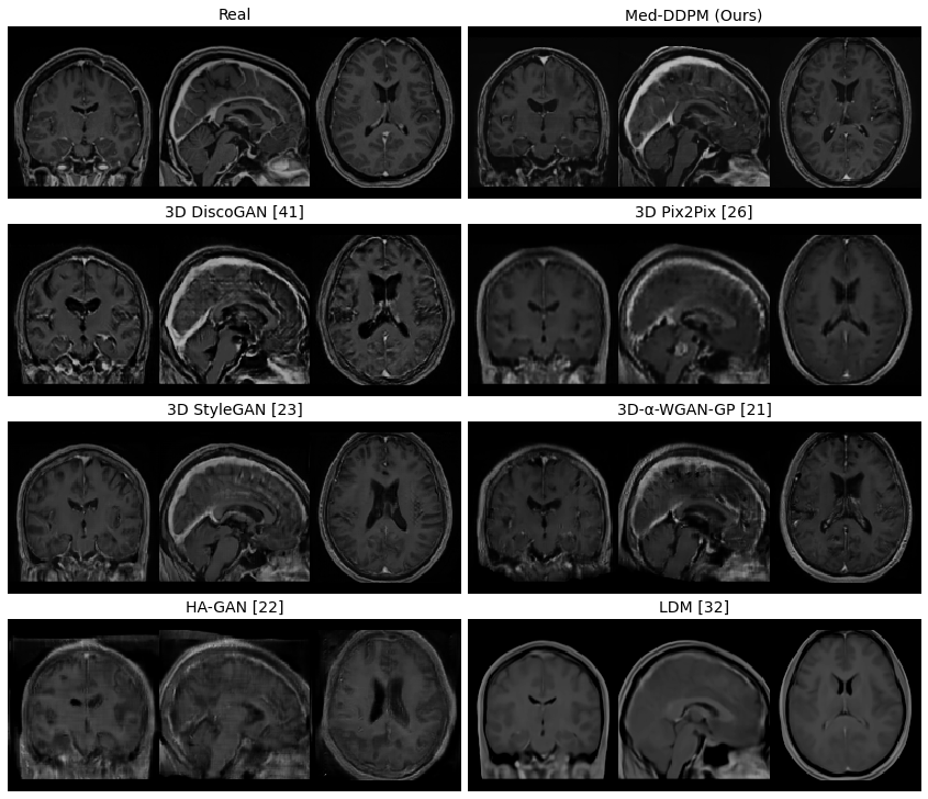
Fig. 2 displays coronal, sagittal, and axial slices of real brain MRI images and those generated by our proposed method and other baseline models. This comparison highlights differences in 3D brain MRI synthesis methods. The 3D StyleGAN generated images are blurry with wire mesh patterns. 3D--WGAN-GP produces even blurrier images with similar textures. HA-GAN images are the blurriest and always asymmetric. LDM sometimes creates images with uniform textures and clearer edges. While none of the models perfectly replicates continuous vessels, 3D--WGAN-GP images show better vessel continuity, but with unnaturally wide vessels. Overall, all GAN models produce less varied images than the two diffusion models, LDM and our Med-DDPM method.
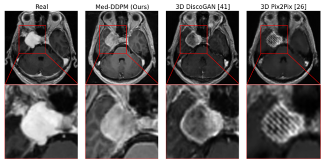
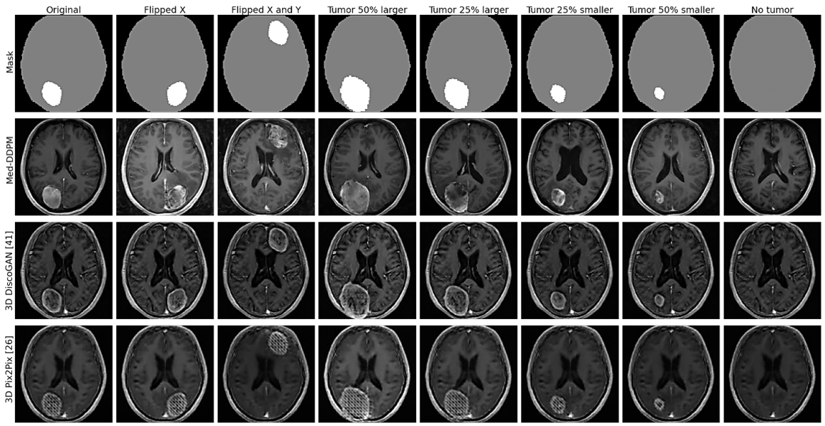
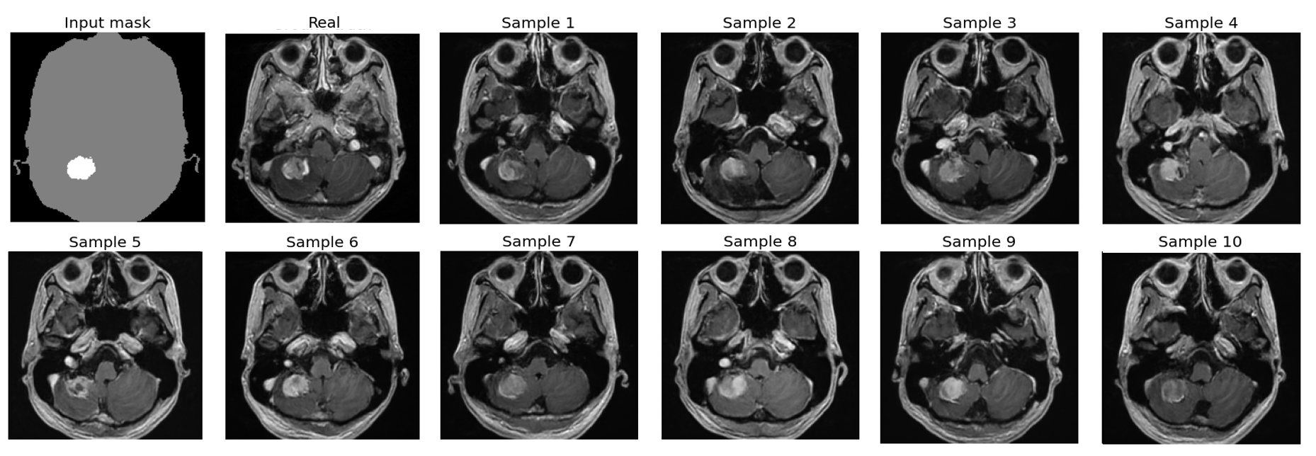
Fig.3 offers a closer look at tumor areas in axial plane images, comparing real and synthetic images from Med-DDPM and two other conditional GAN models. Fig.4 features synthetic images created using manipulated masks, which are generated by two different functions. The first function scales tumor masks from their center, while the second shifts them within the brain in axial, coronal, and sagittal planes. Fig. 5 demonstrates the variety of synthetic images produced from a single input mask. The Pix2Pix model often yields blurry images, especially with unseen test masks. DiscoGAN performs better, producing more realistic images with distinct tumor areas. However, DiscoGAN’s images lack clear brain features and exhibit coarse gyri and sulci.
In contrast, our Med-DDPM model excels in generating highly realistic and detailed images. It captures both brain features and tumor regions clearly, though occasionally it shows incomplete tumor ring enhancement, similar to real images. The peri-tumoral edema rendered by Med-DDPM appears more realistic than the isotropic low-intensity output of DiscoGAN, making these synthesized images closely resemble real ones.
3.4.1 Quantitative results
The comprehensive quantitative evaluation of various generative models, including our proposed Med-DDPM, is detailed in Table 1. This comparison is based on four commonly used metrics: Mean Squared Error (MSE), Maximum Mean Discrepancy (MMD), Multi-Scale Structural Similarity Index (MS-SSIM), and 3D Fréchet Inception Distance (3D-FID). Our Med-DDPM model demonstrated exceptional performance across most metrics. It achieved the lowest MSE value of , highlighting its exceptional ability to preserve intricate details and structures. Additionally, with an MMD value of , Med-DDPM proved effective in matching the distribution of the target domain. However, the Med-DDPM’s 3D-FID score of was higher than most of the GAN models. This suggests that the feature extraction model might not be fully optimized for extracting brain imaging features. Most notably, Med-DDPM achieved an MS-SSIM score of , the closest to the real data score of , underlining its superiority in maintaining structural integrity.
While models like 3D Pix2Pix and 3D StyleGAN excelled in specific areas, such as 3D Pix2Pix having the lowest MMD and 3D StyleGAN achieving the lowest 3D-FID, Med-DDPM consistently maintained a balance across all metrics. This balanced performance highlights its overall effectiveness in medical image generation tasks, adeptly handling the diverse nature of the data.
| Method | MSE | MMD | 3D-FID | MS-SSIM |
|---|---|---|---|---|
| 3D--WGAN-GP [21] | 0.0181 | 92.3998 | 74.5512 | 0.7610 |
| HA-GAN [22] | 0.0331 | 192.9155 | 1788.4518 | 0.4347 |
| 3D StyleGAN [23] | 0.0160 | 35.2398 | 48.9729 | 0.7429 |
| LDM [32] | 0.0349 | 330.3030 | 2730.7849 | 0.6584 |
| 3D Pix2Pix [26] | 0.0171 | 24.9456 | 59.4183 | 0.6966 |
| 3D DiscoGAN [41] | 0.0188 | 44.6890 | 86.3527 | 0.6730 |
| Med-DDPM (Ours) | 0.0146 | 28.2507 | 144.8321 | 0.6132 |
| Real | - | - | - | 0.5864 |
3.4.2 Qualitative results
Regular visual assessment tests were conducted throughout the experimentation phase. The experts were presented with a mixture of real and synthetic 3D images generated by the proposed model and the two baseline models. The experts evaluated the quality of the generated images. It was evident to the experts that the synthetic images produced by the two baseline models exhibited blurriness and lacked realistic-looking brain features. In contrast, the synthetic images generated by our proposed method appeared more realistic. However, upon careful examination of the axial plane, the experts were able to identify the synthetic images due to slight inconsistencies in vessel continuity within the Circle of Willis area. Additionally, the synthetic images did not exhibit the presence of mass effects around large tumors, which typically result in shifts in the ventricles and the midline.
| Experiment | Method | Dice | IoU | Accuracy | Recall | Precision |
| R 1000 | Real images | 0.6531±0.2831 | 0.5383±0.2565 | 0.6860±0.3159 | 0.6860±0.3159 | 0.8800±0.3250 |
| S 1000 | 3D DiscoGAN | 0.4685±0.2816 | 0.3497±0.2393 | 0.4169±0.2765 | 0.4169±0.2765 | 0.9100±0.2862 |
| 3D Pix2Pix | 0.3171±0.2706 | 0.2219±0.2094 | 0.2957±0.2759 | 0.2957±0.2759 | 0.7600±0.4271 | |
| Med-DDPM (Ours) | 0.6207±0.2882 | 0.5040±0.2610 | 0.6137±0.3156 | 0.6137±0.3156 | 0.9000±0.3000 | |
| R 500 + S 500 | 3D DiscoGAN | 0.6098±0.2775 | 0.4888±0.2534 | 0.5458±0.2818 | 0.5458±0.2818 | 0.9200±0.2713 |
| 3D Pix2Pix | 0.6135±0.2989 | 0.5011±0.2742 | 0.5836±0.3161 | 0.5836±0.3161 | 0.9100±0.2862 | |
| Med-DDPM (Ours) | 0.6449±0.2769 | 0.5272±0.2534 | 0.6832±0.3125 | 0.6832±0.3125 | 0.9300±0.2551 | |
| R 1000 + S 1000 | 3D DiscoGAN | 0.6239±0.2627 | 0.4989±0.2404 | 0.5570±0.2678 | 0.5570±0.2678 | 0.9200±0.2713 |
| 3D Pix2Pix | 0.6343±0.2928 | 0.5211±0.2670 | 0.5938±0.3010 | 0.5938±0.3010 | 0.8900±0.3129 | |
| Med-DDPM (Ours) | 0.6561±0.2758 | 0.5401±0.2553 | 0.6602±0.3028 | 0.6602±0.3028 | 0.9200±0.2713 | |
| R 1000 + S 2000 | 3D DiscoGAN | 0.6141±0.2543 | 0.4848±0.2295 | 0.5548±0.2537 | 0.5548±0.2537 | 0.9500±0.2179 |
| 3D Pix2Pix | 0.6010±0.3544 | 0.4915±0.2030 | 0.5820±0.2895 | 0.5820±0.2895 | 0.9100±0.2713 | |
| Med-DDPM (Ours) | 0.6675±0.2623 | 0.5495±0.2489 | 0.6673±0.2949 | 0.6673±0.2949 | 0.9400±0.2375 | |
| Evaluation results of a segmentation model trained on synthetic images. ’R’ denotes real images from the dataset, serving as the baseline for comparison. ’S’ represents synthetic images used for training the segmentation model, while ’R + S’ signifies a combination of real and synthetic data employed in model training. The best outcomes, compared to the baseline, are highlighted in bold. | ||||||
| # | Model | Med-DDPM (Ours) | 3D Pix2Pix [21] | ||||
| Training | Inference | Training Speed | Training | Inference | Training Speed | ||
| (mb) | (mb) | (iter/s) | (mb) | (mb) | (iter/s) | ||
| 1 | Single-modality synthesis | 23981 | 7991 | 0.65 | 17138 | 7046 | 13.21 |
| 2 | Multi-modality synthesis | 80963 | 21159 | 0.61 | 69225 | 18299 | 5.89 |
3.5 Comparison of Segmentation Models Trained on Synthetic Images
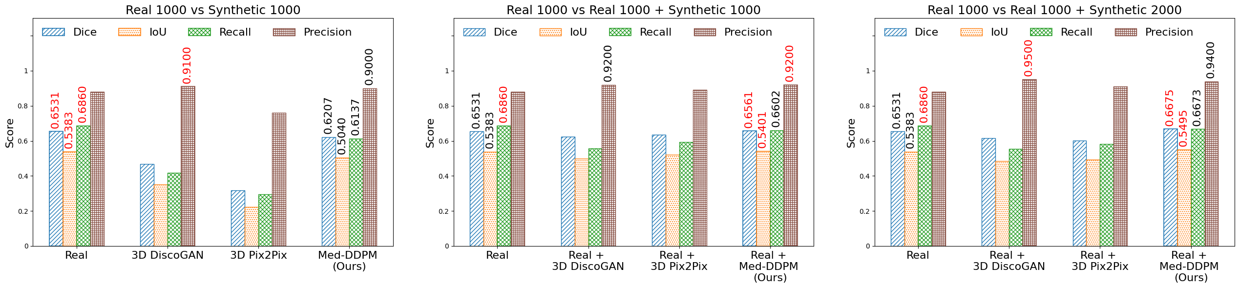
The evaluation presented in Table 2 highlights the performance of segmentation models trained on synthetic images. It particularly emphasizes the exceptional performance of our Med-DDPM model compared to the baseline models, 3D DiscoGAN and 3D Pix2Pix.
In scenarios involving 1000 real images, 1000 synthetic images, and their combinations, Med-DDPM consistently outperformed the baseline models. Specifically, in the experiment with solely 1000 synthetic images, Med-DDPM achieved a Dice score of 0.621, surpassing 3D DiscoGAN (0.468) and 3D Pix2Pix (0.317). In combined scenarios, such as 1000 real images with 1000 synthetic images, Med-DDPM maintained its lead with a Dice score of 0.656, compared to 0.623 for 3D DiscoGAN and 0.634 for 3D Pix2Pix.
Moreover, in the larger dataset comprising 1000 real images with 2000 synthetic images, Med-DDPM’s performance reached its peak with a Dice score of 0.667, surpassing the baseline accuracy of 0.653 for real images and demonstrating its potential of data augmentation capabilities.
While the 3D DiscoGAN and 3D Pix2Pix models showed improvements in mixed data scenarios, they were consistently outclassed by Med-DDPM.
3.6 3D Multimodal MRI Synthesis Experiment
In this section, we present the results of an additional experiment conducted to validate the effectiveness of our proposed method, Med-DDPM, for multimodal MRI synthesis. We utilized the brain-extracted MRI dataset from the BraTS2021 challenge444http://braintumorsegmentation.org/ for this experiment, with the objective of demonstrating the capability of our method in generating high-quality images for all four MRI modalities (T1, T1CE, T2, and Flair) simultaneously from a segmentation mask.
To enhance model training efficiency, we selected 193 high-quality images where all modalities have no distortion and artifacts from the dataset and preprocessed them by applying cropping and padding to a size of 192x192x144. The original labels of the segmentation mask in this dataset are 0, 1, 2, and 4, where 1, 2, and 4 represent tumor parts. For better adaptation to our needs, we modified the mask labels as follows: we changed mask labels from 4 to 3 and introduced one more label (label 4) to represent the brain area, achieved by thresholding the T1 image. Consequently, the final class labels were defined as follows: 0 represents the background, 1 to 3 correspond to the tumor parts, and 4 indicates the brain area.
Next, we performed a one-hot encoding operation, excluding the background class label 0. This operation resulted in a mask image with four channels, each representing one of the classes. During the training process, we applied channel-wise concatenation to combine the input training image (including all four modalities T1, T1CE, T2, and Flair concatenated together, resulting in a four-channel image) with the final mask image, also a four-channel image. The resulting concatenated image contains eight channels, and this configuration was used in the training process. For the Med-DDPM model in this experiment, we used the same experimental setups as in our main model training, except we employed 200,000 iterations for this experiment. Due to the high resolution nature of the eight-channel image, we trained our model on the NVIDIA A100 80GB GPU card for 5 days.
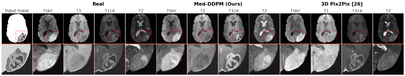
In this experiment, we also employed C.-H.Shin et al.[26] as our comparison baseline model since originally this paper is only one study in the literature investigating 3D semantic brain MRI synthesis especifically 4 modalities synthesis.
Fig. 7 illustrates a comparison between the generated samples from our experiments and the corresponding real images. The images generated by our proposed method exhibit a high level of fidelity and closely resemble the actual MRI modalities than baseline GAN model. This outcome further highlights the robustness and accuracy of our approach in effectively generating multiple modalities from a single segmentation mask.
3.7 Memory efficiency
In this section, we compare the memory efficiency of our model with the baseline model by measuring GPU memory usage and training speed. We adopted the training speed measurement methodology used by L. Sun et al. [22], which involves recording the number of iterations per second during training. For the training of multi-modality synthesis, we employed an NVIDIA Tesla A100 GPU, and for single modality synthesis, a Tesla V100–SXM2 32 GB GPU card was used. Both models were trained with a batch size of 1. Table 3 provides the detailed comparison of memory consumption and training speed. Although our proposed method produces more realistic-looking images than the baseline model, the architecture of our diffusion model necessitates higher memory consumption compared to the baseline 3D pix2pix GAN model.
4 Discussion
This study’s findings underscore the significant advancements in 3D brain MRI synthesis using generative models. The Med-DDPM model, in particular, demonstrates remarkable capabilities in generating realistic and detailed brain images, a critical advancement in medical imaging.
In the comparison of various generative models, Med-DDPM consistently outperformed baseline models like 3D StyleGAN, HA-GAN, and 3D--WGAN-GP, especially in maintaining the structural integrity and realistic representation of both normal brain tissue and pathological features like tumors. This is evident in the quantitative results, where Med-DDPM achieved the lowest Mean Squared Error (MSE) and a close-to-real Multi-Scale Structural Similarity Index (MS-SSIM). However, its higher 3D-FID score compared to other models suggests room for further optimization in feature extraction specific to brain imaging.
Furthermore, the qualitative assessments by experts validated the superiority of Med-DDPM in generating synthetic images that closely resemble real ones. The minor inconsistencies noted in vessel continuity and the lack of mass effects around large tumors highlight areas for future improvement.
The segmentation model evaluations further reinforce the utility of synthetic images generated by Med-DDPM, particularly in data augmentation. The model’s superior performance in datasets with a combination of real and synthetic images demonstrates its potential in enhancing the training of segmentation algorithms, thus contributing significantly to advancements in medical image analysis.
5 Conclusion
In summary, Med-DDPM represents a substantial leap forward in medical imaging, particularly in synthesizing 3D semantic brain MRIs. It showcases the power of AI in addressing challenges like data scarcity and privacy in healthcare domain. The unique semantic conditioning of our proposed model facilitates the generation of diverse and anatomically accurate images. This sets a new standard in image fidelity and opens up new possibilities for image anonymization. The effectiveness of Med-DDPM is evident in its performance in tumor segmentation tasks and its ability to generate all four MRI modalities from a single segmentation mask. This versatility makes it crucial for accurately representing complex brain structures. Overall, Med-DDPM not only demonstrates the capabilities of diffusion models in generating high-quality medical images but also underscores the role of AI in transforming medical imaging amid data and privacy constraints. Future work should focus on expanding its applications and refining its capabilities to further advance medical imaging.
Acknowledgment
This work was supported by the National Science and Technology Council, Taiwan [Grant No. 111-2221-E-002-049-MY3] and National Taiwan University Hospital [Grant No. 110-EDN03].
References
References
- [1] M. Puttagunta and S. Ravi, ”Medical image analysis based on deep learning approach,” Multimedia Tools and Applications, vol. 80, no. 16, pp. 24365-24398, Jul. 2021. doi: 10.1007/s11042-021-10707-4.
- [2] G. Litjens et al., ”A survey on deep learning in medical image analysis,” Medical Image Analysis, vol. 42, pp. 60-88, 2017. doi: 10.1016/j.media.2017.07.005.
- [3] K. He et al., ”Transformers in Medical Image Analysis: A Review,” Intelligent Medicine, vol. 2022, 2022. doi: 10.1016/j.imed.2022.07.002.
- [4] A. R. Luca, T. F. Ursuleanu, L. Gheorghe, R. Grigorovici, S. Iancu, M. Hlusneac, A. Grigorovici, ”Impact of quality, type and volume of data used by deep learning models in the analysis of medical images,” Informatics in Medicine Unlocked, vol. 29, pp. 100911, 2022. doi: 10.1016/j.imu.2022.100911.
- [5] M. H. Hesamian, W. Jia, X. He, and P. Kennedy, ”Deep Learning Techniques for Medical Image Segmentation: Achievements and Challenges,” J Digit Imaging, vol. 32, no. 4, pp. 582-596, Aug. 2019. doi: 10.1007/s10278-018-0159-6.
- [6] L. M. Prevedello et al., ”Challenges Related to Artificial Intelligence Research in Medical Imaging and the Importance of Image Analysis Competitions,” Radiology: Artificial Intelligence, vol. 1, no. 1, p. e180031, 2019. doi: 10.1148/ryai.2019180031.
- [7] Z. Li, K. Kamnitsas, and B. Glocker, ”Overfitting of Neural Nets Under Class Imbalance: Analysis and Improvements for Segmentation,” in Medical Image Computing and Computer Assisted Intervention – MICCAI 2019, D. Shen et al. (Eds.), Cham: Springer International Publishing, 2019, pp. 402–410, ISBN: 978-3-030-32248-9.
- [8] A. Zhang, L. Xing, J. Zou, and J. C. Wu, ”Shifting machine learning for healthcare from development to deployment and from models to data,” Nature Biomedical Engineering, vol. 6, no. 12, pp. 1330-1345, 2022. doi: 10.1038/s41551-022-00898-y.
- [9] X. Yi, E. Walia, and P. Babyn, ”Generative adversarial network in medical imaging: A review,” Medical Image Analysis, vol. 58, p. 101552, 2019. doi: https://doi.org/10.1016/j.media.2019.101552.
- [10] Y. Chen et al., ”Generative Adversarial Networks in Medical Image augmentation: A review,” Computers in Biology and Medicine, vol. 144, p. 105382, 2022. doi: https://doi.org/10.1016/j.compbiomed.2022.105382.
- [11] E. Ahishakiye, M. B. Van Gijzen, J. Tumwiine, R. Wario, and J. Obungoloch, ”A survey on deep learning in medical image reconstruction,” Intelligent Medicine, vol. 1, no. 3, pp. 118-127, 2021. doi: https://doi.org/10.1016/j.imed.2021.03.003.
- [12] B. K. Beaulieu-Jones et al., ”Privacy-Preserving Generative Deep Neural Networks Support Clinical Data Sharing,” Circulation: Cardiovascular Quality and Outcomes, vol. 12, e005122, 2019, doi: https://doi.org/10.1161/CIRCOUTCOMES.118.005122.
- [13] J. Yoon, L. N. Drumright, and M. van der Schaar, ”Anonymization Through Data Synthesis Using Generative Adversarial Networks (ADS-GAN),” IEEE Journal of Biomedical and Health Informatics, vol. 24, no. 8, pp. 2378-2388, 2020. doi: 10.1109/jbhi.2020.2980262. PMID: 32167919.
- [14] Y. Al Khalil, S. Amirrajab, C. Lorenz, J. Weese, J. Pluim, and M. Breeuwer, ”On the usability of synthetic data for improving the robustness of deep learning-based segmentation of cardiac magnetic resonance images,” in Medical Image Analysis, vol. 84, p. 102688, 2023. doi: https://doi.org/10.1016/j.media.2022.102688.
- [15] V. Fernandez et al., ”Can Segmentation Models Be Trained with Fully Synthetically Generated Data?,” in Zhao, C., Svoboda, D., Wolterink, J.M., Escobar, M. (eds) Simulation and Synthesis in Medical Imaging. SASHIMI 2022. Lecture Notes in Computer Science, vol 13570, Springer, Cham, 2022.
- [16] D. Saxena, and J. Cao, ”Generative Adversarial Networks (GANs): Challenges, Solutions, and Future Directions,” in ACM Comput. Surv., vol. 54, no. 3, Art. no. 63, April 2022, pp. 1–42, doi: 10.1145/3446374.
- [17] Y. Skandarani, P.-M. Jodoin, and A. Lalande, ”GANs for Medical Image Synthesis: An Empirical Study,” in Journal of Imaging, vol. 9, no. 3, 2023, p. 69. [Online]. Available: https://www.mdpi.com/2313-433X/9/3/69.
- [18] L. Wang, W. Chen, W. Yang, F. Bi, and F. R. Yu, ”A State-of-the-Art Review on Image Synthesis With Generative Adversarial Networks,” in IEEE Access, vol. 8, 2020, pp. 63514–63537, doi: 10.1109/ACCESS.2020.2982224.
- [19] E. Jung, M. Luna, and S.H. Park, ”Conditional GAN with an Attention-Based Generator and a 3D Discriminator for 3D Medical Image Generation,” in Medical Image Computing and Computer Assisted Intervention – MICCAI 2021, M. de Bruijne et al., Eds., Lecture Notes in Computer Science, vol. 12906, Cham: Springer, 2021, doi: 10.1007/978-3-030-87231-1_31.
- [20] Q. Zhou and H. Zou, ”A layer-wise fusion network incorporating self-supervised learning for multimodal MR image synthesis,” in Frontiers in Genetics, vol. 13, 2022, doi: 10.3389/fgene.2022.937042.
- [21] G. Kwon, C. Han, and Ds. Kim, ”Generation of 3D Brain MRI Using Auto-Encoding Generative Adversarial Networks,” in Medical Image Computing and Computer Assisted Intervention – MICCAI 2019, MICCAI 2019, Lecture Notes in Computer Science, vol. 11766, D. Shen et al., Eds. Cham, Switzerland: Springer, 2019, doi: 10.1007/978-3-030-32248-9_14.
- [22] L. Sun, J. Chen, Y. Xu, M. Gong, K. Yu, and K. Batmanghelich, ”Hierarchical Amortized GAN for 3D High Resolution Medical Image Synthesis,” in IEEE Journal of Biomedical and Health Informatics, vol. 26, no. 8, pp. 3966-3975, Aug. 2022, doi: 10.1109/JBHI.2022.3172976.
- [23] S. Hong et al., ”3D-StyleGAN: A Style-Based Generative Adversarial Network for Generative Modeling of Three-Dimensional Medical Images,” in S. Engelhardt et al., Eds., Deep Generative Models, and Data Augmentation, Labelling, and Imperfections, DGM4MICCAI DALI 2021, Lecture Notes in Computer Science, vol. 13003, Springer, Cham, 2021. [Online]. Available: https://doi.org/10.1007/978-3-030-88210-5_3
- [24] A. B. Qasim et al., ”Red-GAN: Attacking class imbalance via conditioned generation. Yet another medical imaging perspective,” in Proceedings of the Third Conference on Medical Imaging with Deep Learning, vol. 121, 2020, pp. 655–668. [Online]. Available: https://proceedings.mlr.press/v121/qasim20a.html
- [25] T. Park, M.-Y. Liu, T.-C. Wang, and J.-Y. Zhu, ”Semantic Image Synthesis With Spatially-Adaptive Normalization,” in 2019 IEEE/CVF Conference on Computer Vision and Pattern Recognition (CVPR), Long Beach, CA, USA, 2019, pp. 2332-2341, doi: 10.1109/CVPR.2019.00244.
- [26] H.-C. Shin et al., ”Medical image synthesis for data augmentation and anonymization using generative adversarial networks,” in Simulation and Synthesis in Medical Imaging, 1st ed., vol. 11037, M. A. Horsch et al. (Eds.), Springer, 2018, pp. 1–11. ISBN: 978-3-030-00536-8, doi: 10.1007/978-3-030-00536-8_1.
- [27] P. Isola et al., ”Image-to-Image Translation with Conditional Adversarial Networks,” CoRR, vol. abs/1611.07004, 2016.
- [28] D. Prafulla and N. Alexander, ”Diffusion Models Beat GANs on Image Synthesis,” Adv. Neural Inf. Process Syst., vol. 34, 2021.
- [29] L. Yang et al., ”Diffusion Models: A Comprehensive Survey of Methods and Applications,” arXiv preprint arXiv:2209.00796, 2023.
- [30] A. Kazerouni et al., ”Diffusion models in medical imaging: A comprehensive survey,” in Medical Image Analysis, vol. 88, 2023, p. 102846, doi: https://doi.org/10.1016/j.media.2023.102846.
- [31] Z. Dorjsembe, S. Odonchimed, and F. Xiao, ”Three-Dimensional Medical Image Synthesis with Denoising Diffusion Probabilistic Models,” in Medical Imaging with Deep Learning, 2022.
- [32] W. H. L. Pinaya et al., ”Brain Imaging Generation with Latent Diffusion Models,” in Deep Generative Models, A. Mukhopadhyay, I. Oksuz, S. Engelhardt, D. Zhu, Y. Yuan, Eds., vol. 13609, Lecture Notes in Computer Science, Springer, Cham, 2022, doi: 10.1007/978-3-031-18576-2_12.
- [33] M. Özbey et al., ”Unsupervised Medical Image Translation With Adversarial Diffusion Models,” in IEEE Transactions on Medical Imaging, vol. 42, no. 12, pp. 3524-3539, Dec. 2023, doi: 10.1109/TMI.2023.3290149.
- [34] J. Ho, A. Jain, and P. Abbeel, ”Denoising Diffusion Probabilistic Models,” arXiv preprint arXiv:2006.11239, 2020.
- [35] Ö. Çiçek, A. Abdulkadir, S. S. Lienkamp, T. Brox, and O. Ronneberger, ”3D U-Net: Learning Dense Volumetric Segmentation from Sparse Annotation,” in Medical Image Computing and Computer-Assisted Intervention – MICCAI 2016, Springer, Cham, 2016, pp. 424-432.
- [36] J. Ho, C. Saharia, W. Chan, D. J. Fleet, M. Norouzi, and T. Salimans, ”Cascaded Diffusion Models for High Fidelity Image Generation,” J. Mach. Learn. Res., vol. 23, no. 1, pp, Jan. 2022.
- [37] N. Tsourakis, ”Chapter 2,” in Machine Learning Techniques for Text, 1st ed. Birmingham: Packt Publishing, 2022.
- [38] A. Nichol and P. Dhariwal, ”Improved Denoising Diffusion Probabilistic Models,” ArXiv, 2021.
- [39] Wu, Siangruei et al., ”Deep Learning-Based Segmentation of Various Brain Lesions for Radiosurgery,” Applied Sciences, vol. 11, no. 19, article no. 9180, 2021, DOI: 10.3390/app11199180.
- [40] T. Kim, M. Cha, H. Kim, J. K. Lee, and J. Kim, ”Learning to Discover Cross-Domain Relations with Generative Adversarial Networks,” in Proceedings of the 34th International Conference on Machine Learning - Volume 70, Sydney, NSW, Australia, 2017, pp. 1857-1865.
- [41] M. Heusel, H. Ramsauer, T. Unterthiner, B. Nessler, and S. Hochreiter, ”GANs Trained by a Two Time-Scale Update Rule Converge to a Local Nash Equilibrium,” Proceedings of the 31st International Conference on Neural Information Processing Systems, pp. 6629–6640, 2017, ISBN: 9781510860964.
- [42] C. Szegedy, V. Vanhoucke, S. Ioffe, J. Shlens, and Z. Wojna, ”Rethinking the Inception Architecture for Computer Vision,” in 2016 IEEE Conference on Computer Vision and Pattern Recognition (CVPR), pp. 2818–2826, 2016, doi: 10.1109/CVPR.2016.308.
- [43] P. Subramaniam et al., ”Generating 3D TOF-MRA volumes and segmentation labels using generative adversarial networks,” Medical Image Analysis, vol. 78, pp. 102396, 2022, doi: https://doi.org/10.1016/j.media.2022.102396.
- [44] L. Sun, J. Chen, Y. Xu, M. Gong, K. Yu, and K. Batmanghelich, ”Hierarchical Amortized GAN for 3D High Resolution Medical Image Synthesis,” IEEE Journal of Biomedical and Health Informatics, vol. 26, no. 8, pp. 3966–3975, 2022, doi: 10.1109/JBHI.2022.3172976.
- [45] S. Chen, K. Ma, and Y. Zheng, ”Med3D: Transfer Learning for 3D Medical Image Analysis,” CoRR, vol. abs/1904.00625, 2019, arXiv:1904.00625.
- [46] A. Gretton, K. M. Borgwardt, M. J. Rasch, B. Sch”olkopf, and A. Smola, ”A Kernel Two-Sample Test,” Journal of Machine Learning Research, vol. 13, no. 25, pp. 723-773, 2012, url: http://jmlr.org/papers/v13/gretton12a.html.
- [47] Z. Wang, E.P. Simoncelli, and A.C. Bovik, ”Multiscale structural similarity for image quality assessment,” in Proc. The Thrity-Seventh Asilomar Conference on Signals, Systems & Computers, 2003, vol. 2, pp. 1398-1402, 2003, doi: 10.1109/ACSSC.2003.1292216.