Low and High Frequency Vibrations Synergistically Enhance Singlet Exciton Fission Through Robust Vibronic Resonances
Abstract
Singlet exciton fission (SEF) is initiated by ultrafast internal conversion of a singlet exciton into a correlated triplet pair . The ‘reaction coordinates’ for ultrafast SEF even in archetypal systems such as pentacene thin film remain unclear with synthetic design principles broadly relying on tailoring electronic couplings to achieve new templates for efficient SEF materials. Spectroscopic detection of vibrational coherences in the photoproduct has motivated theoretical investigations into a possible role of vibronic resonance in driving SEF, akin to that reported in several photosynthetic proteins. However, a precise understanding of how prominent low-frequency vibrations and their modulation of intermolecular orbital overlaps, equally prominent high-frequency vibrations, and order of magnitude larger Huang-Rhys factors in SEF chromophores compared to photosynthetic pigments, collectively influence the mechanistic details of SEF remains starkly lacking. Here we address this gap and identify previously unrecognized effects which are quite contrasting from those known in photosynthesis excitons, and vitally enhance non-adiabatic internal conversion in SEF. These include broad non-selective vibronic resonances, high-frequency vibrations coupling multiple such resonances, and a synergistic effect of low-frequency vibrations on vibronic resonances. Spectroscopically these effects are reported by quantum beats of mixed polarization signatures in the simulated two-dimensional electronic spectra. We leverage this to propose readily implementable polarization-based experiments to test the above predictions, and unambiguously distinguish vibrations which drive vibronic mixing and promote SEF, against spectator vibrations simply accompanying ultrafast internal conversion. Our findings have direct implications for the broad experimental interest in synthetically tailoring molecules to promote vibronically enhanced internal conversion.
1 Introduction
Ultrafast conversion of a singlet exciton into a correlated triplet pair is the first step in singlet exciton fission (SEF)1, and subsequently leads to the generation of spatially separated triplets with uncorrelated spin wavefunctions on slower timescales. The high efficiency of the overall process in certain materials promises applications in next-generation photovoltaic technologies. However, mechanistic details of even the first step of this reaction remain unsettled ranging from preliminary suggestions of direct2, charge transfer (CT) state mediated3, 4 and ‘superexchange’ mediated5 ultrafast conversion of to to a more recently proposed role for increased charge-transfer couplings at crystal edges and defects6. Molecular design principles to achieve new SEF chromophores have accordingly been generally limited to synthetic tuning of electronic couplings only. A role for mixed vibrational-electronic (vibronic) motions in driving this process has come under close scrutiny7, 8, 9, 10, 11, 12, 13 only recently.
In photosynthesis, likely coincidences between large number of Franck-Condon (FC) active low-frequency intramolecular vibrations of the pigments with excited state electronic energy gaps of the multi-pigment protein are termed as vibronic resonances14, 15. These lead to strongly coupled vibrational-electronic motions creating non-adiabatic energy funnels16 which can efficiently drive photosynthetic energy and charge transfer. Such resonances are increasingly reported in several photosynthetic proteins17 based on readily observed vibronic coherences, or impulsively excited superpositions of vibronically mixed eigenstates. Along similar lines, and as a departure from previously proposed SEF mechanisms, vibronic resonances along high-frequency intramolecular vibrations in SEF chromophores have been recently implicated13, 9, 10 to drive ultrafast SEF. While this may be material specific, even for archetypal systems such as pentacene thin films it is not clear whether vibronic resonance due to a high-frequency intramolecular vibration is robust enough to energetic mismatches9 to enhance the rate of formation. A recent two-state ad hoc model12 also suggests no role for vibronic resonance. While it is not clear whether models with two electronic states, and , adequately capture the essential physics of the first steps of SEF and will be a subject of this presentation, some pertinent questions regarding the physical principles governing the initial steps of SEF may be – 1. Huang-Rhys (HR) factors, which dictate the probability of vibrational excitations accompanying electronic excitation, are order of magnitude larger in acenes than in photosynthetic pigments. How does that modify the vibronic resonances in SEF compared to narrow, selective vibronic resonances18, 19 between photosynthetic excitons ? 2. Prominent low-frequency FC active vibrations, which also tune intermolecular orbital overlaps are known in acene thin films20, 21, 13, 12. How do these vibrations of mixed inter/intramolecular character affect resonant vibronic couplings present along the high-frequency intramolecular vibrations ? 3. Large HR factors in acenes imply that prominent vibrational quantum beats are expected even in a solution of monomers. Can one then decipher mechanistic details solely based on readily observed quantum beats ?
The latter question deserves further motivation. In the archetypal SEF chromophores such as pentacene, large HR factors, with dimensionless displacements of the order of (classical turning points of the zero-point level), imply that ultrafast population transients in time-resolved spectroscopies will also exhibit strong modulations due to oscillating vibrational wavepackets on the ground and excited electronic states, even for pentacene monomers in solution22. Further complicating the interpretation of quantum beats in experiments is the well-known fact that dominant vibrational wavepackets in the photoproduct can simply arise due to transfer of coherently prepared reactant vibrational wavepackets that survive non-adiabatic internal conversion, or due to photoproduct formation within a fraction of vibrational time period23, 24, 25, 26, 27. For instance, Petelenz and co-workers have elucidated28, 29 that experimentally reported30 new vibrational coherences in the product may be those that are simply transferred from to with substantially larger amplitude in the photoproduct. Multilevel Redfield simulations31 of electronic curve crossing from Jean and Fleming further emphasize the above points. They show that vibrational wavepackets along spectator vibrational modes orthogonal to the reaction coordinate can be coherently transferred to the photoproduct without participating in ultrafast internal conversion. In contrast, wavepacket motions along promoter modes that drive vibronic mixing experience significant anharmonicity and dephase during ultrafast internal conversion. The conclusion from above experimental and theoretical reports is that vibrational coherences or new vibrational frequencies in the photoproduct do not a priori identify ‘reaction coordinates’ for internal conversion. This is true even for state-of-the-art two-dimensional electronic spectroscopy (2DES) experiments, which resolve ultrafast dynamics along multiple spectral dimensions – excitation, detection and coherence frequencies. For example, strong modulations reported in 2DES studies13, 12 on pentacene thin films may simply arise from spectator vibrational wavepackets which are not dephased by non-adiabatic anharmonic couplings present along promoter modes, and continue to oscillate through the SEF process. Thus, while much interest is generated by the possibility of selectively exciting promoter modes32, 33 and leveraging vibronic couplings34 to select reaction outcomes, unique spectroscopic markers of promoter modes are vital to decipher the quantum dynamical details of ultrafast SEF.
This manuscript addresses the above questions, identifies previously unrecognized synergistic effects between high- and low-frequency vibrational motions which together enhance non-adiabatic internal conversion, and proposes readily implementable spectroscopic signatures that only arise from coupled vibrational-electronic motions without any ambiguity from spectator vibrations which do not promote SEF and simply accompany ultrafast internal conversion. Our findings have direct implications for synthetic design of molecules to promote vibronically enhanced SEF, as well as for spectroscopy experiments aiming to distill the relevant mechanistic details.
2 Results and Discussion
Analysis of vibronic coupling in a donor-acceptor system provides a useful starting point to distinguish promoter versus spectator vibrational motions. For example, the Hamiltonian of an electronically coupled dimer can be analyzed in terms of in-phase and anti-phase relative motions of the two molecules and along their respective intramolecular vibrational coordinates . These can be represented as , formed by linear combinations of two intramolecular Franck-Condon (FC) modes on the respective molecules. This has precedence in the early works of Witkowskii and Moffit35, Fulton and Gouterman36, and Förster37, and leads to adiabatic separability of in-phase motions along from the rest of the Hamiltonian. In-phase motions do not affect donor-acceptor energy gap and are mere spectators in the dimer energy transfer problem, while anti-phase motions along tune relative donor-acceptor energy gap and act as promoter modes that mix electronic degrees of freedom. At vibronic resonance, this mixing becomes strongly non-adiabatic15, 16 and only anti-phase motions drive the vibronic wavepacket towards the acceptor. Here we adopt this understanding to analyze the SEF Hamiltonian.
Berkelbach and Reichman5, 38 have described ultrafast SEF in a pentacene dimer unit cell as well as supercells using a microsopic exciton model. Their calculations suggest that a dimer description can still qualitatively describe SEF rates. In larger systems39, 38 with growing CT manifold, or with stabilized40 CT states in the crystal, increased CT-mediated mixing of and further enhances SEF rates. Ab initio electronic structure calculations from Martinez 41 et al. have reported similar effects in going from dimer to bulk crystals. Zimmerman et al. have reported2 that the initial excitation at the vertical FC geometry is primarily dimeric, with contributions from neighboring pentacene molecules becoming negligible as the intermolecular distance is reduced by a small fraction (20%) post photoexcitation. Overall, a dimer unit cell description for the archetypal pentacene system is a reasonable starting point. A further entropic enhancement of SEF rates due to a large number of final manifold of states, as suggested by Tempelaar et al.9, will not be captured in the dimer description adopted here.
2.1 Model Hamiltonian and Non-Trivial Role of In-phase Vibrational Motions
Following previous works42, 11, 43, 7, the diabatic basis set for describing the singly excited manifold of coupled identical pentacene molecules and is given by locally excited states and , correlated triplet state and CT states and . The notation adopted is such that denotes a collective state where molecule is excited while molecule is in the ground electronic state, and denotes a state in which is cationic and is anionic. The ground to excited state transition dipoles in pentacene are aligned along the short axis41, 7 such that the transition dipoles to the optically allowed states are 54.3o in the herringbone structure44. The doubly excited state manifold, will be discussed during the discussion of 2DES simulations. The purely electronic part of the singly excited Hamiltonian is given by . Matrix elements arise due to static Coulomb or charge transfer couplings and is the electronic site energy of state . Indices run over the diabatic basis states. We next introduce linear vibronic coupling arising from the set of intramolecular FC active modes on each molecule, with vibrational coordinate operators and for the set. The linear vibronic coupling terms are on where , on , and on . The cationic and anionic stabilization energies for pentacene vibrations in the high frequency region are reported to be approximately equal45. Thus, for simplicity, we assume equal cationic and anionic FC displacements, that is, . Similarly, we have assumed equal7, 8 FC displacements on the triplet states of molecules and . The singly excited state SEF Hamiltonian becomes –
| (1) | |||||
where is the ground state Hamiltonian given by . Equation 1 assumes identical but displaced harmonic oscillators to describe the diabatic electronic potentials. It is important to note that ground state vibrational excitations are not restricted. Vibrational excitations are allowed on electronically unexcited molecule as well because such a molecule can still be part of an overall excited state in which the other molecule is electronically excited. Imposing such a restriction is equivalent to one-particle46 or coherent scattering approximation47. We48, 49 and others19, 50 have previously shown that such an approximation severely underestimates the scope of vibronic resonance, although several studies13, 12 describing vibronic couplings in SEF have the made one-particle approximation (1PA). Low-frequency vibrations in pentacene thin films are FC active20, 51, 21, have mixed inter/intramolecular character, and modulate intermolecular orbital overlaps to affect electronic couplings52. This latter effect is akin to Herzberg-Teller type coupling53, 54 and is termed as Peierls coupling9, 12 in the context of SEF. The interplay of Peierls coupling and linear vibronic coupling will be analyzed in Section 2.3.
Following earlier works 35, 36, 37 on energy transfer in excitonic dimers, we transform Equation 1 with , the in-phase and anti-phase relative motions of the two molecules, –
| (2) | |||||
What becomes immediately evident from Equation 2 is that not just , but also vibrational motions are adiabatically inseparable from the SEF Hamiltonian. Given the intuition from a purely excitonic dimer, mixing between the optically bright excitons, admixtures of and , by anti-phase vibrational motions which tune their relative energy gap is not surprising. However, counter intuitive to this established picture, in-phase motions, which were mere spectators in the case of excitonic dimer, can now drive the coupling between bright excitons and the state.
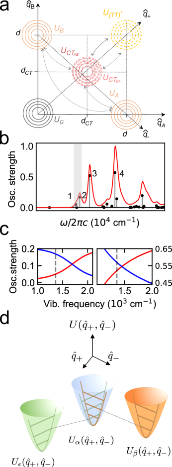
The resultant physical picture is motivated in Figure 1a. Further mathematical analysis of linear vibronic couplings in Equation 2 is presented in Equations S1–S2 and Figure S1. The multidimensional diabatic potentials of Equation 1 are two-dimensional for the simplest case of one FC active intramolecular vibrational mode on each molecule, and denoted as contours along intramolecular vibrational coordinates, and . The diabatic potential for the locally excited states , are shifted by FC displacement along the respective intramolecular vibrational coordinate. Akin to what is expected in an excitonic dimer18, the vector joining the minima of and , is the anti-phase energy gap tuning mode which drives non-adiabatic vibronic mixing between and , while is orthogonal and plays no role in the process. In the SEF problem, however, a striking contrast is seen when considering and diabatic states. Now, both in-phase and anti-phase motions, along and respectively, can tune energy gaps between and states to mix electronic degrees of freedom. This vital mechanistic significance of in-phase vibrational motions in SEF has been unrecognized in literature, except the adiabatic vibronic coupling density analysis of Nakano et al. 55 who remark on the importance of high-frequency in-phase vibrational motions. For example, recent two-state models13, 56 of SEF will miss this central role of in-phase vibrational motions. The mechanistic significance of the adiabatic inseparability of both and from the electronic Hamiltonian in Equation 2 is illustrated below in Section 2.2.
2.2 In-phase and Anti-phase Motions Create Coupled Vibronic Resonances
Prominent vibrational quantum beats in 2DES experiments on acenes, both monomers22 and thin films13, 12, which match FC vibrations in the Raman spectra are well-known. With dimensionless FC displacements close to unity57, large FC displacements in acenes are strikingly different from those in photosynthetic pigments. Larger widths of vibronic resonances, proportional to for an excitonic dimer19, 48, may therefore be naturally expected in case of acene-based systems. is the electronic mixing angle between electronic states which is also expected to be larger in SEF. Below we illustrate how these unique features influence vibronic couplings in SEF.
We consider a high-frequency intramolecular FC mode of frequency 1360 cm-1 as part of the system Hamiltonian, where numerical diagonalization of the Hamiltonian ensures that its non-adiabatic mixing with electronic degrees of freedom is treated exactly15, 16 without making Born-Oppenheimer approximation. This mode corresponds to the symmetric stretching mode of the pentacene ring also included in prior studies7, 44. The remaining vibrational bath is modeled as temperature-dependent underdamped Brownian oscillators which introduce decoherence18 between the ground and excited state electronic energy gap, and consequently homogeneously broadened lineshapes. Parameters for Brownian oscillator lineshapes are derived from fits of experimental linear absorption spectrum of pentacene monomers in ref.58. The total Huang-Rhys (HR) factor in the high-frequency modes on the locally excited states is unity to be consistent with prior models57, 7. Cationic and anionic stabilization energies for pentacene are approximately equal 45, as are the respective vibrational frequencies on cationic and anionic transient states59. Parameters for the electronic Hamiltonian have been taken from ref.41 which serve as a good approximation to a higher-level calculation reported3 earlier by Ananth et al. The five electronic states, without any vibrations, undergo electronic mixing to produce five excitons which are denoted as , , , , and according to increasing excitonic energy. The basis state character of these excitons is listed in Table S5. The two highest energy excitons () in the dimer are CT-dominant while the lowest exciton is -dominant with 7% total bright character from locally excited states. The lineshapes are ensemble averaged over an energetic disorder of 75 cm-1 between excited electronic states, estimated by Rao et al. from fits of 2DES simulations to experimental data. The resulting ensemble averaging only affects experimentally observed dephasing of excited state coherences17 (shown later in Sections 2.4 and 2.5). Inhomogeneity in the 0–1 optical energy gap only broadens the lineshapes without affecting decoherence or dephasing rates. This is not included in order to highlight the rich underlying spectroscopic features. Calculations including the 0–1 inhomogeneity are shown in Sections S6. Further details of model parameters are discussed in Section S3. Convergence checks are described in Section S5.
The consequences of broad vibronic resonance widths expected in acene-based systems are understood by analyzing the oscillator strengths beneath the simulated absorption lineshapes in Figure 1b. The line strengths in the dimer are sufficiently complicated even with one intramolecular vibrational mode. However, a pattern of three unequal intensities, marked as faint gray band, arises due to vibronic mixing between bright exciton and a quantum of vibrational excitation on , denoted as near-resonant manifold. Peaks in the 15200-16550 cm-1 region arise from multiple vibronic mixing channels. Exciton is near-resonant with excitons with one quantum of vibrational excitation. This latter manifold is also near-resonant with the second vibrational excitation on , together leading to a complex set of line strengths. This manifold is denoted as . Similar patterns have been semi-analytically described19, 48 in case of photosynthetic dimers.
To confirm the above intensity borrowing effects, we analyze the widths of these near-resonances. Jonas and co-workers have defined19 a width of vibronic resonance based on the range of frequencies over which intensity borrowing ratio changes from 1:2 to 2:1. For a photosynthetic dimer, expected resonance widths are only 30 cm-1. In comparison when intensities of peaks labelled ‘1’ and ‘2’ in are plotted as a function of , a 10x larger width of 330 cm-1 is seen in Figure 1c. When peaks in manifold are analyzed while the vibrational frequency is scanned, a similar intensity borrowing pattern between ‘3’ and ‘4’ is seen with even broader widths. This confirms the expectation that non-adiabatic vibronic mixing in case of acenes is highly robust to resonance mismatches, and therefore becomes unavoidable over a broad range of vibrational frequencies over which the optically bright excitons intermix, as well as impart bright character to vibrationally excited -dominant exciton. The physical picture that emerges from the above discussion is summarized in Figure 1d. Broad resonance widths are vital for enabling bright excitons () intermixing through anti-phase motions along , as well as their mixing with through in-phase motions along , both caused by a single intramolecular vibration on each molecule, thereby leading to coupled vibronic near-resonances as shown in Figure 1d. Importantly, two-state models with reduced vibrational dimensionality13, 12 will miss out on these previously unrecognized effects that are unique to the SEF Hamiltonian. The mechanistic significance of robust, coupled vibronic resonances and the complementary role of low-frequency vibrations in this picture is elucidated in Section 2.3.
2.3 Peierls Coupling Complements Vibronic Resonance
Micro-Raman spectroscopy20, 21 of acene thin films has reported several prominent low-frequency vibrations of mixed inter/intramolecular character which cause52, 55 non-perturbative modulations of orbital overlap transfer integrals to affect intermolecular Peierls coupling. Huo et al. have suggested60 that a distribution of electronic couplings created by such low-frequency vibrations prevents destructive interference between CT couplings to enhance SEF. On the contrary, Herbert et al. have suggested11 only a minor role for such couplings in enhancing SEF, with dominant role played by linear vibronic couplings arising from high frequency C–C stretching motions. Similarly, Tempelaar et al. have reported9 that Peierls coupling ‘breaks’ vibronic resonance condition to diminish SEF rates. In addition to the above inconsistent understanding of the role of Peierls coupling in SEF, models so far have suggested exclusive roles for Peierls coupling and resonant vibronic couplings. Here we will illustrate the complex interplay of these couplings to introduce new insights in this picture.
Peierls coupling is introduced through a low frequency vibration cm-1, prominent in 2DES13 and resonance Raman20, 21 experiments. The corresponding vibrational coordinates are with FC displacements on locally excited states , on state and on the states. Modulation of overlap integrals due to the low-frequency motions is additionally incorporated through Peierls coupling Hamiltonian45 . Indices and run over all the electronic states similar to , and denotes the coupling constant. Based on the calculations in ref.12, is estimated to be 150 cm-1. As before, all the parameters are directly motivated from experiments as described in Section S3 and listed in Table S4. Now both and vibrations are treated as quantum oscillators in the system Hamiltonian such that each diabatic exciton potential becomes 4-dimensional with diabatic basis states denoted as . runs over all five excitons, with quanta of vibrational excitation. Note that, as before, ground state vibrations of the unexcited molecule are still allowed to be a part of an overall excited electronic state, that is, the basis set is numerically exact without any 1PA. It is crucial to also note that Peierls distortion has been treated as a quantum oscillator in the system Hamiltonian instead of electronically off-diagonal disorder under the ‘frozen mode’ approximation61, 9. Although this makes the four-wavemixing simulations significantly more expensive, we will show that quantum oscillator treatment is necessary in order to describe the complementary nature of Peierls coupling. To best of our knowledge, such effects have not been reported earlier because of either exclusive treatment of both couplings, or approximating low-frequency Peierls distortion as a ’frozen’ mode.
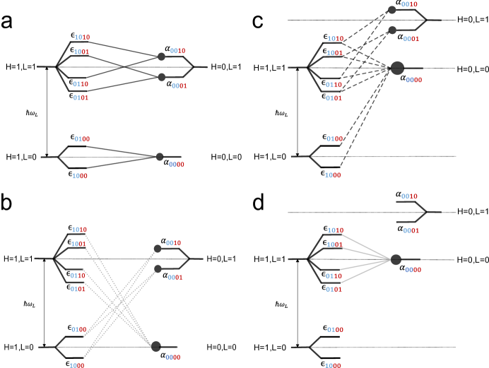
The interplay of linear vibronic and Peierls coupling can be analyzed by considering the two scenarios shown in Figure 2, where the exciton basis states are represented as for brevity. The left column, panels A and B, considers exact resonance due to , while the right column, panels C and D, considers near-resonant where the lack of resonance is compensated by a quantum of excitation along the low frequency vibration . In each column, the top and bottom panels show the dominant linear vibronic and Peierls coupling channels, respectively. Considering Figure 2a, one of the dominant linear vibronic coupling matrix element in the manifold, denoted as , will be proportional to –
.
A similar matrix element can be written along . Non-dominant coupling elements will also arise due to linear vibronic coupling along the low-frequency FC modes , and not shown in the figure. Interestingly, panel A shows that a quantum of excitation along the low-frequency mode, for example , leads to identical resonance conditions in the manifold with twice as many coupling channels, although with an accompanying vibrational overlap factor . These factors are approximately the same as those arising from overlaps owing to large FC displacement in acenes. See Section S1 for calculations of these FC overlaps. Thus, presence of low-frequency FC active vibrations by themselves enhance resonant vibronic mixing by increasing the number of mixing channels.
Figure 2b shows that when Peierls coupling is introduced in the above resonant picture, additional coupling channels open up in the manifold. For example, one of the dominant Peierls coupling matrix element between and along will be proportional to –
.
Note that energetic mismatch between these participating levels will suppress perturbative mixing between these states caused by Peierls coupling. The two unfavorable factors, weaker mixing and 0-1 FC overlaps, are denoted by dotted lines in the panel. However, increased number of mixing channels and substantial FC overlaps due to large intramolecular FC displacements in acenes (unlike in the case of photosynthetic excitons) together ensure that their net effect is consequential (Section 2.4).
Figures 2c,d show how the resonant picture modifies when there is energetic mismatch in the manifold. The dominant coupling matrix elements are marked on the figure. Compared to panel A, the near-resonant vibronic mixing in panel C is weakened (shown in dashed lines) due to energetic mismatch. However, a low frequency vibrational excitation makes up for the mismatch and opens up additional mixing channels between and four possible degenerate states with excitation on . These channels are denoted as dashed line because a 0-1 FC overlap reduces the mixing. With the same reasoning, a comparison of energetic alignment between panels B and D shows that Peierls couplings of panel B become stronger in panel D because the earlier energetic mismatch is now compensated for by low-frequency vibrational excitations. It is important to note that without a vibronic near-resonance, low-frequency vibrational excitations will not by themselves be sufficient to compensate for large energetic mismatch between exciton energies of (TT)1 and S1 manifolds. A subtle point to also note is that in case of only minor energetic mismatches introduced by low-frequency vibrational excitations, increase in the number of mixing channels especially in the presence of Peierls coupling, can more than compensate for it, biasing the overall effect towards enhancement of vibronic mixing. In summary, Peierls coupling through low-frequency vibrations increases the number of vibronic mixing channels and is expected to complement resonant vibronic mixing by making it more robust to energetic mismatches.
Based on above considerations, the width of vibronic resonance (Figure 1b), exciton delocalization and population transfer rates from the locally excited states to the acceptor state , are all expected to be enhanced. These effects are confirmed through numerical calculations in Section 2.4, and are contrary to earlier treatments9, 11, 12 of Peierls coupling where, either an explicit quantum oscillator treatment of low-frequency vibrations was not possible, or Peierls coupling was treated exclusively of linear vibronic coupling.
2.4 Enhanced Vibronic Mixing, Resonance Width and Exciton Delocalization
Intensity borrowing effects in oscillator strengths, such as those in Figure 1b can be directly analyzed to understand delocalization. However oscillator strengths even for the lowest energy excitons are fairly complicated when both and intramolecular vibrations are introduced in the Hamiltonian (see Figure 3a). Instead, we use inverse participation ratio (IPR) as an alternative metric to analyze these effects. IPR is a measure of excitonic delocalization which has been previously extended14 by Moran et al. to quantify delocalization in vibronic excitons. A lower IPR implies more exciton delocalization with perfect delocalization corresponding to an IPR of , for electronic states.
In Figure 3a shows the exact calculation of absorption intensities of a pentacene dimer at 300 K without(left panel) and with(right panel) Peierls coupling. Each molecule has two FC active vibrations – 1360 cm-1 and 265 cm-1. It is evident that with the inclusion of Peierls coupling, oscillator strengths are further distributed among more number of excitons indicating larger delocalization due to increase in inter-exciton mixing channels. The black Gaussian curve covering the low-energy excitons shows the intensity of the laser pulse used for 2D spectroscopy simulations (shown later in Section 2.5). The absorption sticks are overlaid with the lineshapes obtained from underdamped Brownian quantum oscillators62. Further details of the calculation are provided in Section S3.
Enhanced delocalization due to Peierls coupling is further confirmed in Figure 3b by comparing IPR of brightest 20 vibronic eigenvectors in a range of 2300 cm-1 within the lower energy manifold. IPR is compared for the case of with versus without Peierls coupling (). The oscillator strengths are chosen from the simulation of linear absorption strengths in Figure 3a. The dashed horizontal lines in Figure 3b are the IPRs for the lowest three excitons (, , and ) of the purely electronic Hamiltonian , and shown for reference. From a comparison of left and right panels in Figure 3b, it is evident that exciton delocalization is enhanced in the presence of Peierls coupling such that optically bright excitons are also more delocalized on average. Increased delocalization of optically bright excitons implies that optical excitation will directly create vibronic wavepackets with substantial character from states.
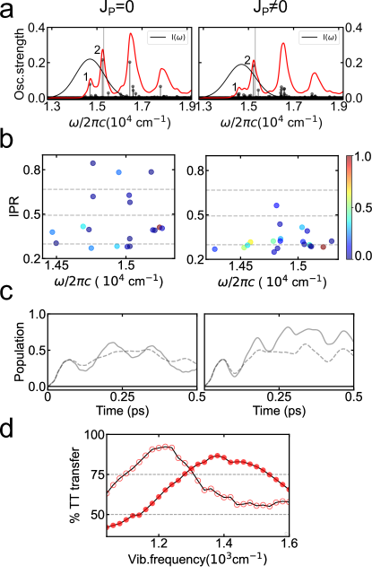
In the Fermi’s Golden rule rate limit, increased vibronic mixing channels (Figure 2) imply faster SEF rates with couplings through individual mixing channels adding18, 49 as . Increased delocalization of vibronic excitons in the presence of Peierls coupling from low-frequency vibrations also suggests enhanced population transfer to when a coherent superposition of bright excitons is impulsively excited. This is confirmed in Figure 3c which compares coherent population transfer to expected from the system Hamiltonian following an impulsive photoexcitation polarized along the exciton. The calculation compares the case of with versus without Peierls coupling with full details in Section S2. Including energetic disorder13 in site energies causes disorder in excitonic energy gaps leading to ensemble dephasing17 of electronic and vibronic quantum beats, with 1/e timescale17 of 100 fs. In case of no Peierls coupling (left panel), 66% of the initial excitation of optically bright excitons is transferred to the optically dark states within 300 fs. This is true even with ensemble averaging which suppresses purely electronic and vibronic quantum beats. The slower beats at 54 cm-1 correspond to vibronic splittings similar to those in Figure 1c, while the faster beats at 265 cm-1 correspond to intramolecular vibration. Corresponding Fourier transforms are shown in Figure S3. The high frequency vibrational beat i.e. 1360 cm-1 cannot be observed because excitations lie outside the laser bandwidth. In contrast, when Peierls coupling is introduced through the low-frequency vibration, the transfer is enhanced to 86%, in line with the theoretical expectation from Figure 2. Interestingly, ensemble dephasing strongly masks this enhancement with apparent transfer approximately similar to that seen without Peierls coupling in the low-frequency mode, even though on an individual system level Peierls coupling and vibronic near-resonance together enhance population transfer. In contrast to vibronically enhanced transfer, a purely electronic picture, with parameters derived from ab-initio models41, predicts only 32% of transfer when a coherent superposition of , and excitons is excited (Figure S2). When Peierls coupling is introduced without a high-frequency near-resonant vibration, population transfer no greater than that possible with purely electronic coupling is seen (Figure S2), underscoring the necessity of a vibronic near-resonance for Peierls coupling to be effective. Interestingly, the complementary effect of low-frequency modulations of intermolecular orbital overlaps in enhancing mixing, also implies enhanced indirect mixing. This is evident from Figure S4 and Table S2 with 2x increase in population transfer to upon selective excitation of the higher energy optically bright exciton.
The synergistic effect of low and high frequency vibrations on vibronic near-resonance is further confirmed by analyzing the effect of Peierls coupling on the width of vibronic resonance. Presence of low-frequency vibrational mode significantly complicates the interpretation of oscillator strengths that was possible with only high-frequency mode in the Hamiltonian (compare Figure 1b with oscillator strengths plotted in Figure 3a). Therefore, we use vibronically enhanced population transfer as a proxy for near-resonant intensity borrowing. Figure 3c plots the % transfer as a function of which, as in Figure 1b, is scanned while keeping unchanged. The width of near-resonant vibrations over which transfer to state remains above 75% increases by 1.24x, from 210 cm-1 to 260 cm-1. This increased width essentially implies that low-frequency modulations in intermolecular orbital overlaps in acene-based SEF systems make vibronic resonances, the width of which are already quite broad due to large HR factors (Figure 1b), highly robust to mismatches between vibrational frequencies and exciton energy gaps. This contrast between selective role of narrow vibronic resonances in photosynthetic excitons63, 15 versus non-selective and robust vibronic resonances expected in SEF is quite striking.
2.5 Distinguishing Vibronically Enhanced From Spectator Quantum Beats Using 2DES
Our findings suggest that strong mixing between excitons through vibrational motions may be expected in SEF upon photoexcitation. Such mixing of excitons can manifest as intensity borrowing effects in linear and non-linear optical spectra. Furthermore, when an impulsive photoexcitation creates a superposition of such vibronic eigenvectors, an amplitude enhancement of quantum beats on the excited and ground electronic state is expected15, 18, 19. Enhanced beating amplitude has been experimentally reported17 in the context of pump-probe and 2DES experiments on photosynthetic excitons. Such quantum beats can serve as a sensitive probe of the underlying mechanism of energy/charge transfer. For example, vibrational wavepackets corresponding to motions which drive internal conversion through a conical intersection64 do not survive on the excited state while orthogonal motions do. In a square symmetric molecule, Jonas and co-workers have shown that65, 66, 67 quantum beat amplitude of promoter vibrations is anisotropic to parallel versus perpendicular relative optical polarization of pump and probe electric fields. Polarization anisotropy of quantum beat amplitudes can therefore serve to distinguish promoter versus spectator vibrations.
In the current context of SEF, our analysis suggests that vibronic states formed by mixing of excitons polarized along different directions will carry highly mixed optical polarization signatures. Furthermore, mixed polarization signatures of vibronic eigenvectors will be caused by not just high-frequency vibrations (as they lead to vibronic near-resonances), but also due to low-frequency vibrations due to their vital complementary role in enhancing vibronic mixing between excitons. However, considering these predictions in the light that large HR factors in acenes will by themselves lead to strongly modulated vibrational quantum beats in 2DES or pump-probe spectra, such as for pentacene monomers in solution22, discriminating vibronically enhanced quantum beats against those arising from spectator vibrational motions becomes imperative in order to make sound judgements regarding the mechanistic details of SEF. Taking motivation from vibrational polarization anisotropy analysis65, 66, 67 of Jonas and co-workers, below we propose a novel polarization-based 2DES approach for unambiguous spectroscopic identification of promoter versus spectator vibrational motions in SEF, suggesting experiments which can readily test the above mechanistic predictions.
Pump-probe spectroscopy (PP) plots evolution of detection frequency of a system caused by pump excitation as a function of waiting time between pump and probe pulses. Akin to pump-probe, 2DES is also a four-wavemixing experiment. However, compared to PP, 2DES involves a pair of pump pulses, the delay between which is scanned in an interferometrically stable fashion to result in an additional Fourier transformed absorption frequency axis . The resulting data is plotted as a 2D contour map that correlates excitation and detection frequency of the system. 2D snapshots as a function of report on rich vibrational-electronic relaxation dynamics of a system. In case of pentacene thin films, prominent quantum beats with a few picosecond dephasing timescale along are reported13, 12, 68 in 2DES and PP studies, and contribute as positive ground state bleach (GSB) and negative excited state absorption (ESA) signals. The excited state emission (ESE) signal is reported69 to exhibit a sub-100 fs decay due to rapid internal conversion of photoexcited singlet states into correlated triplets. Accordingly, following the 2DES simulations of Tempelaar and Reichman8, our 2DES calculations only consider the GSB and ESA signal contributions. In a broader context, it is well known that excited state wavepackets can be generated by impulsive internal conversion26, can exhibit modified beating amplitudes26, 28, 70, rapidly dephase upon internal conversion, or simply undergo coherence transfer31 to the product without promoting any vibronic mixing. Owing to these complications and the availability of clean GSB signals in case of SEF, we focus our analysis on the GSB signals arising from ground state vibrational quantum beats. Only rephasing 2DES signal contributions, where 2DES pulse sequence rephases the optical dephasing that occurred during the first time interval, are considered. Due to simultaneous availability of superior time resolution and excitation frequency information in 2DES, the polarization-based spectroscopic signatures of promoter modes that we propose will be based on 2DES. Equivalent PP signatures are possible, although may be ambiguous due to lack of excitation frequency information and no separation of rephasing and non-rephasing pathways.
Time-resolved studies71 on pentacene thin films have reported weaker featureless ESA contributions on either side of the GSB signal. The negative ESA features correspond to optically allowed transitions polarized along the long axis8 of the molecule, compared to the short axis polarized singlet transitions. The relative oscillator strength and energy shift of the broad and featureless transitions are not precisely determined, with previous estimates8, 13 based on fitting simulations to experimental data. For instance, ESA oscillator strengths , relative to , have ranged from 2.52 in case of 2DES simulations reported in Rao et al.13 to significantly larger value of 152 in the 2DES simulations of Tempelaar et.al.8. The laser bandwidth used for 2D simulations corresponds to a 10 fs Gaussian pulse (field FWHM) with corresponding FWHM spectral range overlaid on the oscillator strengths in Figure 3a. Based on this laser bandwidth, our choice of ESA oscillator strengths of 3.62 and red-shift of 4803 cm-1 is such that the corresponding signal amplitudes in the simulated 2DES spectra are approximately consistent with the experimentally observed71 ratio of GSB signal to the ESA feature at 1.7 eV. With quanta along each vibrational mode on a given diabatic electronic state, the four-wavemixing response function simulations scale expensively with the 4th power of the basis set size as . This necessitates limitations on the number of eigenvectors that can be probed with desired convergence in the resulting non-linear signals. Our choice of the laser bandwidth (corresponding to a 10 fs pulse duration) is partly dictated by this criterion, such that only the lower energy eigenvectors in the manifold are probed with a convergence of 2DES CMs to within 2%. Further details of 2DES simulations and parameters are presented in Section S3. The convergence checks have been detailed in Section S5.
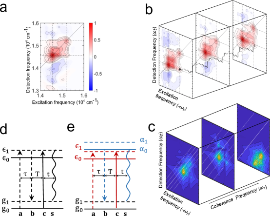
Figure 4a shows the simulated absorptive72 2DES spectrum at fs which reports changes in sample absorption created by the pump as a function of excitation () and detection () frequencies. Prominent GSB features with multiple diagonal peaks and associated cross-peaks can be seen. These arise from the two brightest excitons in the mixed manifold probed by the laser spectrum (marked as ‘1’ and ‘2’ in Figure 3a). The simulated 2D features are more defined (than those observed experimentally) due to intentional absence of inhomogeneity in the 0–1 optical energy gap in our calculations. Calculations of linear and 2DES spectra including this broadening are shown in Section S6 and do not affect the polarization based 2DES signatures of vibronic enhancement proposed here. As also reported13 experimentally by Rao et al., no 2D cross-peak between the bright manifold and is seen because of negligible bright character in . The broad ESA features along the detection axis arise from probe interactions to and from vibrationally excited manifold. Given the impulsive nature of excitation, coherent wavepackets, with purely vibrational or mixed vibronic origins, contribute to the 2DES data as quantum beats. All such ground or excited electronic state wavepackets interfere together and the resultant quantum dynamics can be monitored along the pump-probe waiting time as shown in Fig. 4b. The resultant oscillatory signal contributions can be resolved as 2DES CMs along the coherence axis by removing the incoherent population background and Fourier transformation along the waiting time . This is schematically described in Figure 4b,c with details of the analysis in Section S.5.2. CM at a given reports the 2D amplitude distribution of quantum beats of frequency .
Following ref.73, the third order response function expression that leads to a rephasing 2DES GSB signal contribution depends on transition dipole products as
.
Fields interact with individual systems to generate the transition dipole . The phase-matched sum of dipole-radiated electric fields (signal field) is detected by interference with a known electric field . denotes isotropic orientational average over the four electric field polarization vectors with respect to the molecular frame transition dipole vectors. The Greens’ function time propagator72 part has been suppressed for brevity. Using this notation, the 2DES GSB signal contribution that arises from ground state vibrational coherences in a two-electronic level system can be written as . The diagrammatic representation of this contribution as a wavemixing pathway is shown in Figure 5d. After two interactions, a ground state vibrational coherence between and is created which evolves with . The diagram is explained in detail in Section S4. Simplifying the transition dipole product for an all-parallel polarization sequence with electric field polarization in the lab frame, the coherent GSB contribution can be simplified as , where denotes the transition dipole vector followed by FC overlap factors. The position of this contribution lies at , that is, one vibrational quanta above the 2D diagonal along the excitation axis. This can also be deduced from the length of first and last interaction in the wavemixing diagram in Figure 5d.
When the near-resonant vibronic manifold is considered (Section 2.2), CMs are further complicated with the wavemixing pathways modified due to vibronic resonance as shown in Figure 5e. Considering vibronic mixing to dominantly arise from only within the degenerate manifold, the mixed eigenvectors can be approximated48 as . Wavemixing pathways arising from these eigenvectors lead to dipole products such as in the response function. All the pathways resulting from this dipole product contribute at the 2D cross-peak location between the negligible lower diagonal 2D peak (DPL, arising from direct excitation), and the bright upper diagonal 2D peak (DPU, arising from direct excitation). Expansion of the dipole product shows that even without any vibronic mixing from the bright exciton , a wavemixing pathway equivalent to Figure 5d will arise, however with a negligible contribution due to its dependence ( contains only 7% bright character, see Table S5). Similarly, dominant terms dependent on will also contribute at the same 2D cross peak location. However, it is important to recognize that such terms are not specific to vibronic mixing because they can arise even without any vibronic mixing. Jonas and co-workers have shown15 that non-adiabatic mixing due to vibronic resonance on the excited state leads to enhancement ground state vibrational quantum beats at the 2DES cross peak in case of photosynthetic excitons. Such beats are otherwise weak because small HR factors in photosynthetic pigments render wavemixing pathways such as those in Figure 4d very weak on account of poor FC overlaps. The same is not true in the SEF context however. Large HR factors in acenes imply that dominant beating amplitude at the cross peak can readily arise from wavemixing pathways with non-specific dipole products such as in Figure 4e. Wavemixing pathways starting from vibrationally hot ground state, allowed in the simulations, further complicate the interpretation of quantum beating signals. Thus, any inferences regarding vibronic enhancement of SEF cannot be made on the basis of readily detectable quantum beat amplitude at the 2DES cross-peak location.
A distinction between vibronic mixing dependent pathways can be motivated by considering the dipole product cross-terms in the wavemixing pathway in Figure 5e. Cross-terms such as only arise in the presence of vibronic mixing. To motivate a polarization-based signature of quantum beats arising due to vibronic mixing akin to vibrational polarization anisotropy65, 66, we will first denote dipole products such as in a compact notation as, for an all-parallel electric field polarization in the lab frame. Alternatively, considering a perpendicular pump-probe polarization sequence, the dipole product will be denoted as . The other non-specific dipole product is denoted as . The isotropic orientational averages for the parallel (, ) and perpendicular (, ) sequences are related73 by a factor of 1/3, implying that a 2DES CM evaluated at a mixed polarization sequence will cancel out the non-specific ground state quantum beat pathways because they are isotropic. In contrast, the vibronic mixing specific cross-terms such as in Figure 5e are anisotropic and survive the sequence. These will only arise in the presence of vibronic mixing without any masking from non-specific vibronic mixing contributions. The wavemixing diagram for the most dominant surviving pathway is shown in Figure 5e where interactions utilizing and transition dipole vectors are shown in different color to denote their different directions. All other surviving pathways are analyzed in Section S4.
The above polarization sequence to isolate vibronic mixing specific terms assumes a large laser focal spot, typical of conventional 2DES approaches74, to sample an orientationally averaged response of the thin film. Below we apply this idea to the model SEF Hamiltonian to spectroscopically confirm the synergistic role of low- and high-frequency vibrations in vibronic enhancement of SEF. In order to isolate the GSB wavemixing pathways in Fig. 4d which may arise due to vibronic enhancement, the laser spectrum should cover the lowest energy triplet exciton as well as the low-energy bright excitons ( and in the pentacene dimer). This can be understood by considering the vertical lengths of interactions in Fig. 4d. The laser spectrum in Fig. 3a is chosen accordingly. Such an experiment can be readily implemented to test these predictions and identify which, if any, vibrational modes promote vibronically enhanced SEF with associated enhancement of vibrational quantum beats monitored through spectroscopy.
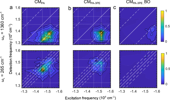
Figure 5 shows 2DES CMs at and . Figure 5a shows 2DES CMs for the Hamiltonian in Equation 1 where both and intramolecular vibrations are included explicitly in the system Hamiltonian and therefore allowed to participate in vibronic mixing. The map is generated by an all-parallel polarization sequence and therefore includes wavemixing pathways which are both specific as well as non-specific to vibronic mixing as explained in Figure 5d and Figure 5e. The beating amplitude appears at the 2D cross-peak location between and diagonal peaks, even though the former is not seen13 in the 2DES map in Figure 5a. This is so because a dipole product dependence renders it negligibly weak. The CMs do however show the oscillatory cross-peak contributions as being quite prominent. The beating amplitude appears at expected location of approximately one vibrational quanta below the diagonal along the detection axis. Similar features have been experimentally reported13 and interpreted as signatures of vibronic mixing. However, as explained earlier in this section, it would be misleading to interpret such quantum beats as vibronically enhanced solely based on their 2D location and prominent amplitude because of dominant non-specific vibronic mixing contributions, such as , at the same 2D cross peak location. For the same reason, the CMs for and cannot confirm the prediction in Section 2.3 that Peierls coupling by itself, without vibronic near-resonance through the high-frequency vibration, is ineffective in promoting vibronic mixing (see Fig.2). 2DES signals arising from vibrationally hot ground states further complicate the CMs.
Next we apply the polarization sequence to eliminate non-specific isotropic quantum beat contributions. Figure 5b plots the resulting CMs for both and . As expected for , cross dipole product contributions such as and survive the sequence to result in the CMs in panel b. Interestingly, as also expected, CM contributions also survive because their complementary role in near-resonant vibronic mixing leads to mixed polarization signatures for quantum beats as well. To further confirm the synergistic effect of low-frequency vibrations on vibronic near-resonance, we remove the mode from the system Hamiltonian and include it in the Brownian oscillator bath while keeping its total stabilization energy same as before. The low-frequency FC active vibration as well as its Peierls coupling is still treated explicitly in the system Hamiltonian as before. The resulting CMs are shown in panel c. Since is not treated explicitly in the system, it can no longer participate in non-adiabatic vibronic mixing. Consequently, as may be expected, its CM does not survive the sequence. Additionally, the CM also does not survive the sequence confirming the complementary enhancement of vibronic mixing through low-frequency modulations of intermolecular orbital overlaps when a vibronic near-resonance due to high-frequency vibrations is expected.
It can be easily shown that experimentally one does not need to perform two different experiments, one each with and sequence. Rather a single experiment with polarization sequence leads to a scaled signal. Such pulse polarization sequences were originally devised75 by Zanni and Hochstrasser in the context of eliminating 2D diagonal peaks in 2DIR spectra. More complex polarization schemes specific to vibronic mixing have also been implemented76. However the sequence is fairly easy to implement compared to other schemes. Zanni and co-workers have recently77 extended this sequence to incorporate rotational diffusion effects and isolate 2D cross-peak in a pump-probe experiment by eliminating diagonal peak contributions. Here we have extended this concept in the context of polarization dependence of vibronic quantum beats to isolate anisotropic quantum beat wavemixing pathways that are specific to vibronic mixing in SEF.
3 Conclusions
Resonances between electronic energy gaps in photosynthetic proteins and intramolecular vibrational frequencies of the pigments leads to a non-adiabatic energy funnel17 driven by anti-phase vibrational motions between the two pigments. Such resonances are narrow and therefore not robust to energetic mismatches. In-phase motions between the two molecules are mere spectators35, 36, 15 in this process. Similar effects have been reported13, 9, 10 in case of SEF. We have introduced fresh, and previously overlooked, mechanistic insights into this emerging understanding of the rapid formation of state. We show that, quite counterintuitively, in-phase vibrational motions between the two pentacene molecules drive non-adiabatic vibronic mixing between the bright exciton and the optically dark manifold, while anti-phase vibrational motions mix the optically bright excitons together, as in an excitonic dimer. Together these vibrational motions lead to a coupled vibronic resonances between different pairs of electronic states, driven by a single high-frequency vibration on each molecule. Significantly, this occurs over a broad non-selective range of vibrational frequencies and is in striking contrast to that in photosynthetic excitons.
Modulations of intermolecular orbital overlaps in pentacene thin films are expected from known20, 21 low-frequency vibrations with mixed inter/intramolecular character. Significantly, introducing such a vibration in the above picture leads to an overall synergistic effect of low- and high-frequency vibrations in SEF. The existing vibronic near-resonances become more robust to energetic mismatches with increased vibronic exciton delocalization, and consequently lead to increased formation of upon impulsive photoexcitation of bright electronic states. Treatment of low-frequency vibrations beyond a ‘frozen’ mode9 leads to these interesting effects and resolves conflicting9, 60 and often exclusive12 roles of vibronic resonance and Peierls’ coupling in enhancing SEF.
Quantum beats in 2DES often carry vital mechanistic insights17 into energy and charge transfer. In SEF, prominent vibrational quantum beats are already expected in pentacene monomers because of large HR factors. We argue that readily observed13, 12 quantum beat amplitudes at 2DES at cross-peak locations therefore cannot unambiguously identify vibronic mixing effects in SEF. Instead, by analyzing the polarization anisotropy of vibrational quantum beats in the clean ground state signals available in SEF, we propose and demonstrate polarization-based 2DES signatures that survive only in the presence of excited state vibronic mixing. Such a polarization scheme is readily implementable, can distinguish vibrational quantum beats that arise from vibronic mixing and promote SEF against those which merely accompany the process as spectator modes, and makes the predictions of our model amenable to experimental scrutiny.
Multiple electronic states, explicit quantum treatment of multiple vibrational modes, and no approximation13 of ground state vibrations in vibronic resonance are together vital in order to capture the previously unrecognized complementary roles of vibronic coupling from low- and high-frequency vibrations, as well as of in-phase and anti-phase vibrational motions. Even though our calculations, with experimentally relevant parameters, are based on a pentacene dimer and do not capture the thermodynamic enhancement9 of SEF rates possible in a crystal, the physical insights about robust, non-selective and coupled vibronic resonances may have significant implications even for a pentacene thin film. For example, coupled resonances, but along a high- and low-frequency mode, seem likely to occur within a Davydov component given its 1200 cm-1 width44. Similarly, experimentally known energetic separation of cm-1 between upper and lower Davydov components in pentacene thin films44 is of the order of a high-frequency vibrational excitation. Ultrafast decay of electronic polarization anisotropy78 in pentacene nanoparticles on the timescale of formation is also suggestive of highly mixed vibronic states at play. Physical insights gained from our model are more general in nature and may be applicable to intramolecular SEF systems as well. Our analysis motivates synthetic design principles that go beyond tuning electronic couplings79 to incorporate high-frequency intramolecular vibrations with large Huang-Rhys factors, exciton energy gaps of the order of high frequency vibrations, and low-frequency vibrations capable of modulating intermolecular orbital overlaps.
References
- Smith and Michl 2013 Smith, M. B.; Michl, J. Recent Advances in Singlet Fission. Annual Review of Physical Chemistry 2013, 64, 361–386
- Zimmerman et al. 2011 Zimmerman, P. M.; Bell, F.; Casanova, D.; Head-Gordon, M. Mechanism for Singlet Fission in Pentacene and Tetracene: From Single Exciton to Two Triplets. Journal of the American Chemical Society 2011, 133, 19944–19952
- Zeng et al. 2014 Zeng, T.; Hoffmann, R.; Ananth, N. The Low-Lying Electronic States of Pentacene and Their Roles in Singlet Fission. Journal of the American Chemical Society 2014, 136, 5755–5764
- Feng et al. 2013 Feng, X.; Luzanov, A. V.; Krylov, A. I. Fission of Entangled Spins: An Electronic Structure Perspective. The Journal of Physical Chemistry Letters 2013, 4, 3845–3852
- Phys et al. 2014 Phys, J. C.; Berkelbach, T. C.; Hybertsen, M. S.; Reichman, D. R. Microscopic theory of singlet exciton fission . II . Application to pentacene dimers and the role of superexchange. 2014, 114103
- Jones et al. 2020 Jones, A. C.; Kearns, N. M.; Ho, J.-J.; Flach, J. T.; Zanni, M. T. Impact of non-equilibrium molecular packings on singlet fission in microcrystals observed using 2D white-light microscopy. Nature Chemistry 2020, 12, 40–47
- Tempelaar and Reichman 2017 Tempelaar, R.; Reichman, D. R. Vibronic exciton theory of singlet fission. I. Linear absorption and the anatomy of the correlated triplet pair state. The Journal of Chemical Physics 2017, 146, 174703
- Tempelaar and Reichman 2017 Tempelaar, R.; Reichman, D. R. Vibronic exciton theory of singlet fission. II. Two-dimensional spectroscopic detection of the correlated triplet pair state. The Journal of Chemical Physics 2017, 146, 174704
- Tempelaar and Reichman 2018 Tempelaar, R.; Reichman, D. R. Vibronic exciton theory of singlet fission. III. How vibronic coupling and thermodynamics promote rapid triplet generation in pentacene crystals. The Journal of Chemical Physics 2018, 148, 244701
- Unger et al. 2022 Unger, F.; Moretti, L.; Hausch, J.; Bredehoeft, J.; Zeiser, C.; Haug, S.; Tempelaar, R.; Hestand, N. J.; Cerullo, G.; Broch, K. Modulating Singlet Fission by Scanning through Vibronic Resonances in Pentacene-Based Blends. Journal of the American Chemical Society 2022, 144, 20610–20619
- Morrison and Herbert 2017 Morrison, A. F.; Herbert, J. M. Evidence for Singlet Fission Driven by Vibronic Coherence in Crystalline Tetracene. The Journal of Physical Chemistry Letters 2017, 8, 1442–1448, PMID: 28277682
- Duan et al. 2020 Duan, H.-G.; Jha, A.; Li, X.; Tiwari, V.; Ye, H.; Nayak, P. K.; Zhu, X.-L.; Li, Z.; Martinez, T. J.; Thorwart, M.; Miller, R. J. D. Intermolecular vibrations mediate ultrafast singlet fission. Science Advances 2020, 6
- Bakulin et al. 2016 Bakulin, A. A.; Morgan, S. E.; Kehoe, T. B.; Wilson, M. W. B.; Chin, A. W.; Zigmantas, D.; Egorova, D.; Rao, A. Real-time observation of multiexcitonic states in ultrafast singlet fission using coherent 2D electronic spectroscopy. Nature Chemistry 2016, 8, 16–23
- Womick and Moran 2011 Womick, J. M.; Moran, A. M. Vibronic Enhancement of Exciton Sizes and Energy Transport in Photosynthetic Complexes. J. Phys. Chem. B 2011, 115, 1347–1356
- Tiwari et al. 2013 Tiwari, V.; Peters, W. K.; Jonas, D. M. Electronic resonance with anticorrelated pigment vibrations drives photosynthetic energy transfer outside the adiabatic framework. Proceedings of the National Academy of Sciences 2013, 110, 1203–1208
- Peters et al. 2017 Peters, W. K.; Tiwari, V.; Jonas, D. M. Nodeless vibrational amplitudes and quantum nonadiabatic dynamics in the nested funnel for a pseudo Jahn-Teller molecule or homodimer. The Journal of Chemical Physics 2017, 147, 194306
- Jonas 2018 Jonas, D. M. Vibrational and Nonadiabatic Coherence in 2D Electronic Spectroscopy, the Jahn–Teller Effect, and Energy Transfer. Annual Review of Physical Chemistry 2018, 69, 327–352
- Tiwari et al. 2017 Tiwari, V.; Peters, W. K.; Jonas, D. M. Electronic energy transfer through non-adiabatic vibrational-electronic resonance. I. Theory for a dimer. The Journal of Chemical Physics 2017, 147, 154308
- Tiwari and Jonas 2018 Tiwari, V.; Jonas, D. M. Electronic energy transfer through non-adiabatic vibrational-electronic resonance. II. 1D spectra for a dimer. The Journal of Chemical Physics 2018, 148, 84308
- Brillante et al. 2002 Brillante, A.; Della Valle, R. G.; Farina, L.; Girlando, A.; Masino, M.; Venuti, E. Raman phonon spectra of pentacene polymorphs. Chemical Physics Letters 2002, 357, 32–36
- Della Valle et al. 2004 Della Valle, R. G.; Venuti, E.; Farina, L.; Brillante, A.; Masino, M.; Girlando, A. Intramolecular and Low-Frequency Intermolecular Vibrations of Pentacene Polymorphs as a Function of Temperature. The Journal of Physical Chemistry B 2004, 108, 1822–1826
- Le et al. 2021 Le, D. V.; de la Perrelle, J. M.; Do, T. N.; Leng, X.; Tapping, P. C.; Scholes, G. D.; Kee, T. W.; Tan, H.-S. Characterization of the ultrafast spectral diffusion and vibronic coherence of TIPS-pentacene using 2D electronic spectroscopy. The Journal of Chemical Physics 2021, 155, 14302
- Zhu et al. 1994 Zhu, L.; Sage, J. T.; Champion, P. M. Observation of Coherent Reaction Dynamics in Heme Proteins. Science 1994, 266, 629–632
- Wynne et al. 1996 Wynne, K.; Reid, G. D.; Hochstrasser, R. M. Vibrational coherence in electron transfer: The tetracyanoethylene–pyrene complex. The Journal of Chemical Physics 1996, 105, 2287–2297
- Wolfseder et al. 1998 Wolfseder, B.; Seidner, L.; Domcke, W.; Stock, G.; Seel, M.; Engleitner, S.; Zinth, W. Vibrational coherence in ultrafast electron-transfer dynamics of oxazine 1 in N,N-dimethylaniline: simulation of a femtosecond pump-probe experiment. Chemical Physics 1998, 233, 323–334
- Kim et al. 2012 Kim, S. Y.; Kim, C. H.; Park, M.; Ko, K. C.; Lee, J. Y.; Joo, T. Coherent Nuclear Wave Packets Generated by Ultrafast Intramolecular Charge-Transfer Reaction. The Journal of Physical Chemistry Letters 2012, 3, 2761–2766
- Rafiq et al. 2021 Rafiq, S.; Fu, B.; Kudisch, B.; Scholes, G. D. Interplay of vibrational wavepackets during an ultrafast electron transfer reaction. Nature Chemistry 2021, 13, 70–76
- Andrzejak et al. 2019 Andrzejak, M.; Skóra, T.; Petelenz, P. Is Vibrational Coherence a Byproduct of Singlet Exciton Fission? The Journal of Physical Chemistry C 2019, 123, 91–101
- Andrzejak et al. 2020 Andrzejak, M.; Skóra, T.; Petelenz, P. Limitations of Generic Chromophore Concept for Femtosecond Vibrational Coherences. The Journal of Physical Chemistry C 2020, 124, 3529–3535
- Stern et al. 2017 Stern, H. L.; Cheminal, A.; Yost, S. R.; Broch, K.; Bayliss, S. L.; Chen, K.; Tabachnyk, M.; Thorley, K.; Greenham, N.; Hodgkiss, J. M.; Anthony, J.; Head-Gordon, M.; Musser, A. J.; Rao, A.; Friend, R. H. Vibronically coherent ultrafast triplet-pair formation and subsequent thermally activated dissociation control efficient endothermic singlet fission. Nature Chemistry 2017, 9, 1205
- Jean and Fleming 1995 Jean, J. M.; Fleming, G. R. Competition between energy and phase relaxation in electronic curve crossing processes. The Journal of Chemical Physics 1995, 103, 2092–2101
- Paulus et al. 2020 Paulus, B. C.; Adelman, S. L.; Jamula, L.; McCusker, J. Leveraging excited-state coherence for synthetic control of ultrafast dynamics. Nature 2020, 582, 214–218
- Delor et al. 2014 Delor, M.; Scattergood, P. A.; Sazanovich, I. V.; Parker, A. W.; Greetham, G. M.; Meijer, A. J. H. M.; Towrie, M.; Weinstein, J. A. Toward control of electron transfer in donor-acceptor molecules by bond-specific infrared excitation. Science 2014, 346, 1492–1495
- Scholes et al. 2017 Scholes, G. D. et al. Using coherence to enhance function in chemical and biophysical systems. Nature 2017, 543, 647–656
- Witkowski and Moffitt 1960 Witkowski, A.; Moffitt, W. Electronic Spectra of Dimers: Derivation of the Fundamental Vibronic Equation. The Journal of Chemical Physics 1960, 33, 872–875
- Fulton and Gouterman 1961 Fulton, R. L.; Gouterman, M. Vibronic Coupling. I. Mathematical Treatment for Two Electronic States. The Journal of Chemical Physics 1961, 35, 1059–1071
- 37 Förster, T. Modern Quantum Chemistry, edited by O. Sinanoglu{u}; Academic Press Inc., New York. (1996), p 93
- Berkelbach et al. 2014 Berkelbach, T. C.; Hybertsen, M. S.; Reichman, D. R. Microscopic theory of singlet exciton fission. III. Crystalline pentacene. The Journal of Chemical Physics 2014, 141, 74705
- Phys et al. 2020 Phys, J. C.; Accomasso, D.; Granucci, G.; Wibowo, M. Delocalization effects in singlet fission : Comparing models with two and three interacting molecules Delocalization effects in singlet fission : Comparing models with two and three interacting molecules. 2020, 244125
- Nagami et al. 2021 Nagami, T.; Miyamoto, H.; Sakai, R.; Nakano, M. Stabilization of Charge-Transfer States in Pentacene Crystals and Its Role in Singlet Fission. The Journal of Physical Chemistry C 2021, 125, 2264–2275
- Phys et al. 2020 Phys, J. C.; Li, X.; Parrish, R. M.; Martínez, T. J.; Li, X. An ab initio exciton model for singlet fission An ab initio exciton model for singlet fission. 2020, 184116
- Mirjani et al. 2014 Mirjani, F.; Renaud, N.; Gorczak, N.; Grozema, F. C. Theoretical Investigation of Singlet Fission in Molecular Dimers: The Role of Charge Transfer States and Quantum Interference. 2014,
- Berkelbach et al. 2013 Berkelbach, T. C.; Hybertsen, M. S.; Reichman, D. R.; Berkelbach, T. C.; Hybertsen, M. S.; Reichman, D. R. Microscopic theory of singlet exciton fission . I . General formulation Microscopic theory of singlet exciton fission . I . General formulation. 2013, 114102
- Hestand et al. 2015 Hestand, N. J.; Yamagata, H.; Xu, B.; Sun, D.; Zhong, Y.; Harutyunyan, A. R.; Chen, G.; Dai, H.-l.; Rao, Y.; Spano, F. C. Polarized Absorption in Crystalline Pentacene : Theory vs Experiment. J. Chem. Phys. 2015, 119, 22137–22147
- Coropceanu et al. 2007 Coropceanu, V.; Demetrio, A.; Filho, S.; Olivier, Y.; Silbey, R.; Bre, J.-l. Charge Transport in Organic Semiconductors. 2007, 926–952
- Philpott 1969 Philpott, M. R. Theory of the Vibrational Structure of Molecular Excitons. Soluble ”One-Phonon” Models. The Journal of Chemical Physics 1969, 51, 2616–2624
- Roden et al. 2008 Roden, J.; Eisfeld, A.; Briggs, J. S. The J- and H-bands of dye aggregate spectra: Analysis of the coherent exciton scattering (CES) approximation. Chemical Physics 2008, 352, 258–266
- Sahu et al. 2020 Sahu, A.; Kurian, J. S.; Tiwari, V. Vibronic resonance is inadequately described by one-particle basis sets. The Journal of Chemical Physics 2020, 153, 224114
- Patra and Tiwari 2022 Patra, S.; Tiwari, V. Vibronic resonance along effective modes mediates selective energy transfer in excitonically coupled aggregates. The Journal of Chemical Physics 2022, 156, 184115
- Bašinskaitė et al. 2014 Bašinskaitė, E.; Butkus, V.; Abramavicius, D.; Valkunas, L. Vibronic models for nonlinear spectroscopy simulations. Photosynthesis Research 2014, 121, 95–106
- Filippini and Gramaccioli 1984 Filippini, G.; Gramaccioli, C. M. Lattice-dynamical calculations for tetracene and pentacene. Chemical Physics Letters 1984, 104, 50–53
- Troisi and Orlandi 2006 Troisi, A.; Orlandi, G. Dynamics of the Intermolecular Transfer Integral in Crystalline Organic Semiconductors. The Journal of Physical Chemistry A 2006, 110, 4065–4070
- Dostál et al. 2014 Dostál, J.; Mančal, T.; Vácha, F.; Pšenčík, J.; Zigmantas, D. Unraveling the nature of coherent beatings in chlorosomes. The Journal of Chemical Physics 2014, 140, 115103
- Seibt and Mančal 2018 Seibt, J.; Mančal, T. Treatment of Herzberg-Teller and non-Condon effects in optical spectra with Hierarchical Equations of Motion. Chemical Physics 2018, 515, 129–140
- Ito et al. 2015 Ito, S.; Nagami, T.; Nakano, M. Density Analysis of Intra- and Intermolecular Vibronic Couplings toward Bath Engineering for Singlet Fission. The Journal of Physical Chemistry Letters 2015, 6, 4972–4977
- Cao et al. 2020 Cao, J. et al. Quantum biology revisited. Science Advances 2020, 6
- Hestand and Spano 2018 Hestand, N. J.; Spano, F. C. Expanded Theory of H- and J-Molecular Aggregates: The Effects of Vibronic Coupling and Intermolecular Charge Transfer. Chemical Reviews 2018, 118, 7069–7163
- Sun et al. 2021 Sun, K.; Liu, X.; Hu, W.; Zhang, M.; Long, G.; Zhao, Y. Singlet fission dynamics and optical spectra of pentacene and its derivatives. Phys. Chem. Chem. Phys. 2021, 23, 12654–12667
- Hart et al. 2018 Hart, S. M.; Silva, W. R.; Frontiera, R. R. Femtosecond stimulated Raman evidence for charge-transfer character in pentacene singlet fission. Chem. Sci. 2018, 9, 1242–1250
- Castellanos and Huo 2017 Castellanos, M. A.; Huo, P. Enhancing Singlet Fission Dynamics by Suppressing Destructive Interference between Charge-Transfer Pathways. The Journal of Physical Chemistry Letters 2017, 8, 2480–2488
- Montoya-Castillo et al. 2015 Montoya-Castillo, A.; Berkelbach, T. C.; Reichman, D. R. Extending the applicability of Redfield theories into highly non-Markovian regimes. The Journal of Chemical Physics 2015, 143, 194108
- Mukamel 1990 Mukamel, S. Femtosecond Optical Spectroscopy: A Direct Look at Elementary Chemical Events. Annual Review of Physical Chemistry 1990, 41, 647–681
- Higgins et al. 2021 Higgins, J. S.; Lloyd, L. T.; Sohail, S. H.; Allodi, M. A.; Otto, J. P.; Saer, R. G.; Wood, R. E.; Massey, S. C.; Ting, P.-C.; Blankenship, R. E.; Engel, G. S. Photosynthesis tunes quantum-mechanical mixing of electronic and vibrational states to steer exciton energy transfer. Proceedings of the National Academy of Sciences 2021, 118, e2018240118
- Yarkony 1996 Yarkony, D. R. Diabolical conical intersections. Reviews of Modern Physics 1996, 68, 985–1013
- Farrow et al. 2008 Farrow, D. A.; Qian, W.; Smith, E. R.; Ferro, A. A.; Jonas, D. M. Polarized pump-probe measurements of electronic motion via a conical intersection. The Journal of Chemical Physics 2008, 128, 144510
- Farrow et al. 2008 Farrow, D. A.; Smith, E. R.; Qian, W.; Jonas, D. M. The polarization anisotropy of vibrational quantum beats in resonant pump- probe experiments : Diagrammatic calculations for square symmetric molecules The polarization anisotropy of vibrational quantum beats in resonant pump-probe experiments : Diagramma. 2008, 174509
- Kitney-Hayes et al. 2014 Kitney-Hayes, K. A.; Ferro, A. A.; Tiwari, V.; Jonas, D. M. Two-dimensional Fourier transform electronic spectroscopy at a conical intersection. The Journal of Chemical Physics 2014, 140, 124312
- Musser et al. 2015 Musser, A. J.; Liebel, M.; Schnedermann, C.; Wende, T.; Kehoe, T. B.; Rao, A.; Kukura, P. Evidence for conical intersection dynamics mediating ultrafast singlet exciton fission. Nature Physics 2015, 11, 352
- Wilson et al. 2011 Wilson, M. W. B.; Rao, A.; Clark, J.; Kumar, R. S. S.; Brida, D.; Cerullo, G.; Friend, R. H. Ultrafast Dynamics of Exciton Fission in Polycrystalline Pentacene. 2011, 11830–11833
- Kim et al. 2020 Kim, J.; Kim, C. H.; Burger, C.; Park, M.; Kling, M. F.; Kim, D. E.; Joo, T. Non-Born–Oppenheimer Molecular Dynamics Observed by Coherent Nuclear Wave Packets. The Journal of Physical Chemistry Letters 2020, 11, 755–761
- Thorsmølle, X. et al. 2009 Thorsmølle, X., V. K.; Averitt, R. D.; Demsar, J.; Smith, D. L.; Tretiak, S.; Martin, R. L.; Chi, X.; Crone, B. K.; Ramirez, A. P.; Taylor, A. J. Morphology Effectively Controls Singlet-Triplet Exciton Relaxation and Charge Transport in Organic Semiconductors. Phys. Rev. Lett. 2009, 102, 17401
- Jonas 2003 Jonas, D. M. Two-Dimensional Femtosecond Spectroscopy. Annu. Rev. Phys. Chem. 2003, 54, 425–463
- Qian and Jonas 2003 Qian, W.; Jonas, D. M. Role of cyclic sets of transition dipoles in the pump–probe polarization anisotropy: Application to square symmetric molecules and perpendicular chromophore pairs. The Journal of Chemical Physics 2003, 119, 1611–1622
- Fuller and Ogilvie 2015 Fuller, F. D.; Ogilvie, J. P. Experimental Implementations of Two-Dimensional Fourier Transform Electronic Spectroscopy. Annual Review of Physical Chemistry 2015, 66, 667–690
- Zanni et al. 2001 Zanni, M. T.; Ge, N.-H.; Kim, Y. S.; Hochstrasser, R. M. Two-dimensional IR spectroscopy can be designed to eliminate the diagonal peaks and expose only the crosspeaks needed for structure determination. Proceedings of the National Academy of Sciences 2001, 98, 11265–11270
- Thyrhaug et al. 2018 Thyrhaug, E.; Tempelaar, R.; Alcocer, M. J. P.; Žídek, K.; Bína, D.; Knoester, J.; Jansen, T. L. C.; Zigmantas, D. Identification and characterization of diverse coherences in the Fenna–Matthews–Olson complex. Nature Chemistry 2018, 10, 780–786
- Farrell et al. 2022 Farrell, K. M.; Yang, N.; Zanni, M. T. A polarization scheme that resolves cross-peaks with transient absorption and eliminates diagonal peaks in 2D spectroscopy. Proceedings of the National Academy of Sciences 2022, 119, e2117398119
- Pensack et al. 2015 Pensack, R. D.; Tilley, A. J.; Parkin, S. R.; Lee, T. S.; Payne, M. M.; Gao, D.; Jahnke, A. A.; Oblinsky, D. G.; Li, P.-F.; Anthony, J. E.; Seferos, D. S.; Scholes, G. D. Exciton Delocalization Drives Rapid Singlet Fission in Nanoparticles of Acene Derivatives. Journal of the American Chemical Society 2015, 137, 6790–6803, PMID: 25946670
- Krishnapriya et al. 2019 Krishnapriya, K. C.; Musser, A. J.; Patil, S. Molecular Design Strategies for Efficient Intramolecular Singlet Exciton Fission. ACS Energy Letters 2019, 4, 192–202