Photo-induced charge-transfer renormalization in NiO
Abstract
Photo-doped states in strongly correlated charge transfer insulators are characterized by - and - interactions and the resulting intertwined dynamics of charge excitations and local multiplets. Here we use femtosecond x-ray absorption spectroscopy in combination with dynamical mean-field theory to disentangle these contributions in NiO. Upon resonant optical excitation across the charge transfer gap, the Ni and O absorption edges red-shift for ps, which is explained by a simultaneous Hartree shift and a renormalization of the local interactions. Furthermore, below the Ni edge an additional signature is identified for ps, which reflects a transient nonthermal population of local many-body multiplets. Overall, the photo-doped state differs significantly from a chemically doped state. Our results demonstrate the ability to reveal excitation pathways in correlated materials by x-ray spectroscopies, which is relevant for ultrafast materials design.
Strongly correlated materials host some of the most intriguing states of matter due to the competition between interaction-induced localization and the itinerant nature of electrons, and they are therefore ideal candidates to realize material control on ultrafast timescales [1, 2]. Paradigmatic examples are Mott and charge-transfer (CT) insulators, whose optical properties are determined by the charge transfer between the ligand (typically ) and the correlated orbital (typically ) states [3, 4, 5]. Element- and site-selective information on many-body states in such materials can be obtained by resonant soft x-ray absorption and emission spectroscopies [6]. A comparison of core level absorption edges and their fine structure with cluster calculations [7] or dynamical mean-field theory (DMFT) [8, 9] can provide detailed information about the CT gap, Coulomb repulsion [7, 10], - multiplet excitations, and the hybridization between the ligand and correlated orbitals [11, 12]. Furthermore, ligand absorption edges probe the itinerant states [11], whose nature is crucial for understanding the low-energy physics in chemically doped systems [13, 14, 15].
Femtosecond time-resolved x-ray spectroscopy is sensitive to low energy excitations and transient energy shifts [16, 17, 18, 19, 20, 21]. It is, thus, a potentially powerful tool for investigating photo-excited non-equilibrium states in Mott and CT insulators. Recently, a strong sub-gap excitation has been shown to modify the gap size during the pulse both in cuprate [22] and nickelate [23, 24] CT insulators. These effects can be attributed to photo-manipulations of the screening environment [22], the magnetic order [23], or the hybridization between correlated and itinerant orbitals [24], coined dynamical Franz-Keldysh effect [25]. A natural open question is how this picture changes in the case of resonant excitations, which create long-lived charge carriers in the conduction band, and lead to so-called photo-doped states which may host various nontrivial quantum phases [26, 27, 28, 29]. To understand these states, and to potentially design targeted excitation pathways, it is important to clarify the different nature of the charge carriers in photo-doped and chemically doped states, and to disentangle effects such as band shifts due to dynamical screening [30, 31] and charge redistribution between orbitals [32, 33, 30, 34] from the dynamics of Mott excitons or electronic - excitations [35, 36, 37, 38].
In this Letter, we demonstrate how time-resolved x-ray absorption spectroscopy (XAS) in combination with non-equilibrium dynamical mean-field theory (DMFT) [40, 41] can help to achieve this goal for the paradigmatic CT insulator NiO. We identify long-lived energy shifts and lineshape modifications in the excitonic peaks at the Ni and O absorption edges, and link them to orbital occupations and the screening environment. The observed spectral changes at the O edge reveal differences in the nature of photo-doped and chemically doped holes. In addition, we resolve a short-lived Ni pre-edge feature that represents many-body multiplets, i.e. Hund’s excitations. Since these phenomena are representative of generic charge transfer insulators, our results show how time-resolved XAS can reveal relevant information on the excitation pathways in this material family.
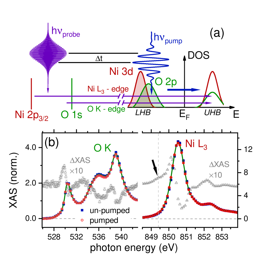
Figure 1(a) shows a sketch of time-resolved XAS at the Ni () and O () edges. The experiments were performed at room temperature at the Spectroscopy and Coherent Scattering (SCS) instrument of European XFEL using a pump-probe setup with an effective time resolution of 80 fs [42, 21, 43]. Laser pulses with 4.7 eV photon energy, 35 fs pulse duration, and 0.8-4 mJ/cm2 fluence were employed to pump NiO above the CT gap. This pumping involves excitations from the O states to the upper Hubbard band (UHB), see Fig. 1(a) and Ref. [39]. Figure 1(b) depicts the O and Ni XAS signals before and after photoexcitation at a pump-probe delay of ps, with the pump-induced difference XAS shown by the gray dots. We find a spectral shift to lower X-ray photon energies, as indicated by the derivative-like shape of XAS. We also measured the time dependence at fixed photon energies at both edges, see Fig. 2(a). Exponential fits to these transients, convoluted with 80 fs time resolution [21], indicate a rise time of fs for 0.8 mJ/cm2 pump fluence at the Ni edge and fs at the O edge. These energy shifts build up, reach a plateau at both edges within less than ps, and decay on longer timescales.
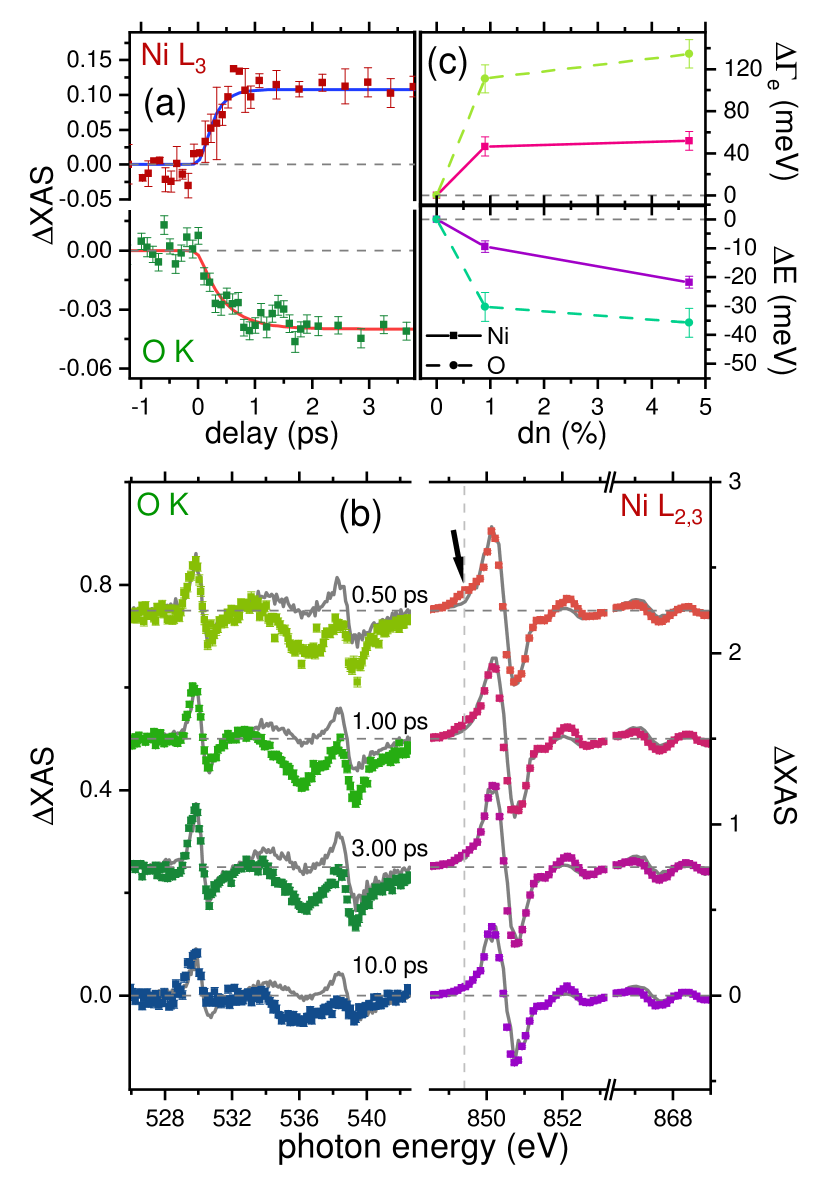
In a first analysis we compare the spectra of the photo-excited system to curves modelled by a red-shift and a broadening of the static spectra, c.f. Fig. 2(b). In Fig. 2(c) we plot the best fit and at ps as a function of the excitation density , i.e., the density of pump-excited electrons relative to all valence electrons (see [39] for the determination of from the pump fluence). The changes in the broadening (or, since , the changes in the lifetime) are larger for the itinerant than for the electrons [21, 39]. While some parts of the spectrum can be reproduced by the shift and broadening, there are important differences: This includes parts of the O spectrum around eV, as well as an additional pump-induced feature in the pre-edge region of the Ni edge (see vertical arrow in Fig. 2(b)), which decays within 3 ps. We will now provide interpretations of the shifts and of these additional features.
We first argue that the shift is a generic consequence of photo-doping in CT insulators, which arises simultaneously from (i) dynamical screening of the Coulomb interaction parameters on the transition metal site, and (ii) nonlocal Coulomb interactions between photo-doped ligand holes and electrons in the core and valence orbitals (Hartree shift). As a first illustration, consider a simple cluster consisting of a valence (), core (), and ligand () orbital, with only density-density Coulomb interactions , , , , but no - hybridization. Upon photo-excitation, the XAS energy for the transition from the core level to the valence level will shift like ; here the first square bracket is due to the change of the local interactions (, ) and level positions (, ) by modified screening [31, 30], while the second term is a Hartree shift due the addition of ligand holes ( is the change in the total ligand occupation).
To demonstrate that such Coulomb shifts prevail beyond the simplistic atomic model, we perform a lattice simulation which includes both a microscopic description of dynamical screening and Hartree shifts. We employ a minimal model for a CT insulator, including one transition metal orbital and two oxygen orbitals per unit cell. The hopping between the and orbitals , the crystal-field splitting and Coulomb interaction parameters are adjusted to match the equilibrium spectral function of NiO, see [39] for details. To capture the strong correlations from the Hubbard interaction and the photo-induced changes in screening, we use the GW+EDMFT formalism [44, 30, 45]. We simulate the photo-excitation by a time-dependent electric field pulse. The XAS signal is calculated by solving an auxiliary impurity problem that includes an additional core-level with a lifetime (); see [46] for details. X-ray energies are measured relative to the position of the main excitonic resonance in the equilibrium spectrum.
In the simulation, the photo-excited charge carriers quickly relax to the edge of the charge-transfer gap and are then trapped due to kinetic constraints. While the full GW+DMFT simulations are restricted to short times, previous DMFT simulations for a comparable gap demonstrated a long (ps) lifetime of the photo-doped charge carriers [47]. We therefore expect that also the XAS signal will not change much on this scale, and can be compared to the long-lived experimental signal in the photo-doped state. The results in Fig. 3 confirm an almost rigid red-shift of the XAS line after photo-doping. The relative importance of the Hartree shift and dynamical screening does not affect this qualitative behavior, but only the magnitude of the shift (compare the results for eV, when the Hartree shifts in the atomic model would cancel, and , in Fig. 3(c)). This indicates that the Coulomb shift of the XAS exciton is a generic feature of photo-doped systems, which arises jointly from dynamical screening of the local interactions and Hartree shifts.
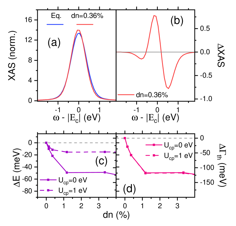
Moreover, the theory analysis shows that the shift increases linearly with the photo-doping density and then saturates (Fig. 3c), which is in qualitative agreement with the behavior in the experiment (Fig. 2(c)). A notable qualitative difference to the experiment is that the spectra in the simulations do not broaden, but become even more narrow (c.f. Figs. 1(a) and Fig. 3(a)). This may be because the simulation does not capture additional scattering channels which can lead to a change of the lineshape of the exciton, such as nonlocal spin fluctuations.
An interesting question is whether time-resolved XAS can disentangle the different contributions to the Coulomb shift. If one would assume a cancellation of the Hartree shifts () and no screening of , XAS would measure mainly the change of the Hubbard interaction , as concluded previously for LSCO [22]. However, the observed energy shifts (see Fig. 2(c)) of less than meV are small compared to the interaction parameters, so that a statement on the detailed origin of the shift would require a precise knowledge of , , and , which is beyond the capability of current first principles approaches such as constrained RPA [48]. Nevertheless, our analysis proves a high sensitivity of XAS to the microscopic interactions, and therefore shows that time-resolved XAS can give information on various Coulomb parameters and their changes upon photo-doping that is not easily obtained otherwise, including the nonlocal terms and which are often neglected even in equilibrium [10, 6].
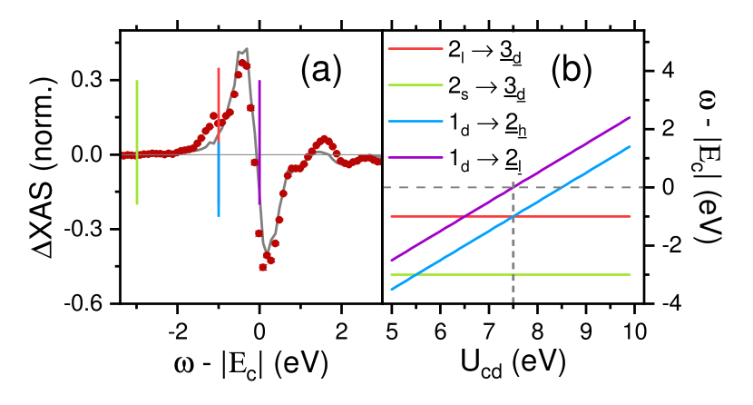
Having interpreted the red-shift of the edge in terms of Coulomb shifts, we now focus on the pronounced additional pre-edge feature in the Ni XAS at ps (Fig. 4(a)), which is absent at the longest delay times (compare Fig. 2(b)). This motivates a theoretical treatment of the actual, richer multiplet structure of NiO beyond our simple charge-transfer insulator model. There are two electrons in two orbitals forming a high-spin state due to the Hund coupling . A full simulation of a multi-orbital charge transfer insulator goes beyond the scope of the current study (some nonequilibrium DMFT results are presented in [39]). However, it was shown in Ref. [46] that the positions of the excitonic resonances in the photo-excited XAS are still well reproduced by atomic limit calculations, and their relative amplitude allows to measure the time-dependent weight of the different local multiplets in the full many body state on the lattice.
In the atomic limit, we label the local many-body multiplets as , where refers to the occupation and to the spin state, with for the high spin, for the low spin and for the intra-orbital singlet. The main XAS resonance in the two-orbital half-filled Hubbard model corresponds to the transition , where the underline indicates the presence of a core hole. After the photo-excitation additional transitions can be activated due to the presence of low spin doublons (2l or 2s) or holes (1d) on the nickel orbital. In particular, the additional photoinduced resonances due to Hund excitations include and and they appear eV or eV below the main excitonic peak (Fig. 4(b)) [35, 37, 49]. Remarkably, the experimental Ni XAS pre-edge feature perfectly matches the energy of the transition , see Fig. 4(a). This observation provides a proof-of-principle that transient XAS allows us to directly monitor the occupations of local many-body states after a photo-excitation. There are no obvious signs of the transition at lower energy, as it presumably would only appear at even shorter and/or higher pump frequencies and photodoping. For the core-valence interaction eV, the Hund side peak is degenerate with the transition . However, one would expect that holes on the Ni site will undergo a CT relaxation to the ligands on the femtosecond timescale, so that the -initial state would have a much shorter lifetime than the observed satellite.
Finally, a comparison of photo- and chemical doping is instructive. Chemical doping leads to the appearance of a pre-edge feature at the O edge [50] while such a pre-edge peak is absent at the Ni edges [51]. Photo-doping produces the opposite result, i.e., absence of a pre-edge feature at the O edge (which by comparison to [50] would be expected at 527 eV), and presence of such a feature at the Ni edge. While the theoretical discussion above can provide an explanation of the Ni pre-edge feature, the two-band Hubbard model does not describe the O edge. Nevertheless, the fact that the O spectra cannot simply be described by a shift and broadening is consistent with the existence of photo-doped holes on the O sites, while the difference between chemically doped and photo-doped spectra is a key observation which shows that photo-doped holes differ in nature from chemically dopes holes: they might occupy different - hybrid orbitals (such as non-bonding configurations instead of the Zhang-Rice singlets), or there may exist excitonic correlations between photo-doped electrons and holes.
In conclusion, we showed that photo-doping by above charge-transfer gap excitations in NiO results in characteristic transient signatures in XAS. These are (i) energy shifts of the excitonic peaks of several 10 meV for photo-doping on the order of 1%, which persist for tens of ps due to the long lifetime of the photo-doped state, and (ii) many-body multiplet excitations, observed here in the Ni pre-edge region, which decay on a sub-ps timescale. We demonstrated a good qualitative agreement between the XAS measurements and a simple model for charge-transfer insulators, which shows that energy shifts resulting from photo-induced changes in the electrostatic Hartree energy are a generic feature in transition metal oxides. We also concluded that screening induced changes in the interaction strengths play a role in determining these shifts. Our work furthermore establishes that XAS of photo-excited states is sensitive to the interaction between core electrons and ligand holes, which is important for a quantitative interpretation of the spectral changes. All these observations reveal generic signatures of charge-transfer insulators, so that our work provides a basis for a systematic analysis of these microscopic phenomena in this important class of materials. Finally, we note that the disentangling of non-equilibrium multiplet effects from time-dependent renormalizations of interaction parameters and Hartree shifts may help in the design of tailored excitation protocols for reaching specific photo-doped states.
Acknowledgments We acknowledge European XFEL in Schenefeld, Germany, for provision of X-ray free-electron laser beamtime at the SCS instrument and thank the staff for their assistance. Funded by the Deutsche Forschungsgemeinschaft (DFG, German Research Foundation) through Project No. 278162697 - SFB 1242. UB, PW, and ME acknowledge support from the Deutsche Forschungsgemeinschaft (DFG, German Science Foundation) through FOR 5249-449872909 (Project P6). D.G. acknowledges the support by the program No. P1-0044 and No. J1-2455 of the Slovenian Research Agency (ARRS).
T. L. performed the experiments and analyzed the data. D. G. did the calculations. Both contributed equally to this work.
References
- Giannetti et al. [2016] C. Giannetti, M. Capone, D. Fausti, M. Fabrizio, F. Parmigiani, and D. Mihailovic, Advances in Physics 65, 58 (2016).
- de la Torre et al. [2021] A. de la Torre, D. M. Kennes, M. Claassen, S. Gerber, J. W. McIver, and M. A. Sentef, Rev. Mod. Phys. 93, 041002 (2021).
- Sawatzky and Allen [1984] G. A. Sawatzky and J. W. Allen, Phys. Rev. Lett. 53, 2339 (1984).
- Zaanen et al. [1985] J. Zaanen, G. A. Sawatzky, and J. W. Allen, Phys. Rev. Lett. 55, 418 (1985).
- Zaanen et al. [1986] J. Zaanen, C. Westra, and G. A. Sawatzky, Phys. Rev. B 33, 8060 (1986).
- de Groot et al. [2021] F. M. de Groot, H. Elnaggar, F. Frati, R.-p. Wang, M. U. Delgado-Jaime, M. van Veenendaal, J. Fernandez-Rodriguez, M. W. Haverkort, R. J. Green, G. van der Laan, Y. Kvashnin, A. Hariki, H. Ikeno, H. Ramanantoanina, C. Daul, B. Delley, M. Odelius, M. Lundberg, O. Kuhn, S. I. Bokarev, E. Shirley, J. Vinson, K. Gilmore, M. Stener, G. Fronzoni, P. Decleva, P. Kruger, M. Retegan, Y. Joly, C. Vorwerk, C. Draxl, J. Rehr, and A. Tanaka, Journal of Electron Spectroscopy and Related Phenomena 249, 147061 (2021).
- van der Laan et al. [1986] G. van der Laan, J. Zaanen, G. A. Sawatzky, R. Karnatak, and J.-M. Esteva, Phys. Rev. B 33, 4253 (1986).
- Haverkort et al. [2014] M. Haverkort, G. Sangiovanni, P. Hansmann, A. Toschi, Y. Lu, and S. Macke, EPL (Europhysics Letters) 108, 57004 (2014).
- Cornaglia and Georges [2007] P. Cornaglia and A. Georges, Physical Review B 75, 115112 (2007).
- Okada and Kotani [1997] K. Okada and A. Kotani, Journal of the Physical Society of Japan 66, 341 (1997).
- Schuler et al. [2005] T. M. Schuler, D. L. Ederer, S. Itza-Ortiz, G. T. Woods, T. A. Callcott, and J. C. Woicik, Phys. Rev. B 71, 115113 (2005).
- Lüder et al. [2017] J. Lüder, J. Schött, B. Brena, M. W. Haverkort, P. Thunström, O. Eriksson, B. Sanyal, I. Di Marco, and Y. O. Kvashnin, Physical Review B 96, 245131 (2017).
- Taguchi et al. [2008] M. Taguchi, M. Matsunami, Y. Ishida, R. Eguchi, A. Chainani, Y. Takata, M. Yabashi, K. Tamasaku, Y. Nishino, T. Ishikawa, Y. Senba, H. Ohashi, and S. Shin, Phys. Rev. Lett. 100, 206401 (2008).
- Kuneš et al. [2007] J. Kuneš, V. Anisimov, A. Lukoyanov, and D. Vollhardt, Physical Review B 75, 165115 (2007).
- Bała et al. [1994] J. Bała, A. M. Oleś, and J. Zaanen, Physical review letters 72, 2600 (1994).
- Stamm et al. [2010] C. Stamm, N. Pontius, T. Kachel, M. Wietstruk, and H. A. Dürr, Phys. Rev. B 81, 104425 (2010).
- Smith et al. [2020] A. D. Smith, T. Balčiunas, Y.-P. Chang, C. Schmidt, K. Zinchenko, F. B. Nunes, E. Rossi, V. Svoboda, Z. Yin, J.-P. Wolf, and H. J. Wörner, J. Phys. Chem. Lett. 11, 1981 (2020).
- Rothenbach et al. [2019] N. Rothenbach, M. E. Gruner, K. Ollefs, C. Schmitz-Antoniak, S. Salamon, P. Zhou, R. Li, M. Mo, S. Park, X. Shen, S. Weathersby, J. Yang, X. J. Wang, R. Pentcheva, H. Wende, U. Bovensiepen, K. Sokolowski-Tinten, and A. Eschenlohr, Phys. Rev. B 100, 174301 (2019).
- Diesen et al. [2021] E. Diesen, H.-Y. Wang, S. Schreck, M. Weston, H. Ogasawara, J. LaRue, F. Perakis, M. Dell’Angela, F. Capotondi, L. Giannessi, E. Pedersoli, D. Naumenko, I. Nikolov, L. Raimondi, C. Spezzani, M. Beye, F. Cavalca, B. Liu, J. Gladh, S. Koroidov, P. S. Miedema, R. Costantini, T. F. Heinz, F. Abild-Pedersen, J. Voss, A. C. Luntz, and A. Nilsson, Phys. Rev. Lett. 127, 016802 (2021).
- Sidiropoulos et al. [2021] T. P. H. Sidiropoulos, N. Di Palo, D. E. Rivas, S. Severino, M. Reduzzi, B. Nandy, B. Bauerhenne, S. Krylow, T. Vasileiadis, T. Danz, P. Elliott, S. Sharma, K. Dewhurst, C. Ropers, Y. Joly, M. E. Garcia, M. Wolf, R. Ernstorfer, and J. Biegert, Phys. Rev. X 11, 041060 (2021).
- Lojewski et al. [2023] T. Lojewski, M. F. Elhanoty, L. L. Guyader, O. Grånäs, N. Agarwal, C. Boeglin, R. Carley, A. Castoldi, C. David, C. Deiter, F. Döring, R. Engel, F. Erdinger, H. Fangohr, C. Fiorini, P. Fischer, N. Gerasimova, R. Gort, F. deGroot, K. Hansen, S. Hauf, D. Hickin, M. Izquierdo, B. E. V. Kuiken, Y. Kvashnin, C.-H. Lambert, D. Lomidze, S. Maffessanti, L. Mercadier, G. Mercurio, P. S. Miedema, K. Ollefs, M. Pace, M. Porro, J. Rezvani, B. Rösner, N. Rothenbach, A. Samartsev, A. Scherz, J. Schlappa, C. Stamm, M. Teichmann, P. Thunström, M. Turcato, A. Yaroslavtsev, J. Zhu, M. Beye, H. Wende, U. Bovensiepen, O. Eriksson, and A. Eschenlohr, Mater. Res. Lett. 11, 655 (2023).
- Baykusheva et al. [2022] D. R. Baykusheva, H. Jang, A. A. Husain, S. Lee, S. F. R. TenHuisen, P. Zhou, S. Park, H. Kim, J.-K. Kim, H.-D. Kim, M. Kim, S.-Y. Park, P. Abbamonte, B. J. Kim, G. D. Gu, Y. Wang, and M. Mitrano, Phys. Rev. X 12, 011013 (2022).
- Wang et al. [2022] X. Wang, R. Y. Engel, I. Vaskivskyi, D. Turenne, V. Shokeen, A. Yaroslavtsev, O. Grånäs, R. Knut, J. O. Schunck, S. Dziarzhytski, et al., Faraday Discussions 237, 300 (2022).
- Grånäs et al. [2022] O. Grånäs, I. Vaskivskyi, X. Wang, P. Thunström, S. Ghimire, R. Knut, J. Söderström, L. Kjellsson, D. Turenne, R. Y. Engel, M. Beye, J. Lu, D. J. Higley, A. H. Reid, W. Schlotter, G. Coslovich, M. Hoffmann, G. Kolesov, C. Schüßler-Langeheine, A. Styervoyedov, N. Tancogne-Dejean, M. A. Sentef, D. A. Reis, A. Rubio, S. S. P. Parkin, O. Karis, J.-E. Rubensson, O. Eriksson, and H. A. Dürr, Phys. Rev. Res. 4, L032030 (2022).
- Tancogne-Dejean et al. [2020] N. Tancogne-Dejean, M. A. Sentef, and A. Rubio, Phys. Rev. B 102, 115106 (2020).
- Stojchevska et al. [2014] L. Stojchevska, I. Vaskivskyi, T. Mertelj, P. Kusar, D. Svetin, S. Brazovskii, and D. Mihailovic, Science 344, 177 (2014).
- Li et al. [2018] J. Li, H. U. R. Strand, P. Werner, and M. Eckstein, Nat. Commun. 9, 4581 (2018).
- Kaneko et al. [2019] T. Kaneko, T. Shirakawa, S. Sorella, and S. Yunoki, Phys. Rev. Lett. 122, 077002 (2019).
- Li et al. [2020] J. Li, D. Golez, P. Werner, and M. Eckstein, Mod. Phys. Lett. B 34, 2040054 (2020).
- Golež et al. [2019a] D. Golež, L. Boehnke, M. Eckstein, and P. Werner, Phys. Rev. B 100, 041111 (2019a).
- Tancogne-Dejean et al. [2018] N. Tancogne-Dejean, M. A. Sentef, and A. Rubio, Phys. Rev. Lett. 121, 097402 (2018).
- Sandri and Fabrizio [2015] M. Sandri and M. Fabrizio, Phys. Rev. B 91, 115102 (2015).
- He and Millis [2016] Z. He and A. J. Millis, Phys. Rev. B 93, 115126 (2016).
- Beaulieu et al. [2021] S. Beaulieu, S. Dong, N. Tancogne-Dejean, M. Dendzik, T. Pincelli, J. Maklar, R. P. Xian, M. A. Sentef, M. Wolf, A. Rubio, et al., Science Advances 7, eabd9275 (2021).
- Strand et al. [2017] H. U. R. Strand, D. Golež, M. Eckstein, and P. Werner, Phys. Rev. B 96, 165104 (2017).
- Rincón et al. [2018] J. Rincón, E. Dagotto, and A. E. Feiguin, Phys. Rev. B 97, 235104 (2018).
- Gillmeister et al. [2020] K. Gillmeister, D. Golez, C.-T. Chiang, N. Bittner, Y. Pavlyukh, J. Berakdar, P. Werner, and W. Widdra, Nature Communications 11, 4095 (2020).
- Müller et al. [2022] A. Müller, F. Grandi, and M. Eckstein, Phys. Rev. B 106, L121107 (2022).
- [39] See the supplement for the calculation of the excitation density and more information on data processing, the modelling to extract spectral shifts and broadening, and the theoretical method.
- Georges et al. [1996] A. Georges, G. Kotliar, W. Krauth, and M. J. Rozenberg, Rev. Mod. Phys. 68, 13 (1996).
- Aoki et al. [2014] H. Aoki, N. Tsuji, M. Eckstein, M. Kollar, T. Oka, and P. Werner, Rev. Mod. Phys. 86, 779 (2014).
- Le Guyader et al. [2023] L. Le Guyader, A. Eschenlohr, M. Beye, W. Schlotter, F. Döring, C. Carinan, D. Hickin, N. Agarwal, C. Boeglin, U. Bovensiepen, J. Buck, R. Carley, A. Castoldi, A. D’Elia, J.-T. Delitz, W. Ehsan, R. Engel, F. Erdinger, H. Fangohr, P. Fischer, C. Fiorini, A. Föhlisch, L. Gelisio, M. Gensch, N. Gerasimova, R. Gort, K. Hansen, S. Hauf, M. Izquierdo, E. Jal, E. Kamil, S. Karabekyan, T. Kluyver, T. Laarmann, T. Lojewski, D. Lomidze, S. Maffessanti, T. Mamyrbayev, A. Marcelli, L. Mercadier, G. Mercurio, P. S. Miedema, K. Ollefs, K. Rossnagel, B. Rösner, N. Rothenbach, A. Samartsev, J. Schlappa, K. Setoodehnia, G. Sorin Chiuzbaian, L. Spieker, C. Stamm, F. Stellato, S. Techert, M. Teichmann, M. Turcato, B. Van Kuiken, H. Wende, A. Yaroslavtsev, J. Zhu, S. Molodtsov, C. David, M. Porro, and A. Scherz, Journal of Synchrotron Radiation 30, 284 (2023).
- [43] Data recorded for the experiment at the European XFEL are available at doi:10.22003/xfel.eu-data-002589-00.
- Golež et al. [2017] D. Golež, L. Boehnke, H. U. R. Strand, M. Eckstein, and P. Werner, Phys. Rev. Lett. 118, 246402 (2017).
- Golež et al. [2019b] D. Golež, M. Eckstein, and P. Werner, Phys. Rev. B 100, 235117 (2019b).
- Werner et al. [2022] P. Werner, D. Golez, and M. Eckstein, Phys. Rev. B 106, 165106 (2022).
- Dasari et al. [2021] N. Dasari, J. Li, P. Werner, and M. Eckstein, Phys. Rev. B 103, L201116 (2021).
- Aryasetiawan et al. [2004] F. Aryasetiawan, M. Imada, A. Georges, G. Kotliar, S. Biermann, and A. I. Lichtenstein, Phys. Rev. B 70, 195104 (2004).
- Kuneš et al. [2007] J. Kuneš, V. I. Anisimov, A. V. Lukoyanov, and D. Vollhardt, Phys. Rev. B 75, 165115 (2007).
- Kuiper et al. [1989] P. Kuiper, G. Kruizinga, J. Ghijsen, G. A. Sawatzky, and H. Verweij, Phys. Rev. Lett. 62, 221 (1989).
- van Elp et al. [1991] J. van Elp, B. Searle, G. Sawatzky, and M. Sacchi, Solid State Communications 80, 67 (1991).