X-ray absorption spectroscopy using an ultrafast laboratory-scale laser-plasma accelerator source
Abstract
The absorption profile of the copper K-edge was measured over a 250 eV window using ultrashort X-rays from a laser-plasma wakefield accelerator. For the first time with a femtosecond probe, Extended X-ray Absorption Fine Structure (EXAFS) features were observed in a single shot, detailing the local atomic structure. This unique capability will allow the investigation of novel ultrafast processes, and in particular probing high energy density matter and physics far-from-equilibrium. A perspective on the additional strengths of a laboratory-based ultrafast X-ray absorption source is presented.
In recent years laser-plasma based particle accelerators have provided access to gigaelectronvolt electron energies within the small-scale laboratory environment Esarey et al. (2009); Albert et al. (2021); Gonsalves et al. (2019). This has led to new research involving strong field QED studies Cole et al. (2018); Poder et al. (2018), electron-positron pair generation Kettle et al. (2021); Sarri et al. (2015) and X-ray and gamma ray applications Albert et al. (2014); Cole et al. (2015); Sarri et al. (2014); Schumaker et al. (2014). One growing area makes use of the X-rays generated in tandem with the accelerated beam as the electrons wiggle in the back of the plasma wakefield bubble Kneip et al. (2010); Corde et al. (2013). The X-rays have a pulse duration comparable to the emitted electron bunch; usually 10’s of femtoseconds Debus et al. (2010); Lundh et al. (2011); Mangles et al. (2006).
A key strength of this X-ray source is that it has a smooth, broadband synchrotron-like spectrum, making it ideal for X-ray absorption spectroscopy (XAS) techniques such as X-ray Absorption Near-Edge Structure (XANES) and Extended X-ray Absorption Fine Structure (EXAFS) spectroscopy. In these techniques the absorption, scattering and interference of ejected photoelectrons from neighboring atoms manifest as modulations in the absorption profile near resonant edges. These modulations are directly linked to the local electronic and ionic structure of the sample Rehr and Albers (2000); Koningsberger and Prins (1988). In fact the electron temperature, ion temperature, ionisation state and local ionic positions can be simultaneously measured Dorchies and Recoules (2016).
Over the last few decades XANES and EXAFS has been used with great success on synchrotron facilities to measure the structure of materials, across a wide range of sciences Syn (2018); Bilderback et al. (2005). However, when the duration of the X-ray probe is important, measurements are limited to the pulse duration of the synchrotron; generally on the order of hundreds of picoseconds Torchio et al. (2016), unless additional slicing techniques are implemented (that sacrifice flux). X-ray Free Electron Laser (XFEL) facilities operate at femtosecond pulse duration but provide a narrow bandwidth, requiring a scan over many shots to obtain a broad absorption spectrum Lemke et al. (2013). The combined ultrashort and broadband capabilities of a laser-plasma accelerator X-ray source makes it uniquely suited to investigate novel ultrafast processes on a femtosecond scale through XAS techniques. These include femtochemistry phenomena, such as photodissociation, spin crossover processes Bressler et al. (2008); Gawelda et al. (2007); Chen (2001, 2005), photobiology Bressler and Chergui (2010); Jonas et al. (1996); van Grondelle et al. (1994); Arcovito et al. (2005), or photocatalysts and photoelectrodes Uemura et al. (2017); Hu et al. (2021); Park et al. (2022), which are becoming increasingly important, for example in energy storage research. Another active area is that of spintronics for data storage and transfer, with specific interest in antiferromagnets Hirohata et al. (2020); Li et al. (2019); Johnson et al. (2015); Strungaru et al. (2022). Other ultrafast phenomena such as non-thermal phase changes in semi-conductors Rousse et al. (2001) or bond-hardening effects Ernstorfer et al. (2009) are also of interest.
A major application in physics is the study of extreme states far from equilibrium Mahieu et al. (2018), or those with high energy density, such as the tremendous pressures and temperatures of planetary formations Koenig et al. (2004), or inertial confinement fusion experiments Betti and Hurricane (2016). For example, nanosecond EXAFS has been performed on high-energy laser systems to investigate materials at high pressures Yaakobi et al. (2004); Ping et al. (2013). For these experiments a single-shot measurement is of major benefit, as driving the sample to the appropriate conditions requires extensive resources and shot rate is limited.
Until now, the photon flux from laser-plasma accelerator based XAS measurements has been lacking, requiring 10’s to 100’s of shots to form a full absorption profile, which often suffers from noise and limited spectral range Ta Phuoc et al. (2007); Mahieu et al. (2018). Recently single-shot XANES measurements using a laser-plasma accelerator have been demonstrated Kettle et al. (2019), although due to the noise levels of the absorption profile, the spectral range was limited to 40 eV. In this article, using a new experimental geometry, we demonstrate a marked increase in signal-over-noise and extend the single-shot spectral range to over 250 eV. This allows access to the EXAFS region for the first time, an important capability for performing pump-probe experiments of high-energy-density physics and extreme states. Using these developments, we present a platform for a laser-plasma accelerator based XAS system that will enable high quality laboratory-scale measurements, while providing the unique capability of measuring ultrafast processes on a femtosecond timescale.
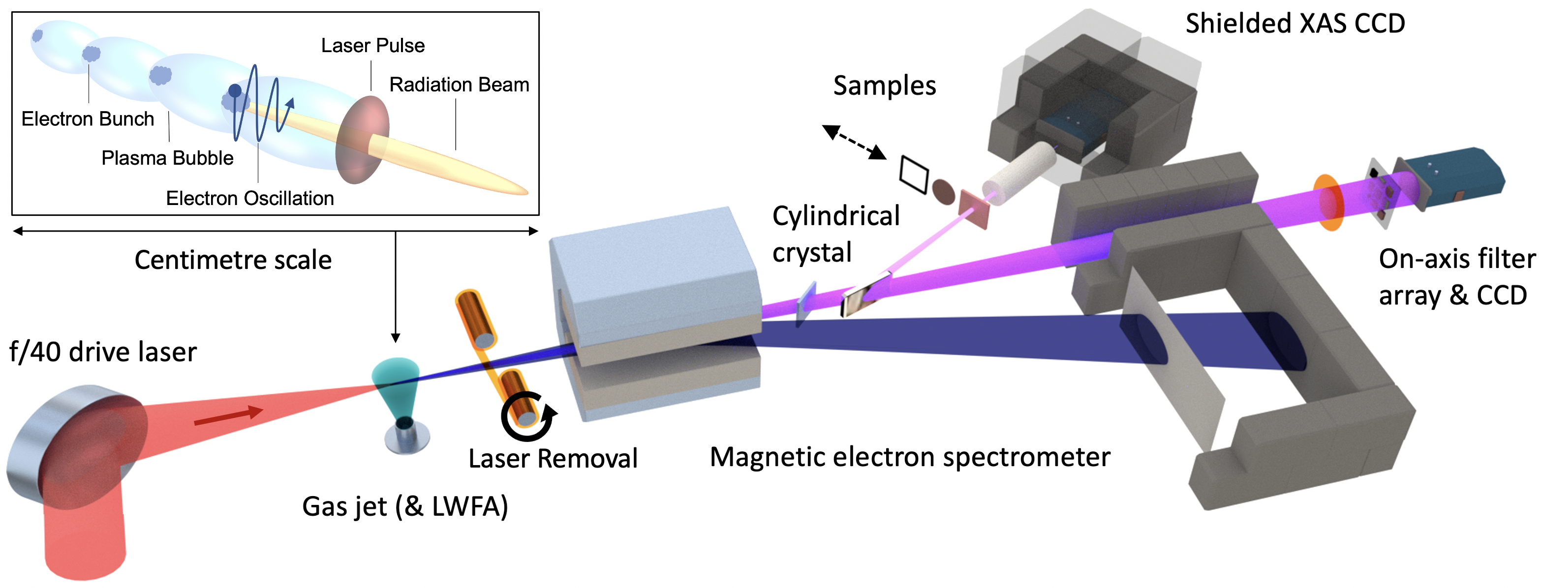
The experiment was conducted using the Gemini Laser at the Central Laser Facility (U.K.). An overview of the setup can be seen in Fig. 1 Our (2023). The drive laser was focused using an /40 geometry onto a 15 mm diameter gas jet nozzle. Each laser pulse (provided at 0.05 Hz) had a duration of fs and contained J, corresponding to a peak laser power of TW. This provided an on-target intensity of ( and a laser strength parameter of , before any self-focusing effects. As this pulse propagates through the gas, it drives a laser wakefield accelerator (LWFA), expelling electrons from the atoms and creating a charge cavity in the wake of the laser pulse. The electrons are accelerated to relativistic energies in this wake, emitting X-ray synchrotron light as they oscillate at the back of the cavity. A 99% He and 1% mix was used to promote ionisation injection of electrons into the charge cavity Pak et al. (2010); McGuffey et al. (2010). The nozzle provided a plasma density of . After the LWFA the residual drive laser was blocked by a refreshable tape drive (125 Kapton), and the electron beam was diagnosed with a 0.3 magnetic spectrometer that dispersed the beam onto an imaged scintillating Lanex screen. The electron beam was then dumped into a lead shielded cavity. The broadband X-ray beam is reflected off a cylindrical HAPG (Highly Annealed Pyrolytic Graphite) crystal, onto a well shielded X-ray CCD. The crystal spectrally disperses the X-rays in one axis, while focusing them spatially in the other axis. The crystal was tilted 11.8∘ from the laser and X-ray axis to observe X-rays in the 9 keV region. Various copper samples were placed on a translation stage before the CCD, so that their absorption could be measured. By temporarily moving the crystal, it was also possible to infer the full broadband spectral shape of the X-rays using an on-axis CCD with an elemental filter array placed in front Rousse et al. (2004); Kneip et al. (2008, 2010); King et al. (2019).
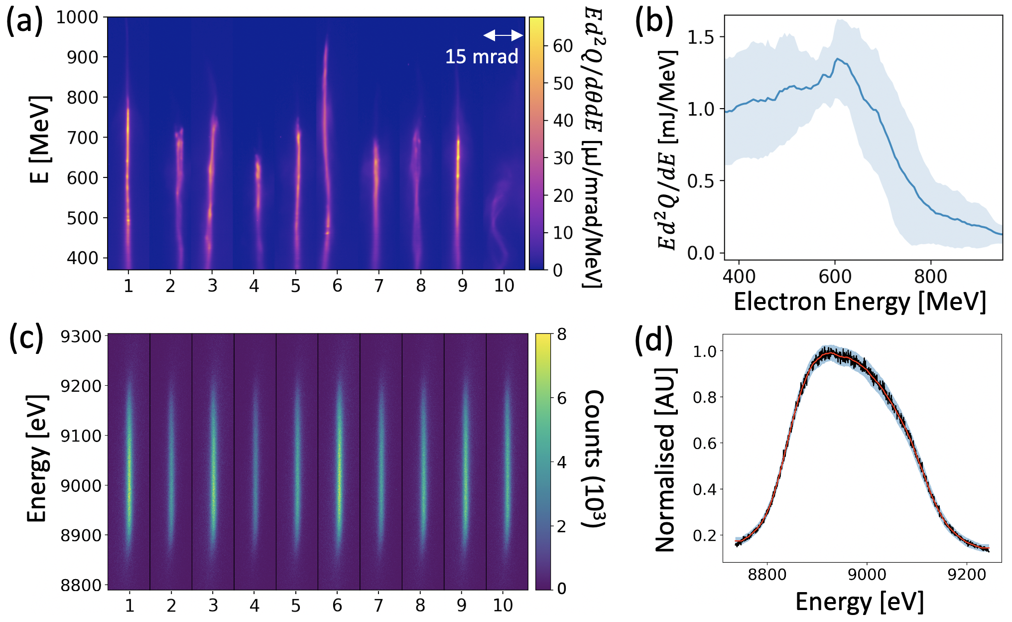
For the data discussed here, the LWFA was operated in a high-charge mode (as opposed to high electron energy), as this has previously been seen to produce more photons in our desired spectral range Kettle et al. (2019). Fig. 2 (a) depicts the electron spectra for 10 consecutive example shots. The 90th percentile electron energy was MeV, with a charge of pC per shot. This led to the generation of an on-axis X-ray beam with an average best-fit critical energy of , and a standard deviation of (inferred over 10 shots in the run prior to the data of Fig. 2 (a)). Fig. 2 (c) depicts the X-ray signal for the same 10 shots of Fig. 2 (a). On average photons/eV were emitted in the 9 keV region per shot. It is important to note that while the spectral content of the electron beam is somewhat erratic, with the 90th percentile electron beam energy varying by 150 MeV (22%) across the run and a shot-to-shot variation in total charge of 29%, the measured portion of 9 keV radiation is seen to be spectrally smooth, and can be easily scaled in magnitude. This is because the crystal spectrometer observes a small slice of the broadband synchrotron spectrum, a relatively slowly varying function with electron beam energy. By averaging over the profiles of the X-ray data in Fig. 2 (c), a reference spectral profile shape is found (each single shot is normalised to its peak level, and a rolling average smoothing function is applied). This profile is given in Fig. 2 (d). The variation from the measured signal is on order of a few percent and so it is possible for the absorption profile of a sample to be measured without an on-shot reference (but rather an assumed profile). In summary, the stability of the X-ray source for absorption measurements is evident, despite the fluctuations of the electron beam properties.
The absorption profile of a copper sample has been measured, assuming the reference profile of Fig. 2 (d) as the unattenuated signal (the known transmission before the copper K-edge is used to scale the magnitude). Once an absorption profile is found, a normalised absorption is calculated by fitting below and above the copper K-edge, subtracting the former, before dividing by the latter. The process is a standard XAS procedure, and we follow the method outlined in our previous work Kettle et al. (2019), and that of Cook and Sayers Cook and Sayers (1981).
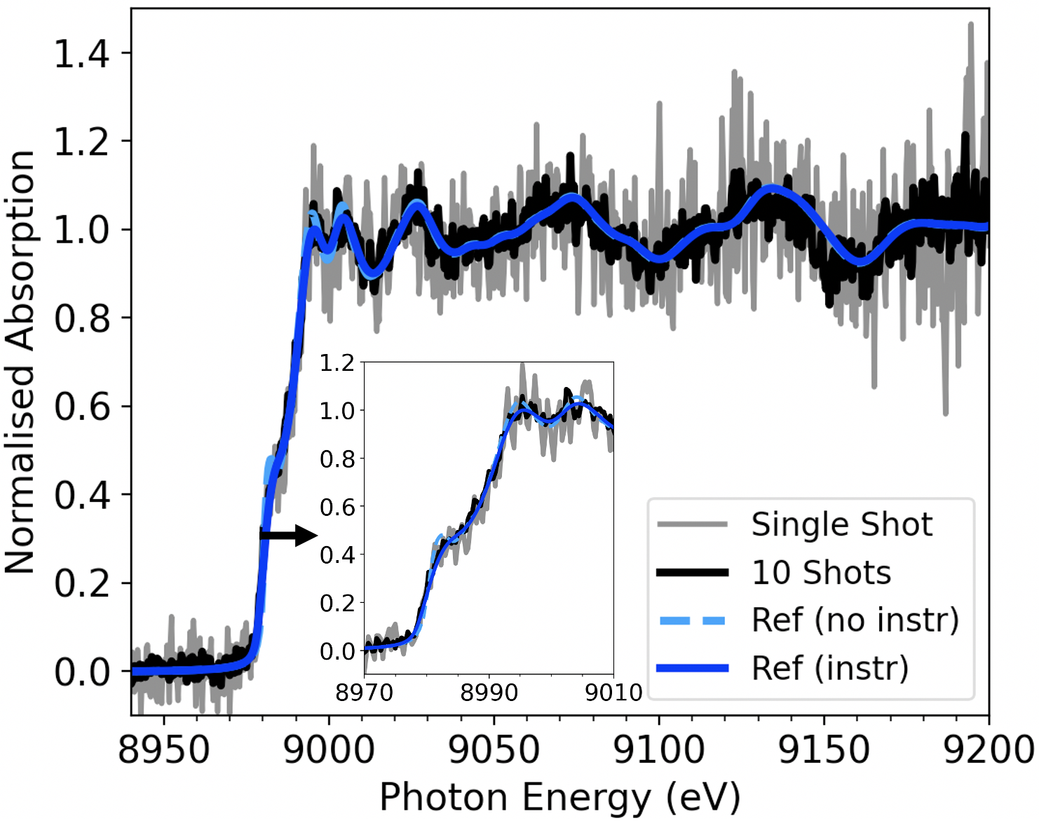
Fig. 3 depicts the resulting normalised absorption data for the copper sample. The inset gives the XANES profile close to the edge. The profile for a single shot (grey) and the average profile for a 10 shot run (black) is shown. Both are compared to the absorption profile of a similar copper foil sample measured at B18, the general purpose XAS beamline at the Diamond Light Source synchrotron facility. This reference is presented both with the simulated instrument function of our system applied (blue solid), and the unaltered synchrotron data (light blue dashed). Our instrument response was simulated using the MMPXRT code Šmíd et al. (2021) and indicates a spectral resolution of eV (FWHM). Note that the reference data without our instrument function applied differs only slightly at the edge (inset). The measured profile stretches over a 250 eV spectral range, allowing access to the EXAFS region of the profile. It demonstrates a marked increase in signal-to-noise over our previous work Kettle et al. (2019), as well as overall photon number from other LWFA X-ray absorption measurements Mahieu et al. (2018). The two most important alterations to the experimental setup were to use a spatially focusing cylindrical crystal, and to mitigate the noise on the X-ray detector from the electron beam by sweeping the beam into a shielded dump and separately enclosing the X-ray CCD. Both these factors contributed to greatly increased signal-to-noise. In fact, we infer that the noise level on a single shot is close to Poisson limited (where for peak signal we have photons per spectral pixel, with a standard deviation of photons, or ). This indicates we have removed the majority of any environmental noise sources, and are limited by the measured photon number of our signal. The CCD is cooled to C and readout noise is on order of 1 count per spectral pixel, or of a single photon.
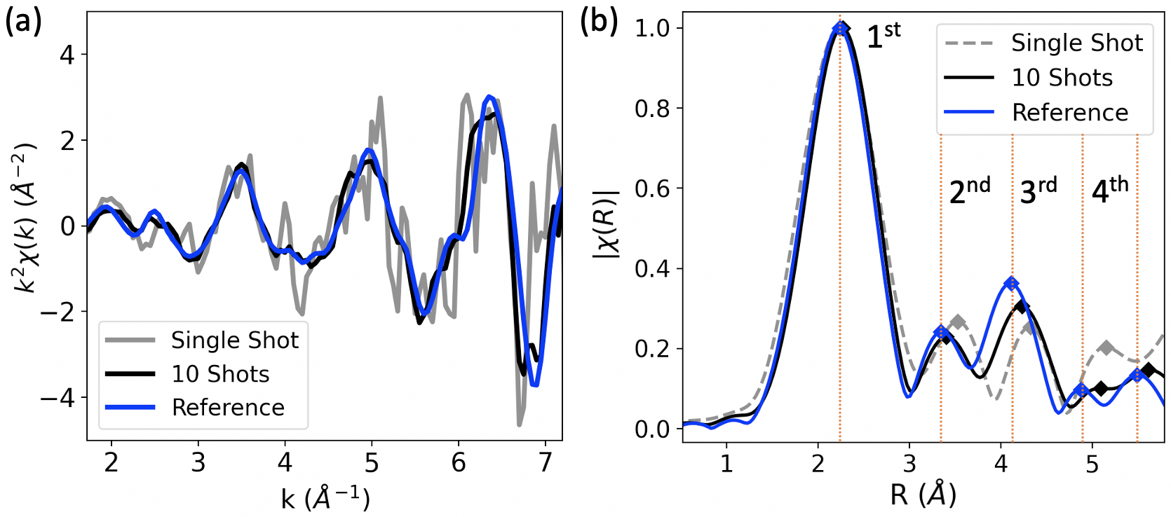
In Fig. 3 the EXAFS features after the edge are visible on a single shot, a first of its kind measurement for a laser-plasma accelerator XAS system. We investigate these features further by subtracting a spline fit to the post-edge background and converting to , the EXAFS fine-structure function, where is wavenumber and is the absorption threshold energy. See Fig. 4 (a). Note that while our spectral resolution results in a small amount of broadening on the fine XANES features, there is no meaningful effect on the broader EXAFS structure. The next step is to Fourier transform to . See Fig. 4 (b) for a comparison of our data to the synchrotron reference data, processed under the same conditions. is similar to a radial distribution function, and the peaks correspond to the distance to neighboring atoms (or coordination shells). The scattering from the first four shells are labelled Zabinsky et al. (1995). Our single shot data matches the first shell of the reference data to within 1.5%, and shells 2-4 within 5% (a peak finding algorithm was used to determine their location). The 10 shot data improves on this, with the second, third and fourth shells matching within 2%, 3% and 5% respectively. This analysis is a direct measurement of the local atomic structure, and unique to the species of a sample. Interpretation of this structure also allows access to information on the ion temperature, or any sample compression or phase changes.
It is evident that the signal-to-noise ratio we have achieved has made it possible to produce high quality measurements of both the XANES and EXAFS features in a single shot using a femtosecond probe. This enables XAS probing of ultrafast processes, but especially those samples in extreme conditions which are limited to a low shot rate. The development of this ultrafast XAS platform is on-going, with clear routes for further improvements on flux, photon energy and spectral resolution. Petawatt-class laser systems are becoming more commonplace, providing an order of magnitude more laser power than the experiment we detail. These systems can access higher electron energies Gonsalves et al. (2019); Thomas (2010), and an estimated order of magnitude increase in the associated X-ray flux Bloom et al. (2020); Thomas (2010). Novel targetry techniques can also be used to increase the source X-ray flux. Recent results Rakowski et al. (2022); Kozlova et al. (2020); Ta Phuoc et al. (2008) have shown that introducing a density gradient into the target, using a wire or razor blade obstruction, can increase the electron oscillation radius and frequency, enhancing the X-ray emission by an order of magnitude. Combining the above improvements in laser technology and targetry one could speculate two orders of magnitude improvement in signal-to-noise. See Fig. 5 (a) for a simulated comparison of these improvements, based on the noise being Poisson-limited by the photon number. The spectral resolution can be increased by using a perfect crystal (such as Si or Ge) instead of a mosaic crystal, if a sacrifice in X-ray flux can be allowed. Simulations using MMPXRT Šmíd et al. (2021) suggest sub-eV resolution in this configuration. The small source size of the X-rays (sub-micron Kneip et al. (2012)) can be harnessed in conjunction with a suitable focusing optic, such as a toroidal crystal, to allow micron focusing of the probe beam. This can be used to probe small sample volumes (for example in HED targets) or high-resolution scanning of inhomogeneities in industrial samples such as batteries or spintronics materials. Accessing higher electron beam energies will also generate synchrotron spectra with higher critical energies, and thus provide photons of 100’s keV Kneip et al. (2012); Hojbota et al. (2022). These photon energies cannot be accessed easily by conventional synchrotron facilities, and will provide the capability to probe higher-Z elements and inner atomic edges. Important, for example, in nuclear research Lind et al. (2007). The stability of the electron accelerator can be improved through laser techniques Döpp et al. (2017); Ferran Pousa et al. (2022), and recent studies have shown that machine learning can be used to optimise the X-ray emission Shalloo et al. (2020); Ye et al. (2022).
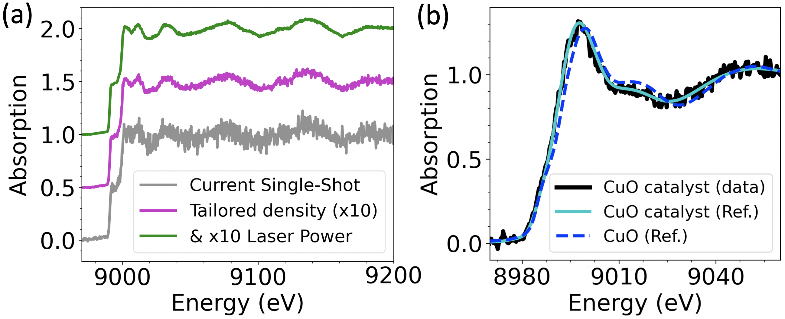
In addition to the above, petawatt lasers can now operate at repetition rates of 1 Hz, providing an increased rate of data taking ( increase from this experiment). This would allow laser-plasma accelerator based XAS platforms to complement synchrotron systems, i.e. making a similar quality scan in a few minutes. As an example, using our current source capability, Fig. 5 (b) depicts an ex situ absorption profile of a copper based hydrogenation catalyst provided by Johnson Matthey, UK. Our data (black), and the profile for the same sample scanned at the Diamond Light Source (cyan), is compared to a CuO reference sample (dashed blue). The characteristic features of this industrial catalyst are clear, indicated by a shifted edge at 8995 eV and an altered near-edge shape at 9015 eV, even with the Cu loading at 13 wt% as expressed as an oxide. The oxidation state shows the Cu to be oxidic and predominantly in the Cu2+ oxidation state in comparison to the CuO reference sample. The data shown took 10 minutes of shots, but with the improvements discussed in the previous paragraph, a profile with an improved signal-over-noise could be generated in under a minute. The benefit of a laser-plasma based XAS system as described here is that it could be achieved in a relatively small laboratory environment, in comparison to a conventional synchrotron light source that requires a large national facility. This reduced scale makes it an accessible tool for single institutions and makes long-term programmatic research possible. For example detailed studies of catalysts (which can improve conversion rates, lower energy consumption, or tune the selectivity of chemical reactions) Fernández-García (2002), or new energy storage technologies, including long term effects such as repeated charge cycle degradation of batteries Jahrman et al. (2019); Preger et al. (2020); Bi et al. (2019); Wang et al. (2016); Nowack et al. (2016). A laboratory scale instrument also allows dedicated user access for immediate probing of delicate samples or the appropriate handling of hazardous samples, for example biological hazards, or the analysis of radiological materials for environmental remediation (nuclear waste management) Zhong et al. (2021); Mottram et al. (2020).
In conclusion, we have demonstrated that the X-rays from a LWFA source are sufficiently bright and stable to measure the absorption profile around the copper K-edge, over a 250 eV spectral window in a single shot. The absorption profile is of sufficient quality to make single-shot EXAFS measurements with a LFWA-based source for the first time, providing direct access to the local atomic structure of a sample using a femtosecond probe. In particular EXAFS is sensitive to short-range order and can provide the ion temperature of a sample, both difficult measurements to make. Single shot capability of an ultrafast probe is of significant benefit for studying transient pump-probe samples of high-energy-density physics such as warm dense matter and other non-equilibrium states, as well as other ultrafast electronic and ionic processes. The compact size will also allow the instrument to be co-located with other laboratories more easily, especially XFEL’s or high-power laser facilities (where it can be inherently synchronised on a femtosecond level), acting as an auxiliary probe. In addition, by implementing an XAS source on a laboratory scale, the breadth and depth of both industrial and programmatic scientific research that can be achieved on a wide-scale basis is vastly increased.
We wish to acknowledge the support of the staff at the Central Laser Facility. This project has received funding from the European Research Council (ERC) under the European Union’s Horizon 2020 research and innovation programme (grant agreement no 682399), as well as support from the John Adams Institute for Accelerator Science (STFC: ST/P002021/1, ST/V001639/1), the EPSRC (EP/S001379/1, EP/V044397/1, EP/N027175/1), the US DOE grant No. DE-SC0022109, the Knut and Alice Wallenberg Foundation (KAW 2019.0318), the Swedish Research Council (2019-04784), and the Helmholtz Association (VH-NG-1338). The data that support the findings of this study are openly available Our (2023).
References
- Esarey et al. (2009) E. Esarey, C. B. Schroeder, and W. P. Leemans, Rev. Mod. Phys. 81, 1229 (2009).
- Albert et al. (2021) F. Albert, M. E. Couprie, A. Debus, M. C. Downer, J. Faure, A. Flacco, L. A. Gizzi, T. Grismayer, A. Huebl, C. Joshi, M. Labat, W. P. Leemans, A. R. Maier, S. P. D. Mangles, P. Mason, F. Mathieu, P. Muggli, M. Nishiuchi, J. Osterhoff, P. P. Rajeev, U. Schramm, J. Schreiber, A. G. R. Thomas, J.-L. Vay, M. Vranic, and K. Zeil, New Journal of Physics 23, 031101 (2021).
- Gonsalves et al. (2019) A. J. Gonsalves, K. Nakamura, J. Daniels, C. Benedetti, C. Pieronek, T. C. H. de Raadt, S. Steinke, J. H. Bin, S. S. Bulanov, J. van Tilborg, C. G. R. Geddes, C. B. Schroeder, C. Tóth, E. Esarey, K. Swanson, L. Fan-Chiang, G. Bagdasarov, N. Bobrova, V. Gasilov, G. Korn, P. Sasorov, and W. P. Leemans, Phys. Rev. Lett. 122, 084801 (2019).
- Cole et al. (2018) J. M. Cole, K. T. Behm, E. Gerstmayr, T. G. Blackburn, J. C. Wood, C. D. Baird, M. J. Duff, C. Harvey, A. Ilderton, A. S. Joglekar, K. Krushelnick, S. Kuschel, M. Marklund, P. McKenna, C. D. Murphy, K. Poder, C. P. Ridgers, G. M. Samarin, G. Sarri, D. R. Symes, A. G. R. Thomas, J. Warwick, M. Zepf, Z. Najmudin, and S. P. D. Mangles, Phys. Rev. X 8, 011020 (2018).
- Poder et al. (2018) K. Poder, M. Tamburini, G. Sarri, A. Di Piazza, S. Kuschel, C. D. Baird, K. Behm, S. Bohlen, J. M. Cole, D. J. Corvan, M. Duff, E. Gerstmayr, C. H. Keitel, K. Krushelnick, S. P. D. Mangles, P. McKenna, C. D. Murphy, Z. Najmudin, C. P. Ridgers, G. M. Samarin, D. R. Symes, A. G. R. Thomas, J. Warwick, and M. Zepf, Phys. Rev. X 8, 031004 (2018).
- Kettle et al. (2021) B. Kettle, D. Hollatz, E. Gerstmayr, G. M. Samarin, A. Alejo, S. Astbury, C. Baird, S. Bohlen, M. Campbell, C. Colgan, D. Dannheim, C. Gregory, H. Harsh, P. Hatfield, J. Hinojosa, Y. Katzir, J. Morton, C. D. Murphy, A. Nurnberg, J. Osterhoff, G. Pérez-Callejo, K. Põder, P. P. Rajeev, C. Roedel, F. Roeder, F. C. Salgado, G. Sarri, A. Seidel, S. Spannagel, C. Spindloe, S. Steinke, M. J. V. Streeter, A. G. R. Thomas, C. Underwood, R. Watt, M. Zepf, S. J. Rose, and S. P. D. Mangles, New Journal of Physics 23, 115006 (2021).
- Sarri et al. (2015) G. Sarri, K. Poder, J. M. Cole, W. Schumaker, A. Di Piazza, B. Reville, T. Dzelzainis, D. Doria, L. A. Gizzi, G. Grittani, S. Kar, C. H. Keitel, K. Krushelnick, S. Kuschel, S. P. D. Mangles, Z. Najmudin, N. Shukla, L. O. Silva, D. Symes, A. G. R. Thomas, M. Vargas, J. Vieira, and M. Zepf, Nature Communications 6, 6747 (2015).
- Albert et al. (2014) F. Albert, A. G. R. Thomas, S. P. D. Mangles, S. Banerjee, S. Corde, A. Flacco, M. Litos, D. Neely, J. Vieira, and Z. Najmudin, Plasma Phys. Control. Fusion 56, 084015 (2014).
- Cole et al. (2015) J. M. Cole, J. C. Wood, N. C. Lopes, K. Poder, R. L. Abel, S. Alatabi, J. S. J. Bryant, A. Jin, S. Kneip, K. Mecseki, et al., Sci. Rep. 5 (2015), 10.1038/srep13244.
- Sarri et al. (2014) G. Sarri, D. J. Corvan, W. Schumaker, J. M. Cole, A. Di Piazza, H. Ahmed, C. Harvey, C. H. Keitel, K. Krushelnick, S. P. D. Mangles, Z. Najmudin, D. Symes, A. G. R. Thomas, M. Yeung, Z. Zhao, and M. Zepf, Phys. Rev. Lett. 113, 224801 (2014).
- Schumaker et al. (2014) W. Schumaker, G. Sarri, M. Vargas, Z. Zhao, K. Behm, V. Chvykov, B. Dromey, B. Hou, A. Maksimchuk, J. Nees, V. Yanovsky, M. Zepf, A. G. R. Thomas, and K. Krushelnick, Physics of Plasmas 21, 056704 (2014), https://doi.org/10.1063/1.4875336 .
- Kneip et al. (2010) S. Kneip, C. McGuffey, J. L. Martins, S. F. Martins, C. Bellei, V. Chvykov, F. Dollar, R. Fonseca, C. Huntington, G. Kalintchenko, et al., Nat. Phys. 6, 980 (2010).
- Corde et al. (2013) S. Corde, K. Ta Phuoc, G. Lambert, R. Fitour, V. Malka, A. Rousse, A. Beck, and E. Lefebvre, Rev. Mod. Phys. 85, 1 (2013).
- Debus et al. (2010) A. D. Debus, M. Bussmann, U. Schramm, R. Sauerbrey, C. D. Murphy, Z. Major, R. Hörlein, L. Veisz, K. Schmid, J. Schreiber, K. Witte, S. P. Jamison, J. G. Gallacher, D. A. Jaroszynski, M. C. Kaluza, B. Hidding, S. Kiselev, R. Heathcote, P. S. Foster, D. Neely, E. J. Divall, C. J. Hooker, J. M. Smith, K. Ertel, A. J. Langley, P. Norreys, J. L. Collier, and S. Karsch, Phys. Rev. Lett. 104, 084802 (2010).
- Lundh et al. (2011) O. Lundh, J. Lim, C. Rechatin, L. Ammoura, A. Ben-Ismaïl, X. Davoine, G. Gallot, J.-P. Goddet, E. Lefebvre, V. Malka, and J. Faure, Nat. Phys. 7, 219–222 (2011).
- Mangles et al. (2006) S. P. D. Mangles, A. G. R. Thomas, M. C. Kaluza, O. Lundh, F. Lindau, A. Persson, F. S. Tsung, Z. Najmudin, W. B. Mori, C.-G. Wahlström, and K. Krushelnick, Phys. Rev. Lett. 96, 215001 (2006).
- Rehr and Albers (2000) J. J. Rehr and R. C. Albers, Rev. Mod. Phys. 72, 621–654 (2000).
- Koningsberger and Prins (1988) D. C. Koningsberger and R. Prins, X-Ray Absorption: Principles, Applications, Techniques of EXAFS, SEXAFS and XANES (Wiley, 1988).
- Dorchies and Recoules (2016) F. Dorchies and V. Recoules, Phys. Rep. 657, 1 (2016).
- Syn (2018) Nature Reviews Materials 3, 281 (2018).
- Bilderback et al. (2005) D. H. Bilderback, P. Elleaume, and E. Weckert, Journal of Physics B: Atomic, Molecular and Optical Physics 38, S773 (2005).
- Torchio et al. (2016) R. Torchio, F. Occelli, O. Mathon, A. Sollier, E. Lescoute, L. Videau, T. Vinci, A. Benuzzi-Mounaix, J. Headspith, W. Helsby, S. Bland, D. Eakins, D. Chapman, S. Pascarelli, and P. Loubeyre, Scientific Reports 6, 26402 (2016).
- Lemke et al. (2013) H. T. Lemke, C. Bressler, L. X. Chen, D. M. Fritz, K. J. Gaffney, A. Galler, W. Gawelda, K. Haldrup, R. W. Hartsock, H. Ihee, J. Kim, K. H. Kim, J. H. Lee, M. M. Nielsen, A. B. Stickrath, W. Zhang, D. Zhu, and M. Cammarata, The Journal of Physical Chemistry A 117, 735 (2013), pMID: 23281652, https://doi.org/10.1021/jp312559h .
- Bressler et al. (2008) C. Bressler, R. Abela, and M. Chergui, Zeitschrift für Kristallographie - Crystalline Materials 223, 307 (2008).
- Gawelda et al. (2007) W. Gawelda, A. Cannizzo, V.-T. Pham, A. El Nahhas, C. J. Milne, R. van der Veen, C. Bressler, and M. Chergui, CHIMIA 61, 179 (2007).
- Chen (2001) L. X. Chen, Journal of Electron Spectroscopy and Related Phenomena 119, 161 (2001), chemical Applications of Synchrotron Radiation.
- Chen (2005) L. X. Chen, Annual Review of Physical Chemistry 56, 221 (2005), pMID: 15796701.
- Bressler and Chergui (2010) C. Bressler and M. Chergui, Annual Review of Physical Chemistry 61, 263 (2010), pMID: 20055677.
- Jonas et al. (1996) D. M. Jonas, M. J. Lang, Y. Nagasawa, S. E. Bradforth, S. N. Dikshit, R. Jimenez, T. Joo, and G. R. Fleming, in The Reaction Center of Photosynthetic Bacteria, edited by M.-E. Michel-Beyerle (Springer Berlin Heidelberg, Berlin, Heidelberg, 1996) pp. 187–198.
- van Grondelle et al. (1994) R. van Grondelle, J. P. Dekker, T. Gillbro, and V. Sundstrom, Biochimica et Biophysica Acta (BBA) - Bioenergetics 1187, 1 (1994).
- Arcovito et al. (2005) A. Arcovito, D. Lamb, G. Nienhaus, J. Hazemann, M. Benfatto, and S. D. Longa, Biophysical Journal 88, 2954 (2005).
- Uemura et al. (2017) Y. Uemura, D. Kido, A. Koide, Y. Wakisaka, Y. Niwa, S. Nozawa, K. Ichiyanagi, R. Fukaya, S.-i. Adachi, T. Katayama, T. Togashi, S. Owada, M. Yabashi, K. Hatada, A. Iwase, A. Kudo, S. Takakusagi, T. Yokoyama, and K. Asakura, Chem. Commun. 53, 7314 (2017).
- Hu et al. (2021) Y. Hu, C. Gao, and Y. Xiong, Solar RRL 5, 2000468 (2021), https://onlinelibrary.wiley.com/doi/pdf/10.1002/solr.202000468 .
- Park et al. (2022) S. H. Park, A. Katoch, K. H. Chae, S. Gautam, P. Miedema, S. W. Cho, M. Kim, R.-P. Wang, M. Lazemi, F. de Groot, and S. Kwon, Nature Communications 13, 2531 (2022).
- Hirohata et al. (2020) A. Hirohata, K. Yamada, Y. Nakatani, I.-L. Prejbeanu, B. Diény, P. Pirro, and B. Hillebrands, Journal of Magnetism and Magnetic Materials 509, 166711 (2020).
- Li et al. (2019) J. Li, R. Wang, H. Guo, Y. Zhu, Y. Cao, J. Liu, H. Ding, H. Wen, and X. Liu, Journal of Physics: Condensed Matter 31, 255801 (2019).
- Johnson et al. (2015) J. A. Johnson, T. Kubacka, M. C. Hoffmann, C. Vicario, S. de Jong, P. Beaud, S. Grübel, S.-W. Huang, L. Huber, Y. W. Windsor, E. M. Bothschafter, L. Rettig, M. Ramakrishnan, A. Alberca, L. Patthey, Y.-D. Chuang, J. J. Turner, G. L. Dakovski, W.-S. Lee, M. P. Minitti, W. Schlotter, R. G. Moore, C. P. Hauri, S. M. Koohpayeh, V. Scagnoli, G. Ingold, S. L. Johnson, and U. Staub, Phys. Rev. B 92, 184429 (2015).
- Strungaru et al. (2022) M. Strungaru, M. Augustin, and E. J. G. Santos, npj Computational Materials 8, 169 (2022).
- Rousse et al. (2001) A. Rousse, C. Rischel, S. Fourmaux, I. Uschmann, S. Sebban, G. Grillon, P. Balcou, E. Förster, J. P. Geindre, P. Audebert, J. C. Gauthier, and D. Hulin, Nature 410, 65 (2001).
- Ernstorfer et al. (2009) R. Ernstorfer, M. Harb, C. T. Hebeisen, G. Sciaini, T. Dartigalongue, and R. J. D. Miller, Science 323, 1033 (2009), https://www.science.org/doi/pdf/10.1126/science.1162697 .
- Mahieu et al. (2018) B. Mahieu, N. Jourdain, K. T. Phuoc, F. Dorchies, J.-P. Goddet, A. Lifschitz, P. Renaudin, and L. Lecherbourg, Nat. Commun. 9 (2018), 10.1038/s41467-018-05791-4.
- Koenig et al. (2004) M. Koenig, E. Henry, G. Huser, A. Benuzzi-Mounaix, B. Faral, E. Martinolli, S. Lepape, T. Vinci, D. Batani, M. Tomasini, B. Telaro, P. Loubeyre, T. Hall, P. Celliers, G. Collins, L. DaSilva, R. Cauble, D. Hicks, D. Bradley, A. MacKinnon, P. Patel, J. Eggert, J. Pasley, O. Willi, D. Neely, M. Notley, C. Danson, M. Borghesi, L. Romagnani, T. Boehly, and K. Lee, Nuclear Fusion 44, S208 (2004).
- Betti and Hurricane (2016) R. Betti and O. A. Hurricane, Nature Physics 12, 435 (2016).
- Yaakobi et al. (2004) B. Yaakobi, D. D. Meyerhofer, T. R. Boehly, J. J. Rehr, B. A. Remington, P. G. Allen, S. M. Pollaine, and R. C. Albers, Phys. Rev. Lett. 92, 095504 (2004).
- Ping et al. (2013) Y. Ping, F. Coppari, D. G. Hicks, B. Yaakobi, D. E. Fratanduono, S. Hamel, J. H. Eggert, J. R. Rygg, R. F. Smith, D. C. Swift, et al., Phys. Rev. Lett. 111, 065501 (2013).
- Ta Phuoc et al. (2007) K. Ta Phuoc, R. Fitour, A. Tafzi, T. Garl, N. Artemiev, R. Shah, F. Albert, D. Boschetto, A. Rousse, et al., Physics of Plasmas 14, 080701 (2007).
- Kettle et al. (2019) B. Kettle, E. Gerstmayr, M. J. V. Streeter, F. Albert, R. A. Baggott, N. Bourgeois, J. M. Cole, S. Dann, K. Falk, I. Gallardo González, A. E. Hussein, N. Lemos, N. C. Lopes, O. Lundh, Y. Ma, S. J. Rose, C. Spindloe, D. R. Symes, M. Šmíd, A. G. R. Thomas, R. Watt, and S. P. D. Mangles, Phys. Rev. Lett. 123, 254801 (2019).
- Our (2023) https://doi.org/10.5281/zenodo.7876186 (2023), ultrafast X-ray absorption spectroscopy using a laboratory-scale laser-plasma accelerator [Data set].
- Pak et al. (2010) A. Pak, K. A. Marsh, S. F. Martins, W. Lu, W. B. Mori, and C. Joshi, Phys. Rev. Lett. 104, 025003 (2010).
- McGuffey et al. (2010) C. McGuffey, A. G. R. Thomas, W. Schumaker, T. Matsuoka, V. Chvykov, F. J. Dollar, G. Kalintchenko, V. Yanovsky, A. Maksimchuk, K. Krushelnick, et al., Phys. Rev. Lett. 104, 025004 (2010).
- Rousse et al. (2004) A. Rousse, K. T. Phuoc, R. Shah, A. Pukhov, E. Lefebvre, V. Malka, S. Kiselev, F. Burgy, J.-P. Rousseau, D. Umstadter, et al., Phys. Rev. Lett. 93, 135005 (2004).
- Kneip et al. (2008) S. Kneip, S. R. Nagel, C. Bellei, N. Bourgeois, A. E. Dangor, A. Gopal, R. Heathcote, S. P. D. Mangles, J. R. Marquès, A. Maksimchuk, P. M. Nilson, K. T. Phuoc, S. Reed, M. Tzoufras, F. S. Tsung, L. Willingale, W. B. Mori, A. Rousse, K. Krushelnick, and Z. Najmudin, Phys. Rev. Lett. 100, 105006 (2008).
- King et al. (2019) P. M. King, N. Lemos, J. L. Shaw, A. L. Milder, K. A. Marsh, A. Pak, B. M. Hegelich, P. Michel, J. Moody, C. Joshi, and F. Albert, Review of Scientific Instruments 90, 033503 (2019).
- Cook and Sayers (1981) J. W. Cook and D. E. Sayers, Journal of Applied Physics 52, 5024 (1981), https://doi.org/10.1063/1.329444 .
- Šmíd et al. (2021) M. Šmíd, X. Pan, and K. Falk, Computer Physics Communications 262, 107811 (2021).
- Zabinsky et al. (1995) S. I. Zabinsky, J. J. Rehr, A. Ankudinov, R. C. Albers, and M. J. Eller, Phys. Rev. B 52, 2995 (1995).
- Thomas (2010) A. G. R. Thomas, Physics of Plasmas 17, 056708 (2010), https://doi.org/10.1063/1.3368678 .
- Bloom et al. (2020) M. S. Bloom, M. J. V. Streeter, S. Kneip, R. A. Bendoyro, O. Cheklov, J. M. Cole, A. Döpp, C. J. Hooker, J. Holloway, J. Jiang, N. C. Lopes, H. Nakamura, P. A. Norreys, P. P. Rajeev, D. R. Symes, J. Schreiber, J. C. Wood, M. Wing, Z. Najmudin, and S. P. D. Mangles, Phys. Rev. Accel. Beams 23, 061301 (2020).
- Rakowski et al. (2022) R. Rakowski, P. Zhang, K. Jensen, B. Kettle, T. Kawamoto, S. Banerjee, C. Fruhling, G. Golovin, D. Haden, M. S. Robinson, D. Umstadter, B. A. Shadwick, and M. Fuchs, Scientific Reports 12, 10855 (2022).
- Kozlova et al. (2020) M. Kozlova, I. Andriyash, J. Gautier, S. Sebban, S. Smartsev, N. Jourdain, U. Chaulagain, Y. Azamoum, A. Tafzi, J.-P. Goddet, K. Oubrerie, C. Thaury, A. Rousse, and K. Ta Phuoc, Phys. Rev. X 10, 011061 (2020).
- Ta Phuoc et al. (2008) K. Ta Phuoc, E. Esarey, V. Leurent, E. Cormier-Michel, C. G. R. Geddes, C. B. Schroeder, A. Rousse, and W. P. Leemans, Physics of Plasmas 15, 063102 (2008), https://doi.org/10.1063/1.2918657 .
- Kneip et al. (2012) S. Kneip, Z. Najmudin, and A. G. Thomas, High Energy Density Physics 8, 133 (2012).
- Hojbota et al. (2022) C. I. Hojbota, M. Mirzaie, D. Y. Kim, T. G. Pak, M. Rezaei-Pandari, V. B. Pathak, J. H. Jeon, J. W. Yoon, J. H. Sung, S. K. Lee, C. M. Kim, K. Y. Kim, and C. H. Nam, “High-energy betatron source driven by a 4-pw laser with applications to non-destructive imaging,” (2022).
- Lind et al. (2007) O. Lind, B. Salbu, K. Janssens, K. Proost, M. García-León, and R. García-Tenorio, Science of The Total Environment 376, 294 (2007).
- Döpp et al. (2017) A. Döpp, B. Mahieu, A. Lifschitz, C. Thaury, A. Doche, E. Guillaume, G. Grittani, O. Lundh, M. Hansson, J. Gautier, M. Kozlova, J. P. Goddet, P. Rousseau, A. Tafzi, V. Malka, A. Rousse, S. Corde, and K. Ta Phuoc, Light: Science & Applications 6, e17086 (2017).
- Ferran Pousa et al. (2022) A. Ferran Pousa, I. Agapov, S. A. Antipov, R. W. Assmann, R. Brinkmann, S. Jalas, M. Kirchen, W. P. Leemans, A. R. Maier, A. Martinez de la Ossa, J. Osterhoff, and M. Thévenet, Phys. Rev. Lett. 129, 094801 (2022).
- Shalloo et al. (2020) R. J. Shalloo, S. J. D. Dann, J.-N. Gruse, C. I. D. Underwood, A. F. Antoine, C. Arran, M. Backhouse, C. D. Baird, M. D. Balcazar, N. Bourgeois, J. A. Cardarelli, P. Hatfield, J. Kang, K. Krushelnick, S. P. D. Mangles, C. D. Murphy, N. Lu, J. Osterhoff, K. Põder, P. P. Rajeev, C. P. Ridgers, S. Rozario, M. P. Selwood, A. J. Shahani, D. R. Symes, A. G. R. Thomas, C. Thornton, Z. Najmudin, and M. J. V. Streeter, Nature Communications 11, 6355 (2020).
- Ye et al. (2022) H. Ye, Y. Gu, X. Zhang, S. Wang, F. Tan, J. Zhang, Y. Yang, Y. Yan, Y. Wu, W. Huang, and W. Zhou, Results in Physics 43, 106116 (2022).
- Fernández-García (2002) M. Fernández-García, Catalysis Reviews 44, 59 (2002), https://doi.org/10.1081/CR-120001459 .
- Jahrman et al. (2019) E. P. Jahrman, L. A. Pellerin, A. S. Ditter, L. R. Bradshaw, T. T. Fister, B. J. Polzin, S. E. Trask, A. R. Dunlop, and G. T. Seidler, Journal of The Electrochemical Society 166, A2549 (2019).
- Preger et al. (2020) Y. Preger, H. M. Barkholtz, A. Fresquez, D. L. Campbell, B. W. Juba, J. Romàn-Kustas, S. R. Ferreira, and B. Chalamala, Journal of The Electrochemical Society 167, 120532 (2020).
- Bi et al. (2019) W. Bi, E. Jahrman, G. Seidler, J. Wang, G. Gao, G. Wu, M. Atif, M. AlSalhi, and G. Cao, ACS Applied Materials & Interfaces 11, 16647 (2019), https://doi.org/10.1021/acsami.9b03830 .
- Wang et al. (2016) J. Wang, Y.-c. Karen Chen-Wiegart, C. Eng, Q. Shen, and J. Wang, Nature Communications 7, 12372 (2016).
- Nowack et al. (2016) L. Nowack, D. Grolimund, V. Samson, F. Marone, and V. Wood, Scientific Reports 6, 21479 (2016).
- Zhong et al. (2021) M.-X. Zhong, B. Walkley, D. J. Bailey, L. R. Blackburn, H. Ding, S.-Q. Wang, W.-C. Bao, L. J. Gardner, S.-K. Sun, M. C. Stennett, C. L. Corkhill, and N. C. Hyatt, Journal of Nuclear Materials 556, 153198 (2021).
- Mottram et al. (2020) L. M. Mottram, M. C. Dixon Wilkins, L. R. Blackburn, T. Oulton, M. C. Stennett, S. K. Sun, C. L. Corkhill, and N. C. Hyatt, MRS Advances 5, 27 (2020).