† People involved in the organization of the challenge.
‡ People contributing data from their institutions.
§ People involved in annotation process.
¶ Equal senior authors.
** Corresponding author: 4545email: {hongwei.li@uzh.ch}
The Brain Tumor Segmentation (BraTS) Challenge 2023:
Brain MR Image Synthesis for Tumor Segmentation (BraSyn)
Abstract
Automated brain tumor segmentation methods have become well-established and reached performance levels offering clear clinical utility. These methods typically rely on four input magnetic resonance imaging (MRI) modalities: T1-weighted images with and without contrast enhancement, T2-weighted images, and FLAIR images. However, some sequences are often missing in clinical practice due to time constraints or image artifacts, such as patient motion. Consequently, the ability to substitute missing modalities and gain segmentation performance is highly desirable and necessary for the broader adoption of these algorithms in the clinical routine. In this work, we present the establishment of the Brain MR Image Synthesis Benchmark (BraSyn) in conjunction with the Medical Image Computing and Computer-Assisted Intervention (MICCAI) 2023. The primary objective of this challenge is to evaluate image synthesis methods that can realistically generate missing MRI modalities when multiple available images are provided. The ultimate aim is to facilitate automated brain tumor segmentation pipelines. The image dataset used in the benchmark is diverse and multi-modal, created through collaboration with various hospitals and research institutions.
Keywords:
BraTS, challenge, MRI, brain, tumor, segmentation, machine learning, image synthesis1 Introduction
Manual segmentation of brain tumors in magnetic resonance images (MRI) is a tedious task with high variability among raters. However, for the objective assessment of tumor response (as outlined in the RANO criteria [1], reliable volumetry of tumors are essential. A recent study showed that AI-based decision support through automated tumor segmentation clearly benefits even expert clinicians in this task [2]. Many recent works have developed automated segmentation methods to address this using deep learning (DL) [3, 4, 5]. These algorithms mostly require four input MRI modalities, typically T1-weighted (T1-w) images with and without contrast enhancement, T2-weighted (T2-w) images, and FLAIR images during the inference stage. However, a common challenge in clinical routines is missing MR sequences, e.g., because of time constraints and/or image artifacts caused by patient motion. Some sequences, especially FLAIR and T1, are often missing from routine MRI examinations [6]. Therefore, the synthesis of missing modalities is desirable and necessary for the more widespread use of such algorithms in clinical routine.
This challenge calls for modality synthesis algorithms of MRI volumes, enabling a straightforward application of BraTS routines in centers with less extensive imaging protocols or for analyzing legacy datasets. As BraTS focuses on brain tumor image analysis, this modality synthesis task will enable the application of the downstream image segmentation routines even in incomplete datasets.
Generating missing MRI sequences holds promise to facilitate image segmentation and has attracted growing attention in recent years [6, 7, 8]. For example, deep learning networks based on generative adversarial networks (GANs) have been explored for this task with promising results [9, 10, 11]. From a technical standpoint, these algorithms need to overcome a multitude of challenges: First, the image resolutions of the individual sequences might differ; for example, FLAIR images tend to be acquired using 2D sequences, leading to anisotropic resolution, and thus matching the resolution of other 3D imaging sequences only poorly. Second, motion artifacts may be present in some of the sequences. At the same time, MRI bias fields may differ in their local impact on the different image modalities, leading to spatially inconstant artifacts. And third, a general domain shift between the training and test sets due to different acquisition settings and types of scanners can be expected in almost any large and multi-institutional dataset [12]. All these effects must be considered when developing methods for synthesizing MR images. Questions about how to deal with these challenges, for example, by choosing adequate metrics or invariance properties of the algorithms and network architecture, have yet to be answered.
In previous BraTS challenges, we have set up publicly available datasets – and algorithms – for multi-modal brain glioma segmentation [13, 14, 15]. In our whole-volume MRI synthesis task, we will build on these efforts (and the previously generated data sets) to further the development of much-needed computational tools for data integration and homogenization. This new challenge will enable a broader application of the tumor segmentation algorithms developed in previous BraTS editions (that require a fixed set of image modalities) and better integration with other downstream routines used for quantitative neuro-image analysis (that only work well for brain images without perturbations from artifacts or lesion). The resulting MRI synthesis is essential to develop effective, generalizable, reproducible methods for analyzing high-resolution MRI of brain tumors. BraSyn will include well-established data from multiple sites from previous BraTS challenges, adding new inference tasks beyond image segmentation. Resulting algorithms will have the potential to benefit automated brain (tumor) image processing and improve the clinical risk stratification tools for early interventions, treatments, and care management decisions across hospitals and research institutions worldwide.
2 Materials
2.1 Dataset
The BraSyn-2023 dataset is based on the RSNA-ASNR-MICCAI BraTS 2021 dataset [13] and involves the retrospective collection of multi-parametric MRI (mpMRI) scans of brain tumors from various institutions. These scans were acquired under standard clinical conditions but with different imaging equipment and protocols, resulting in a wide range of image quality due to variations in clinical practices across institutions. Expert neuroradiologists meticulously reviewed and approved ground truth annotations for each tumor subregion. The annotated tumor subregions are based on observed features visible to trained radiologists (referred to as VASARI features) and include the Gd-enhancing tumor (ET - labeled as 4), peritumoral edematous/infiltrated tissue (ED - labeled as 2), and necrotic tumor core (NCR - labeled as 1). The ET represents the enhancing part of the tumor, characterized by areas with both strong and weak enhancement on T1Gd MRI. The NCR represents the necrotic core of the tumor, which appears hypointense on T1Gd MRI. The ED refers to the peritumoral edematous and infiltrated tissue, identified by the abnormal hyperintense signal observed on the T2 FLAIR volumes, encompassing both the non-enhancing infiltrative tumor and vasogenic edema in the peritumoral region.
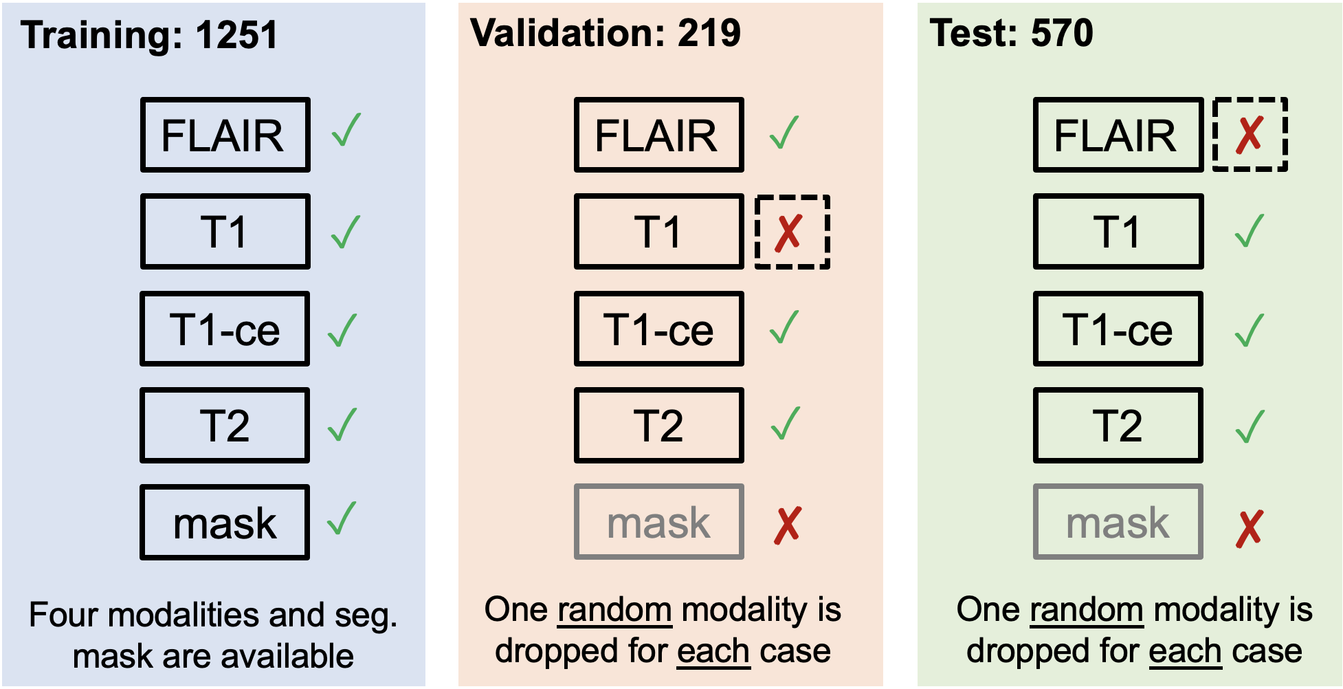
In line with the approach of algorithmic evaluation in machine learning, the data utilized in the BraSyn-2023 challenge is partitioned into training, validation, and private testing datasets. For the training data, participants are provided with four complete image modalities along with their respective segmentation labels, following a setup akin to the segmentation challenge. In the validation and test sets, a single modality out of the four sequences will be randomly omitted for each case. This deliberate omission aims to assess the effectiveness and performance of the submitted image synthesis methods.
Imaging data description. The mpMRI scans included in the BraTS 2023 challenge contain a) pre-contrast (T1) and b) post-contrast T1-weighted (T1Gd (Gadolinium)), c) T2-weighted (T2), and d) T2 Fluid Attenuated Inversion Recovery (T2-FLAIR) volumes, acquired with different protocols and various scanners from multiple institutions.
All the BraTS mpMRI scans have undergone standardized pre-processing, which includes the conversion of DICOM files to the NIfTI file format[16], co-registration to the same anatomical template (SRI24) [17], resampling to a uniform isotropic resolution (), and finally skull-stripping. The pre-processing pipeline is publicly available through the Cancer Imaging Phenomics Toolkit (CaPTk) 111https://cbica.github.io/CaPTk/ [18, 19, 20] and Federated Tumor Segmentation (FeTS) tool 222https://github.com/FETS-AI/Front-End/. Conversion to NIfTI strips the accompanying metadata from the DICOM images and essentially removes all Protected Health Information (PHI) from the DICOM headers. Furthermore, skull-stripping mitigates potential facial reconstruction/recognition of the patient [21, 22]. The specific approach we have used for skull stripping is based on a novel DL approach that accounts for the brain shape prior and is agnostic to the MRI sequence input [23].
All imaging volumes have then been segmented using the STAPLE [24] fusion of previous top-ranked BraTS algorithms, namely, nnU-Net [25], DeepScan [26], and DeepMedic [3]. Subsequently, volunteer neuro-radiologists with different levels of rank and experience undertook the manual refinement of these fused labels, adhering to a consistently communicated annotation protocol. The refined annotations, after this manual refinement process, were ultimately reviewed and approved by highly experienced board-certified attending neuro-radiologists who possess over 15 years of expertise in glioma-related work
2.2 Evaluation Metrics
The inference task that submitted algorithms have to solve is the following: When presented with a test set, one of the four modalities will be missing. The algorithm must predict a plausible brain tumor image for the missing modality. The image will be evaluated in terms of general image quality as well as by the performance of a downstream tumor segmentation algorithm that will be applied to the completed image set. For ranking the contributions, we will be using the following set of metrics: First, we calculate the structural similarity index measure (SSIM) as a direct image quality metric to quantify how realistic the synthesized images are compared to clinically acquired real images. SSIMs will be calculated in the tumor area and in the healthy part of the brain, resulting in two scores for each test subject. Second, we will evaluate whether the synthesized images are helpful for a segmentation algorithm. To this end, we will segment the four modalities including a synthesized volume with a state-of-the-art BraTS segmentation algorithm, and calculate Dice scores for three tumor structures as ‘indirect’ metrics. The automated segmentation will be performed by the final FeTS algorithm [27], leveraging the model pre-trained in the FETS brain tumor segmentation initiative. Participants will have access to the algorithm to fine-tune their methods and optimize their performance for our evaluation scenario. To assess image quality and segmentation accuracy, a total of five ranking metrics will be utilized.
2.3 Ranking strategy
For the final ranking of the participants, an equally weighted rank-sum is computed for each case of the test set, considering all the aforementioned metrics. This will rank algorithms according to each metric and sum up all ranks. The synthesis task will have five ranking scores: three Dice scores for each tumor tissue and two SSIMs for the image quality of tumor and non-tumor regions. The participating team with the best rank-sum will win each challenge task.
2.4 Participation Policy and Timeline
The challenge will begin by releasing a training dataset containing imaging data and corresponding labels for ground truth. Participants are allowed to start developing and training their methods using this dataset.
After three weeks, the validation data will be released, allowing participants to obtain initial results on unseen data. They can include these results in their submitted short MICCAI LNCS papers, along with their cross-validated results on the training data. While the ground truth for the validation data will not be disclosed, participants can make multiple submissions on the online evaluation platforms. The top-performing teams in the validation phase will be invited to prepare slides for a brief oral presentation of their methods during the BraTS challenge at MICCAI 2023.
During training, participants can utilize publicly available brain scans from healthy individuals. However, to ensure a fair comparison among methods, participants must explicitly mention any additional data in their submitted manuscripts and report results using only the training data provided by the organizers, focusing on potential differences in outcomes.
Finally, all participants will be assessed and ranked based on an unseen testing dataset, which will not be accessible once they have uploaded their containerized method to the evaluation platforms. The final rankings and winners will be announced at MICCAI 2023, with the top-ranked teams receiving monetary prizes.
Acknowledgments
We thank all the data contributors, annotators, and approvers for their time and efforts.
Funding
Research reported in this publication was partly supported by the National Institutes of Health (NIH) under award numbers: NIH/NCI/ITCR:U01CA242871. The content of this publication is solely the responsibility of the authors and does not represent the official views of the NIH.
References
- [1] P. Y. Wen, D. R. Macdonald, D. A. Reardon, T. F. Cloughesy, A. G. Sorensen, E. Galanis, J. DeGroot, W. Wick, M. R. Gilbert, A. B. Lassman, et al., “Updated response assessment criteria for high-grade gliomas: response assessment in neuro-oncology working group,” Journal of clinical oncology, vol. 28, no. 11, pp. 1963–1972, 2010.
- [2] P. Vollmuth, M. Foltyn, R. Y. Huang, N. Galldiks, J. Petersen, F. Isensee, M. J. van den Bent, F. Barkhof, J. E. Park, Y. W. Park, et al., “Artificial intelligence (ai)-based decision support improves reproducibility of tumor response assessment in neuro-oncology: An international multi-reader study,” Neuro-Oncology, vol. 25, no. 3, pp. 533–543, 2023.
- [3] K. Kamnitsas, C. Ledig, V. F. Newcombe, J. P. Simpson, A. D. Kane, D. K. Menon, D. Rueckert, and B. Glocker, “Efficient multi-scale 3d cnn with fully connected crf for accurate brain lesion segmentation,” Medical image analysis, vol. 36, pp. 61–78, 2017.
- [4] S. Pereira, A. Pinto, V. Alves, and C. A. Silva, “Brain tumor segmentation using convolutional neural networks in mri images,” IEEE transactions on medical imaging, vol. 35, no. 5, pp. 1240–1251, 2016.
- [5] G. Wang, W. Li, S. Ourselin, and T. Vercauteren, “Automatic brain tumor segmentation based on cascaded convolutional neural networks with uncertainty estimation,” Frontiers in computational neuroscience, vol. 13, p. 56, 2019.
- [6] G. M. Conte, A. D. Weston, D. C. Vogelsang, K. A. Philbrick, J. C. Cai, M. Barbera, F. Sanvito, D. H. Lachance, R. B. Jenkins, W. O. Tobin, et al., “Generative adversarial networks to synthesize missing t1 and flair mri sequences for use in a multisequence brain tumor segmentation model,” Radiology, vol. 299, no. 2, pp. 313–323, 2021.
- [7] J. E. Iglesias, E. Konukoglu, D. Zikic, B. Glocker, K. Van Leemput, and B. Fischl, “Is synthesizing mri contrast useful for inter-modality analysis?,” in Medical Image Computing and Computer-Assisted Intervention–MICCAI 2013: 16th International Conference, Nagoya, Japan, September 22-26, 2013, Proceedings, Part I 16, pp. 631–638, Springer, 2013.
- [8] S. M. Anwar, M. Majid, A. Qayyum, M. Awais, M. Alnowami, and M. K. Khan, “Medical image analysis using convolutional neural networks: a review,” Journal of medical systems, vol. 42, pp. 1–13, 2018.
- [9] H. Li, J. C. Paetzold, A. Sekuboyina, F. Kofler, J. Zhang, J. S. Kirschke, B. Wiestler, and B. Menze, “Diamondgan: unified multi-modal generative adversarial networks for mri sequences synthesis,” in Medical Image Computing and Computer Assisted Intervention–MICCAI 2019: 22nd International Conference, Shenzhen, China, October 13–17, 2019, Proceedings, Part IV 22, pp. 795–803, Springer, 2019.
- [10] M. F. Thomas, F. Kofler, L. Grundl, T. Finck, H. Li, C. Zimmer, B. Menze, and B. Wiestler, “Improving automated glioma segmentation in routine clinical use through artificial intelligence-based replacement of missing sequences with synthetic magnetic resonance imaging scans,” Investigative Radiology, vol. 57, no. 3, pp. 187–193, 2022.
- [11] J. E. Iglesias, B. Billot, Y. Balbastre, A. Tabari, J. Conklin, R. G. González, D. C. Alexander, P. Golland, B. L. Edlow, B. Fischl, et al., “Joint super-resolution and synthesis of 1 mm isotropic mp-rage volumes from clinical mri exams with scans of different orientation, resolution and contrast,” Neuroimage, vol. 237, p. 118206, 2021.
- [12] Q. Hu, H. Li, and J. Zhang, “Domain-adaptive 3d medical image synthesis: An efficient unsupervised approach,” in Medical Image Computing and Computer Assisted Intervention–MICCAI 2022: 25th International Conference, Singapore, September 18–22, 2022, Proceedings, Part VI, pp. 495–504, Springer, 2022.
- [13] U. Baid, S. Ghodasara, S. Mohan, M. Bilello, E. Calabrese, E. Colak, K. Farahani, J. Kalpathy-Cramer, F. C. Kitamura, S. Pati, et al., “The rsna-asnr-miccai brats 2021 benchmark on brain tumor segmentation and radiogenomic classification,” arXiv preprint arXiv:2107.02314, 2021.
- [14] S. Bakas, M. Reyes, A. Jakab, S. Bauer, M. Rempfler, A. Crimi, R. T. Shinohara, C. Berger, S. M. Ha, M. Rozycki, et al., “Identifying the best machine learning algorithms for brain tumor segmentation, progression assessment, and overall survival prediction in the brats challenge,” arXiv preprint arXiv:1811.02629, 2018.
- [15] B. H. Menze, A. Jakab, S. Bauer, J. Kalpathy-Cramer, K. Farahani, J. Kirby, Y. Burren, N. Porz, J. Slotboom, R. Wiest, et al., “The multimodal brain tumor image segmentation benchmark (brats),” IEEE transactions on medical imaging, vol. 34, no. 10, pp. 1993–2024, 2014.
- [16] R. Cox, J. Ashburner, H. Breman, K. Fissell, C. Haselgrove, C. Holmes, J. Lancaster, D. Rex, S. Smith, J. Woodward, et al., “A (sort of) new image data format standard: Nifti-1: We 150,” Neuroimage, vol. 22, 2004.
- [17] T. Rohlfing, N. M. Zahr, E. V. Sullivan, and A. Pfefferbaum, “The sri24 multichannel atlas of normal adult human brain structure,” Human brain mapping, vol. 31, no. 5, pp. 798–819, 2010.
- [18] C. Davatzikos, S. Rathore, S. Bakas, S. Pati, M. Bergman, R. Kalarot, P. Sridharan, A. Gastounioti, N. Jahani, E. Cohen, et al., “Cancer imaging phenomics toolkit: quantitative imaging analytics for precision diagnostics and predictive modeling of clinical outcome,” Journal of medical imaging, vol. 5, no. 1, p. 011018, 2018.
- [19] S. Rathore, S. Bakas, S. Pati, H. Akbari, R. Kalarot, P. Sridharan, M. Rozycki, M. Bergman, B. Tunc, R. Verma, et al., “Brain cancer imaging phenomics toolkit (brain-captk): an interactive platform for quantitative analysis of glioblastoma,” in International MICCAI Brainlesion Workshop, pp. 133–145, Springer, 2017.
- [20] S. Pati, A. Singh, S. Rathore, A. Gastounioti, M. Bergman, P. Ngo, S. M. Ha, D. Bounias, J. Minock, G. Murphy, et al., “The cancer imaging phenomics toolkit (captk): Technical overview,” in International MICCAI Brainlesion Workshop, pp. 380–394, Springer, 2019.
- [21] C. G. Schwarz, W. K. Kremers, T. M. Therneau, R. R. Sharp, J. L. Gunter, P. Vemuri, A. Arani, A. J. Spychalla, K. Kantarci, D. S. Knopman, R. C. Petersen, and C. R. Jack, “Identification of anonymous mri research participants with face-recognition software,” New England Journal of Medicine, vol. 381, no. 17, pp. 1684–1686, 2019. PMID: 31644852.
- [22] C. G. Schwarz, W. K. Kremers, T. M. Therneau, R. R. Sharp, J. L. Gunter, P. Vemuri, A. Arani, A. J. Spychalla, K. Kantarci, D. S. Knopman, R. C. Petersen, and C. R. Jack, “Identification from mri with face-recognition software,” New England Journal of Medicine, vol. 382, no. 5, pp. 489–490, 2020. PMID: 31995706.
- [23] S. Thakur, J. Doshi, S. Pati, S. Rathore, C. Sako, M. Bilello, S. M. Ha, G. Shukla, A. Flanders, A. Kotrotsou, et al., “Brain extraction on mri scans in presence of diffuse glioma: Multi-institutional performance evaluation of deep learning methods and robust modality-agnostic training,” NeuroImage, vol. 220, p. 117081, 2020.
- [24] S. K. Warfield, K. H. Zou, and W. M. Wells, “Simultaneous truth and performance level estimation (staple): an algorithm for the validation of image segmentation,” IEEE transactions on medical imaging, vol. 23, no. 7, pp. 903–921, 2004.
- [25] F. Isensee, P. F. Jaeger, S. A. Kohl, J. Petersen, and K. H. Maier-Hein, “nnu-net: a self-configuring method for deep learning-based biomedical image segmentation,” Nature Methods, pp. 1–9, 2020.
- [26] R. McKinley, R. Meier, and R. Wiest, “Ensembles of densely-connected cnns with label-uncertainty for brain tumor segmentation,” in International MICCAI Brainlesion Workshop, pp. 456–465, Springer, 2018.
- [27] S. Pati, U. Baid, B. Edwards, M. J. Sheller, P. Foley, G. A. Reina, S. Thakur, C. Sako, M. Bilello, C. Davatzikos, et al., “The federated tumor segmentation (fets) tool: an open-source solution to further solid tumor research,” Physics in Medicine & Biology, vol. 67, no. 20, p. 204002, 2022.