† People involved in the organization of the challenge.
‡ People contributing data from their institutions.
Corresponding author: 4949email: {florian.kofler@tum.de}
The Brain Tumor Segmentation (BraTS) Challenge 2023:
Local Synthesis of Healthy Brain Tissue via Inpainting
Abstract
A myriad of algorithms for the automatic analysis of brain MR images is available to support clinicians in their decision-making. For brain tumor patients, the image acquisition time series typically starts with a scan that is already pathological. This poses problems, as many algorithms are designed to analyze healthy brains and provide no guarantees for images featuring lesions. Examples include but are not limited to algorithms for brain anatomy parcellation, tissue segmentation, and brain extraction. To solve this dilemma, we introduce the BraTS 2023 inpainting challenge. Here, the participants’ task is to explore inpainting techniques to synthesize healthy brain scans from lesioned ones. The following manuscript contains the task formulation, dataset, and submission procedure. Later it will be updated to summarize the findings of the challenge. The challenge is organized as part of the BraTS 2023 challenge hosted at the MICCAI 2023 conference in Vancouver, Canada.
Keywords:
BraTS, challenge, brain, tumor, segmentation, machine learning, artificial intelligence, AI, infill, in-painting, challenge specific keyword1 Introduction
The focus of this year’s BraTS [1, 2, 3, 4] cluster of challenges spans across various tumor entities, missing data, and tumor image processing tasks. Specifically, in this challenge, we focus on local brain magnetic resonance imaging (MRI) synthesis. We define local synthesis as image synthesis within the brain area affected by a lesion, and the task is to fill in (inpaint) this area with image contrasts that would represent healthy tissue.
Local inpainting enables a synthetic “removal” of the tumor area from the image, which, in turn, offers means for an application of standard brain image segmentation algorithms in tumor patients (i.e. brain parcellation algorithms) – without any extra need for modification. This might bring a deeper understanding of the relationship between different brain tumor regions (brain parcellation) and abnormal brain tissue (brain tumors). Brain tissue segmentation would be of high importance for downstream tasks such as brain tumor modeling [5, 6]. Furthermore, inpainting would allow to deal with local artifacts, such as those arising from B-field inhomogeneities that occasionally degrade brain tumor images.
The clinical impact of our infill challenge will be in enabling the use of BraTS algorithms in case of non-standard imaging protocols [7], and in directly using brain parcellation tools, such as [8, 9], for treatment planning and the localization of areas of risk. Complementing the clinical perspective of improved algorithmic deployment, we – as the BraTS organizers – also envision technical advancements from a post-challenge use of the algorithms of this challenge: we will be able to further enrich the BraTS (training) data set by offering different whole brain parcellation masks from established neuroimaging tools for all BraTS cases. Both contributions will enable new lines of technical and neuroimaging research.

Challenge goal.
In this challenge, we encourage participants to offer with algorithmic solutions to synthesize 3D healthy brain tissue in the area affected by glioma. We cast the problem of synthesizing the healthy tissue as inpainting within the tumor area. Inpainting is a well-researched problem in the computer vision community 111In this manuscript, we use the term infilling synonymously for inpainting.. Numerous algorithms are proposed to realistically infill missing parts for 2D natural images [10, 11, 12, 13, 14] and a smaller number for brain images [15]. However, it is still a question whether such algorithms would translate to three-dimensional inpainting of MRI scans. To explore this, we provide a forum to discuss and a platform to benchmark the newly developed methods.
2 Challenge Description
2.1 Task and Data
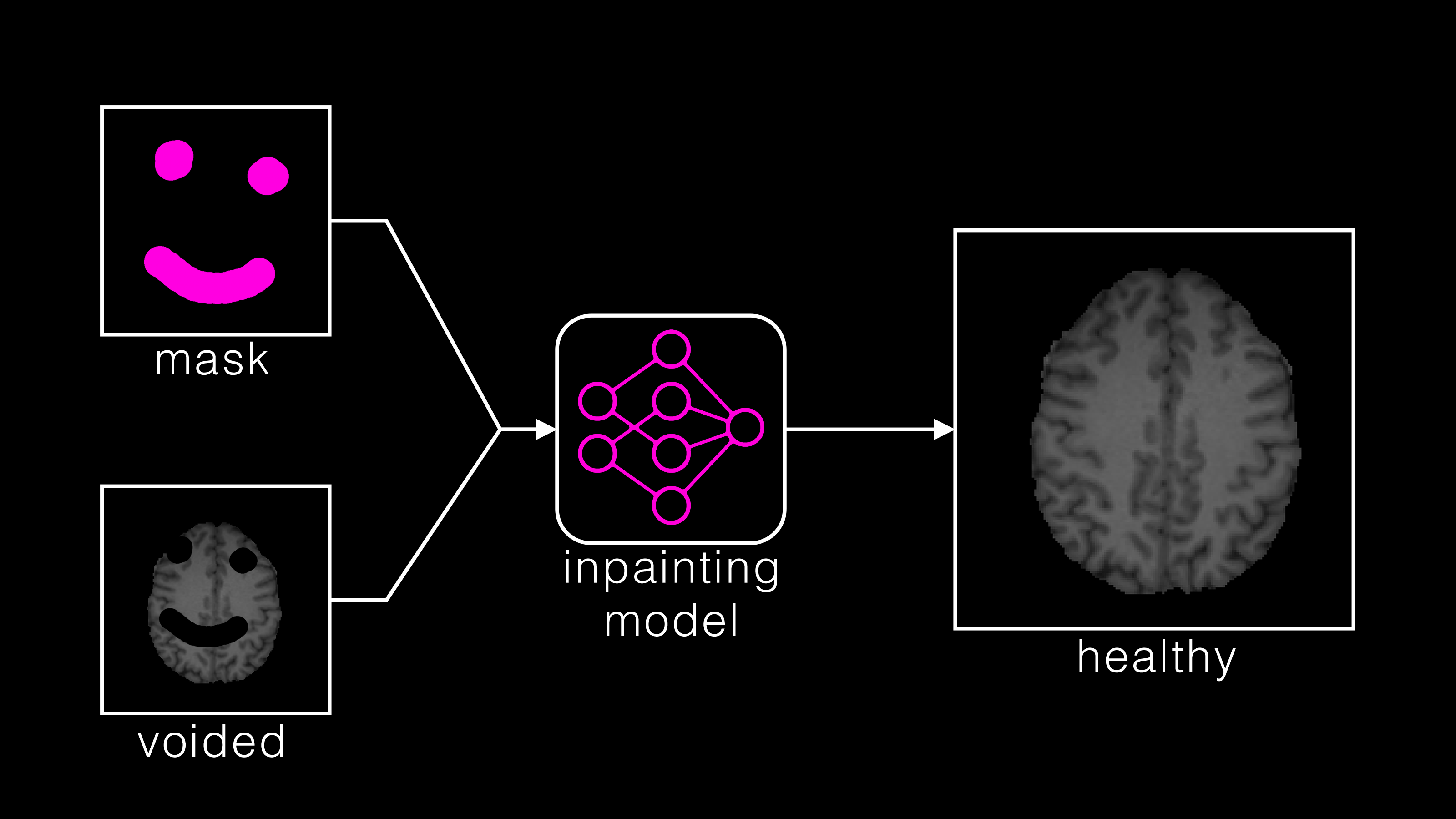
In the BraTS 2023 Inpainting Challenge, participants need to develop algorithms that synthesize healthy versions of brain MR images with pathologies. Given an image and masks corresponding to inpainting targets, the task is to infill the masked areas with healthy tissue. The challenge exclusively employs T1 scans from the multi-modal BraTS 2022 segmentation challenge. This challenge provides a retrospective collection of brain tumor MRI scans acquired from multiple different institutions under standard clinical conditions, but with different equipment and imaging protocols [1, 2, 3]. Additionally, the original BraTS challenges provide multi-class tumor delineations that are approved by expert neuroradiologists. These sub-regions are united to a single area of interest and dilated to account for mass effect.
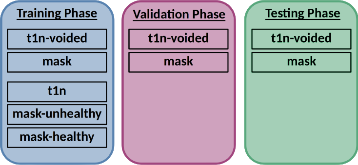
Following the paradigm of algorithmic evaluation in machine learning, the data included in the BraTS 2023 inpainting challenge are divided into training, validation, and testing datasets. Since there is no healthy ground truth tissue available for the tumor regions, surrogate inpainting masks are generated in the healthy part of the tumor and can be used for training supervised infill algorithms, as well as in the final evaluation.

To maintain a low barrier to participation, we provide participants with suggestions of healthy regions they can use to train their algorithms on (training set). This generation protocol for healthy masks is described in more detail in Section 2.2 and is publicly available. Participants are highly encouraged to iterate on it and use it to extend the quality and quantity of their training dataset. To enable this process, we provide the original T1 scans for all brains in the training dataset, see Fig. 2. For inference, the algorithms are provided with the inpainting masks and the T1 scans with voided masked areas. We apply this voiding, defined as setting voxels to an intensity of zero to avoid revealing target information. The testing data are kept hidden from the participants at all times. Only for training purposes the full T1 images are provided. A more detailed tutorial on an exemplary training and submission pipeline, including dataset handling and generation, will soon be provided at the challenge GitHub page.
2.2 Healthy Inpainting Mask Generation
We work on T1 sequence images and provide two types of masks to participants. The first type of mask delineates areas that are with high likelihood affected by the tumor or mass effect. In contrast, the second type marks areas that are likely to be healthy. Consequently, non-masked areas can be considered as contested territory.
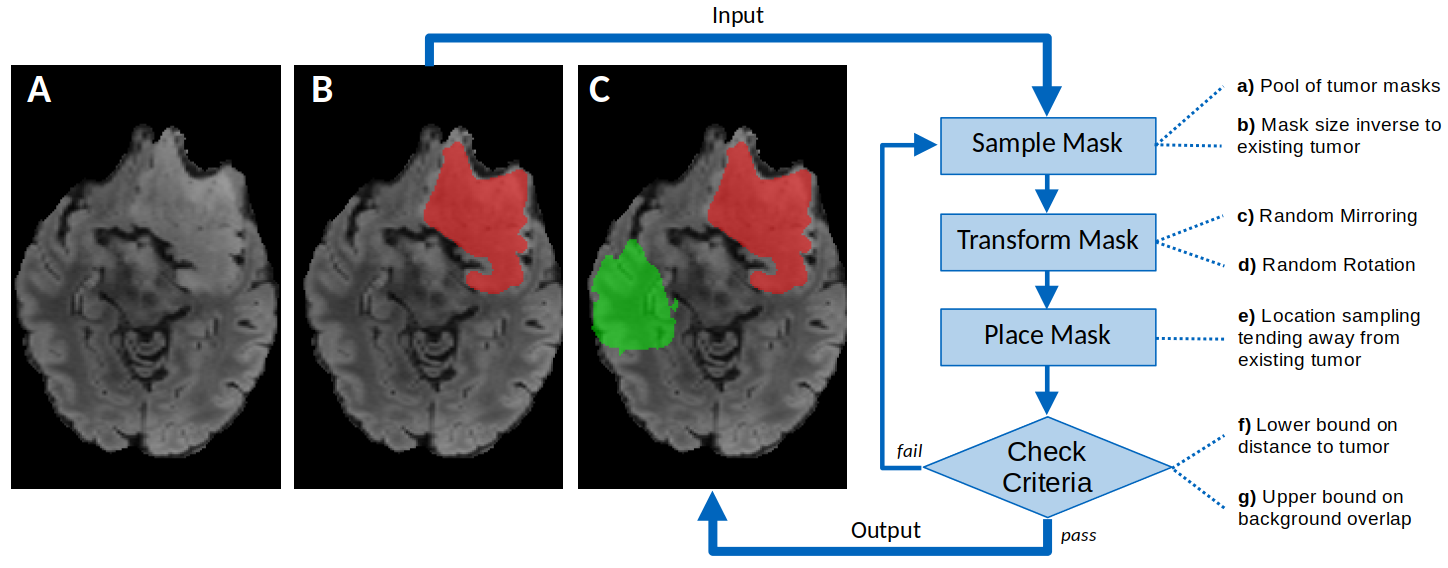
To create the masks for healthy brain regions, we sample from existing tumor shapes and place masks in regions that are distant from the tumor. This enables training and evaluating the inpainting algorithms on realistic shapes. It is important to note that, unlike tumors, the inpainting targets can overlap with ventricles, CSF, and out-of-brain background, as the algorithms are supposed to correctly inpaint all of such areas. Fig 3 visualizes the procedure to generate inpainting masks.

First, a pool of potential masks is created by taking all disconnected compartments of existing tumor annotations which consist of at least 800 voxels (Fig 3, a). A pool of 1429 masks is extracted from 1251 different brains. In the next step, a mask is randomly chosen from the previously created pool biased by the size of the tumor that is already present in the respective brain (Fig 3, b). Small masks are chosen for big tumors and vice versa. This is achieved by selecting masks on the other side of the side distribution of the existing tumor. For example, if our existing tumor annotation size is in the percentile (big tumor), we take a mask around the percentile (small mask). Respectively, if the tumor size is around the median (e.g. percentile), a healthy mask with a similar size (also percentile) is used.
After a mask is chosen from the pool, it is transformed using random mirroring (Fig 3, c) (mirror probability of 50% independently in each dimension) and rotation (Fig 3, d). For rotation, a random angle between 0° and 360° is used for both the X-Y and the Y-Z planes. After the transformations, the mask is placed at a semi-random position (Fig 3, e) based on the distance to the existing tumor. For that, two random points are sampled within the brain (non-zero T1) but not in the tumor. According to Euclidean voxel distance, the point furthest away from the tumor is chosen as the position for the healthy tissue mask.
Afterward, the mask’s validity is checked based on a minimal distance (Fig 3, f) to the tumor annotation of 5 voxels Euclidean distance and that at most 25% overlap (Fig 3, g) with the background (zero T1). If one of these conditions is violated, the sampling process with subsequent transformation and placement is repeated. This mask sampling procedure is carried out until one valid configuration is found. The resulting healthy mask, as well as the (dilated) tumor mask, is provided to participants.
The above-described sampling approach is applied only once per brain for the training, validation, and testing dataset. Of course, more than one mask can be generated per brain. We encourage participants to improve this procedure and generate more healthy in-painting masks for training purposes.
2.3 Participation
The challenge will commence with the release of the training dataset, which will consist of imaging data, the corresponding inpainting masks, and the underlying tumor and healthy tissue masks.
Participants can start designing and training their methods using this training dataset. The validation data will be released within three weeks after the training data is released. This will allow participants to obtain preliminary results in unseen data and also report these in their submitted short MICCAI LNCS papers, in addition to their cross-validated results on the validation data.
Finally, all participants will be evaluated and ranked on the same unseen testing data, which will not be made available to the participants, after uploading their containerized method to the evaluation platforms. The final top-ranked participating teams will be announced at the 2023 MICCAI Annual Meeting. The top-ranked participating teams in both tasks will receive material prizes.
2.4 Performance Evaluation
To measure the performance of the contributions, we will evaluate the quality of the infilled regions. Since ground truth data is only available for the masked regions with healthy tissue, the evaluation will be restricted to these. We will use the following set of well-established metrics to quantify how realistic the synthesized image regions are compared to real ones: structural similarity index measure (SSIM) [16], peak-signal-to-noise-ratio (PSNR), and mean-square-error (MSE). For the final ranking of the MICCAI challenge, an equally weighted rank-sum is computed across all three metrics. To compute the rank within each metric, we rank the participants for each case and again compute a rank-sum.
2.5 Baseline Model
Our baseline model implementation serves as a benchmark and illustration of the task at hand. The reference model can be accessed on the challenge GitHub site, which utilizes a pix2pix architecture [17]. This model predicts solely the missing mask and concatenates it to the masked image. pix2pix represents an autoencoder that utilizes an added GAN loss to enhance image fidelity. Performance evaluation for the baseline model will be done using the same measurements as mentioned in 2.4.
3 Results
This section will contain the challenge rankings on the internal test set. Furthermore, more sophisticated qualitative and quantitative analyses will be conducted.
References
- [1] B. H. Menze, A. Jakab, S. Bauer, J. Kalpathy-Cramer, K. Farahani, J. Kirby, Y. Burren, N. Porz, J. Slotboom, R. Wiest, et al., “The multimodal brain tumor image segmentation benchmark (brats),” IEEE transactions on medical imaging, vol. 34, no. 10, pp. 1993–2024, 2014.
- [2] S. Bakas, H. Akbari, A. Sotiras, M. Bilello, M. Rozycki, J. S. Kirby, J. B. Freymann, K. Farahani, and C. Davatzikos, “Advancing the cancer genome atlas glioma mri collections with expert segmentation labels and radiomic features,” Scientific data, vol. 4, no. 1, pp. 1–13, 2017.
- [3] S. Bakas, M. Reyes, A. Jakab, S. Bauer, M. Rempfler, A. Crimi, R. T. Shinohara, C. Berger, S. M. Ha, M. Rozycki, et al., “Identifying the best machine learning algorithms for brain tumor segmentation, progression assessment, and overall survival prediction in the brats challenge,” arXiv preprint arXiv:1811.02629, 2018.
- [4] U. Baid, S. Ghodasara, S. Mohan, M. Bilello, E. Calabrese, E. Colak, K. Farahani, J. Kalpathy-Cramer, F. C. Kitamura, S. Pati, et al., “The rsna-asnr-miccai brats 2021 benchmark on brain tumor segmentation and radiogenomic classification,” arXiv preprint arXiv:2107.02314, 2021.
- [5] I. Ezhov, T. Mot, S. Shit, J. Lipkova, J. C. Paetzold, F. Kofler, C. Pellegrini, M. Kollovieh, F. Navarro, H. Li, et al., “Geometry-aware neural solver for fast bayesian calibration of brain tumor models,” IEEE Transactions on Medical Imaging, vol. 41, no. 5, pp. 1269–1278, 2021.
- [6] I. Ezhov, K. Scibilia, K. Franitza, F. Steinbauer, S. Shit, L. Zimmer, J. Lipkova, F. Kofler, J. C. Paetzold, L. Canalini, et al., “Learn-morph-infer: a new way of solving the inverse problem for brain tumor modeling,” Medical Image Analysis, vol. 83, p. 102672, 2023.
- [7] J. E. Villanueva-Meyer, M. C. Mabray, and S. Cha, “Current clinical brain tumor imaging,” Neurosurgery, vol. 81, no. 3, pp. 397–415, 2017.
- [8] M. Jenkinson, C. Beckmann, T. Behrens, M. Woolrich, and S. Smith, “Fsl neuroimage 62 (2): 782–790,” 2012.
- [9] B. Fischl, “Freesurfer,” Neuroimage, vol. 62, no. 2, pp. 774–781, 2012.
- [10] S. Zhao, J. Cui, Y. Sheng, Y. Dong, X. Liang, E. I. Chang, and Y. Xu, “Large scale image completion via co-modulated generative adversarial networks,” in International Conference on Learning Representations (ICLR), 2021.
- [11] A. Romero, A. Castillo, J. Abril-Nova, R. Timofte, R. Das, S. Hira, Z. Pan, M. Zhang, B. Li, D. He, et al., “Ntire 2022 image inpainting challenge: Report,” in Proceedings of the IEEE/CVF Conference on Computer Vision and Pattern Recognition, pp. 1150–1182, 2022.
- [12] X. Guo, H. Yang, and D. Huang, “Image inpainting via conditional texture and structure dual generation,” in Proceedings of the IEEE/CVF International Conference on Computer Vision (ICCV), pp. 14134–14143, October 2021.
- [13] J. Yu, Z. Lin, J. Yang, X. Shen, X. Lu, and T. S. Huang, “Free-form image inpainting with gated convolution,” arXiv preprint arXiv:1806.03589, 2018.
- [14] J. Yu, Z. Lin, J. Yang, X. Shen, X. Lu, and T. S. Huang, “Generative image inpainting with contextual attention,” arXiv preprint arXiv:1801.07892, 2018.
- [15] P. Rouzrokh, B. Khosravi, S. Faghani, M. Moassefi, S. Vahdati, and B. J. Erickson, “Multitask brain tumor inpainting with diffusion models: A methodological report,” arXiv preprint arXiv:2210.12113, 2022.
- [16] Z. Wang, A. C. Bovik, H. R. Sheikh, and E. P. Simoncelli, “Image quality assessment: from error visibility to structural similarity,” IEEE transactions on image processing, vol. 13, no. 4, pp. 600–612, 2004.
- [17] P. Isola, J.-Y. Zhu, T. Zhou, and A. A. Efros, “Image-to-image translation with conditional adversarial networks,” in Proceedings of the IEEE conference on computer vision and pattern recognition, pp. 1125–1134, 2017.