Learning to Learn Unlearned Feature for Brain Tumor Segmentation
Abstract
We propose a fine-tuning algorithm for brain tumor segmentation that needs only a few data samples and helps networks not to forget the original tasks. Our approach is based on active learning and meta-learning. One of the difficulties in medical image segmentation is the lack of datasets with proper annotations, because it requires doctors to tag reliable annotation and there are many variants of a disease, such as glioma and brain metastasis, which are the different types of brain tumor and have different structural features in MR images. Therefore, it is impossible to produce the large-scale medical image datasets for all types of diseases. In this paper, we show a transfer learning method from high grade glioma to brain metastasis, and demonstrate that the proposed algorithm achieves balanced parameters for both glioma and brain metastasis domains within a few steps.
1 Introduction
The performance of semantic segmentation using deep neural networks has been improved recently. These segmentation networks are applied to the medical image analysis to help doctors to save time in diagnosis. The state-of-the-art networks still require large amounts of training data points for pre-training and fine-tuning. Gathering medical image datasets, however, is an expensive and time-consuming so that there are fewer datasets than the datasets for common objects [1, 3, 7]. In particular, in brain tumor segmentation, there is a proper dataset called BRaTS for the High Grade Glioma (HGG) and the Low Grade Glioma (LGG) [5], and no well-annotated dataset for brain metastasis.
HGG and brain metastasis have the different structural feature but the similar contrast feature. In contrast enhanced MR image, the region of brain tumor is highlighted because of the contrast media. In both cases, tumors have the similar contrast. These brain tumors have different pathological properties, so they have different structural characteristics. Therefore, a pre-trained network using the HGG dataset can not generate perfect segmentation for brain metastasis. In this paper, We learn the unlearned feature of brain metastasis without forgetting the pre-trained feature in order to optimize the balanced parameters between HGG and metastasis.
We first pre-trained the fully convolutional network (FCN) [4] with HGG in the dataset [5]. The gradient descent based fine tuning, which we call naive tune, uses many selected data points which have balanced instances per each class to produce the optimal fine-tuning results [3, 7]. We propose two novel fine-tuning methods, passive meta-tune and active meta-tune, to optimize the pre-trained network. These two methods decide which training data points are first learned and update the network with [2]. The orders of training dataset is determined with two active learning based rules, the passive learning and the active learning [6]. In this work, we produce the annotated brain metastasis data samples with 30 patients. Similar to the BraTS dataset, for a patient, there are 4 MR sequences, FLAIR, T1, T1 contrast-enhanced, T2, and 25 slices per each sequence.
2 Proposed methodology
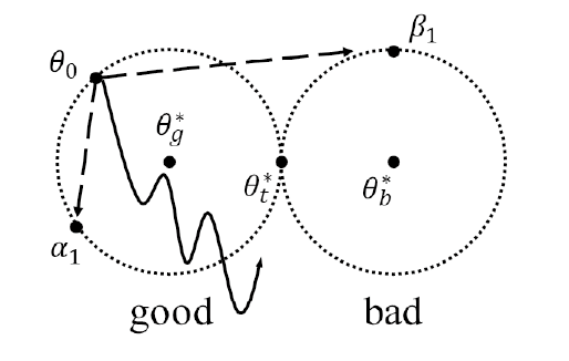
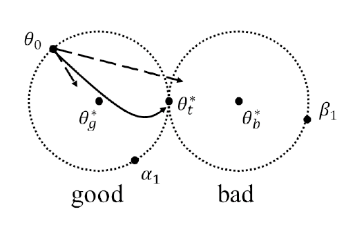
We propose two active learning based meta-tune methods, each of which is based on random sampling and a variant of uncertainty sampling [6]. We define the meta-tune as a fine-tune method using MAML algorithm [2]. The meta-tune generalize the unlearned training examples more quickly than the graident-based fined tune method (naive tune). As shown in Figure (1), we train the model with the learned features continuously as well as the unlearned features not to forget the learned features.
3 Experimental Results
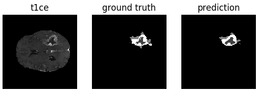
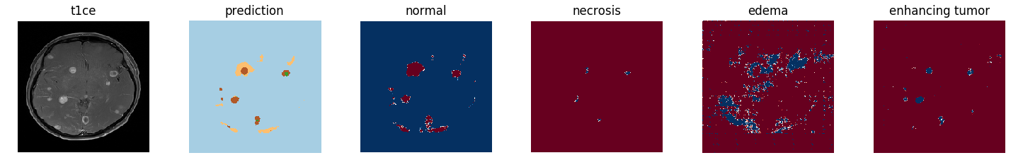
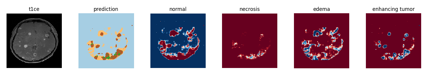
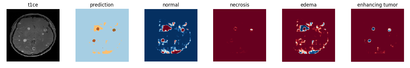
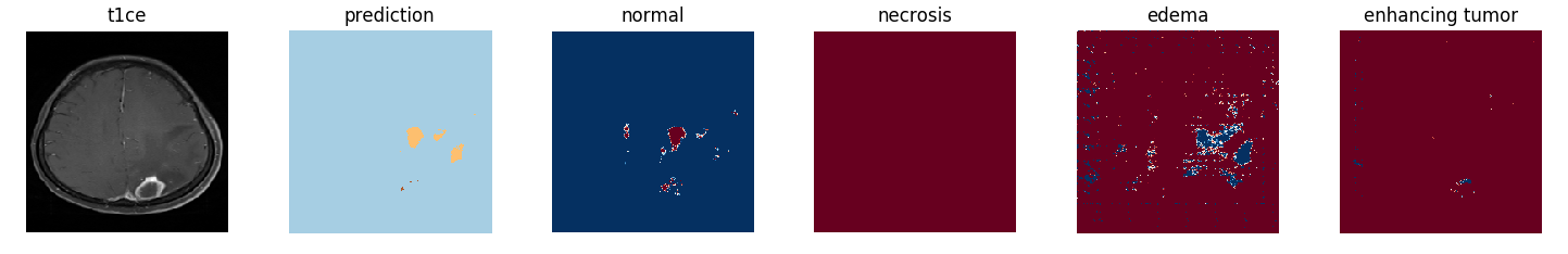
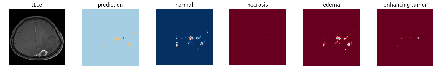
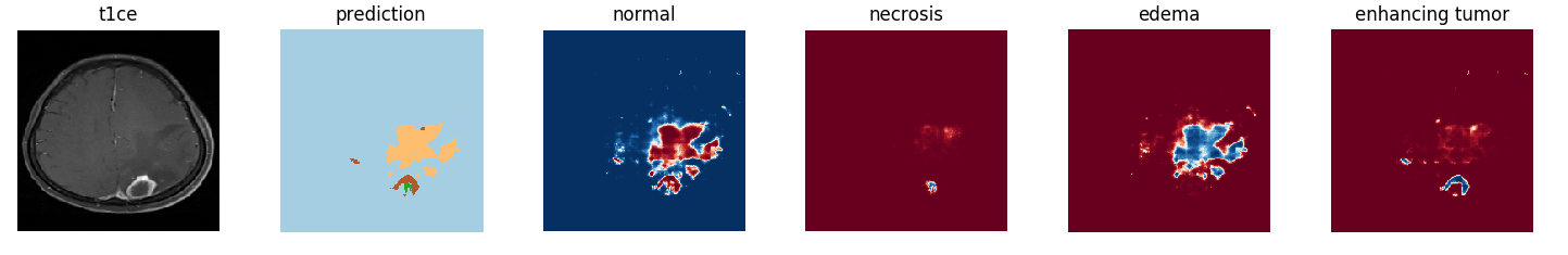
We clinically-acquire multimodal MRI scans and produce all the ground truth annotations by neuroradiologists. We use a VGG-16 based FCN network as the pre-train network. Then, we test our algorithms. (yellow: edema, green: necrosis, brown: ehnacing tumor: brown, red : high probability).
| Method | Dice Score (std) |
|---|---|
| baseline | 0.66 (on BRaTS) |
| naive | 0.33 0.3413 |
| passive | 0.41 0.2752 |
| active | 0.45 0.2317 |
4 Conclusion
We proposed an active meta-tune method which learns unlearned feature without forgetting the original task. We show that our method have a generalization effect within the target domain segmentation (brain metastasis).We expect our method can be extended to other medical lesion applications.
Acknowledgments
This work is in part supported by SNU Eng-Med Collaboration Grant, Basic Science Research Program (NRF-2017R1A2B2007102) through NRF funded by MSIP, Technology Innovation Program (10051928) funded by MOTIE, Bio-Mimetic Robot Research Center funded by DAPA (UD130070ID), INMAC, and BK21-plus.
References
- [1] M. "Everingham, L. Van Gool, C. K. I. Williams, J. Winn, and A." Zisserman. "the PASCAL Visual Object Classes Challenge 2011 (VOC2011) Results". "http://www.pascal-network.org/challenges/VOC/voc2011/workshop/index.html".
- [2] Chelsea Finn, Pieter Abbeel, and Sergey Levine. Model-agnostic meta-learning for fast adaptation of deep networks. In International Conference on Machine Learning, pages 1126–1135, 2017.
- [3] Tsung-Yi Lin, Michael Maire, Serge J. Belongie, Lubomir D. Bourdev, Ross B. Girshick, James Hays, Pietro Perona, Deva Ramanan, Piotr Dollár, and C. Lawrence Zitnick. Microsoft COCO: common objects in context. CoRR, abs/1405.0312, 2014.
- [4] Jonathan Long, Evan Shelhamer, and Trevor Darrell. Fully convolutional networks for semantic segmentation. In The IEEE Conference on Computer Vision and Pattern Recognition (CVPR), June 2015.
- [5] Bjoern H Menze, Andras Jakab, Stefan Bauer, Jayashree Kalpathy-Cramer, Keyvan Farahani, Justin Kirby, Yuliya Burren, Nicole Porz, Johannes Slotboom, Roland Wiest, et al. The multimodal brain tumor image segmentation benchmark (brats). IEEE transactions on medical imaging, 34(10):1993, 2015.
- [6] Stephen Mussmann and Percy Liang. On the relationship between data efficiency and error for uncertainty sampling. In Jennifer Dy and Andreas Krause, editors, Proceedings of the 35th International Conference on Machine Learning, volume 80 of Proceedings of Machine Learning Research, pages 3674–3682, Stockholmsmässan, Stockholm Sweden, 10–15 Jul 2018. PMLR.
- [7] Bolei Zhou, Hang Zhao, Xavier Puig, Sanja Fidler, Adela Barriuso, and Antonio Torralba. Scene parsing through ade20k dataset. In Proceedings of the IEEE Conference on Computer Vision and Pattern Recognition, 2017.