† People involved in the organization of the challenge.
‡ People contributing data from their institutions.
§ People involved in annotation process.
* First author.
¶ Senior authors.
** Corresponding author: 3434email: {evan.calabrese@duke.edu}
The ASNR-MICCAI Brain Tumor Segmentation (BraTS) Challenge 2023: Intracranial Meningioma
Abstract
Meningiomas are the most common primary intracranial tumor in adults and can be associated with significant morbidity and mortality. Radiologists, neurosurgeons, neuro-oncologists, and radiation oncologists rely on multiparametric MRI (mpMRI) for diagnosis, treatment planning, and longitudinal treatment monitoring; yet automated, objective, and quantitative tools for non-invasive assessment of meningiomas on mpMRI are lacking. The BraTS meningioma 2023 challenge will provide a community standard and benchmark for state-of-the-art automated intracranial meningioma segmentation models based on the largest expert annotated multilabel meningioma mpMRI dataset to date. Challenge competitors will develop automated segmentation models to predict three distinct meningioma sub-regions on MRI including enhancing tumor, non-enhancing tumor core, and surrounding nonenhancing T2/FLAIR hyperintensity. Models will be evaluated on separate validation and held-out test datasets using standardized metrics utilized across the BraTS 2023 series of challenges including the Dice similarity coefficient and Hausdorff distance. The models developed during the course of this challenge will aid in incorporation of automated meningioma MRI segmentation into clinical practice, which will ultimately improve care of patients with meningioma.
Keywords:
BraTS, challenge, brain, tumor, segmentation, machine learning, deep learning, artificial intelligence, AI, meningioma1 Introduction
Meningiomas are the most common primary intracranial tumor in adults and can result in significant morbidity and mortality for affected patients [1, 2]. Most meningiomas (80%) are World Health Organization (WHO) grade 1 benign tumors and are typically well controlled with observation, surgical resection, and/or radiation therapy. However, higher grade meningiomas (WHO grades 2 and 3) are associated with significantly higher morbidity and mortality rates and often recur despite optimal management. Currently there is no reliable noninvasive method for identifying meningioma grade, assessing aggressiveness, or predicting recurrence and survival. Traditional MRI features used by clinicians to guide treatment strategy, such as meningioma volume, or degree of peritumoral edema, may not represent tumor WHO grade, or expected clinical course. As such, there is a need for improved radiographic assessment of meningiomas, such that it can guide patient-specific treatment strategy.
Automated segmentation on brain magnetic resonance imaging (MRI) has matured into a clinically viable tool that can provide objective assessments of tumor volume and can assist in surgical planning, radiotherapy planning, and treatment response assessment. However, to date most tumor segmentation studies have focused on gliomas. Meningiomas, while typically more circumscribed than gliomas, provide additional technical challenges for segmentation given their extra-axial location, multiplicity, and propensity for skull-base involvement. In addition, unlike other intracranial tumors, meningiomas are commonly diagnosed by imaging alone, which increases the importance of MRI for treatment planning. The ability to predict meningioma aggressiveness pre-operatively would confer the benefit of guiding surgical strategy, as maximal resection remains the mainstay of effective treatment to mitigate the risk of recurrence[3].
The purpose of the Brain Tumor Segmentation (BraTS) 2023 meningioma challenge is to develop an automated multi-compartment brain MRI segmentation algorithm for intracranial meningiomas. This algorithm, if successful, will provide an important tool for objective assessment of tumor volume for surgical and radiotherapy planning. In addition, this algorithm will provide a starting point for future studies focused on identifying meningioma grade, assessing aggressiveness, and predicting risk of recurrence based on MRI findings alone. This manuscript describes the annotation protocol used to prepare the data used in the BraTS 2023 meningioma challenge, and it outlines the challenge tasks and the evaluation metrics used. Additionally, the manuscript explores the advantages, objectives, and constraints of the challenge, and outlines future directions currently under consideration.
2 Materials & Methods
2.1 Defining intracranial meningioma
For the purposes of this challenge, intracranial meningioma was defined as any grade or subtype of meningioma occurring within the cranial vault. Meningiomas arise from the arachnoid layer of the meninges between the dura mater and pia mater. Intracranial meningiomas commonly present at supratentorial sites of dural reflection, along the sphenoid sinus, and the skull base. Less commonly, meningiomas occur in intraventricular and suprasellar regions, the olfactory groove, and in the posterior fossa along the petrous bone. Approximately 80% of meningioma are classified as WHO grade 1 and include meningothelial, fibroblastic, transitional, psammomatous, angiomatous, microcystic, secretory, metaplastic, and lymphoplasmacyte rich subtypes. Histologically, WHO grade 1 meningiomas typically have psammoma bodies, cellular whirls and calcifications. Approximately 18% of meningiomas are classified as WHO grade 2 and include atypical, chordoid, clear cell, and subtypes. Histologically, WHO grade 2 meningiomas have 4 mitoses per high-power field (HPF), brain invasion, or 3 of the following: hypercellularity, small cells, prominent nucleoli, patternless or sheet like growth, foci of spontaneous necrosis on hematoxylin and eosin staining. Only about 2% of meningiomas are classified as WHO grade 3 and include anaplastic, papillary, and rhabdoid subtypes. Histologically, WHO grade 3 meningiomas have 10 mitoses/HPF and/or carcinomatous features, sarcomatous features, melanomatous features, loss of usual growth pattern, or multifocal necrotic foci. Radiographic findings vary greatly amongst meningioma subtypes, and to date, there are no reliable imaging features that can accurately distinguish meningioma grade or aggressiveness.
2.2 Defining Meningioma MRI Sub-Components
Similar to prior BraTS glioma challenges [4, 5, 6, 7, 8], a key aspect of the challenge is the subdivision of the different tumor compartments that are apparent on MRI [9, 10, 4, 11]. To date, there are no published guidelines about the specific target volume delineation for intracranial meningioma based on prospective studies [12]. However, many different meningioma MRI appearances and sub-regions have been previously described. In 2022, Association des Neuro-oncologues d’Expression Française (ANOCEF) outlined consensus guidelines for meningioma gross tumor volume after 20 experts from 17 radiotherapy centers participated in a three round modified Delphi consensus [12, 13]. The ANOCEF committee defined the enhancing gross tumor to include MRI T1 contrast-enhancing lesions, thickened meninges, and directly invaded bone.
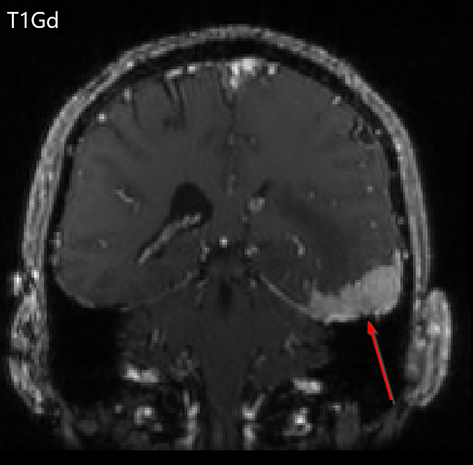
Watt et al. described that meningioma non-enhancing calcification is consistent with low signal intensity on T2 imaging [14]. The microcystic meningioma subtype comprises 1.6% of meningiomas and demonstrates a uniform low-signal intensity on T1 and high signal intensity of T2. Bitzer et al found that peritumoral edema is found in 60% of meningioma cases and is demonstrated by a hyperintense FLAIR signal [15]. En-plaque meningioma is described as asymmetric thickened sheets of enhancing dura (Fig. 1) [14]. Dural tail involvement is defined as thickening and enhancement of the dura infiltrating away from the lesion (Fig. 2) and is used to help radiographically distinguish meningioma from other enhancing CNS lesions owing to its high prevalence in meningioma (72%) and absence in meningioma mimics like intracranial Schwannomas [14, 16, 17].

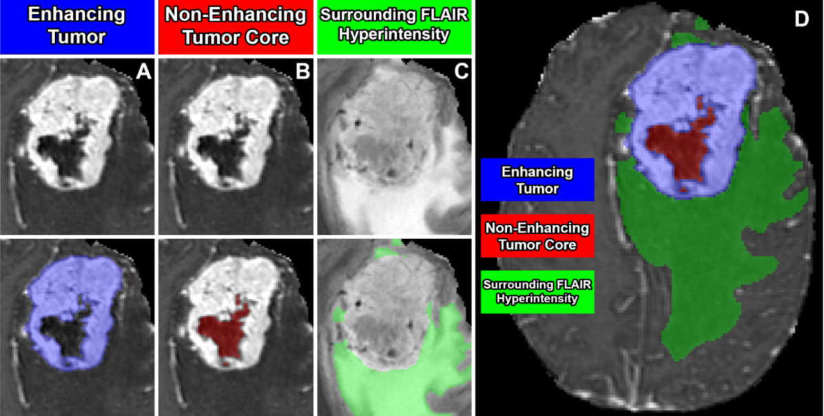
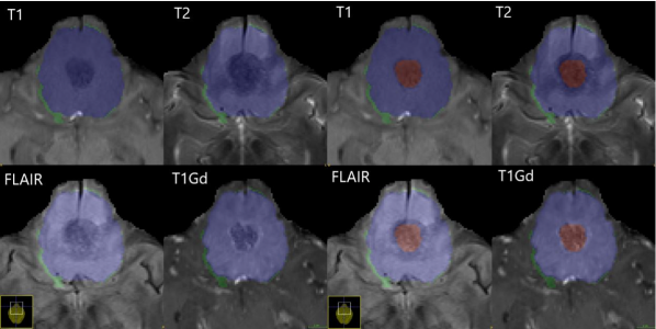
Based on this prior work and others, the BraTS 2023 meningioma challenge defines 3 distinct and non-overlapping segmentation labels (Fig. 3). These include “enhancing tumor”, “nonenhancing tumor core”, and surrounding non-enhancing T2/FLAIR hyperintensity (SNFH). The enhancing tumor label includes all contrast enhancing meningioma, focally thickened meninges (including dural tail), as well as en-plaque meningiomas. This label approximates the region of active, viable tumor. The non-enhancing tumor core label includes all calcification, hyperostosis, necrosis, degeneration, and any other atypical non-enhancing tumor radiographic findings. This label along with the enhancing tumor label (together comprising the “tumor core”) approximately corresponds to the portion of tumor related imaging abnormality that would typically be removed in a gross total resection. The SNFH label includes the entire extent of tumor related T2/FLAIR hyperintensity surrounding the tumor core. This label is distinct from the other labels in that it is composed entirely of brain parenchyma and is not expected to contain any tumor cells, but rather represents irritated, inflamed, and/or edematous brain tissue resulting from adjacent tumor. Importantly, non-tumor related T2/FLAIR signal abnormality, commonly related to chronic microvascular ischemic white matter changes (e.g. leukoaraiosis) or other vascular pathology, was not included in the SNFH label.
2.3 Data Description
All MRI studies in the BraTS meningioma challenge were performed in the pre-operative and pre-treatment setting and were included if one or more tumors radiographically or pathologically consistent with meningioma were included within the field of view. MRI studies containing any intracranial tumor that was not radiographically or pathologically consistent with meningioma were excluded (including cases of neurofibromatosis type 2 with intracranial Schwannomas). All cases include multiparametric MRI (mpMRI) consisting of pre-contrast T1-weighted, post-contrast T1-weighted, T2-weighted, and T2-weighted Fluid Attenuated Inversion Recovery (FLAIR) series.
2.4 Participating sites
Preoperative mpMRI data for the BraTS Meningioma Challenge were contributed by academic medical centers across the United States (Table 1). Cases were identified based on histopathologic assessment following resection or biopsy or based on a formal clinical and radiographic diagnosis of meningioma, often identified based on the International Classification of Diseases Tenth Revision (ICD-10) code D32.9, ”benign neoplasm of the meninges”. This differs from prior BraTS glioma challenges where inclusion was based on histopathologic diagnosis alone; however, unlike high-grade gliomas, meningiomas are commonly diagnosed by imaging alone and may be observed and/or treated without a definitive tissue diagnosis. The specific case inclusion methods (pathologic, clinical/radiologic, or both) and case collection methods (i.e. retrospective, prospective, consecutive) were chosen by each participating site independently, often on the basis of pre-existing curated datasets. Imaging parameters including field strength, echo/repetition time, slice resolution, and slice thickness varied considerably between and within sites. In an effort to encourage data contribution, data contributors were not required to disclose data collection methods or MRI protocol information.
Table 1: Total meningioma case contributions from each participating site. Only meningiomas that are pre-operative, within the intracranial brain mask, and radiographically or pathologically consistent with meningioma are included.
2.5 Image data preprocessing
All mpMRI data underwent standardized image pre-processing steps including conversion from DICOM to Neuroimaging Informatics Technology Initiative (NIfTI) image file format; co-registration of individual image series (T1-weighted, T2-weighted, T2-FLAIR, T1Gd) to the SRI24 atlas space including uniform 1 mm isotropic resampling, and automated skull-stripping using a deep convolutional neural network approach. These basic image pre-processing steps are implemented in the open-source and publicly available Federated Tumor Segmentation (FeTS) tool111https://fets-ai.github.io/Front-End/. It should be noted that meningioma can extend through the skull and/or skull-base foramina and that any extra-cranial portions of tumors were implicitly excluded by the skull-stripping process. Despite this limitation, skull-stripping was included in the pre-processing to preserve patient anonymity (by preventing face reconstruction) and to ensure consistency with other BraTS challenges.
2.6 Pre-segmentation
Prior to manual segmentation, a deep convolutional neural network-based automated segmentation model was used for automated multi-compartment pre-segmentation. This model, implemented in nnU-Net222https://github.com/MIC-DKFZ/nnUNet was initially trained on a sample of 73 manually labeled studies from a single participating institution [18]. Of note, this initial sample consisted entirely of meningiomas that subsequently underwent surgical resection, which may bias the model to poorer performance for non-surgical meningiomas. During the manual correction phase of the challenge preparation, the automated segmentation algorithm was periodically retrained using additional manually corrected cases from other participating sites, including sites that contributed non-surgical meningioma cases. The purpose of iteratively retraining the model with new data was to improve its generalizability to different MRI appearances of meningioma and reduce pre-segmentation bias. Model weights for each of the different pre-segmentation models will be made publicly available at the conclusion of the challenge.
2.7 Manual corrections
For each meningioma case, manual review and correction of pre-segmented labels was performed by individual volunteer “annotators” with widely varying experience levels spanning from medical students to fellowship trained neuroradiologists with 10+ years of experience. Manual corrections were performed using ITK-SNAP, a free, open-source, multi-platform software application used to segment structures in 3D and 4D biomedical images [19]. Annotators were provided with each of the following: 1) basic instruction on using ITK-SNAP for meningioma segmentation, 2) written descriptions of the composition of each tumor sub-compartment, and 3) a list (with examples) of common pre-segmentation errors to identify and address. These steps were designed to reduce inter-observer variability in tumor segmentation correction. After manual correction by annotators, each case was manually reviewed by a fellowship trained neuroradiologist “approver” before inclusion in the challenge dataset. In cases where an approver identified an inaccurate or incomplete segmentation, the case was returned to a different annotator for further refinement until the approver verified the manual correction.
2.8 Common errors of automated segmentation
Based on subjective review of pre-segmented meningioma cases by challenge approvers, a set of commonly encountered automated segmentation errors were identified and provided to challenge annotators in an effort to improve inter-observer variability. These commonly encountered errors included:
-
1.
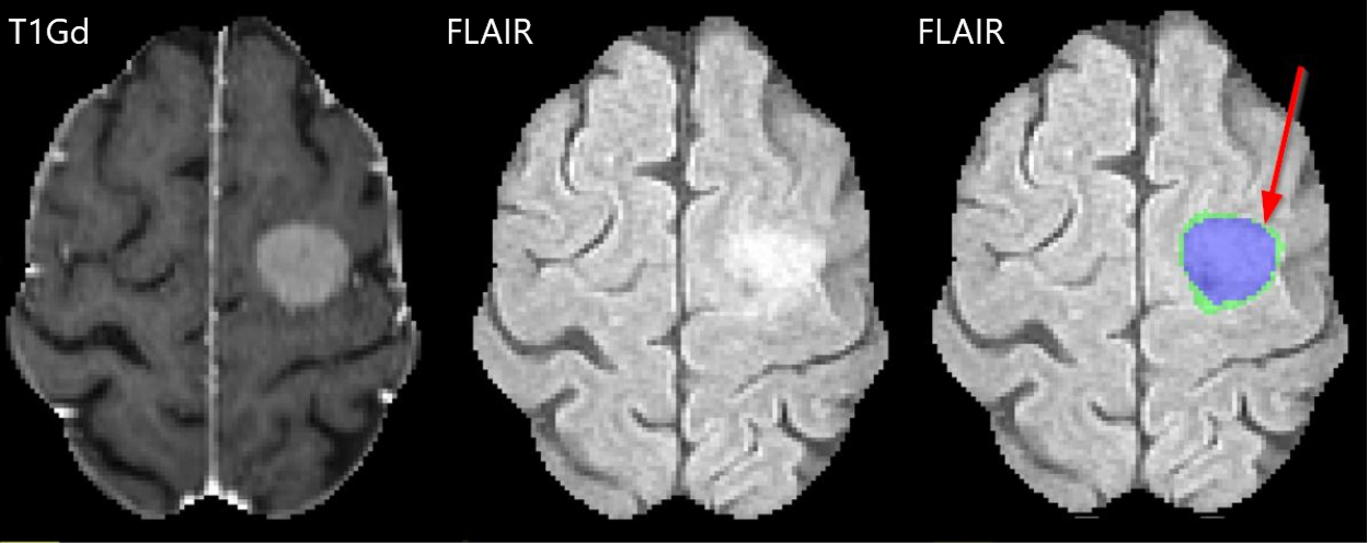
A thin rim of erroneously assigned SNFH label immediately surrounding smaller meningiomas without any true associated SNFH (Fig. 4a).
-
2.
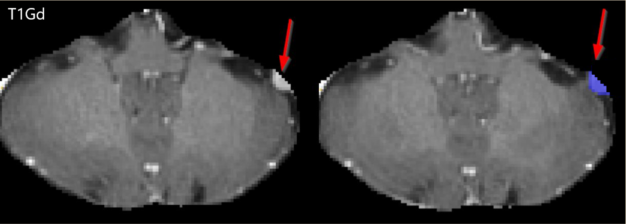
Incomplete or absent segmentation of small convexity meningiomas composed entirely of enhancing tumor, particularly when more than 1 meningioma was included in the field of view (Fig. 4b)
-
3.
Improper assignment or incomplete segmentation of non-enhancing tumor regions, including exophytic hyperostosis, cystic spaces, and areas of intrinsic T1 hyperintensity, which were sometimes erroneously labeled as enhancing tumor or SNFH rather than non-enhancing tumor core (Fig. 4c)
-
4.
Inclusion of non-tumor related brain parenchymal T2/FLAIR signal abnormality, most commonly chronic microvascular ischemic white matter changes (e.g. leukoaraiosis) within the SNFH label (Fig. 4d).
(a) Erroneously marked a thin rim of edema that does not exist.
 (b) Missed small convexity meningioma.
 (c) Improper classification of non-enhancing tumor.
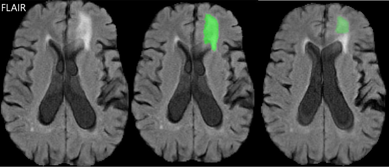 (d) Edema bleeds into periventricular FLAIR abnormalities.
3 Discussion
3.1 Potential benefits of the challenge
The BraTS meningioma 2023 challenge will allow competitors to develop automated segmentation models based on the largest multi-label annotated meningioma mpMRI dataset known to date [20]. By including an expert annotated, large, multi-site, heterogenous, multi-parametric MRI meningioma dataset, there is increased variability in the challenge training dataset. This variability comes from different acquisition and processing protocols across each of the contributing sites, termed “batch effects” [21]. Having variation in the training dataset is crucial because it closely mirrors the actual underlying data distribution, which may ultimately improve the tool’s overall generalizability [21]. Generalizability is a leading factor for the slow adoption of automated segmentation models into the clinical workflow. Automated segmentation models developed using large and diverse mpMRI datasets are expected to have increased performance and increased generalizability when deployed to new sites, which is in turn expected to increase likelihood of clinical adoption.
3.2 Clinical relevance
Accurate, reproducible, and automated meningioma segmentation models have several potential clinical uses. In the short term, automated segmentations are immediately applicable to radiation oncologists who already generate similar segmentations for radiation treatment planning. In the intermediate term, automated segmentations will be useful to radiologists for objective volumetric assessment of tumor growth on serial imaging studies and to neurosurgeons for aiding in operative planning. In the longer term, these automated segmentations will serve as the starting point for predictive models aimed at non-invasive identification of meningioma grade, subtype, aggressiveness, and response to therapy.
In radiation oncology, physicians spend significant time segmenting anatomical structures during radiation therapy planning [22, 23]. Anatomical structures include gross tumor volume (GTV), clinical tumor volume (CTV) and organs-at-risk (OARs) [24]. Modern segmentation methods, sometimes using fused CT and MRI, have demonstrated “clinically acceptable” automated segmentation performance for several OARs and can reduce manual segmentation time by up to 43% [25, 26, 27]. “Clinical acceptability” as defined by Baroudi et al roughly corresponds to a Dice overlap coefficient of 0.90 for automated segmentation compared to manual segmentation, which often corresponds with inter-observer agreement of determining a clinically acceptable segmentation volume [28]. However, despite success with automated segmentation of OARs, GTV segmentation models have yet to be widely implemented into clinical practice. State-of-the-art deep learning models trained on diverse multi-institution data, like those that will be generated by the BraTS meningioma 2023 challenge, have the potential to approach or exceed the “clinically acceptable” range for GTV segmentation and therefore may be immediately applicable to clinical radiation planning.
In image guided neurosurgery, surgeons utilize co-registered preoperative anatomical, functional, and diffusion tensor imaging to assist with tumor localization intraoperatively [29]. Automated segmentations could provide neurosurgeons with additional information to aid in surgical planning, particularly with respect to determining tumor proximity to eloquent white matter tracts identified by DTI tractography and eloquent cerebral cortex identified by functional MRI.
In radiology, automated segmentation has had a growing role in recent years. For example, Lee et al. describe a deep learning algorithm that computes longitudinal volume measurement of vestibular schwannomas following Gamma Knife radiosurgery (GKRS) [30]. The model described by Lee had an average of -0.44% to +1.76% relative volume difference between the AI prediction and expert radiologist measurement over 861 patients in follow-up, which highlights the potential role for automated segmentation methods in longitudinal volumetric assessment. Rudie et al. trained a multichannel multiclass segmentation nnU-Net model on posttreatment diffuse gliomas as well as a separate longitudinal change nnU-Net model to help localize and quantify changes in treatment change tissue and active tumor [31]. They found that the accuracy levels of the longitudinal change networks to predict an increase, decrease, or no change in peritumoral edema or active tumor volumes were not significantly different from those of three neuroradiologists. By automatically computing the volume of tumor at follow-up appointments, providers can more accurately determine progression, pseudo-progression, or regression of the treated disease. In addition, automated segmentations have been used for radiogenomic studies aimed at non-invasive prediction of clinically relevant tumor characteristics such as genetic subtypes of gliomas [32, 33]. One retrospective study analyzed pre-operative MRIs from patients with WHO grade IV diffuse astrocytic gliomas and showed that combining radiomics with CNN features improved genetic biomarker prediction [33]. To date, there has been relatively little investigation into radiogenomics for meningiomas. Clinically, the WHO grade of a meningioma is one of the main determinants of treatment recommendations, and determination by resection or biopsy is not always desirable. If a deep radiogenomics model could accurately identify subtypes, and therefore prognosticate a meningioma prior to intervention, then patients could be spared the risks associated with biopsy, resection, or radiation therapy.
3.3 Limitations of the challenge
While great care and attention was provided during challenge design and dataset preparation, there are several important limitations that should be addressed. First, pre-processing of the mpMRI images introduces loss of radiographic information that would otherwise be analyzed in clinical practice. For example, skull-stripping implicitly removes any portion of a meningioma that extends beyond the confines of the cranial vault, which limits potential clinical utility for meningiomas that are not entirely intracranial. In addition, series co-registration and resampling to a standardized atlas space obscures the native image acquisition resolution and inherently changes the image data through interpolation. These pre-processing steps also introduce an additional step (and therefore potential barrier) to clinical implementation of automated tumor segmentation algorithms. These limitations, while significant, were deemed necessary in order to protect patient privacy, to remain consistent with other BraTS challenges, and to lower potential data-related barriers to challenge participation. Second, the annotator-approval model for manual segmentation corrections, while consistent with prior BraTS challenges, does not address the issue of inter-observer variability, which could fundamentally limit the performance of any model trained on these data. Of specific concern with regards to meningioma, dural tail enhancement segmentation has significant inter-observer variability due to close proximity to normal enhancing structures (e.g. normal dura, dural vasculature) as well as historically ill-defined definitions of what constitutes a dural tail. In order to reduce this variability, annotators were provided with specific written instructions and examples of commonly encountered pre-segmentation errors, and an expert approver reviewed all segmentations before inclusion in the challenge dataset. Ultimately, expert clinical judgment on meningioma boundaries will likely never be perfectly aligned between different observers, and these issues will only be fully addressed by the labor- and time-intensive process of multi-observer consensus segmentation.
3.4 Goals for future challenges
There are many potential ways to expand and improve the BraTS meningioma challenge in future iterations. Perhaps the most straightforward extension would be using the current challenge dataset to predict tumor WHO grade, which would not require any new imaging data and could provide substantial clinical utility for determination of prognosis and optimal treatment course. Existing data could also be used to detect (rather than merely segment), localize, and quantify (with linear or volumetric measurements) intracranial meningiomas, which would be useful for improving radiologist’s workflows and improving objectivity in the longitudinal follow up setting.
Several additional potential extensions of the challenge would require new inclusion/exclusion criteria and additional data, but also offer improved clinical utility. For example, while the BraTS meningioma 2023 challenge is focused on preoperative imaging, meningioma imaging assessment and treatment planning are frequently performed in the postoperative and/or post-treatment setting. In addition, while most meningiomas are predominantly intracranial, extracranial meningiomas involving the face, skull-base, or spinal canal are frequently encountered in clinical practice. Future BraTS meningioma and related challenges should incorporate non-skullstripped mpMRI images of previously treated meningiomas to allow development of automated segmentation models that could be clinically useful for a wider variety of patients with meningioma.
Another important clinical dilemma that could be addressed by future challenges is determination of recurrence versus treatment related changes in patients who have undergone definitive surgical resection and/or radiotherapy for meningioma. Non-pathologic treatment related changes, especially following radiation therapy, have overlapping radiographic findings with recurrent meningioma. Therefore, developing a model to help distinguish recurrence from treatment changes could provide substantial clinical utility for developing optimal patient management plans.
4 Conclusion
The BraTS meningioma 2023 challenge will provide a community standard and benchmark for automated intracranial meningioma segmentation models based on the largest expert annotated multilabel mpMRI image dataset to date. The state-of-the-art models developed during the course of this competition will lead to clinically viable automated meningioma segmentation methods that can aid in objective tumor monitoring, surgical and radiotherapy planning, and non-invasive assessments of clinically relevant tumor characteristics that will ultimately improve care of patients with meningiomas.
Acknowledgments
Developing large and well curated mpMRI datasets for auto-segmentation model development requires significant time and expertise from neuro-radiology experts. We are grateful to everyone who contributed to the development and review of the tumor volume labels including volunteer annotators/approvers from the American Society of Neuroradiology (ASNR).
Funding
Research reported in this publication was partly supported by the National Institutes of Health (NIH) under award numbers: NCI K08CA256045 and NCI/ITCR U01CA242871. The content of this publication is solely the responsibility of the authors and does not represent the official views of the NIH.
References
- [1] C. Ogasawara, B. D. Philbrick, and D. C. Adamson, “Meningioma: a review of epidemiology, pathology, diagnosis, treatment, and future directions,” Biomedicines, vol. 9, no. 3, p. 319, 2021.
- [2] K. Huntoon, A. M. S. Toland, and S. Dahiya, “Meningioma: a review of clinicopathological and molecular aspects,” Frontiers in Oncology, vol. 10, p. 579599, 2020.
- [3] O. Khanna, A. F. Kazerooni, C. J. Farrell, M. P. Baldassari, T. D. Alexander, M. Karsy, B. A. Greenberger, J. A. Garcia, C. Sako, J. J. Evans, et al., “Machine learning using multiparametric magnetic resonance imaging radiomic feature analysis to predict ki-67 in world health organization grade i meningiomas,” Neurosurgery, vol. 89, no. 5, pp. 928–936, 2021.
- [4] S. Bakas, M. Reyes, A. Jakab, S. Bauer, M. Rempfler, A. Crimi, R. Shinohara, C. Berger, S. Ha, M. Rozycki, et al., “Identifying the best machine learning algorithms for brain tumor segmentation,” Progression Assessment, and Overall Survival Prediction in the BRATS Challenge, vol. 10, 2018.
- [5] B. H. Menze, A. Jakab, S. Bauer, J. Kalpathy-Cramer, K. Farahani, J. Kirby, Y. Burren, N. Porz, J. Slotboom, R. Wiest, et al., “The multimodal brain tumor image segmentation benchmark (brats),” IEEE transactions on medical imaging, vol. 34, no. 10, pp. 1993–2024, 2014.
- [6] S. Bakas, H. Akbari, A. Sotiras, M. Bilello, M. Rozycki, J. S. Kirby, J. B. Freymann, K. Farahani, and C. Davatzikos, “Advancing the cancer genome atlas glioma mri collections with expert segmentation labels and radiomic features,” Scientific data, vol. 4, no. 1, pp. 1–13, 2017.
- [7] S. Bakas, H. Akbari, A. Sotiras, et al., “Segmentation labels for the pre-operative scans of the tcga-gbm collection.,” The cancer imaging archive, 2017.
- [8] S. Bakas, H. Akbari, A. Sotiras, M. Bilello, M. Rozycki, J. Kirby, J. Freymann, K. Farahani, and C. Davatzikos, “Segmentation labels and radiomic features for the pre-operative scans of the tcga-lgg collection,” The cancer imaging archive, vol. 286, 2017.
- [9] S. Bakas, C. Sako, H. Akbari, M. Bilello, A. Sotiras, G. Shukla, J. D. Rudie, N. F. Santamaría, A. F. Kazerooni, S. Pati, et al., “The university of pennsylvania glioblastoma (upenn-gbm) cohort: Advanced mri, clinical, genomics, & radiomics,” Scientific data, vol. 9, no. 1, p. 453, 2022.
- [10] U. Baid, S. Ghodasara, S. Mohan, M. Bilello, E. Calabrese, E. Colak, K. Farahani, J. Kalpathy-Cramer, F. C. Kitamura, S. Pati, et al., “The rsna-asnr-miccai brats 2021 benchmark on brain tumor segmentation and radiogenomic classification,” arXiv preprint arXiv:2107.02314, 2021.
- [11] U. Baid, S. Talbar, S. Rane, S. Gupta, M. H. Thakur, A. Moiyadi, N. Sable, M. Akolkar, and A. Mahajan, “A novel approach for fully automatic intra-tumor segmentation with 3d u-net architecture for gliomas,” Frontiers in computational neuroscience, p. 10, 2020.
- [12] N. Martz, J. Salleron, F. Dhermain, G. Vogin, J. Daisne, R. M. Audouard, R. Tanguy, G. Noel, M. Peyre, I. Lecouillard, et al., “Anocef consensus guideline on target volume delineation for meningiomas radiotherapy,” International Journal of Radiation Oncology, Biology, Physics, vol. 114, no. 3, p. e46, 2022.
- [13] P. Nasa, R. Jain, and D. Juneja, “Delphi methodology in healthcare research: how to decide its appropriateness,” World Journal of Methodology, vol. 11, no. 4, p. 116, 2021.
- [14] J. Watts, G. Box, A. Galvin, P. Brotchie, N. Trost, and T. Sutherland, “Magnetic resonance imaging of meningiomas: a pictorial review,” Insights into imaging, vol. 5, no. 1, pp. 113–122, 2014.
- [15] M. Bitzer, H. Opitz, J. Popp, M. Morgalla, A. Gruber, E. Heiss, and K. Voigt, “Angiogenesis and brain oedema in intracranial meningiomas: influence of vascular endothelial growth factor,” Acta neurochirurgica, vol. 140, pp. 333–340, 1998.
- [16] S. O’leary, W. Adams, R. Parrish, and W. Mukonoweshuro, “Atypical imaging appearances of intracranial meningiomas,” Clinical radiology, vol. 62, no. 1, pp. 10–17, 2007.
- [17] R. Dorent, A. Kujawa, M. Ivory, S. Bakas, N. Rieke, S. Joutard, B. Glocker, J. Cardoso, M. Modat, K. Batmanghelich, et al., “Crossmoda 2021 challenge: Benchmark of cross-modality domain adaptation techniques for vestibular schwannoma and cochlea segmentation,” Medical Image Analysis, vol. 83, p. 102628, 2023.
- [18] F. Isensee, P. F. Jaeger, S. A. Kohl, J. Petersen, and K. H. Maier-Hein, “nnu-net: a self-configuring method for deep learning-based biomedical image segmentation,” Nature methods, vol. 18, no. 2, pp. 203–211, 2021.
- [19] P. A. Yushkevich, J. Piven, H. C. Hazlett, R. G. Smith, S. Ho, J. C. Gee, and G. Gerig, “User-guided 3d active contour segmentation of anatomical structures: significantly improved efficiency and reliability,” Neuroimage, vol. 31, no. 3, pp. 1116–1128, 2006.
- [20] A. Vassantachart, Y. Cao, M. Gribble, S. Guzman, J. C. Ye, K. Hurth, A. Mathew, G. Zada, Z. Fan, E. L. Chang, et al., “Automatic differentiation of grade i and ii meningiomas on magnetic resonance image using an asymmetric convolutional neural network,” Scientific Reports, vol. 12, no. 1, p. 3806, 2022.
- [21] M. Bento, I. Fantini, J. Park, L. Rittner, and R. Frayne, “Deep learning in large and multi-site structural brain mr imaging datasets,” Frontiers in Neuroinformatics, vol. 15, p. 82, 2022.
- [22] H. K. Byun, J. S. Chang, M. S. Choi, J. Chun, J. Jung, C. Jeong, J. S. Kim, Y. Chang, S. Y. Chung, S. Lee, et al., “Evaluation of deep learning-based autosegmentation in breast cancer radiotherapy,” Radiation Oncology, vol. 16, no. 1, pp. 1–8, 2021.
- [23] E. Cha, S. Elguindi, I. Onochie, D. Gorovets, J. O. Deasy, M. Zelefsky, and E. F. Gillespie, “Clinical implementation of deep learning contour autosegmentation for prostate radiotherapy,” Radiotherapy and Oncology, vol. 159, pp. 1–7, 2021.
- [24] A. K. Berthelsen, J. Dobbs, E. Kjellén, T. Landberg, T. R. Möller, P. Nilsson, L. Specht, and A. Wambersie, “What’s new in target volume definition for radiologists in icru report 71? how can the icru volume definitions be integrated in clinical practice?,” Cancer Imaging, vol. 7, no. 1, p. 104, 2007.
- [25] T. Vrtovec, D. Močnik, P. Strojan, F. Pernuš, and B. Ibragimov, “Auto-segmentation of organs at risk for head and neck radiotherapy planning: from atlas-based to deep learning methods,” Medical physics, vol. 47, no. 9, pp. e929–e950, 2020.
- [26] D. Močnik, B. Ibragimov, L. Xing, P. Strojan, B. Likar, F. Pernuš, and T. Vrtovec, “Segmentation of parotid glands from registered ct and mr images,” Physica Medica, vol. 52, pp. 33–41, 2018.
- [27] J. S. Ginn, H. A. Gay, J. Hilliard, J. Shah, N. Mistry, C. Möhler, G. D. Hugo, and Y. Hao, “A clinical and time savings evaluation of a deep learning automatic contouring algorithm,” Medical Dosimetry, vol. 48, no. 1, pp. 55–60, 2023.
- [28] H. Baroudi, K. K. Brock, W. Cao, X. Chen, C. Chung, L. E. Court, M. D. El Basha, M. Farhat, S. Gay, M. P. Gronberg, et al., “Automated contouring and planning in radiation therapy: What is ‘clinically acceptable’?,” Diagnostics, vol. 13, no. 4, p. 667, 2023.
- [29] F. Drakopoulos, C. Tsolakis, A. Angelopoulos, Y. Liu, C. Yao, K. R. Kavazidi, N. Foroglou, A. Fedorov, S. Frisken, R. Kikinis, et al., “Adaptive physics-based non-rigid registration for immersive image-guided neuronavigation systems,” Frontiers in Digital Health, vol. 2, p. 613608, 2021.
- [30] C.-c. Lee, W.-K. Lee, C.-C. Wu, C.-F. Lu, H.-C. Yang, Y.-W. Chen, W.-Y. Chung, Y.-S. Hu, H.-M. Wu, Y.-T. Wu, et al., “Applying artificial intelligence to longitudinal imaging analysis of vestibular schwannoma following radiosurgery,” Scientific reports, vol. 11, no. 1, p. 3106, 2021.
- [31] J. D. Rudie, E. Calabrese, R. Saluja, D. Weiss, J. B. Colby, S. Cha, C. P. Hess, A. M. Rauschecker, L. P. Sugrue, and J. E. Villanueva-Meyer, “Longitudinal assessment of posttreatment diffuse glioma tissue volumes with three-dimensional convolutional neural networks,” Radiology: Artificial Intelligence, vol. 4, no. 5, p. e210243, 2022.
- [32] M. Buda, E. A. AlBadawy, A. Saha, and M. A. Mazurowski, “Deep radiogenomics of lower-grade gliomas: Convolutional neural networks predict tumor genomic subtypes using mr images,” Radiology: Artificial Intelligence, vol. 2, no. 1, p. e180050, 2020.
- [33] E. Calabrese, J. D. Rudie, A. M. Rauschecker, J. E. Villanueva-Meyer, J. L. Clarke, D. A. Solomon, and S. Cha, “Combining radiomics and deep convolutional neural network features from preoperative mri for predicting clinically relevant genetic biomarkers in glioblastoma,” Neuro-Oncology Advances, vol. 4, no. 1, p. vdac060, 2022.