Electron energy loss and angular asymmetry induced by elastic scattering in helium droplets
Abstract
Helium nanodroplets are ideal model systems to unravel the complex interaction of condensed matter with ionizing radiation. Here we study the effect of purely elastic electron scattering on angular and energy distributions of photoelectrons emitted from He nanodroplets of variable size (- atoms per droplets). For large droplets, photoelectrons develop a pronounced anisotropy along the incident light beam due to a shadowing effect within the droplets. In contrast, the detected photoelectron spectra are only weakly perturbed. This opens up possibilities for photoelectron spectroscopy of dopants embedded in droplets provided they are smaller than the penetration depth of the light and the trapping range of emitted electrons.
Irradiation of condensed matter with ionizing radiation induces various secondary processes in addition to the primary photoionization event. In biological matter, secondary processes such as electron-impact ionization, radical reactions and dissociative attachment of low-energy electrons (LEEs) to biomolecules are the main causes for radiation damage leading to mutations or cell death [1, 2]. Deciphering the mechanisms of radiation damage is crucial for devising improved schemes for radiotherapy for cancer [3, 4, 5, 6]. LEEs are mainly produced by scattering of the primary and secondary electrons in the medium [7]. In bulk molecular systems, complex interactions of electrons with intra- and intermolecular degrees of freedom make it hard to unravel the various elastic and inelastic scattering channels. Furthermore, the tendency of slow electrons to remain trapped in the material hinders their detection. Therefore, solid or liquid nanoparticles (NPs) have been used to study the dynamics of electron scattering [8] and other properties making NPs relevant for atmospheric, astrophysical, and technical sciences [9, 10, 11, 12, 13].
As atoms or molecules condense into clusters and NPs, photoelectron angular distributions (PADs) tend to become more isotropic because emitted electrons scatter on the cluster constituents and thereby change their emission direction. A reduction of the detected electron kinetic energy for increasing NP size is typically ascribed to inelastic scattering; energy transfer is small in elastic electron-molecule collisions due to the large mass mismatch (). Nevertheless, elastic scattering can lead to diffusion-like, damped electron motion in extended media, as we show in this work. At particle sizes nm, electron distributions are additionally altered by optical confinement effects such as nanofocusing and shadowing [13]. The latter occurs when the particle size exceeds both the penetration depth of the radiation and the mean path traveled by an emitted electron before it is trapped in the droplet (‘trapping range’). Electrons are then preferentially emitted from that side of the particle facing toward the incident light [14, 12]. In electron imaging experiments, the degree of shadowing is often specified by the anisotropy parameter , where is the electron intensity in the forward/backward half plane of the electron image with respect to the incoming light. So far, the various phenomena related to electron scattering have been studied separately for different types of NPs [13]. The continuous evolution of photoelectron distributions from individual atoms up to large NPs has not been reported. In particular, the effect of elastic scattering on electron distributions has not been demonstrated explicitly.
In this work, we present a comprehensive study of photoemission spectra (PES) and PAD of large superfluid helium nanodroplets (HNDs). HNDs are particularly well suited model systems to study electron scattering as they feature an extraordinarily wide gap in their excitation spectrum reaching from eV (phonons) up to 20 eV (excitons) [15]. This gives us the unique opportunity to investigate purely elastic electron-atom scattering in a condensed-phase system in a wide energy range. Additional complications due to nano-focusing effects [13] are negligible as the refractive index of He only weakly deviates from 1 by [16, 17]. Due to their superfluid nature, HNDs are homogeneous, spherically symmetric particles featuring a flat, nearly size-independent density distribution [18, 19]. The droplet size can be varied continuously in a wide range from a few nm up to m [20, 21]. HNDs are widely used as cold, inert and transparent cryo-matrices to form tailored molecular complexes, metal clusters and NPs which can be probed by spectroscopy and mass spectrometry [15, 22]. Extending the HND technique to extreme-ultraviolet or X-ray photoelectron spectroscopy (UPS, XPS) of embedded (‘dopant’) species is an intriguing prospect, in particular for probing novel types of nano-aggregates formed in HNDs [23, 24, 25, 26, 22]. However, it has remained an open question to what extent PES from dopants are distorted by the interaction of emitted electrons with the He matrix. Previous PES measurements of dopants using ns and fs multi-photon laser-ionization schemes appeared to be largely unperturbed by the surrounding He [27, 28, 29, 30, 31, 32, 33], while Penning ionization electron spectra are notoriously broad and structure-less [34, 35, 36, 31]. Likewise, recent experiments investigating above-threshold ionization of atoms and molecules in HNDs indicated that electron-He scattering substantially impacts PES and PAD [37, 38, 39, 40, 41]. The relaxation of hot electrons at energies eV in HNDs has been described by electron-atom binary collisions [28]. Below eV, electrons localize in void bubbles [42, 43, 44] which then scatter at elementary modes of the HND (phonons and rotons) before emerging to the droplet surface where electrons are eventually released into the vacuum [44, 45]. Here we show experimentally and using classical scattering simulations that electron-He elastic scattering is an important mechanism leading to substantial electron-energy loss (EEL) and drastically altered PADs of emitted electrons. Large HNDs with radii nm containing He atoms feature a pronounced anisotropy of the electron PAD along the light propagation axis, but the PES of detected electrons are only weakly perturbed.
HNDs were formed by continuous expansion of He at high pressure (30-50 bar) into vacuum through a cryogenically cooled (7-32 K) nozzle of diameter m. By controlling the expansion pressure and temperature, the droplet mean radius was varied in the range 1-190 nm (-). The droplet size was determined by titration measurements [20] and by comparison to literature values [15]. The XENIA (XUV electron-ion spectrometer for nanodroplets in Aarhus) endstation [46] located at the AMOLine of the ASTRID2 synchrotron at Aarhus University, Denmark [47], was used to record velocity-map images (VMIs) of photoelectrons. PES and PAD were inferred from the VMIs by Abel inversion using the MEVELER reconstruction method [48]. Additionally, high resolution PES were measured using a hemispherical analyzer (VG-220i) mounted under the magic angle at the GasPhase beamline at the synchrotron Elettra in Trieste, Italy [49].
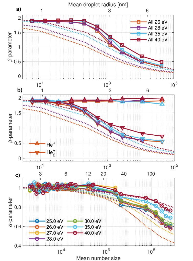
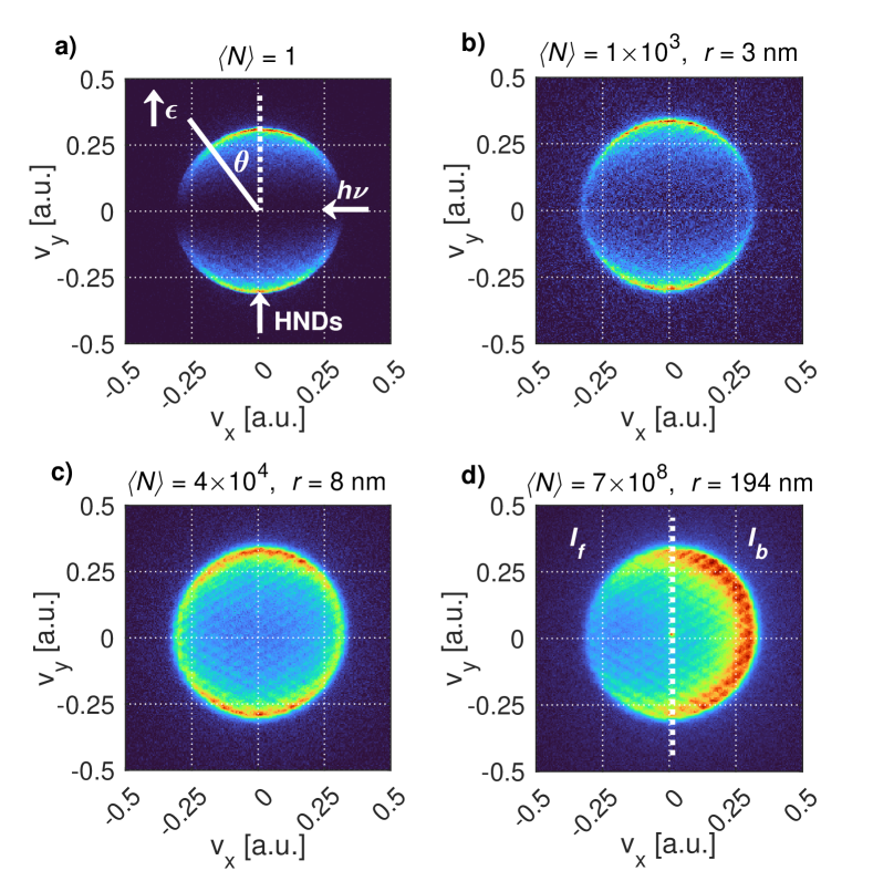
Fig. 1 shows VMIs recorded at a photon energy eV for various HND sizes. Photoelectrons emitted from free He atoms [Fig. 1 a)] by absorption of one photon are emitted according to the PAD corresponding to the asymmetry parameter , as conventionally defined by the angular intensity distribution [50]. Here, is the angle between the light polarization and the emission direction. For increasing HND size, the angular distribution becomes more isotropic () due to the increasing number of elastic collisions the electrons undergo on their way out of the droplet [Fig. 1 b) & c)]. Fig. 1 d) shows the VMI in the case where the HNDs are on average larger than the penetration depth of the incident light. At eV, the photoionization cross-section of He is 6.79 Mb [51] corresponding to a penetration depth of nm into liquid helium. For large HNDs, the PAD is dominated by the shadowing effect and Abel inversion is ill-defined due to breaking of the cylindrical symmetry about the polarization of the light meaning that we cannot define from the VMI. Additional VMIs recorded for different droplet sizes and photon energies are shown in Suppl. Fig. 1 in the Supplementary Information (SI) [52].
Fig. 2 a) shows for all emitted electrons as a function of the HND size in the range - atoms where the shadowing effect is absent. is constant up to . Thereafter, it drops to 0.5 for . However, does not significantly depend on the photon energy, as previously observed [53]. Fig. 2 b) shows as a function of HND size for electrons detected in coincidence with He+ and He ions, respectively. Electrons coincident with He+ feature nearly constant anisotropy with for all droplet sizes; These electrons and He+ ions stem from free He atoms accompanying the droplet beam even under expansion conditions (low nozzle temperature K) when large HNDs are formed. In contrast, for electrons detected in coincidence with He, decreases from 2 for to 0.5 for . As He are formed by fragmentation of He clusters and HNDs, the electrons’ PADs recorded in coincidence with He are indicative for HND photoionization. Note that for these electrons, drops below 2 for droplet sizes whereas for all electrons [Fig. 2 a)], up to . This shows that in the regime of small droplets, , free He atoms are more abundant in the supersonic jet than He droplets. The electron mean-free path (EMFP) for elastic scattering calculated from the total electron scattering cross-section [54] and the number density of bulk liquid He [18] is 8 Å for an electron kinetic energy of 1 eV and it is 16 Å at 10 eV. This value is consistent with the He droplet radius nm up to which [Fig. 2 b)].
For larger He droplets, the -anisotropy due to the shadowing effect dominates the PADs. Fig. 2 c) shows that decreases (increasing shadowing effect) as the droplets grow larger than ( nm). The size-dependence of for electrons detected in coincidence with He (correlated with HNDs) closely follows the one for all electrons because the abundance of free He atoms in the jet is small under these conditions (see Suppl. Fig. 3 [52]).
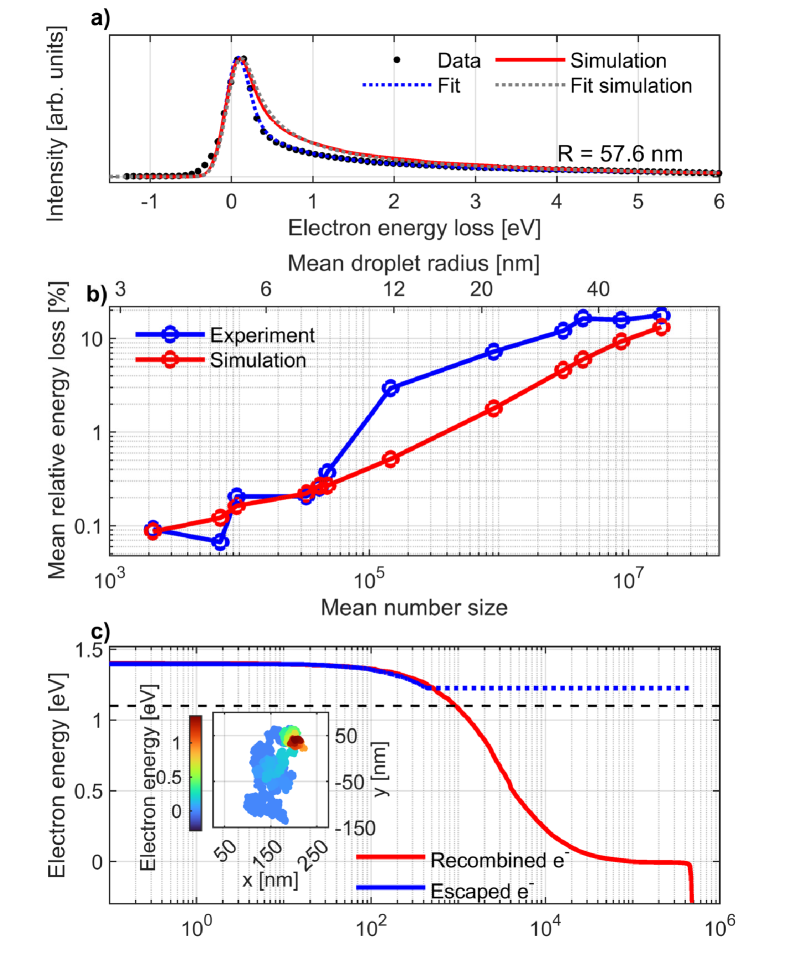
To investigate the energy loss of the photoelectrons for large HNDs, we recorded high-resolution PESs using a hemispherical analyzer. Fig. 3 a) shows an EEL spectrum recorded at eV (see electron spectra for other droplet sizes in Suppl. Fig. 11 [52]). As the droplet size increases, the photoemission line develops a tail toward higher EEL (lower electron energy). Similar asymmetric line broadening was previously reported for small HNDs (- nm) doped with aniline molecules [28]. While the line shape was described by an exponentially decaying function , here we find empirically the best fit with the function (see dotted lines in Fig. 3 a) and details in the SI [52]). From these fits we infer the mean relative EEL (see Suppl. Fig. 11 [52]), which is shown in Fig. 3 b) as function of HND size. Note that the EEL only pertains to the electrons emitted from the droplets. The total EEL is in fact higher if all electrons trapped in the droplets are taken into account. For a droplet of nm, % of the electron energy is lost on average due to elastic scattering. This corresponds to binary collisions according to simple estimates of the kinematics assuming head-on collisions (see SI [52]).
To obtain a better understanding of the electron-He interaction leading to changes in the PAD, the experimental results are compared with classical electron–He elastic scattering simulations based on differential (energy, scattering angle) scattering cross sections. See the SI [52] for a detailed description. In the simulation, an electron recombines with its parent ion when it turns around and binds to the ion with a binding energy exceeding its kinetic energy. A simulated electron trajectory for this case is displayed in Fig. 3 c) as an inset. The corresponding time evolution of the kinetic energy is shown as a red line. For comparison, a trajectory where the electron escapes the droplet is included (blue line). Large HNDs, which have similar properties as bulk superfluid He, feature a potential energy barrier of 1.1 eV below which electrons localize in bubbles [black dashed line in Fig. 3 c] [55, 56, 34]. Thus, in the simulated PAD we exclude those LEEs with kinetic energy eV. Obviously, this leads to an inconsistency between the simulation and the experiment at eV indicating that not all low-energy electrons are actually trapped, e. g. those formed near the surface. We do not have a direct measure of the number of photoelectrons evading detection due to trapping in the HNDs, but can only make a rough estimate based on the He flux measured by the pressure increase in the HND beam dump (see Suppl. Fig. 6 [52]).
The results of the simulations including the 1.1 eV cutoff are shown as dotted lines in Fig. 2. For reference, the results of the simulation without energy cutoff are shown in the Suppl. Fig 10 [52]. The simulated values follow a similar size-dependence as those inferred from electrons detected in coincidence with He in the experiment. The PAD becomes more isotropic as due to elastic electron-He scattering. Deviations from atomic PAD have been observed for inner-valence ionization of the clusters of heavier rare-gas atoms in a size range from - [57, 58, 59, 60]. The conclusion was that PAD are significantly changed when the cluster size reaches the magnitude of the EMFP for elastic scattering [57], which is consistent with our findings for HNDs. The shadowing effect for large HNDs is also well reproduced by the scattering simulations, see Fig. 2 c). The corresponding drop of for is more pronounced near the He ionization threshold (24.59 eV) where both the absorption cross-section (7.40 Mb [51]) and the electron-He scattering cross-section (600 Mb [54]) are highest. Note that at higher photon energies eV additional features appear in the electron VMIs due to inelastic scattering of photoelectrons on He atoms in the droplets, see Suppl Fig. 2 [52]. Different values for these features inform about the mechanisms of generating these electrons.
The value measured for nm at eV is close to that reported for solid salt NPs (NaCl [12], KCl [61]) of similar size.
The fact that shadowing becomes the dominant effect when the HND size approaches the photon penetration depth ( nm) implies that the latter is the upper bound for the electron trapping range. A conservative estimate of the lower bound is the EMFP for elastic scattering ( nm). Thus, the trapping range is in order of 10 nm, which is consistent with the rise in electrons evading detection which we observe in the size range - nm (see the Suppl. Fig 6 [52]). From the simulations we determine the trapping range by evaluating the maximum distance travelled by an electron from the cation in bulk liquid He, see Suppl. Fig. 7-9 [52]. For eV, the trapping range is smaller than the photon penetration depth and it becomes similar in magnitude for eV.
When excluding the droplet barrier for electron trapping, the simulated values of the -parameter closely follow those found when the 1.1 eV-barrier is taken into account, but the trapping range is largely overestimated. As the the trapping range and the potential barrier are closely correlated, an alternative condition for LEE trapping in He droplets could be a finite escape depth at the droplet surface, see Suppl. Fig. 9 [52]. The higher efficiency of electron-He elastic scattering observed in experiments as compared to the classical scattering simulations is likely due to quantum effects becoming important at low energies eV where the de Broglie wavelength of the electron reaches the average distance between He atoms in a HND (3.6 Å) or even the size of the whole HND ( eV). However, performing quantum-scattering simulations goes beyond the scope of this work.
The experimental and simulated electron spectra show a similar tail extending from the photoline toward higher EEL [see Fig. 3 a)]. However, given that the resulting average relative EEL is only 15 % and the peak of the photoemission line remains nearly unshifted, we conclude that photoelectron spectroscopy is possible even for large droplets () provided the electron energy well exceeds 1.1 eV. The larger EEL found in the experiment with aniline-doped HNDs [28] is likely due to the low energy of emitted electrons ( eV), at which scattering should be treated quantum mechanically and electron localization by formation of bubbles can no longer be neglected.
In summary, we have demonstrated the effect of elastic scattering on the energy and angular distributions of photoelectrons emitted from HNDs in wide ranges of droplet size and electron energy. We identify two regimes of anisotropic electron emission. For small droplets (), the cylindrical symmetry with respect to the polarization of the light is retained. However, electron emission becomes more isotropic compared to free He atoms when the HND radius nm exceeds the EMFP for elastic scattering. Large droplets () exhibit a pronounced shadowing effect and the PAD becomes anisotropic along the light propagation direction. For typical experimental conditions () used in spectroscopy experiments, the average EEL is only %; for large droplets () the average EEL of the detected photoelectrons rises but stays below %. This makes HNDs a suitable matrix for UPS and XPS of embedded species, but a significant loss of electron angular information should be expected. Likewise, a large number of electrons remain undetected as multiple elastic scattering leads to trapping of the electrons in the droplets where they recombine with their parent ions. In molecular systems such as water, electron scattering is affected by the higher mass of the molecular constituents and by their internal (ro-vibrational) degrees of freedom which open up additional inelastic scattering channels. Nevertheless, scattering of electrons on molecules where no internal modes are excited will contribute to EEL and should be taken into account as a mechanism generating highly reactive LEEs.
I Acknowledgements
J.D.A. and M.M. acknowledge financial support by the Carlsberg Foundation. We thank the Danish Agency for Science, Technology, and Innovation for funding the instrument center DanScatt. T.F. acknowledges support by the Deutsche Forschungsgemeinschaft (DFG, German Research Foundation) via SFB 1477 “light–matter interactions at interfaces” (project number 441234705) and via the Heisenberg program (project number 436382461). S.R.K. thanks Dept. of Science and Technology, Govt. of India, for support through the DST-DAAD scheme and Science and Eng. Research Board. S.R.K., K.S. and S.D. acknowledge the support of the Scheme for Promotion of Academic Research Collaboration, Min. of Edu., Govt. of India, and the Institute of Excellence programme at IIT-Madras via the Quantum Center for Diamond and Emergent Materials. S.R.K. gratefully acknowledges support of the Max Planck Society’s Partner group programme. M.M. and S.R.K. gratefully acknowledge funding from the SPARC Programme, MHRD, India. A.R.A. acknowledges with gratitude for the support from the Marie Skłodowska-Curie Postdoctoral Fellowship project Photochem-RS-RP (Grant Agreement No. 101068805) provided by the European Union’s Horizon 2020 Research and Innovation Programme. The research leading to this result has been supported by the project CALIPSOplus under grant agreement 730872 from the EU Framework Programme for Research and Innovation HORIZON 2020 and by the COST Action CA21101 “Confined Molecular Systems: From a New Generation of Materials to the Stars (COSY)”.
References
- Gómez-Tejedor and Fuss [2012] G. G. Gómez-Tejedor and M. C. Fuss, Radiation damage in biomolecular systems (Springer Science & Business Media, 2012).
- Alizadeh et al. [2015] E. Alizadeh, T. M. Orlando, and L. Sanche, Biomolecular damage induced by ionizing radiation: the direct and indirect effects of low-energy electrons on DNA, Ann. Rev. Phys. Chem. 66, 379 (2015).
- Hanel et al. [2003] G. Hanel, B. Gstir, S. Denifl, P. Scheier, M. Probst, B. Farizon, M. Farizon, E. Illenberger, and T. Märk, Electron attachment to uracil: Effective destruction at subexcitation energies, Phys. Rev. Lett. 90, 188104 (2003).
- Martin et al. [2004] F. Martin, P. D. Burrow, Z. Cai, P. Cloutier, D. Hunting, and L. Sanche, DNA strand breaks induced by 0–4 eV electrons: The role of shape resonances, Phys. Rev. Lett. 93, 068101 (2004).
- Gokhberg et al. [2014] K. Gokhberg, P. Kolorenč, A. I. Kuleff, and L. S. Cederbaum, Site-and energy-selective slow-electron production through intermolecular coulombic decay, Nature 505, 661 (2014).
- Stumpf et al. [2016] V. Stumpf, K. Gokhberg, and L. S. Cederbaum, The role of metal ions in X-ray-induced photochemistry, Nat. Chem. 8, 237 (2016).
- Bass and Sanche [1998] A. D. Bass and L. Sanche, Absolute and effective cross-sections for low-energy electron-scattering processes within condensed matter, Radiation and Environmental Biophysics 37, 243 (1998).
- Ban et al. [2020] L. Ban, B. L. Yoder, and R. Signorell, Photoemission from free particles and droplets, Annu. Rev. Phys. Chem. 71, 315 (2020).
- Niessner et al. [1989] R. Niessner, W. Robers, and P. Wilbring, Laboratory experiments on the determination of polycyclic aromatic hydrocarbon coverage of submicrometer particles by laser-induced aerosol photoemission, Analytical Chemistry 61, 320 (1989).
- Ziemann and McMurry [1998] P. J. Ziemann and P. H. McMurry, Secondary electron yield measurements as a means for probing organic films on aerosol particles, Aerosol Science and Technology 28, 77 (1998).
- Wilson et al. [2006] K. R. Wilson, D. S. Peterka, M. Jimenez-Cruz, S. R. Leone, and M. Ahmed, VUV photoelectron imaging of biological nanoparticles: Ionization energy determination of nanophase glycine and phenylalanine-glycine-glycine, Phys. Chem. Chem. Phys. 8, 1884 (2006).
- Wilson et al. [2007] K. R. Wilson, S. Zou, J. Shu, E. Rühl, S. R. Leone, G. C. Schatz, and M. Ahmed, Size-dependent angular distributions of low-energy photoelectrons emitted from NaCl nanoparticles, Nano Letters 7, 2014 (2007).
- Signorell et al. [2016] R. Signorell, M. Goldmann, B. L. Yoder, A. Bodi, E. Chasovskikh, L. Lang, and D. Luckhaus, Nanofocusing, shadowing, and electron mean free path in the photoemission from aerosol droplets, Chem. Phys. Lett. 658, 1 (2016).
- Watson [1973] W. D. Watson, Photoelectron emission from small spherical particles, JOSA 63, 164 (1973).
- Toennies and Vilesov [2004] J. P. Toennies and A. F. Vilesov, Superfluid helium droplets: A uniquely cold nanomatrix for molecules and molecular complexes, Angewandte Chemie International Edition 43, 2622 (2004).
- Henke et al. [1993] B. L. Henke, E. M. Gullikson, and J. C. Davis, X-ray interactions: Photoabsorption, scattering, transmission, and reflection at E= 50-30,000 eV, z= 1-92, Atomic Data and Nuclear Data Tables 54, 181 (1993).
- Chantler [1995] C. T. Chantler, Theoretical form factor, attenuation, and scattering tabulation for Z= 1–92 from E= 1–10 eV to E= 0.4–1.0 MeV, Journal of Physical and Chemical Reference Data 24, 71 (1995).
- Harms et al. [1998] J. Harms, J. P. Toennies, and F. Dalfovo, Density of superfluid helium droplets, Phys. Rev. B 58, 3341 (1998).
- Gomez et al. [2014] L. F. Gomez, K. R. Ferguson, J. P. Cryan, C. Bacellar, R. M. P. Tanyag, C. Jones, S. Schorb, D. Anielski, A. Belkacem, C. Bernando, et al., Shapes and vorticities of superfluid helium nanodroplets, Science 345, 906 (2014).
- Gomez et al. [2011] L. F. Gomez, E. Loginov, R. Sliter, and A. F. Vilesov, Sizes of large He droplets, J. Chem. Phys. 135, 154201 (2011).
- Kolatzki et al. [2022] K. Kolatzki, M. L. Schubert, A. Ulmer, T. Möller, D. Rupp, and R. M. P. Tanyag, Micrometer-sized droplets from liquid helium jets at low stagnation pressures, Physics of Fluids 34, 012002 (2022).
- Slenczka and Toennies [2022] A. Slenczka and J. P. Toennies, Molecules in Superfluid Helium Nanodroplets: Spectroscopy, Structure, and Dynamics (Springer Nature, 2022).
- Boatwright et al. [2013] A. Boatwright, C. Feng, D. Spence, E. Latimer, C. Binns, A. M. Ellis, and S. Yang, Helium droplets: a new route to nanoparticles, Faraday discussions 162, 113 (2013).
- Haberfehlner et al. [2015] G. Haberfehlner, P. Thaler, D. Knez, A. Volk, F. Hofer, W. E. Ernst, and G. Kothleitner, Formation of bimetallic clusters in superfluid helium nanodroplets analysed by atomic resolution electron tomography, Nat. Commun. 6, 8779 (2015).
- Schiffmann et al. [2020] A. Schiffmann, T. Jauk, D. Knez, H. Fitzek, F. Hofer, F. Lackner, and W. E. Ernst, Helium droplet assisted synthesis of plasmonic Ag@ZnO core@shell nanoparticles, Nano Research 13, 2979 (2020).
- Messner et al. [2020] R. Messner, W. E. Ernst, and F. Lackner, Shell-isolated Au nanoparticles functionalized with Rhodamine B fluorophores in helium nanodroplets, J. Phys. Chem. Lett. 12, 145 (2020).
- Radcliffe et al. [2004] P. Radcliffe, A. Przystawik, T. Diederich, T. Döppner, J. Tiggesbäumker, and K.-H. Meiwes-Broer, Excited-state relaxation of Ag8 clusters embedded in helium droplets, Phys. Rev. Lett. 92, 173403 (2004).
- Loginov et al. [2005] E. Loginov, D. Rossi, and M. Drabbels, Photoelectron spectroscopy of doped helium nanodroplets, Phys. Rev. Lett. 95, 163401 (2005).
- Kazak et al. [2019] L. Kazak, S. Göde, K.-H. Meiwes-Broer, and J. Tiggesbäumker, Photoelectron spectroscopy on magnesium ensembles in helium nanodroplets, J. Phys. Chem. A 123, 5951 (2019).
- Ltaief et al. [2020] L. B. Ltaief, M. Shcherbinin, S. Mandal, S. R. Krishnan, R. Richter, T. Pfeifer, and M. Mudrich, Direct inner-shell photoionization of Xe atoms embedded in helium nanodroplets, Journal of Physics B: Atomic, Molecular and Optical Physics 53, 204001 (2020).
- Ltaief et al. [2021] L. B. Ltaief, M. Shcherbinin, S. Mandal, S. Krishnan, R. Richter, S. Turchini, N. Zema, and M. Mudrich, Photoelectron spectroscopy of coronene molecules embedded in helium nanodroplets, J. Low Temp. Phys. 202, 444 (2021).
- Dozmorov et al. [2018] N. Dozmorov, A. Baklanov, J. Von Vangerow, F. Stienkemeier, J. Fordyce, and M. Mudrich, Quantum dynamics of Rb atoms desorbing off the surface of He nanodroplets, Phys. Rev. A 98, 043403 (2018).
- Thaler et al. [2018] B. Thaler, S. Ranftl, P. Heim, S. Cesnik, L. Treiber, R. Meyer, A. W. Hauser, W. E. Ernst, and M. Koch, Femtosecond photoexcitation dynamics inside a quantum solvent, Nat. Commun. 9, 1 (2018).
- Wang et al. [2008] C. C. Wang, O. Kornilov, O. Gessner, J. H. Kim, D. S. Peterka, and D. M. Neumark, Photoelectron imaging of helium droplets doped with Xe and Kr atoms, J. Phys. Chem. A 112, 9356 (2008).
- Shcherbinin et al. [2018] M. Shcherbinin, A. LaForge, M. Hanif, R. Richter, and M. Mudrich, Penning ionization of acene molecules by helium nanodroplets, J. Phys. Chem. A 122, 1855 (2018).
- Mandal et al. [2020] S. Mandal, R. Gopal, M. Shcherbinin, A. D’Elia, H. Srinivas, R. Richter, M. Coreno, B. Bapat, M. Mudrich, S. Krishnan, et al., Penning spectroscopy and structure of acetylene oligomers in He nanodroplets, Phys. Chem. Chem. Phys. 22, 10149 (2020).
- Treiber et al. [2021] L. Treiber, B. Thaler, P. Heim, M. Stadlhofer, R. Kanya, M. Kitzler-Zeiler, and M. Koch, Observation of laser-assisted electron scattering in superfluid helium, Nat. Commun. 12, 1 (2021).
- Michiels et al. [2021] R. Michiels, M. Abu-Samha, L. Madsen, M. Binz, U. Bangert, L. Bruder, R. Duim, A. Wituschek, A. LaForge, R. Squibb, et al., Enhancement of above threshold ionization in resonantly excited helium nanodroplets, Physical Review Letters 127, 093201 (2021).
- Krebs et al. [2022] B. S. Krebs, V. Tulsky, L. Kazak, M. Zabel, D. Bauer, and J. Tiggesbäumker, Phase-of-the-phase electron momentum spectroscopy on single metal atoms in helium nanodroplets, J. Phys. Chem. Lett. 13, 1526 (2022).
- Treiber et al. [2022] L. Treiber, R. Kanya, M. Kitzler-Zeiler, and M. Koch, Dynamics of above-threshold ionization and laser-assisted electron scattering inside helium nanodroplets, The Journal of Physical Chemistry A 126, 8380 (2022).
- Zhou et al. [2023] L. Zhou, X. Hu, Y. Peng, J. Qiang, P. Lu, K. Lin, S. Pan, X. Gong, W. Jiang, Z. Jiang, et al., Enhancing strong-field dissociation of H2+ in helium nanodroplets, Phys. Rev. Lett. 130, 033201 (2023).
- Onn and Silver [1969] D. G. Onn and M. Silver, Attenuation and lifetime of hot electrons injected into liquid helium, Physical Review 183, 295 (1969).
- Onn and Silver [1971] D. G. Onn and M. Silver, Injection and thermalization of hot electrons in solid, liquid, and gaseous helium at low temperatures, Phys. Rev. A 3, 1773 (1971).
- Sethumadhavan et al. [2004] B. Sethumadhavan, W. Yao, H. Eguchi, Y. Huang, Y. Kim, R. Lanou, H. Maris, A. Mocharnuk-Macchia, and G. Seidel, Detection of a single electron produced inside bulk superfluid helium, Nuclear Instruments and Methods in Physics Research Section A: Accelerators, Spectrometers, Detectors and Associated Equipment 520, 142 (2004).
- Aitken et al. [2017] F. Aitken, F. Volino, L. G. Mendoza-Luna, K. von Haeften, and J. Eloranta, A thermodynamic model to predict electron mobility in superfluid helium, Physical Chemistry Chemical Physics 19, 15821 (2017).
- Bastian et al. [2022] B. Bastian, J. D. Asmussen, L. Ben Ltaief, A. Czasch, N. C. Jones, S. V. Hoffmann, H. B. Pedersen, and M. Mudrich, A new endstation for extreme-ultraviolet spectroscopy of free clusters and nanodroplets, Rev. Sci. Instrum. 93, 075110 (2022).
- Hertel and Hoffmann [2011] N. Hertel and S. V. Hoffmann, Astrid2: A new Danish low-emittance SR source, Synchrotron Radiation News 24, 19 (2011).
- Dick [2014] B. Dick, Inverting ion images without Abel inversion: maximum entropy reconstruction of velocity maps, Phys. Chem. Chem. Phys. 16, 570 (2014).
- Blyth et al. [1999] R. Blyth, R. Delaunay, M. Zitnik, J. Krempasky, R. Krempaska, J. Slezak, K. Prince, R. Richter, M. Vondracek, R. Camilloni, et al., The high resolution gas phase photoemission beamline, Elettra, J Electron Spectros Relat Phenomena 101, 959 (1999).
- Cooper and Zare [1968] J. Cooper and R. N. Zare, Angular distribution of photoelectrons, J. Chem. Phys. 48, 942 (1968).
- Samson and Stolte [2002] J. Samson and W. C. Stolte, Precision measurements of the total photoionization cross-sections of He, Ne, Ar, Kr, and Xe, Journal of Electron Spectroscopy and Related Phenomena 123, 265 (2002).
- [52] See supplemental material at [url will be inserted by publisher] for additional information.
- Buchta et al. [2013] D. Buchta, S. R. Krishnan, N. B. Brauer, M. Drabbels, P. O’Keeffe, M. Devetta, M. Di Fraia, C. Callegari, R. Richter, M. Coreno, et al., Extreme ultraviolet ionization of pure he nanodroplets: Mass-correlated photoelectron imaging, Penning ionization, and electron energy-loss spectra, J. Chem. Phys. 139, 084301 (2013).
- Adibzadeh and Theodosiou [2005] M. Adibzadeh and C. E. Theodosiou, Elastic electron scattering from inert-gas atoms, Atomic Data and Nuclear Data Tables 91, 8 (2005).
- Broomall et al. [1976] J. R. Broomall, W. D. Johnson, and D. G. Onn, Density dependence of the electron surface barrier for fluid He3 and He4, Physical Review B 14, 2819 (1976).
- Henne and Toennies [1998] U. Henne and J. P. Toennies, Electron capture by large helium droplets, J. Chem. Phys. 108, 9327 (1998).
- Zhang et al. [2008] H. Zhang, D. Rolles, Z. Pešić, J. Bozek, and N. Berrah, Angular distributions of inner-shell photoelectrons from rare-gas clusters, Phys. Rev. A 78, 063201 (2008).
- Pflüger et al. [2011] T. Pflüger, A. Senftleben, X. Ren, A. Dorn, and J. Ullrich, Observation of multiple scattering in (e, 2 e) experiments on small argon clusters, Phys. Rev. Lett. 107, 223201 (2011).
- Öhrwall et al. [2003] G. Öhrwall, M. Tchaplyguine, M. Gisselbrecht, M. Lundwall, R. Feifel, T. Rander, J. Schulz, R. Marinho, A. Lindgren, S. Sorensen, et al., Observation of elastic scattering effects on photoelectron angular distributions in free Xe clusters, J. Phys. B: At., Mol. Opt. Phys. 36, 3937 (2003).
- Rolles et al. [2007] D. Rolles, H. Zhang, Z. Pešić, R. Bilodeau, A. Wills, E. Kukk, B. Rude, G. Ackerman, J. Bozek, R. D. Muiño, et al., Size effects in angle-resolved photoelectron spectroscopy of free rare-gas clusters, Phys. Rev. A 75, 031201 (2007).
- Goldmann et al. [2015] M. Goldmann, J. Miguel-Sánchez, A. H. West, B. L. Yoder, and R. Signorell, Electron mean free path from angle-dependent photoelectron spectroscopy of aerosol particles, J. Chem. Phys. 142, 224304 (2015).