MIDLMedical Imaging with Deep Learning
\jmlrpages
\jmlryear2023
\midlauthor\NameXiongchao Chen\nametag1,2 \Emailxiongchao.chen@yale.edu
\NameZhigang Peng\nametag1 \Emailzhigang.peng@siemens-healthineers.com
\NameGerardo Hermosillo Valadez\nametag1 \Emailgerardo.hermosillovaladez@siemens-healthineers.com
\addr1 Siemens Healthineers, Malvern, PA 19355, USA
\addr2 Department of Biomedical Engineering, Yale University, New Haven, CT 06511, USA
DD-CISENet: Dual-Domain Cross-Iteration Squeeze and Excitation Network for Accelerated MRI Reconstruction
Abstract
Magnetic resonance imaging (MRI) is widely employed for diagnostic tests in neurology. However, the utility of MRI is largely limited by its long acquisition time. Acquiring fewer k-space data in a sparse manner is a potential solution to reducing the acquisition time, but it can lead to severe aliasing reconstruction artifacts. In this paper, we present a novel Dual-Domain Cross-Iteration Squeeze and Excitation Network (DD-CISENet) for accelerated sparse MRI reconstruction. The information of k-spaces and MRI images can be iteratively fused and maintained using the Cross-Iteration Residual connection (CIR) structures. This study included 720 multi-coil brain MRI cases adopted from the open-source fastMRI Dataset [Zbontar et al.(2018)Zbontar, Knoll, Sriram, Murrell, Huang, Muckley, Defazio, Stern, Johnson, and Bruno]. Results showed that the average reconstruction error by DD-CISENet was , which outperformed existing deep learning methods including image-domain prediction (, ), k-space synthesis (, ), and dual-domain feature fusion approaches (, ).
keywords:
Deep learning, MRI reconstruction, dual-domain, multi-coil parallel imaging1 Introduction
Magnetic resonance imaging (MRI) is an essential clinical diagnosis tool of neurology. However, the long scanning time of MRI might induce many problems including patient discomfort, high exam cost, and motion artifacts. One potential approach for accelerated MRI scanning is downsampling k-space measurements. However, the reconstructed images using the downsampled k-space data will display severe aliasing artifacts.
Deep learning has shown great potentials in the accelerated sparse reconstruction of MRI. Existing deep learning approaches can be generally classified into three categories. The first category applied the sparsely reconstructed MRI images as input of neural networks to predict the synthetic fully reconstructed images. The second category utilized the sparse k-space as input of networks to generate the synthetic full-view k-space. The third category combines the features of k-space and images in a dual-domain manner to restore the full-view k-space [Eo et al.(2018)Eo, Jun, Kim, Jang, Lee, and Hwang]. However, the cross-iteration features were typically ignored in previous dual-domain methods. In this study, we present a novel Dual-Domain Cross-Iteration Squeeze-Excitation Network (DD-CISENet) for the accelerated sparse reconstruction of brain MRI. The incorporated Cross-Iteration Residual (CIR) connections enable data fusion across iterations to enhance the reconstruction accuracy.
2 Methods
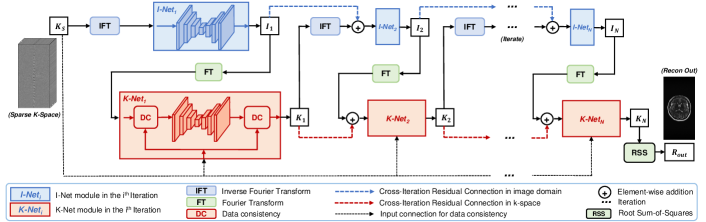
The diagram of DD-CISENet is presented in Fig. 1. The sparse k-space data is first input to I-Net1 module after reconstruction using Inverse Fourier Transform (IFT), generating the output , where refers to a dual Squeeze-Excitation Network (SENet) [Chen et al.(2021)Chen, Zhou, Shi, Liu, Pang, Wang, Miller, Sinusas, and Liu] in I-Net1. refers to the IFT operator.
Then, was input to the K-Net1 module after forward projection using Fourier Transform (FT), generating the output . Then, the IFT of is input to I-Net2 of the 2nd iteration. Meanwhile, is also added to I-Net2 using CIR connections to produce the output . Thus, the image-domain features of the iteration is retained and transmitted to the next iteration to better incorporate the image features.
Similarly, is added to K-Net2 to retain the k-space features. The output k-space of the ith (i2) iteration can be formulated as:
| (1) |
where is a data consistency module. is SENet in K-Neti. is the FT operator. Then, the predicted is reconstructed into the final MRI image as the output.
The overall end-to-end loss function of DD-CISENet is , where represents the combined loss of the iteration. is the loss weight that was empirically set as 0.5 in our study. or is the loss between the ground-truth fully sampled and predicted images or k-spaces.
3 Results and Conclusions
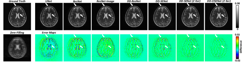
Fig. 2 shows the qualitative comparison of the reconstructed MRI images by multiple approaches. The proposed DD-CISENet outputs more accurate MRI images than existing image-domain prediction techniques (ResNet-Image), k-space synthesis methods (UNet, ResNet), and dual-domain feature fusion approaches (DD-ResNet, DD-SENet). Table LABEL:tab:img lists the quantitative comparison of the generated k-space and reconstructed MRI images by multiple approaches using normalized mean square error (NMSE) and structural similarity (SSIM). It can be observed that DD-CISENet presents quantitatively more accurate k-space data and reconstructed MRI images than existing methods. Paired t-tests further validated the statistical significance of the quantification results (). Thus, the proposed DD-CISENet demonstrated state-of-the-art performance in MRI sparse reconstruction, superior to existing image-domain, k-space, and dual-domain methods.
tab:img
| Methods | Generated K-space Data | Reconstructed MRI Images | |||
| NMSE | SSIM | NMSE | SSIM | P-value† | |
| Zero-Filling | – | ||||
| UNet | |||||
| ResNet | |||||
| ResNet-Image | – | – | |||
| DD-ResNet | |||||
| DD-SENet | |||||
| DD-SENet (2 Iter) | |||||
| DD-CISENet (2 Iter) | |||||
| †P-value of paired t-test on NMSE of Images between the current and previous group. | |||||
This work was funded by Siemens Medical Solutions USA, Inc.
References
- [Chen et al.(2021)Chen, Zhou, Shi, Liu, Pang, Wang, Miller, Sinusas, and Liu] Xiongchao Chen, Bo Zhou, Luyao Shi, Hui Liu, Yulei Pang, Rui Wang, Edward J Miller, Albert J Sinusas, and Chi Liu. Ct-free attenuation correction for dedicated cardiac spect using a 3d dual squeeze-and-excitation residual dense network. Journal of Nuclear Cardiology, pages 1–16, 2021.
- [Eo et al.(2018)Eo, Jun, Kim, Jang, Lee, and Hwang] Taejoon Eo, Yohan Jun, Taeseong Kim, Jinseong Jang, Ho-Joon Lee, and Dosik Hwang. Kiki-net: cross-domain convolutional neural networks for reconstructing undersampled magnetic resonance images. Magnetic resonance in medicine, 80(5):2188–2201, 2018.
- [Zbontar et al.(2018)Zbontar, Knoll, Sriram, Murrell, Huang, Muckley, Defazio, Stern, Johnson, and Bruno] Jure Zbontar, Florian Knoll, Anuroop Sriram, Tullie Murrell, Zhengnan Huang, Matthew J Muckley, Aaron Defazio, Ruben Stern, Patricia Johnson, and Mary Bruno. fastmri: An open dataset and benchmarks for accelerated mri. arXiv preprint arXiv:1811.08839, 2018.