Mask-conditioned latent diffusion for generating gastrointestinal polyp images
Abstract.
In order to take advantage of artificial intelligence (AI) solutions in endoscopy diagnostics, we must overcome the issue of limited annotations. These limitations are caused by the high privacy concerns in the medical field and the requirement of getting aid from experts for the time-consuming and costly medical data annotation process. In computer vision, image synthesis has made a significant contribution in recent years, as a result of the progress of generative adversarial networks (GANs) and diffusion probabilistic models (DPMs). Novel DPMs have outperformed GANs in text, image, and video generation tasks. Therefore, this study proposes a conditional DPM framework to generate synthetic gastrointestinal (GI) polyp images conditioned on given generated segmentation masks. Our experimental results show that our system can generate an unlimited number of high-fidelity synthetic polyp images with the corresponding ground truth masks of polyps. To test the usefulness of the generated data we trained binary image segmentation models to study the effect of using synthetic data. Results show that the best micro-imagewise intersection over union (IOU) of was achieved from DeepLabv3+ when the training data consists of both real data and synthetic data. However, the results reflect that achieving good segmentation performance with synthetic data heavily depends on model architectures.
1. Introduction
The human digestive system can experience a range of abnormal tissue changes, from minor discomforts to severe, life-threatening illnesses (Kaminski et al., 2010). Endoscopy, colonoscopy, and pilcams (wireless capsule endoscopy) (Iddan et al., 2000) are the most common methods for examining the gastrointestinal (GI) tract for diagnosis. However, its effectiveness is greatly impacted by the variability in the performance of the operator (inter-rater reliability) (Borgli et al., 2020). In this regard, artificial intelligence (AI) techniques are researched to build computer-aided diagnosis (CAD) systems to aid gastroenterologists (Le Berre et al., 2020; Riegler et al., 2016; Vinsard et al., 2019; Thambawita et al., 2020).
Supervised machine learning models have become popular in many applications, such as image classification, image detection, and image segmentation. However, AI models require vast amounts of data to train. Especially supervised machine learning techniques need annotated datasets to train. In the medical field, however, acquiring a large annotated dataset is challenging. The challenges include not only privacy concerns but also costly and timely medical data labeling and annotation. In comparison with other applications of machine learning in the health area, we have limited annotated datasets to train machine learning (ML) models. GI-tract datasets are also typically small and mostly limited to polyps (Min et al., 2019). To overcome this issue, one solution is to expand training datasets by generating synthetic data (Jordon et al., 2022; Thambawita et al., 2021a).
Endoscopy diagnostics are being enhanced by AI solutions. Especially, image synthesis has made a significant contribution to overcome the issue of the limited dataset (Rombach et al., 2022). It is now common to use generative adversarial networks (GANs) to generate synthetic images because GANs produce realistic images and achieve impressive results in a wide range of applications (Creswell et al., 2018; Alqahtani et al., 2021). Thus, a GAN is a powerful generative model, however, it suffers from convergence instability.
To overcome the convergence issue in GANs, in recent years, diffusion models (Ho et al., 2020) have gained attention as a potential method for their ability to synthesize natural images. In this study, we introduce a framework consisting of two different diffusion models to create synthetic GI-tract images and corresponding masks. Our contributions are listed as follows:
-
•
We introduce a fully synthetic polyp generation system.
-
•
Our system is able to generate realistic-looking synthetic polyp masks using an improved diffusion model.
-
•
Based on the generated masks, we are able to generate high-fidelity synthetic polyp images conditioned on pre-generated synthetic polyp masks using a conditional latent diffusion model.
-
•
We provide a comprehensive evaluation of using synthetic polyp and mask data to train polyp segmentation models and overall results.
The source code of all the experiments is available at https://github.com/simulamet-host/conditional-polyp-diffusion and the pre-generated synthetic masks and the corresponding conditional synthetic polyp images are available at https://huggingface.co/datasets/deepsynthbody/conditional-polyp-diffusion.
2. Related work
There are many GI image analysis datasets available for machine learning tasks. Some of the commonly used datasets in human GI tract are: ETIS-Larib (Silva et al., 2014), CVC-ClinicDB (Bernal et al., 2015), ASU-Mayo Clinic Polyp database (Tajbakhsh et al., 2015), Kvasir (Pogorelov et al., 2017), Kvasir-SEG (Jha et al., 2020) and Hyperkvasir (Borgli et al., 2020). A few datasets containing manually annotated segmentation masks for polyps. However, these real-world datasets (not limited to GI-tract data) have some limitations. The limitations include:
- •
-
•
Annotation quality: the accuracy and consistency of the annotations of the dataset can impact the performance of machine learning algorithms. Annotations are dependent on annotator and normally high inter-rater variability is there.
-
•
Diversity: the diversity of the images in the dataset is important for the generalization of machine learning algorithms. If the dataset is limited to a narrow range of images, the algorithm may not perform well on new, unseen images.
-
•
Accessibility: legal and privacy constraints can limit the accessibility of medical image datasets, making it difficult to obtain large and diverse datasets for machine learning tasks.
These limitations highlight the need for ongoing development and improvement of medical image datasets to support the advancement of machine learning in medical imaging. To overcome these limitations of real-world datasets, synthetic datasets(Thambawita et al., 2021b, c, 2022, d; Chen et al., 2021) have been increasingly used in medical image analysis. For instance, to generate synthetic polyps, a GAN framework has been proposed to generate a polyp mask and then synthesize the generated polyp with the real polyp image without the use of additional datasets and processes (Qadir et al., 2022). There has also been research on the augmenting of colonoscopy images with polyps by using synthetic samples (Adjei et al., 2022). Fagereng et al. (Fagereng et al., 2022) present a solution to overcome the challenge of a lack of annotated data when building CAD systems for detecting polyps in the GI-tract. The authors propose a pipeline called PolypConnect, which can convert non-polyp images into polyp images to increase the size of training datasets for machine learning models. The results show improvement in mean intersection over union (IOU) when using synthetic data in addition to real data for training. Dhariwal et al. (Dhariwal and Nichol, 2021) compare the performance of diffusion models and GANs for image synthesis tasks. As a result, the authors found that diffusion models outperformed GANs in terms of image quality and stability of the generated images. The results of the paper indicate that diffusion models are a promising alternative to GANs in the field of image synthesis. This work provides valuable insights into the strengths and limitations of both diffusion models and GANs.
3. Methodology
In this section, first, we describe the improved diffusion model which is used to generate realistic synthetic polyp mask images (the blue box of Figure 1). Then, we present the latent diffusion model which is used for generating synthetic polyp images conditioned on the input masks generated from our aforementioned mask generator (the green box of Figure 1). We evaluate the quality of the generated synthetic data and quantify similarity between generated and real data, representing using the last section of Figure 1. Finally, we present our methods used to check the quality of synthetic data using image segmentation models.
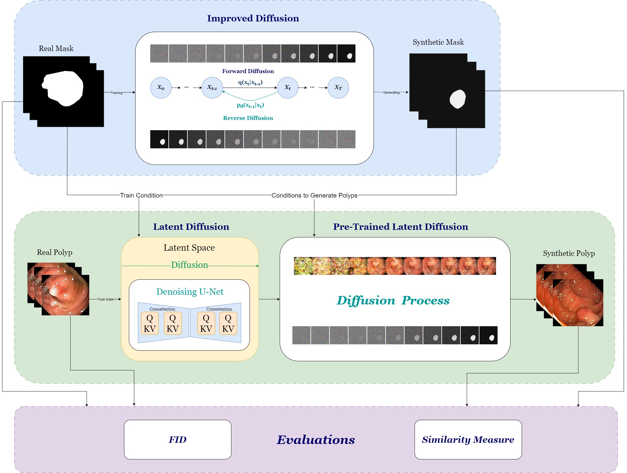
3.1. Improved diffusion model
In our pipeline, we use an improved diffusion model (Nichol and Dhariwal, 2021) to generate synthetic polyp masks which looks realistic to capture the distribution of the masks of the Kvasir-SEG dataset. The improved diffusion model is a type of generative model that uses a gradual, multi-step process to match a data distribution and generate synthetic images. In the context of generating synthetic mask images for the GI-tract, improved diffusion models can be used to generate synthetic mask images that closely resemble real images. Therefore, these models overcome the issue of limited annotated data (Nichol and Dhariwal, 2021; Sohl-Dickstein et al., 2015).
To achieve our goal, the first step is to obtain a training set for real mask images of the GI tract indicating the location of polyps. Then, the mask dataset is used to train the improved diffusion model to generate synthetic polyp images that closely resemble the real images. The improved diffusion model generates synthetic mask images by first adding noise to a randomly selected mask image from the training set. This noise would then be gradually reversed through multiple steps until a synthetic mask image is generated.
The advantage of using improved diffusion models to generate synthetic images is that we can overcome the issue of limited annotated data and train machine learning models more effectively. This can lead on to improvements in the accuracy and efficiency of CAD systems for detecting polyps in the GI-tract (Fagereng et al., 2022)
3.2. Latent diffusion model
The Latent Diffusion model (Rombach et al., 2022), developed by CompVis and trained on the LAION-400M dataset (Schuhmann et al., 2021), operates through a series of denoising autoencoders and diffusion models. This model has been utilized to generate images based on text prompts, and has shown exceptional results in tasks related to image inpainting (Elharrouss et al., 2020) and various other applications, surpassing the current state of the art (Rombach et al., 2021).
Latent diffusion models are a suitable choice for generating synthetic images of the GI tract for several reasons. Firstly, they possess the ability to model intricate and non-linear patterns in the data, crucial for producing convincing images of the GI tract. Secondly, they are capable of generating a large diversity of high-quality synthetic images, which enhances the generalizability of machine learning models. Lastly, they can be trained with a limited amount of real data points, which is important in medical imaging where annotated data is often scarce.
3.3. Mask similarity
To assess the quality of generated images for generative models, the Fréchet inception distance (FID) (Heusel et al., 2017) metric is typically used, which compares the distribution of the generated images compared to real images. Because the improved diffusion model are generating binary masks of polyps, we can also introduce the similarity measure (SIM) metric that is analogous to accuracy. Consider real image together with generated image of the same size, then the similarity is defined as number of pixels that are same for both images divided by the total area of image:
To measure the similarity of generated images to our training real images , we simply take average of the closest pairs (high similarities). If a generated image is and associated closest real image is , we can calculate largest similarity using,
The idea here is that even if the generated images are highly similar to some of the training images (same size, position), they should differ in another aspects, such as rotation.
3.4. Segmentation models
We have used three different well-known image segmentation models, namely UNet++ (Zhou et al., 2018), feature pyramid network (FPN) (Lin et al., 2017), and DeepLabv3+ (Chen et al., 2017) for evaluating the effect of using synthetic data for training polyp segmentation tasks. Initially, we trained these three models using three different approaches, i.e., we trained the system using i) of real polyp images; ii) using synthetic polyp images; and iii) a combination of real and synthetic polyp images. To further analyze the effect of synthetic data, we trained these three models with another set of real and synthetic data combinations. In these combinations, we fixed the number of real images to samples, and we increased the number of synthetic samples from to sequentially in steps of . The main objective of this experiment is to identify the effect of a number of synthetic samples included in the training data. However, we limited this experiment to using only real images because of the time limitation, but in the future we will test with different number of real images from to to find the optimal combination to get better performance.
We tested these models with real images and masks (from the segmentation data of HyperKvasir dataset (Borgli et al., 2020)) which were not used to train either the diffusion model or the segmentation models. Then, we measured micro and micro-imagewise IOU, F1, Accuracy, and Precision from the test dataset for all the segmentation models. Micro values were calculated by summing true positive (TP), false positive (FP), false negative (FN), and true negative (TN) pixels over all images and all classes and then computing scores. In contrast, the micro-imagewise matrices were calculated by summing TP, FP, FN, and TN pixels for each image and then computing scores for each image. Finally, average scores over the dataset were calculated. In the micro-imagewise calculations, all images contributed equally to the final score. However, the second method takes into account class imbalance for each image.
4. Results and discussion
In this section, we discuss experiment setup and the result collected from generative models and segmentation models. A server with Nvidia A100 graphic processing units (GPUs), AMD EPYC 7763 64-cores processor with RAM were used for all the experiments of this study. Additionally, we used Pytorch (Paszke et al., 2019), the Pytorch-lightning libraries, and the Pytorch segmentation library (Iakubovskii, 2019) as development frameworks.
4.1. Diffusion experiments and results
For mask generator, that is improved diffusion model we have used FID and SIM values to quantify and select appropriate model. We have generated masks for each of our saved model, and we compare them with real training masks in Table 1.
| Iter | 0 | 50k | 100k | 150k | 200k | 230k |
|---|---|---|---|---|---|---|
| FID | 140.14 | 128.95 | 117.14 | 105.63 | 88.41 | 141.44 |
| SIM | 88.22 | 89.46 | 90.81 | 91.31 | 92.49 | 88.38 |
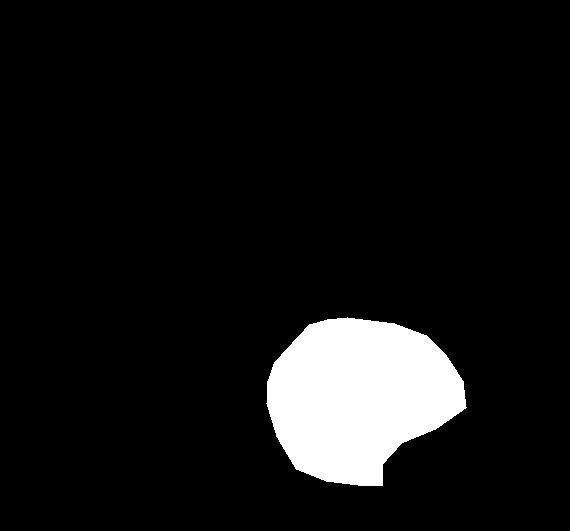
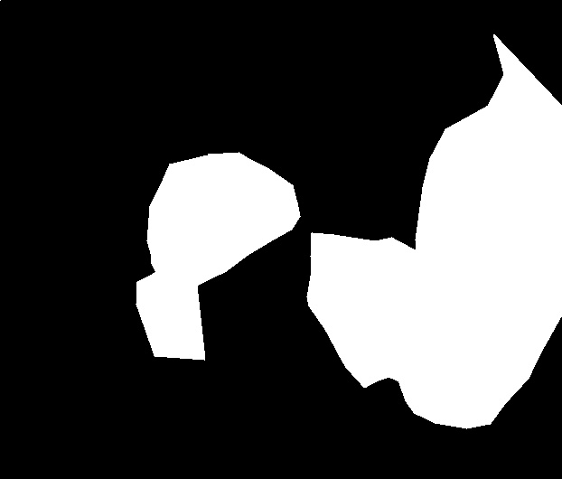
We selected the model from iteration based on the results from Table 1. The reason is that the model achieves lowest FID value together with high SIM values, indicating diverse and quality masks. We also inspected the generated masks visually to confirm this conclusion.
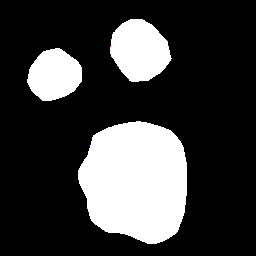
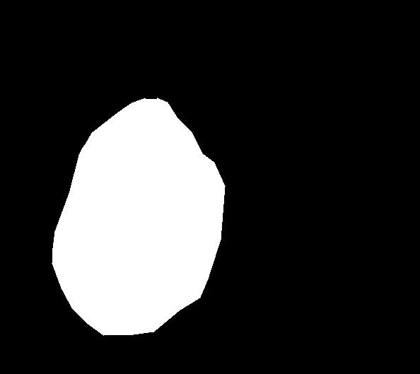
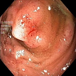
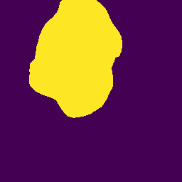
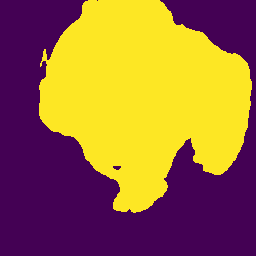
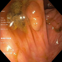
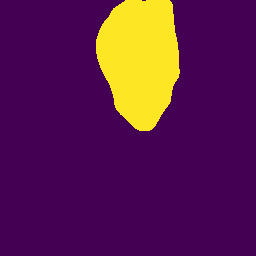
| Real |  |
 |
 |
 |
 |
|---|---|---|---|---|---|
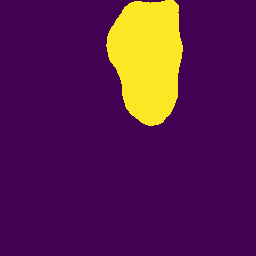
| Generated |  |
 |
 |
 |
 |
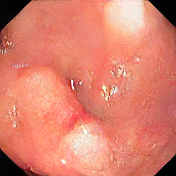
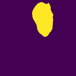
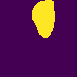
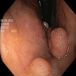
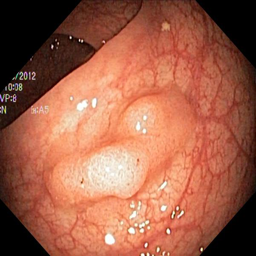
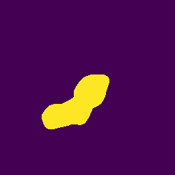
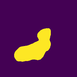
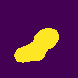
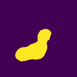
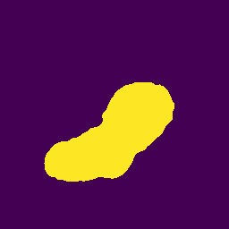
Examples of generated masks in comparison with real masks can be seen in Figure 2. We can see different masks with different shapes and numer of polyps indicating capability to generate diverse synthetic masks. Further discussion with medical professional would be required in order to determine if masks are correct.
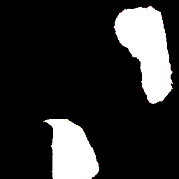
| SIM | 95.89 | 98.11 | 93.45 | 84.62 | 87.98 |
|---|---|---|---|---|---|
| Real |  |
 |
 |
 |
 |
| Generated |  |
 |
 |
 |
 |
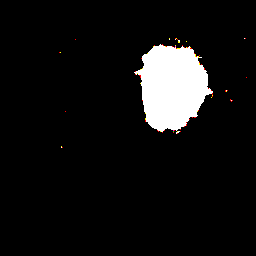
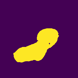
Interestingly, high score doesn’t necessary imply that model is producing identical masks, as can be seen in the Figure 3. For instance, masks may be located in similar positions but have different, smaller shapes, therefore achieving higher similarities.
We have used the generated masks made by the selected model as conditions to our latent polyp diffusion model and produced generated images which we used for further evaluation in Table 2.
| Epoch | 88 | 103 | 135 | 892 | 913 | 922 |
|---|---|---|---|---|---|---|
| FID | 119.34 | 113.83 | 104.78 | 112.66 | 150.97 | 150.85 |
| Condition | Epoch=88 | Epoch=103 | Epoch=135 | Epoch=892 | Epoch=913 | Epoch=922 |
|---|---|---|---|---|---|---|
 |
 |
 |
 |
 |
 |
|
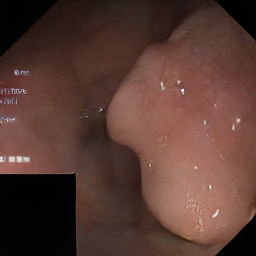
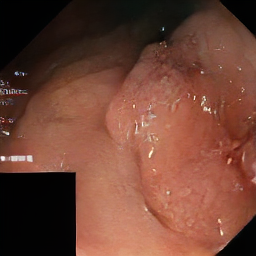
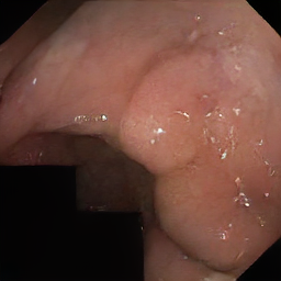
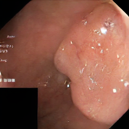
We can see from Table 2 the model which achieved lowest FID score is at . We inspected the generated images, similarly as in Figure 4. It can be seen that the quality of generated images deteriorates at later stages of training, reason may be overfitting. This may lead to problems while generating different samples with same condition which would be more similar.
| (a) |  |
 |
 |
 |
 |
|---|---|---|---|---|---|
| (b) |  |
 |
 |
 |
 |
| (c) |  |
 |
 |
 |
 |
| (d) |  |
 |
 |
 |
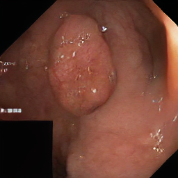 |
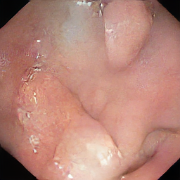
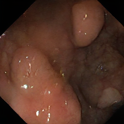
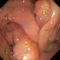
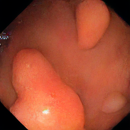
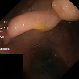
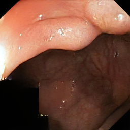
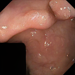
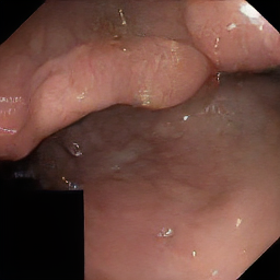
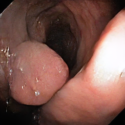
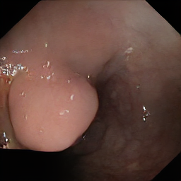
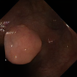
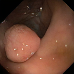
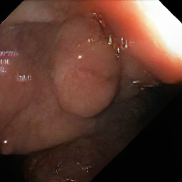
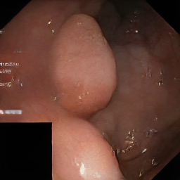
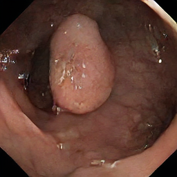
Therefore, we selected the model from earlier stages that achieved lowest FID. We conditioned the model on one mask and generated multiple samples to see if the model generalizes well, results of this experiment can be seen in Figure 5.
4.2. Segmentation experiments and results
We used a learning rate of with the Adam optimizer (Kingma and Ba, 2014) to train the three segmenation models, UNet++, FPN and DeepLabv3+. DiceLoss (Sudre et al., 2017) was used in the training process as the loss function to update the weights. The encoder model of resnet34 was input as the encoder network for all three models (for more details of these encoder networks, please refer to the documentation (Iakubovskii, 2019)). Micro metrics and micro-imagewise metrics (as discussed in the Pytorch segmentation library) were calculated from the best checkpoints and the test dataset after training epochs for all the models. The calculated micro metrics are tabulated in Table 3, and micro-imagewise values are tabulated in Table 4.
| Model: | Unet++ (26.1M) | FPN (23.2M) | DeepLabv3plus (22.4M) | ||||||||||
|---|---|---|---|---|---|---|---|---|---|---|---|---|---|
| # R | # Syn | IOU | F1 | Acc: | Prec: | IOU | F1 | Acc: | Prec: | IOU | F1 | Acc: | Prec: |
| 700 | 0 | 0.7471 | 0.8552 | 0.9509 | 0.8535 | 0.7663 | 0.8677 | 0.9571 | 0.8623 | 0.7457 | 0.8543 | 0.9492 | 0.8699 |
| 0 | 1000 | 0.6852 | 0.8132 | 0.9375 | 0.8009 | 0.6784 | 0.8084 | 0.9276 | 0.7685 | 0.6580 | 0.7938 | 0.9301 | 0.7658 |
| 700 | 1000 | 0.7151 | 0.8339 | 0.9421 | 0.8235 | 0.7300 | 0.8439 | 0.9481 | 0.8323 | 0.7401 | 0.8506 | 0.9492 | 0.8252 |
| 100 | 0 | 0.6970 | 0.8215 | 0.9400 | 0.7912 | 0.6840 | 0.8123 | 0.9371 | 0.8209 | 0.6983 | 0.8224 | 0.9404 | 0.8304 |
| 100 | 100 | 0.6937 | 0.8192 | 0.9382 | 0.7692 | 0.7304 | 0.8442 | 0.9501 | 0.8509 | 0.7200 | 0.8372 | 0.9466 | 0.8305 |
| 100 | 200 | 0.7066 | 0.8281 | 0.9418 | 0.8804 | 0.7382 | 0.8494 | 0.9466 | 0.8763 | 0.7040 | 0.8263 | 0.9429 | 0.8383 |
| 100 | 300 | 0.7309 | 0.8445 | 0.9488 | 0.8536 | 0.7269 | 0.8419 | 0.9459 | 0.8219 | 0.7556 | 0.8608 | 0.9500 | 0.8521 |
| 100 | 400 | 0.6830 | 0.8116 | 0.9386 | 0.8333 | 0.7304 | 0.8442 | 0.9459 | 0.8375 | 0.7298 | 0.8438 | 0.9450 | 0.8342 |
| 100 | 500 | 0.6815 | 0.8106 | 0.9366 | 0.8152 | 0.7244 | 0.8402 | 0.9421 | 0.8209 | 0.7212 | 0.8380 | 0.9454 | 0.8427 |
| 100 | 600 | 0.7083 | 0.8292 | 0.9432 | 0.8287 | 0.7284 | 0.8429 | 0.9491 | 0.8668 | 0.7037 | 0.8261 | 0.9392 | 0.8405 |
| 100 | 700 | 0.7195 | 0.8369 | 0.9460 | 0.8420 | 0.7436 | 0.8530 | 0.9498 | 0.8107 | 0.7083 | 0.8292 | 0.9457 | 0.8347 |
| 100 | 800 | 0.6752 | 0.8061 | 0.9402 | 0.8495 | 0.7387 | 0.8497 | 0.9462 | 0.8278 | 0.7338 | 0.8465 | 0.9476 | 0.8770 |
| 100 | 900 | 0.7069 | 0.8283 | 0.9441 | 0.8319 | 0.7290 | 0.8432 | 0.9463 | 0.8234 | 0.7116 | 0.8315 | 0.9413 | 0.8171 |
| 100 | 1000 | 0.7513 | 0.8580 | 0.9506 | 0.8468 | 0.7214 | 0.8382 | 0.9457 | 0.8126 | 0.7154 | 0.8341 | 0.9401 | 0.8337 |
| Model: | Unet++ (26.1M) | FPN (23.2M) | DeepLabv3plus (22.4M) | ||||||||||
|---|---|---|---|---|---|---|---|---|---|---|---|---|---|
| # R | # Syn | IOU | F1 | Acc: | Prec: | IOU | F1 | Acc: | Prec: | IOU | F1 | Acc: | Prec: |
| 700 | 0 | 0.7551 | 0.8222 | 0.9509 | 0.8742 | 0.7681 | 0.8429 | 0.9571 | 0.8761 | 0.7528 | 0.8317 | 0.9492 | 0.8678 |
| 0 | 1000 | 0.7232 | 0.8013 | 0.9375 | 0.8128 | 0.6977 | 0.7903 | 0.9276 | 0.7757 | 0.7018 | 0.7896 | 0.9301 | 0.8062 |
| 700 | 1000 | 0.7442 | 0.8185 | 0.9421 | 0.8517 | 0.7371 | 0.8128 | 0.9481 | 0.8504 | 0.7751 | 0.8465 | 0.9492 | 0.8628 |
| 100 | 0 | 0.7136 | 0.8005 | 0.9400 | 0.7985 | 0.6587 | 0.7613 | 0.9371 | 0.8039 | 0.7116 | 0.8040 | 0.9404 | 0.8506 |
| 100 | 100 | 0.7146 | 0.7976 | 0.9382 | 0.7931 | 0.7357 | 0.8212 | 0.9501 | 0.8483 | 0.7183 | 0.8048 | 0.9466 | 0.8420 |
| 100 | 200 | 0.7433 | 0.8193 | 0.9418 | 0.8768 | 0.7000 | 0.7842 | 0.9466 | 0.8856 | 0.7197 | 0.8031 | 0.9429 | 0.8554 |
| 100 | 300 | 0.7392 | 0.8168 | 0.9488 | 0.8570 | 0.7302 | 0.8126 | 0.9459 | 0.8357 | 0.7337 | 0.8097 | 0.9500 | 0.8503 |
| 100 | 400 | 0.7097 | 0.7867 | 0.9386 | 0.8444 | 0.7512 | 0.8268 | 0.9459 | 0.8683 | 0.7366 | 0.8153 | 0.9450 | 0.8604 |
| 100 | 500 | 0.7088 | 0.7874 | 0.9366 | 0.8551 | 0.7376 | 0.8200 | 0.9421 | 0.8410 | 0.7264 | 0.8085 | 0.9454 | 0.8369 |
| 100 | 600 | 0.7238 | 0.7987 | 0.9432 | 0.8584 | 0.7348 | 0.8147 | 0.9491 | 0.8639 | 0.7054 | 0.7944 | 0.9392 | 0.8287 |
| 100 | 700 | 0.7230 | 0.7988 | 0.9460 | 0.8471 | 0.7319 | 0.8147 | 0.9498 | 0.8010 | 0.7317 | 0.8135 | 0.9457 | 0.8502 |
| 100 | 800 | 0.7081 | 0.7884 | 0.9402 | 0.8692 | 0.7502 | 0.8274 | 0.9462 | 0.8622 | 0.7548 | 0.8322 | 0.9476 | 0.8725 |
| 100 | 900 | 0.7244 | 0.8025 | 0.9441 | 0.8329 | 0.7441 | 0.8242 | 0.9463 | 0.8534 | 0.7314 | 0.8115 | 0.9413 | 0.8570 |
| 100 | 1000 | 0.7385 | 0.8145 | 0.9506 | 0.8495 | 0.7302 | 0.8086 | 0.9457 | 0.8386 | 0.7343 | 0.8125 | 0.9401 | 0.8574 |
According to the results in Tables 3 and 4, it is clear that adding synthetic data can improve the results of segmentation models. However, it is not always true because some models like FPN and UNet++ show the best IOU, F1, and accuracy when the training data consists of only the real data. In contrast, DeepLabv3 shows the best performance when some synthetic data is included in the training data. Overall, the best micro-imagewise IOU of is achieved from DeepLabv3+ when the training data contains the maximum number of images from both the real data and the synthetic data. Therefore, it is a clear evidence that synthetic data has a direct influence on the final performance of segmentation models. Moreover, we noticed that precision is always better when the synthetic data is in the training data than using only the real data. This implies that synthesized TP can improve the TP predictions which is more important in the medical domain. More visual comparisons are presented in Figure 6. This figure compares the model predictions from the three segmentation models between baseline performance and improved performance (marked using * in Figure 6) using synthetic data. The improved versions of the models were selected using the metrics of Tables 3 and 4.
Another interesting finding of these segmentation experiments is that we get the best values of precision, accuracy, F1 and IOU when we use a smaller number of real images and synthetic images. For example, Unet++ and FPN shows best precision values (micro and micro-imagewise) when the training data consist of real samples and synthetic samples . However, this implies that there is a direct correlation between synthetic data, models, and the number of parameters. Therefore, researchers should not conclude performance gain or degrade of using synthetic data to train segmentation models just by evaluating a single model.
| Image | GT | UNet++(base) | UNet++(*) | FPN (base) | FPN(*) | DLab (base) | DLab (*) |
|---|---|---|---|---|---|---|---|
 |
 |
 |
 |
 |
 |
 |
 |
 |
 |
 |
 |
 |
 |
 |
 |
 |
 |
 |
 |
 |
 |
 |
 |
 |
 |
 |
 |
 |
 |
 |
 |
5. Conclusion and future works
In this study, we used a probabilistic diffusion model-based method to generate synthetic polyp images conditioned on synthetic polyp masks. Our visual and quantitative comparisons show that the generated synthetic data are unique and realistic and not a simple copy of the training data used for the training. Our further analysis of using synthetic data to train polyp segmentation models shows that synthetic data can be used to improve the performance of segmentation models while these improvements are correlated with model architectures. In this regard, we can clearly conclude that synthetic data help to improve the performance of segmentation models. However, deep evaluations should be performed with multiple model architecture to see the real gain of using synthetic data.
In future studies, we will perform more segmentation experiments to get a complete result set for Tables 3 and 4, for example, increasing the synthetic training data gradually with the full real dataset. Moreover, we will generate more synthetic data using our model to train the segmentation models with large synthetic datasets to evaluate the effect of using synthetic data deeply. Generating multiple images conditioned on the same input to train the segmentation models are an another limitation of the presented segmentation experiments. Furthermore, the quality of generated images can be improved using the style-transfer technique (Gatys et al., 2016) as used in the SinGAN-Seg study (Thambawita et al., 2022). Cross-dataset evaluations should be performed to measure the effect of using synthetic data to train segmentation models to improve robustness and generalizability.
Acknowledgements.
The research presented in this paper has benefited from the Experimental Infrastructure for Exploration of Exascale Computing (eX3), which is financially supported by the Research Council of Norway under contract 270053.References
- (1)
- Adjei et al. (2022) Prince Ebenezer Adjei, Zenebe Markos Lonseko, Wenju Du, Han Zhang, and Nini Rao. 2022. Examining the effect of synthetic data augmentation in polyp detection and segmentation. International Journal of Computer Assisted Radiology and Surgery 17, 7 (2022), 1289–1302.
- Alqahtani et al. (2021) Hamed Alqahtani, Manolya Kavakli-Thorne, and Gulshan Kumar. 2021. Applications of generative adversarial networks (gans): An updated review. Archives of Computational Methods in Engineering 28 (2021), 525–552.
- Bernal et al. (2015) Jorge Bernal, F Javier Sánchez, Gloria Fernández-Esparrach, Debora Gil, Cristina Rodríguez, and Fernando Vilariño. 2015. WM-DOVA maps for accurate polyp highlighting in colonoscopy: Validation vs. saliency maps from physicians. Computerized medical imaging and graphics 43 (2015), 99–111.
- Borgli et al. (2020) Hanna Borgli, Vajira Thambawita, Pia H Smedsrud, Steven Hicks, Debesh Jha, Sigrun L Eskeland, Kristin Ranheim Randel, Konstantin Pogorelov, Mathias Lux, Duc Tien Dang Nguyen, et al. 2020. HyperKvasir, a comprehensive multi-class image and video dataset for gastrointestinal endoscopy. Scientific data 7, 1 (2020), 283.
- Chen et al. (2017) Liang-Chieh Chen, George Papandreou, Florian Schroff, and Hartwig Adam. 2017. Rethinking atrous convolution for semantic image segmentation. arXiv preprint arXiv:1706.05587 (2017).
- Chen et al. (2021) Richard J Chen, Ming Y Lu, Tiffany Y Chen, Drew FK Williamson, and Faisal Mahmood. 2021. Synthetic data in machine learning for medicine and healthcare. Nature Biomedical Engineering 5, 6 (2021), 493–497.
- Creswell et al. (2018) Antonia Creswell, Tom White, Vincent Dumoulin, Kai Arulkumaran, Biswa Sengupta, and Anil A Bharath. 2018. Generative adversarial networks: An overview. IEEE signal processing magazine 35, 1 (2018), 53–65.
- Dhariwal and Nichol (2021) Prafulla Dhariwal and Alexander Nichol. 2021. Diffusion models beat gans on image synthesis. Advances in Neural Information Processing Systems 34 (2021), 8780–8794.
- Elharrouss et al. (2020) Omar Elharrouss, Noor Almaadeed, Somaya Al-Maadeed, and Younes Akbari. 2020. Image inpainting: A review. Neural Processing Letters 51 (2020), 2007–2028.
- Fagereng et al. (2022) Jan Andre Fagereng, Vajira Thambawita, Andrea M Storås, Sravanthi Parasa, Thomas de Lange, Pål Halvorsen, and Michael A Riegler. 2022. PolypConnect: Image inpainting for generating realistic gastrointestinal tract images with polyps. In 2022 IEEE 35th International Symposium on Computer-Based Medical Systems (CBMS). IEEE, 66–71.
- Gatys et al. (2016) Leon A Gatys, Alexander S Ecker, and Matthias Bethge. 2016. Image style transfer using convolutional neural networks. In Proceedings of the IEEE conference on computer vision and pattern recognition. 2414–2423.
- Heusel et al. (2017) Martin Heusel, Hubert Ramsauer, Thomas Unterthiner, Bernhard Nessler, and Sepp Hochreiter. 2017. Gans trained by a two time-scale update rule converge to a local nash equilibrium. Advances in neural information processing systems 30 (2017).
- Ho et al. (2020) Jonathan Ho, Ajay Jain, and Pieter Abbeel. 2020. Denoising diffusion probabilistic models. Advances in Neural Information Processing Systems 33 (2020), 6840–6851.
- Iakubovskii (2019) Pavel Iakubovskii. 2019. Segmentation Models Pytorch. https://github.com/qubvel/segmentation_models.pytorch.
- Iddan et al. (2000) Gavriel Iddan, Gavriel Meron, Arkady Glukhovsky, and Paul Swain. 2000. Wireless capsule endoscopy. Nature 405, 6785 (2000), 417–417.
- Jha et al. (2020) Debesh Jha, Pia H Smedsrud, Michael A Riegler, Pål Halvorsen, Thomas de Lange, Dag Johansen, and Håvard D Johansen. 2020. Kvasir-seg: A segmented polyp dataset. In MultiMedia Modeling: 26th International Conference, MMM 2020, Daejeon, South Korea, January 5–8, 2020, Proceedings, Part II 26. Springer, 451–462.
- Jordon et al. (2022) James Jordon, Lukasz Szpruch, Florimond Houssiau, Mirko Bottarelli, Giovanni Cherubin, Carsten Maple, Samuel N Cohen, and Adrian Weller. 2022. Synthetic Data–what, why and how? arXiv preprint arXiv:2205.03257 (2022).
- Kaminski et al. (2010) Michal F Kaminski, Jaroslaw Regula, Ewa Kraszewska, Marcin Polkowski, Urszula Wojciechowska, Joanna Didkowska, Maria Zwierko, Maciej Rupinski, Marek P Nowacki, and Eugeniusz Butruk. 2010. Quality indicators for colonoscopy and the risk of interval cancer. New England journal of medicine 362, 19 (2010), 1795–1803.
- Kingma and Ba (2014) Diederik P Kingma and Jimmy Ba. 2014. Adam: A method for stochastic optimization. arXiv preprint arXiv:1412.6980 (2014).
- Krizhevsky et al. (2017) Alex Krizhevsky, Ilya Sutskever, and Geoffrey E Hinton. 2017. Imagenet classification with deep convolutional neural networks. Commun. ACM 60, 6 (2017), 84–90.
- Le Berre et al. (2020) Catherine Le Berre, William J Sandborn, Sabeur Aridhi, Marie-Dominique Devignes, Laure Fournier, Malika Smail-Tabbone, Silvio Danese, and Laurent Peyrin-Biroulet. 2020. Application of artificial intelligence to gastroenterology and hepatology. Gastroenterology 158, 1 (2020), 76–94.
- Lin et al. (2017) Tsung-Yi Lin, Piotr Dollár, Ross Girshick, Kaiming He, Bharath Hariharan, and Serge Belongie. 2017. Feature pyramid networks for object detection. In Proceedings of the IEEE conference on computer vision and pattern recognition. 2117–2125.
- Lin et al. (2014) Tsung-Yi Lin, Michael Maire, Serge Belongie, James Hays, Pietro Perona, Deva Ramanan, Piotr Dollár, and C Lawrence Zitnick. 2014. Microsoft coco: Common objects in context. In Computer Vision–ECCV 2014: 13th European Conference, Zurich, Switzerland, September 6-12, 2014, Proceedings, Part V 13. Springer, 740–755.
- Min et al. (2019) Min Min, Song Su, Wenrui He, Yiliang Bi, Zhanyu Ma, and Yan Liu. 2019. Computer-aided diagnosis of colorectal polyps using linked color imaging colonoscopy to predict histology. Scientific reports 9, 1 (2019), 1–8.
- Nichol and Dhariwal (2021) Alexander Quinn Nichol and Prafulla Dhariwal. 2021. Improved denoising diffusion probabilistic models. In International Conference on Machine Learning. PMLR, 8162–8171.
- Paszke et al. (2019) Adam Paszke, Sam Gross, Francisco Massa, Adam Lerer, James Bradbury, Gregory Chanan, Trevor Killeen, Zeming Lin, Natalia Gimelshein, Luca Antiga, Alban Desmaison, Andreas Kopf, Edward Yang, Zachary DeVito, Martin Raison, Alykhan Tejani, Sasank Chilamkurthy, Benoit Steiner, Lu Fang, Junjie Bai, and Soumith Chintala. 2019. PyTorch: An Imperative Style, High-Performance Deep Learning Library. In Advances in Neural Information Processing Systems 32. Curran Associates, Inc., 8024–8035. http://papers.neurips.cc/paper/9015-pytorch-an-imperative-style-high-performance-deep-learning-library.pdf
- Pogorelov et al. (2017) Konstantin Pogorelov, Kristin Ranheim Randel, Carsten Griwodz, Sigrun Losada Eskeland, Thomas de Lange, Dag Johansen, Concetto Spampinato, Duc-Tien Dang-Nguyen, Mathias Lux, Peter Thelin Schmidt, et al. 2017. Kvasir: A multi-class image dataset for computer aided gastrointestinal disease detection. In Proceedings of the 8th ACM on Multimedia Systems Conference. 164–169.
- Qadir et al. (2022) Hemin Ali Qadir, Ilangko Balasingham, and Younghak Shin. 2022. Simple U-net based synthetic polyp image generation: Polyp to negative and negative to polyp. Biomedical Signal Processing and Control 74 (2022), 103491.
- Riegler et al. (2016) Michael Riegler, Konstantin Pogorelov, Pål Halvorsen, Thomas de Lange, Carsten Griwodz, Peter Thelin Schmidt, Sigrun Losada Eskeland, and Dag Johansen. 2016. Eir—efficient computer aided diagnosis framework for gastrointestinal endoscopies. In 2016 14th International Workshop on Content-Based Multimedia Indexing (CBMI). IEEE, 1–6.
- Rombach et al. (2021) Robin Rombach, Andreas Blattmann, Dominik Lorenz, Patrick Esser, and Björn Ommer. 2021. High-Resolution Image Synthesis with Latent Diffusion Models. arXiv:2112.10752 [cs.CV]
- Rombach et al. (2022) Robin Rombach, Andreas Blattmann, Dominik Lorenz, Patrick Esser, and Björn Ommer. 2022. High-resolution image synthesis with latent diffusion models. In Proceedings of the IEEE/CVF Conference on Computer Vision and Pattern Recognition. 10684–10695.
- Schuhmann et al. (2021) Christoph Schuhmann, Richard Vencu, Romain Beaumont, Robert Kaczmarczyk, Clayton Mullis, Aarush Katta, Theo Coombes, Jenia Jitsev, and Aran Komatsuzaki. 2021. Laion-400m: Open dataset of clip-filtered 400 million image-text pairs. arXiv preprint arXiv:2111.02114 (2021).
- Silva et al. (2014) Juan Silva, Aymeric Histace, Olivier Romain, Xavier Dray, and Bertrand Granado. 2014. Toward embedded detection of polyps in wce images for early diagnosis of colorectal cancer. International journal of computer assisted radiology and surgery 9 (2014), 283–293.
- Sohl-Dickstein et al. (2015) Jascha Sohl-Dickstein, Eric Weiss, Niru Maheswaranathan, and Surya Ganguli. 2015. Deep unsupervised learning using nonequilibrium thermodynamics. In International Conference on Machine Learning. PMLR, 2256–2265.
- Sudre et al. (2017) Carole H Sudre, Wenqi Li, Tom Vercauteren, Sebastien Ourselin, and M Jorge Cardoso. 2017. Generalised dice overlap as a deep learning loss function for highly unbalanced segmentations. In Deep Learning in Medical Image Analysis and Multimodal Learning for Clinical Decision Support: Third International Workshop, DLMIA 2017, and 7th International Workshop, ML-CDS 2017, Held in Conjunction with MICCAI 2017, Québec City, QC, Canada, September 14, Proceedings 3. Springer, 240–248.
- Tajbakhsh et al. (2015) Nima Tajbakhsh, Suryakanth R Gurudu, and Jianming Liang. 2015. Automated polyp detection in colonoscopy videos using shape and context information. IEEE transactions on medical imaging 35, 2 (2015), 630–644.
- Thambawita et al. (2021a) Vajira Thambawita, Steven A Hicks, Jonas Isaksen, Mette Haug Stensen, Trine B Haugen, JØrgen Kanters, Sravanthi Parasa, Thomas de Lange, Håvard D Johansen, Dag Johansen, et al. 2021a. DeepSynthBody: the beginning of the end for data deficiency in medicine. In 2021 International Conference on Applied Artificial Intelligence (ICAPAI). IEEE, 1–8.
- Thambawita et al. (2021b) Vajira Thambawita, Steven A. Hicks, Jonas Isaksen, Mette Haug Stensen, Trine B. Haugen, JØrgen Kanters, Sravanthi Parasa, Thomas de Lange, Håvard D. Johansen, Dag Johansen, Hugo L. Hammer, Pål Halvorsen, and Michael A. Riegler. 2021b. DeepSynthBody: the beginning of the end for data deficiency in medicine. In 2021 International Conference on Applied Artificial Intelligence (ICAPAI). 1–8. https://doi.org/10.1109/ICAPAI49758.2021.9462062
- Thambawita et al. (2021c) Vajira Thambawita, Jonas L Isaksen, Steven A Hicks, Jonas Ghouse, Gustav Ahlberg, Allan Linneberg, Niels Grarup, Christina Ellervik, Morten Salling Olesen, Torben Hansen, et al. 2021c. DeepFake electrocardiograms using generative adversarial networks are the beginning of the end for privacy issues in medicine. Scientific reports 11, 1 (2021), 21896.
- Thambawita et al. (2020) Vajira Thambawita, Debesh Jha, Hugo Lewi Hammer, Håvard D Johansen, Dag Johansen, Pål Halvorsen, and Michael A Riegler. 2020. An extensive study on cross-dataset bias and evaluation metrics interpretation for machine learning applied to gastrointestinal tract abnormality classification. ACM Transactions on Computing for Healthcare 1, 3 (2020), 1–29.
- Thambawita et al. (2022) Vajira Thambawita, Pegah Salehi, Sajad Amouei Sheshkal, Steven A Hicks, Hugo L Hammer, Sravanthi Parasa, Thomas de Lange, Pål Halvorsen, and Michael A Riegler. 2022. SinGAN-Seg: Synthetic training data generation for medical image segmentation. PloS one 17, 5 (2022), e0267976.
- Thambawita et al. (2021d) Vajira L Thambawita, Inga Strümke, Steven Hicks, Michael A Riegler, Pål Halvorsen, and Sravanthi Parasa. 2021d. ID: 3523524 Data augmentation using generative adversarial networks for creating realistic artificial colon polyp images: validation study by endoscopists. Gastrointestinal Endoscopy 93, 6 (2021), AB190.
- Vinsard et al. (2019) Daniela Guerrero Vinsard, Yuichi Mori, Masashi Misawa, Shin-ei Kudo, Amit Rastogi, Ulas Bagci, Douglas K Rex, and Michael B Wallace. 2019. Quality assurance of computer-aided detection and diagnosis in colonoscopy. Gastrointestinal endoscopy 90, 1 (2019), 55–63.
- Zhou et al. (2018) Zongwei Zhou, Md Mahfuzur Rahman Siddiquee, Nima Tajbakhsh, and Jianming Liang. 2018. Unet++: A nested u-net architecture for medical image segmentation. In Deep Learning in Medical Image Analysis and Multimodal Learning for Clinical Decision Support: 4th International Workshop, DLMIA 2018, and 8th International Workshop, ML-CDS 2018, Held in Conjunction with MICCAI 2018, Granada, Spain, September 20, 2018, Proceedings 4. Springer, 3–11.