Graph Representation Learning for
Interactive Biomolecule Systems
Abstract
Advances in deep learning models have revolutionized the study of biomolecule systems and their mechanisms. Graph representation learning, in particular, is important for accurately capturing the geometric information of biomolecules at different levels. This paper presents a comprehensive review of the methodologies used to represent biological molecules and systems as computer-recognizable objects, such as sequences, graphs, and surfaces. Moreover, it examines how geometric deep learning models, with an emphasis on graph-based techniques, can analyze biomolecule data to enable drug discovery, protein characterization, and biological system analysis. The study concludes with an overview of the current state of the field, highlighting the challenges that exist and the potential future research directions.
keywords:
Graph Neural Networks , Geometric Deep Learning , Protein Design , Molecule Interaction1 Introduction
Studying molecular mechanisms and interactions at different levels of the biological systems is fundamental for understanding organisms, discovering disease pathways, developing novel drugs, and designing functional proteins like enzymes and antibodies. The emergence of deep learning techniques has facilitated the automatic analysis of biological entities by transforming large datasets into tractable numerical representations for trainable algorithms.
One prevalent strategy for capturing the interactions among subunits, e.g., amino acids in a protein or atoms in a chemical compound, is to construct a graph where nodes denote the subunits, and edges indicate the pair-wise interactions between them. For example, a molecule graph represents atoms as nodes connected by chemical bonds. Similarly, a protein graph often employs amino acids as nodes, while edges can reveal sequential neighborhoods and/or spatial proximity. Moreover, graphs can depict higher-level interactions, such as gene expression and metabolic pathway prediction, where a graph captures information exchange between different groups or systems, and nodes stand for molecules, cells, or more complex entities.
Proteins are essential biomolecules with diverse functions in living organisms, including transport, storage, membrane composition, and enzymatic activity. To perform these functions, proteins need to interact physically with small molecules, DNA/RNA, and other proteins. Understanding the function of proteins is crucial for gaining biological insights into protein engineering for disease treatment and prevention, among other life sciences applications.
Protein function is determined by its structure, which has been studied through techniques such as X-ray crystallography, NMR spectroscopy, and Cryo-EM since the 1960s. With the increasing availability of protein structures, particularly with the emergence of in silico folding methods [1, 2, 3, 4], graphs have become a popular tool for characterizing the spatial organization of proteins. A graph is a two-dimensional representation of structured data that encodes pairwise topology between node objects, such as atomic connectivity through chemical bonds.
One significant breakthrough in AI methods for predicting protein structure comes with AlphaFold 2 [3]. This algorithm achieves precision comparable to wet lab experiments by combining Transformer and structure learning models to extract general protein features from large protein sequences. Another notable method is RosettaFold [1] with SE(3) equivariant neural networks and achieves slightly lower performance. Both methods utilize geometric deep learning as a key component of their success. Since then, structure deep learning has been a tool for protein-related prediction tasks, e.g., protein folding and inverse folding, directed evolution, protein-protein docking, protein-small molecule docking, and de novo protein sequence generation. These advances are revolutionizing the traditional pharmaceutical industry, which searches for target protein-based drugs by enumerating drug-like molecules on high-throughput screening (HTS) technologies at a high cost of time and labor [5].
The remainder of the paper is organized as follows: Sections 2 and 3 introduce the learning representation of biological systems and micro- and macromolecules. Section 4 reviews typical graph machine learning tasks on biomolecules. Section 5 reviews a variety of representation learning methods for extracting and processing biomolecule data and accomplishing downstream tasks. Section 6 reviews various applications of geometric representation learning in drug discovery, protein understanding, and biological system analysis. Section 7 summarizes popular benchmark datasets on protein complexes, protein-ligand binding, antibody-antigen structures, and knowledge graphs. Section 8 discusses and concludes the paper with industrial applications.
2 Geometric Representation in Biological Systems
Interactions play a crucial role in depicting the complexity of biological systems, and they can be represented as graphs that are amenable to computational analysis. The nature and scale of the interactions dictate the categorization of the graphs into four distinct types, as illustrated in Figure 1.
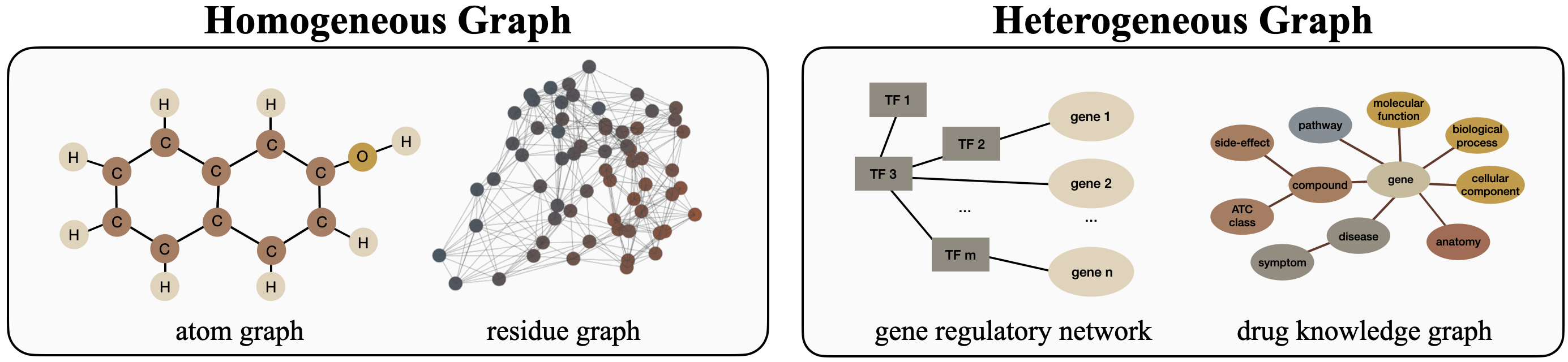
2.1 Homogeneous Graph
When representing structured entities using graphs, it is customary to utilize homogeneous graphs, in which all nodes and edges are of the same type. In the field of structural biology, such homogeneous graphs are commonly employed to depict chemical compounds, proteins, or peptide chains, as we will discuss presently.
2.1.1 Atom Graphs for Chemical Compounds
In the realm of computational biology, chemical compounds are the smallest unit of interest in drug design. Typically, drug candidates are small molecules composed of several tens or hundreds of atoms. The proliferation of high-throughput screening technologies and the advent of computer technology has paved the way for the computational analysis of bioactive drug-like molecules. To this end, it is necessary to represent these compounds using syntactic structures that can be processed by algorithms. One widely used representation is the Simplified Molecular Input Line Entry System (SMILES) [6], which encodes isomeric information into a one-dimensional sequence. While SMILES does not encode stereochemistry, an alternative choice is the molecular graph representation, which represents a compound as a graph, with atoms as nodes and covalent bonds as edges between pairs of nodes.
The utilization of structural information representation has been extensively employed in the field of drug discovery, particularly in drug-target interaction (DTI) [7, 8, 9], binding affinity prediction [10, 11], and drug generation [12, 13]. Depending on the specific formulation of the learning task, it can be a learning task at the node level (e.g., determine which nodes of the target drug will interface with the pocket) or the graph level (e.g., forecast whether the drug will bind to the protein with a given pocket). A comprehensive discourse of the prevalent learning tasks will be expounded in Section 4.
2.1.2 Residue Graphs for Proteins
Proteins perform diverse functions in living systems, such as transport, storage, membrane composition, and enzymatic action. The function of a protein is determined by its unique structure, which serves as a natural foundation for incorporating structural information into algorithms for protein analysis. Graphs have become a popular tool for characterizing protein spatial arrangements. In a residue graph, nodes usually represent amino acids, and edges are constructed to express the chemical or geometrical relationships between node pairs. Alternatively, nodes can be defined as heavy atoms on the main chain to capture more detailed information at finer scales. For a comprehensive discussion on protein graph construction, we recommend readers to [14, 15].
Predicting unseen interactions between a protein and one or more other biological entities has caught great attention in recent studies. One such application is vaccine design, which studies to identify effective interactions between antigens and antibodies to combat pathogenic activity [16, 17, 18]. RNA-protein interaction (RPI) identifies the binding sites and binding affinities between ribonucleic acid (RNA) and RNA binding proteins to understand biological processes such as post-transcriptional regulation of genes [19, 20, 21, 22]. Constructing the quaternary structure of a protein complex is another vital perspective to understanding the protein functions in the living system, which also requires examining the protein-protein interactions [23, 24].
2.2 Heterogeneous Network
Heterogeneous graphs are frequently used to represent integrated networks that depict relationships among diverse entities such as gene expression data and drug-disease association networks. These graphs differ from homogeneous graphs in that they comprise various node and/or edge types, providing a wealth of information and multiple perspectives for the underlying system.
2.2.1 Molecular Interaction Network
A biological system, whether it be a living organism or a human body, is comprised of numerous entities that interact with each other to form a complex network. Nodes in these networks may represent genes, enzymes, proteins, metabolites, other biomolecules, or environmental factors, depending on the network’s properties [25]. In the context of graph representations, different nodes are connected by (directed or undirected) edges that indicate their interactions with each other. For example, a gene regulatory network [26] might feature a directed edge from gene A to gene B, to imply that gene A directly regulates the expression of gene B without the intervention of other genes. Similarly, a metabolism network [27] studies enzymatic reactions (edges) that catalyze one metabolite (node) to another.
The prediction of edge connections, which represent the interactions between molecules, is often the primary goal of graph analysis. In gene regulatory networks, interpreting links between unconnected nodes from gene expression time series data provides insight into the mechanistic regulation of gene expression and allows researchers to infer the phenotype from observed events, given that not all regulatory relationships are adequately captured. In metabolism networks, predicting the metabolic pathway or the pathway dynamics of a specific compound is essential for identifying key metabolites and associated genes and prioritizing potential targets for metabolic engineering.
2.2.2 Drug Knowledge Graph
The prediction of interactions between drugs and targets is crucial for understanding the mechanisms of drug action and identifying potential adverse effects, thus facilitating cost-efficient and safe drug discovery. Network-based approaches usually take existing drugs and diseases as a big interactive network to interpret drug-target associations and propagate the target drug. Biomedical knowledge graphs provide rich and diverse information about various entities and their relationships, such as drug-target interactions and semantic relations between drug entities, to facilitate the interpretation of unknown relationships [28, 29]. Adverse drug reaction prediction is also important, as unexpected interactions between drugs can lead to polypharmacy side effects that undermine the intended therapeutic effects. Accurate prediction of drug-drug interactions can thus increase the success rate of clinical trials and reduce costs in drug discovery. Interpreting the unknown relationships of given entities also plays a key role in adverse drug reaction prediction, which labels undesirable interactions of drugs and targets that fail to meet the intended therapeutic requirements. For instance, a common factor that affects phase III clinical trials is polypharmacy side effects raised by unexpected interactions between drugs [30]. Therefore, effective prediction of drug-drug interactions is substantial for sparing the cost and increasing the success rate of clinical trials for drug discovery.
3 Characterization of Molecules
In the context of deep learning, numerical representations of biological entities such as atoms, compounds, proteins, genes, or cells are required. To set up the problem, different descriptions of input biomolecules are used to reflect the preferred inductive bias, which is closely related to the selection of deep learning techniques. For instance, protein sequences are typically processed with language models to extract contextual representations along one-dimensional chains, allowing for capturing long-range relationships between different atoms or amino acids along the same chain. On the other hand, grid-like representations are handled by convolutional networks, where atoms or amino acids within smaller receptive fields are more influential to the central instance. This section provides a summary of the common rules for characterizing molecules.
3.1 Node Types
As mentioned earlier, biomolecules possess a well-defined hierarchy. For example, proteins consist of amino acids, which in turn are composed of multiple atoms. In this section, we will discuss the construction of a biological system in three hierarchies: atoms, amino acids, and macromolecules.
Atom
A chemical entity comprises a set of atoms that are interconnected by means of chemical bonds. The atomic characteristics dictate the shape and properties of the molecule, rendering it sensible to represent a chemical compound with its constituent atomic symbols, such as C for carbon, O for oxygen, and N for nitrogen. The description of atoms can be supplemented by their physicochemical properties, such as the charges of atoms and the number of attached hydrogen atoms [31]. The shape or conformation of the molecule is crucial for studying its functionality. For small molecules, the torsion angles (or dihedral angles) of four consecutive covalently bonded atoms are usually used.
Amino Acid
The primary structure of a protein refers to the specific arrangement of amino acids that make up its structural foundation. Amino acids, excluding those that are uncommon or non-traditional, can be classified into different groups. Each type of amino acid is commonly designated by a unique one-letter code, such as R for Arginine and K for Lysine, and these codes correspond to various characteristics including hydrophobicity, polarizability, and net charge. For an extensive overview of the diverse properties of amino acids, please refer to [24].
Molecule
When investigating the intermolecular interactions within a biological system or between different systems, it is commonplace to model molecules and their interactions using nodes and edges in a graph [32, 33]. The choice of nodes depends on the particular system under investigation. For example, in gene regulatory networks, nodes could represent transcription factors and target genes [34]. In metabolic networks, nodes might be metabolites-reactions [35] or enzyme-substrate pairs [36]. Alternatively, in drug knowledge graphs, nodes may be disease, gene, compound, protein, and other relevant entities [37, 38].
3.2 Molecule Modalities
There exist various modalities to represent a given biomolecule. For instance, a protein’s chemical composition can be expressed using its amino acid sequence, and its folded tertiary structure can be used to describe the inter-residue spatial relationships. We provide a brief summary of some commonly used methods for representing biomolecules.
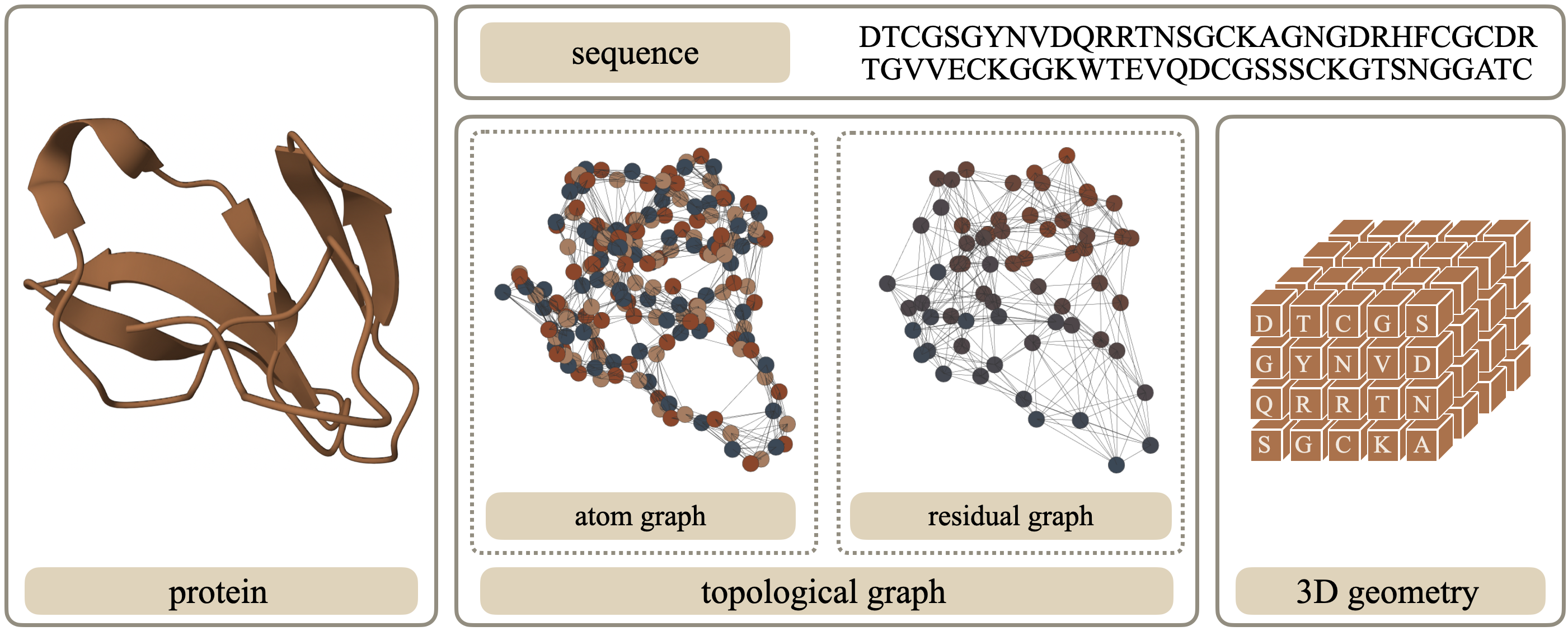
1D Sequence
As previously discussed, one approach for representing a molecule is through character-level tokenization [39], where each component (e.g., an atom or amino acid) is represented by a character in a string. For example, complex compounds can be described using SMILES [6] or self-referencing embedded strings (SELFIES) [40], while the primary structure of a protein can be encoded as a unique sentence composed of different amino acid types. In proteins, amino acids, motifs, and domains are comparable to words, phrases, and sentences in human languages, where sequential representations are typically encoded using language models such as 1D CNNs and attention mechanisms.
2D Graph
While the sequential representation of a molecule provides a detailed breakdown of its components, it fails to consider the intramolecular steric interactions between them, such as the force fields between atoms. Graph data presents an alternative way to describe a molecule’s geometric connectivity by reducing its conformation without explicitly defining a 3D coordinate system. In essence, a graph is composed of a set of nodes that represent the characters in the 1D sequence (atoms or residues), with edges connecting nodes based on identified relationships. These relationships can be either explicitly defined by biological connections (e.g., chemical bonds) [41, 42, 43] or implicitly defined by geometric connections (e.g., pair-wise Euclidean distances in 3D space) [44, 45, 46]. The edge connectivity is stored in an adjacency matrix, which is critical for constructing aggregation rules that establish node representations. However, constructing an appropriate adjacency matrix can be challenging, particularly for large molecules like proteins. Sparse connectivity may fail to capture the complete geometry of the underlying topological manifold, while an overly dense adjacency matrix may impair computational efficiency and potentially dilute valuable information for learning tasks.
3D Geometry
The spatial relationships between internal entities (e.g., amino acids or non-hydrogen atoms) in molecular structures can be represented in 3D space by their 3D coordinates. There are several variants of 3D geometric representation. If neglecting the pair-wise connections, the group of entities is called a point cloud [47, 48]; if the nodes are assumed aligning to the grid, it can be viewed as a 3D box [49]; if a molecule’s shape is a major concern over its internal structure, a 3D mesh can be leveraged to describe the surface [50, 51]; otherwise, if the nodes are connected discretely in the space, we take it as a 3D graph [45]. The choice of representation depends on the specific properties of interest, and all of them require consideration of rotation and translation equivariance in 3D space by geometric deep learning [52, 53].
3.3 Feature Preparation
The features of nodes and edges in a network are informed by domain-specific knowledge, which may exert a significant influence on learning outcomes. Some widely-used input functions are enumerated below.
Atom Feature
An in-depth examination of proteins at the atomic level provides a more nuanced understanding, as the energy of inter-residue interactions is influenced by the number of atoms in contact [54]. These atoms can be identified by their specific types, with some common examples including N, C, O, and S [55]. Atom types also exhibit various physicochemical attributes, such as charge (positive, negative, or neutral) [56], polarity (polar or non-polar) [57], and aromaticity (aliphatic, aromatic, or neutral) [58], which augment their distinctive characteristics. In addition, researchers have defined spatial attributes for atom nodes, such as the number of attached non-hydrogen or hetero atoms representing heavy-valence or hetero-valence properties [31, 59]. Rather than hand-crafting atom-level feature vectors, an alternative is to employ fingerprints, a type of sparse descriptor containing thousands of values, to represent the structural and physicochemical properties of atoms [8].
Residue Feature
When considering an amino acid within a biomolecule, such as a protein, its physicochemical and geometric characteristics play a critical role in determining its potential for interaction within the molecule structure. A protein is typically represented by one-hot encoding the residue types to or dimensions [24]. The rigidity, flexibility, and internal motion of the residue can be identified by the B-factor, which is determined through crystallization processes [60]. Geometric properties, such as the 3D position of the residue, are typically represented by its -carbon (or C) coordinates [61], while the solvent-accessible surface area (SASA) is used to identify whether the residue is on the protein’s surface or in the core [62]. The relative positions of amino acids in the protein chain can be expressed using trigonometric values of backbone dihedral angles derived from the positions of the backbone atoms N, C and C [63]. In addition to scalar features, vector features can also be informative, such as the direction of C-C [3, 64]. Incorporating such directional attributes requires designing specialized propagation rules, such as graph vector perceptrons (GVP) [65].
Edge Feature
The attributes of edges describe the relationship between interconnected nodes. The inter-residue distances of adjacent nodes can be derived directly using their Euclidean distance in 3D space. Additionally, Gaussian radial basis functions (RBF) can be used to project scalar distance into higher dimensions for better expressivity [66, 67]. Relative positional encoding represents the sequential distance of closely located residues on the amino acid chain [3, 64, 68]. The positions of heavy atoms in two connected residues encode local interactions with fine-grained local frame orientation [69, 45].
4 Geometric Learning Tasks
A graph can describe the structure of a given molecule with a set of nodes and edges , where indicates node is connected to and otherwise. For computational convenience, the pair-wise graph connection is stored in an adjacency matrix , which is a symmetric matrix for undirected graph, i.e., , . The numerical representation for nodes and edges, if applicable, are respectively described by and .
4.1 Graph Representation Learning
In graph representation learning, the main objective is to learn a powerful and informative latent representation of the graph or nodes, which can be further utilized in various downstream applications such as node- or graph-level attribute predictions.
Discriminative Learning
Supervised or semi-supervised discriminative learning models are usually applied to predict the optimal labels from input using large sets of labeled data. The discriminative models essentially learn to estimate the probability distribution , where represents the target variable specific to the task at hand.
Generative Learning
Despite the considerable progress achieved by supervised learning models in predicting labels, their reliance on extensive manual labeling poses challenges such as generalization errors, spurious correlations, and adversarial attacks. In computational biology, the limited availability of labeled data necessitates the development of approaches that can learn from fewer manual labels, samples, and trials. In some cases, the goal of learning is to generate new molecule samples from the joint distribution with specified .
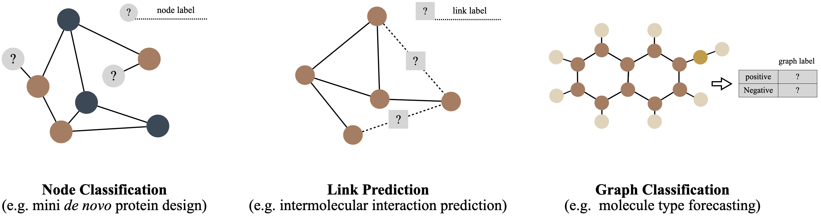
4.2 Downstream Tasks
Graph data is composed of nodes, edges, and the graph itself. Typically, representation learning is done at the node level, but the resulting vector representation can be utilized for all three levels of label assignment tasks.
Node-level Properties Prediction
At the node level, graph convolutional layers encode the topology of an attributed graph by creating a matrix , where each of the nodes is represented by an -dimensional vector that aggregates information about its neighborhood. Then, readout layers assign each node a label . The learning tasks are named node classification or regression, depending on whether the label is discrete or continuous. For example, identifying the type of an amino acid in a protein is a node classification task, while predicting the SASA of a node is a node regression task.
Link Prediction
The process of learning edge properties is similar to node-level learning, except that the subjects of labeling become edges instead of nodes. Typically, a label is a binary indicator for the existence or non-existence of a connection between nodes or a decimal weight indicating the strength of the relationship. The objective of relationship inference or link prediction is to estimate the pairwise connections of nodes, represented as , given a set of node representations . The concatenation of hidden representations of node pairs is used to predict whether there exists a connection between and through binary classification. Link prediction tasks are commonly used in analyzing heterophilic graphs, such as molecular interaction networks [70] and drug repurposing graphs [71].
Graph-level Properties Prediction
Graph-level tasks refer to the prediction of labels for a collection of graphs, denoted as , with corresponding labels . Graphs can differ in size and structure, in contrast to image classification where the number of pixels is consistent across different images. For example, in D&D [72, 73] that classify proteins into enzyme and non-enzyme structures, a protein structure can consist of dozens to over amino acids. Developing graph pooling strategies that can unify varying-sized graphs into a low-dimensional representation is beneficial for these tasks. Moreover, when predicting interactions between pairs of graphs, concatenation layers may be necessary to combine the representations of different graphs.
5 Representation Learning Strategies
This section discusses three main approaches to establishing a learning pipeline for biomolecules. We will begin with basic encoding modules for sequential or spatial molecular representation learning. Since encoding strategies can be combined in parallel or subsequently, we will also provide examples of multimodal inputs that offer more than one modality or scale of biomolecules. Given that labeled biomolecule datasets are limited in practice, we will summarize some essential principles for model augmentation. While we will use residue graphs or protein sequences as examples for explaining the models, they can be easily adapted or generalized to other types of molecules such as chemical compounds.
5.1 Topological Encoding
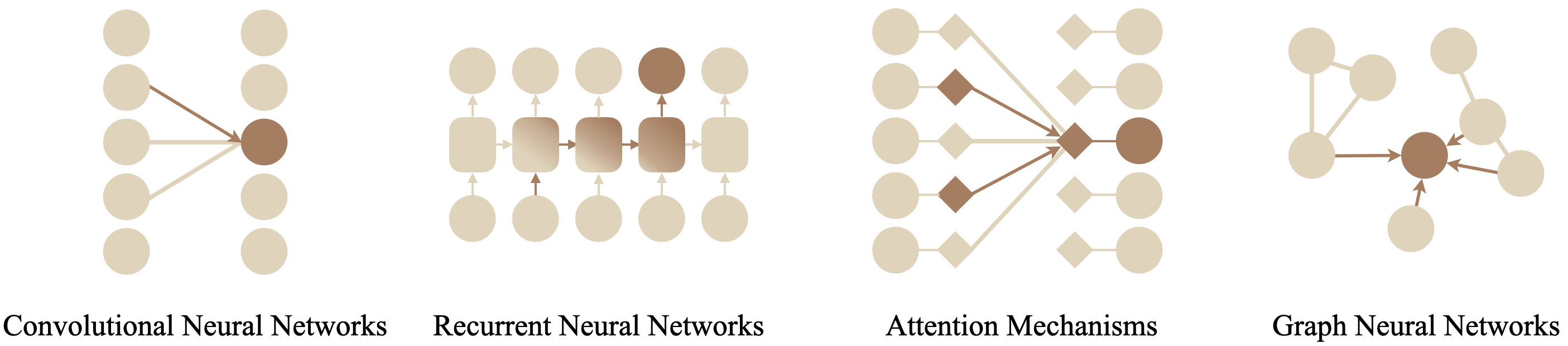
We present three commonly used neural networks in the field of molecular learning: graph networks, recurrent networks, and attention-based networks. Although not all introduced modules were initially designed for graph inputs, they can be used jointly or subsequently to fulfill various roles in a learning pipeline. For instance, IEGMN [66, 69] utilizes attention mechanisms in designing equivariant message passing for residue graphs; GVP-GNN [65, 74] designs attention-like vector perceptron to transmit both scalar and vector nature across graph nodes; DeepFRI [75] learns LSTM-extracted residue-level features with GCN.
5.1.1 Neural Message Passing
As discussed in Section 3, the topology of proteins is crucial for understanding their functionality. To characterize protein data, various inductive biases can be incorporated, such as 3D density mappings with CNNs [76, 77, 78] and interactive propagation with GNNs [64]. The latter is typically formulated under the framework of (neural) message passing [79]. For a biomolecular graph, the hidden representation for the th node in the th layer is updated by
| (1) |
where the information from all one-hop neighbors in is aggregated using a differentiable and permutation-invariant function , such as summation, average, or maximization. The represent another two differentiable functions for transformation.
As a protein’s atomic dynamics remain unchanged regardless of its translation or rotation within a system [80], some propagation rules incorporate the inductive bias of symmetry into structure-based models, requiring the learned 3D protein transformations to belong to the Special Euclidean group SE(3) [53, 81], which is the set of rotation and translation equivariant transformations in 3D space, i.e., with some rotation matrix and translation vector . These algorithms avoid expensive data augmentation and typically need the propagation to consider positional encoding of 3D coordinates [45, 82, 83].
5.1.2 Autoregressive Sequential Propagation
Sequence-based inferences frequently assume a conditional relationship for . In de novo protein design, autoregressive methods are used to predict the next masked amino acid token based on previously inferred amino acids [84, 85, 86]. Such an assumption satisfies the memory embedding criteria of recurrent neural networks , which joins the historical embeddings and current observation to determine its hidden representation:
| (2) |
Since the vanilla recurrent mechanism dampens the importance of early encoded states, gating mechanisms [87] introduce additional memory cells to guide the transmission of useful memory in previous steps. Gated recurrent networks have been widely applied in time series analysis for directional relationship inference. Because sequence representations of proteins neither specify an origin or termination nor a query direction in nature, BiLSTM [88] becomes a popular choice due to its enhanced performance in forward and reverse long-short-term memory encoding [89, 90, 91].
5.1.3 Pair-wise Relationship by Attention Mechanism
Inferring connections between tokens (i.e., amino acids) in either direction along the protein sequence using a recurrent network may miss important relationships between tokens that are sequentially far apart but spatially close. Attention mechanisms provide an alternative by considering all pair-wise relationships in a protein to infer the most comprehensive interactions between contact tokens. Transformer [92] as an advanced self-attention mechanism, has gained popularity due to its ability to capture global dependencies among input instances. Transformer, an advance in self-attention, has seen a surge in recent breakthroughs. For instance, AlphaFold 2 [3] leverages evolutionary Transformers (Evoformer) for pair-wise residue representations, and ESM-if1 [93] applies Transformers on scalar and vector features of residues. Fundamentally, a self-attention layer captures global dependencies among input instances by
| (3) |
with , , and . The entire mechanism can be intuitively understood by considering and as the questions of the input sequence and the capability scores of every input token to answer each question. In return, the inner product measures the weighted similarity of input tokens. Compared to recurrent networks, self-attention layers investigate pair-wise relationships along the entire chain to avoid squashing long-range dependencies in a vector. It then becomes theoretically possible to train extra-long sequences with little information loss. However, the massive size of learnable parameters limits the input sequence length to no more than or tokens in most models.
5.2 Multimodal and Multiscale Molecular Representations
While this review mainly discusses the graph representation of proteins, there is potential for integrating protein descriptions in various modalities and scales to uncover the mechanisms behind protein functionalities [94].
A handful of previous literature features different morphologies of proteins, of which sequence and structure representations are arguably the two most popular options. Large pre-trained language models such as ProtBERT [95] and ESM-2 [96] are used for sequential representations, while GNNs and CNNs can encode local environment information for structure representations. In downstream tasks, various algorithms such as MLPs [97, 98], autoencoders [48, 99], or other specially-designed methods [100, 101] can integrate separate embeddings from different modalities into a single-vector representation. Multiple sequence alignment (MSA) can also enhance sequential inputs to identify conserved and variable regions of proteins from homologous sequences [3, 102, 103]. Other research seeks versatile modalities. HOLOPROT [51] represents a protein’s surface and structure, where the former generates the spherical relationship of residue nodes through a triangulation algorithm, and the latter captures four levels of a protein structure by a residue graph with multi-scale message-passing networks. Transformer-M [104] establishes atomic-level descriptions for a protein by incorporating 1D (e.g., physiochemical properties), 2D (e.g., sequential distance), and 3D (e.g., Euclidean distance) channels. Beyond conventional topological or physiochemical characteristics from sole proteins, some work explores feature representation from residue-centric meta-paths in interactive networks, such as PPI networks [98, 105], drug-protein-disease networks [106] or combining six STRING networks for human and yeast [107].
Describing proteins from different scales and obtaining more detailed representations while minimizing costs can be highly advantageous. Several methods have been proposed to extract feature representations for proteins and ligands at different levels. For example, DeepFusion [108] generates feature representations for drugs and proteins using pre-computed global structural similarity matrices and local chemical substructure semantic features. To encode structure representations at multiple scales, a coarse-grained GNN handles node features and edge connections at coarser levels with gated recurrent units [109]. AGL-Score [110] and MWCG [111] construct multiscale weighted colored subgraphs with atomic types and extended connectivity interactive features (ECIF) to capture intramolecular interactions between proteins and ligands. iEdgeDTA [112] extracts both node-level and sequence-level protein features by language models and graph convolutions. MSSA-Mixup [113] captures internal dependencies among different scales of graph signals by graph wavelet theory using self-attention, and [114] develops the persistent path-spectral model that characterizes the data at multiple scales during the filtration process.
5.3 Model Augmentation
The availability of accurately labeled biomolecular data is often limited, and the ability of a trained model to generalize to new data is often a major concern. Self-supervised learning [115, 116] strategies are becoming increasingly popular as they offer a promising solution to these problems. These strategies train algorithms to recover partially corrupted or incomplete original inputs without any additional manual labels or supervision. Pre-trained models can then be directly applied to downstream tasks, and in situations where there is a small set of labeled training samples, self-supervised learning can replace random initialization, as seen in many computer vision tasks. Though, when fitness to a particular prediction task is the only concern and the number of labeled instances is sufficiently big, a supervised learning approach remains the preferred choice. This section introduces two broad categories of self-supervised learning strategies, which are generative and contrastive methods.
5.3.1 Generative Learning Models
Generative models aim at summarizing observed probability density landscapes in a hidden space and sample new molecules or molecular representations conditioned on protein properties, functions, and/or structures [117, 118].
Variational Autoencoders (VAE)
VAE refers to a general class of networks that map the inputs by an encoder network to a low-dimensional latent space that follows a specific distribution, e.g., . A decoder network then reconstructs samples from the latent space to in the original space. The training goal is to find the optimal network parameters that minimize the distance between the reconstructed and original samples, i.e.,
| (4) |
where is a tunable parameter. The reconstruction loss accounts for inductive biases that are added to the inputs, such as the molecule’s rigidity [119].
Generative adversarial networks (GANs)
GANs are a subset of energy-based models that comprise a generator that maps random noise to instances in the data space and an adversarial discriminator that distinguishes between real and generated examples. A min-max loss function pushes the generator and discriminator toward the dynamic equilibrium by
where is sampled from ground truth input, and is generated using . Maximizing the objective function with a fixed guides toward the optimal discriminator. There are various options for reformulating the discriminator loss, and a comprehensive discussion can be found in [120].
Generative Diffusion Models
The explosive success in image synthesis has attracted increasing attention in generative diffusion models [121, 122, 123]. Similar to hierarchical VAE [124], a diffusion model steps into latent states progressively by . Compared to hierarchical VAE that learns an implicit transformation from to , diffusion models add random Gaussian noise during the generation steps by , . Consequently, the loss function involves an additional noise matching term over Eq. (4). Generative diffusion models have been applied in several recent works for molecule generation or protein design, where noise is added to atoms’ type [125] or structure [43, 126, 127].
5.3.2 Contrastive Learning
Contrastive learning is another technique utilized to enhance the representation of proteins [128, 129]. Given a protein instance that is similar to and dissimilar to , a function is used to encode the three graphs in order to maximize the similarity between and while minimizing the similarity between and . To achieve this, pseudo data labels are required during self-supervised learning, where positive pairs ( and ) can be jointly sampled by sequence-based [130, 131] or Euclidean distance-based [132] croppings. Negative pairs ( and ), on the other hand, are sampled independently. When measuring the pair-wise similarity of hidden representations, a scoring function is learned on the encoded representation. For instance, InfoNCE [133, 134, 130] and NT-Xent [131] evaluate the agreements between the positive pair and all other negative pairs . Formally,
| (5) |
with batch size . The similarity between two representations is usually implemented by the inner products or the cosine similarity . The minimized loss attains a lower bound of the mutual information for the hidden representations. Moreover, researchers may consider integrating previous strategies, such as MoHeG [135], which leveraged a weighted loss function that combined Eq. (4) and Eq. (5). This approach led to a substantial improvement over using either equation individually.
6 Applications
Proteins that possess functional activity are typically composed of multiple polypeptide chains or require the presence of other biomolecules, such as coenzymes and cofactors, to perform their biological roles. A number of routine tasks involving protein functionality have been investigated in the scientific literature. In accordance with Section 2, we categorize three levels of protein interactions with compounds, proteins, and biological networks, respectively.
6.1 Drug Discovery
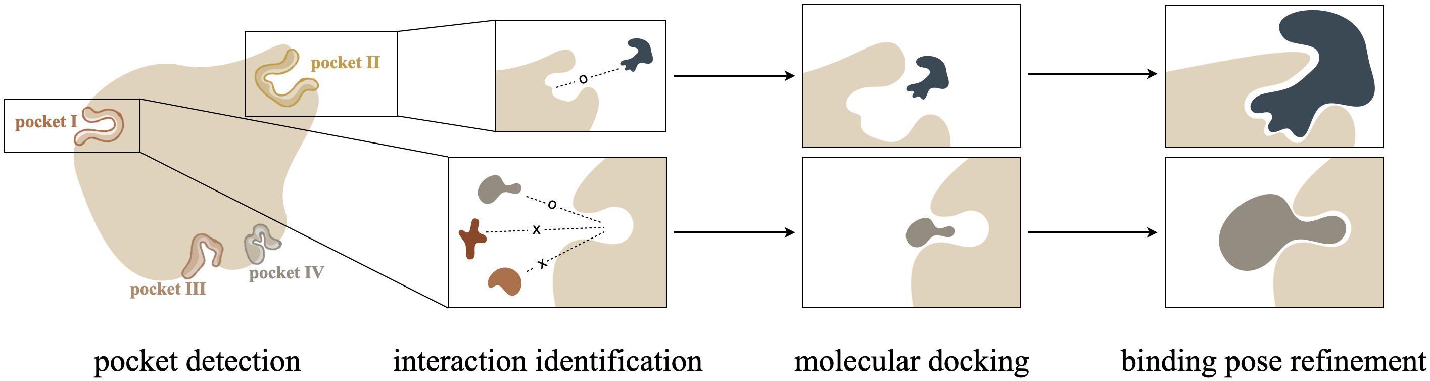
Drugs (ligands) exert their effects by interacting with target viral proteins (receptors). A ligand molecule binds specifically and reversibly to a receptor protein, such as a virus, to alter its activity or function. Therefore, designing a new drug involves identifying potential ligands from drug-like molecules and studying their interactions with specific receptors. Figure 5 demonstrates the four computational steps for a ligand binding to a given receptor protein [136]: 1) pocket detection finds feasible binding site(s) on the receptor. 2) interaction identification recognizes candidate ligands that may interface with the current pocket. 3) molecular docking generates binding poses for the ligand-receptor docking pairs. 4) binding pose refinement dynamically fine-tunes the conformations at the binding sites.
6.1.1 Pocket Detection
Detecting new binding pockets is a crucial and challenging task that initiates the entire process of drug discovery. The identification of binding regions is typically achieved by locating heavy atoms in molecule sequences [137, 138] or voxels of 3D protein images [139, 140, 141] or surfaces [142, 141, 143], where Transformer [92] or 3DCNN [76, 78] are frequently used encoding blocks.
Since proteins do not naturally distribute atoms on a grid, recent work has explored using graph representations to capture protein structures. Binding site prediction is then converted into a classification task on graph nodes. ScanNet [48] detects both protein-protein binding sites and B-cell epitopes (i.e., antibody binding sites) to SARS-CoV-2 spike proteins with attention-based graph convolutions on both atom-level and residue-level protein representations. GraphPPIS [144] extracts node representations of residue graphs with deep GCNs to identify PPI sites with protein-interacting probabilities. PocketMiner [145] modifies GVP-GNN to find cryptic pockets of protein targets where binding sites are only exposed on the surface during binding.
Another line of research focuses on designing pocket matching algorithms to pair relative pockets for binding similar ligands, which can be assessed by the shape of molecular surfaces or sub-graphs that reveals similarities in their physical and biological properties [146, 147, 148]. In graph representation, the objective then turns to finding the maximum similarity between local sub-graphs, such as the pocket size, solvent accessibility, and flexibility [149, 150]. The topological characteristics of pockets are usually captured by PDEs [151, 152], which evolve a convex hull surface inward until it touches the protein surface everywhere. The algorithm not only estimates the surface area, volume, and depth of the pocket but also describes sub-pockets characterizations that determine multi-ligand synergistic interactions.
6.1.2 Interaction Identification
The identification of potential interactions between drugs and their targets is crucial for the development of safe and effective drugs [11, 153]. Such interactions can involve various types of binding, activation, and inhibition. Researchers have designed different computational approaches that can predict interactions between compound-protein, protein-protein, and antibody-antigen.
Compound-Protein Interaction (CPI)
The interaction of drug molecules with target proteins is essential for identifying hit and lead compounds [154] and recognizing potential drug side effects [155]. Due to the limited availability of protein structures in the pre-AlphaFold era, earlier research encodes protein sequences by language models and represents compounds as sequences [156, 157, 158] or images [159], or graphs [160, 161, 162]. Later on, the increasingly available number of protein structures urges the prevalence of graph convolutions for protein receptors, such as GCN, GraphSAGE, and GAT, for CPI prediction. These approaches aim to predict binary interaction estimations [163, 164] or numerical binding affinities[165].
Predicting protein-protein interactions can be approached on different levels, such as atom, residue, or protein levels, for binding score prediction or binary interface label assignment. Graphs can also be constructed for interface regions for protein-protein complexes instead of individual proteins.
Protein-Protein Interaction (PPI)
In contrast to small molecules that interact with a single target receptor, proteins act in a network of complex molecular interactions, investigating which is vital to prevent side effects from false-binding [166]. Predicting protein-protein interactions can be approached on different levels, such as atom [167], residue [168], or protein [169] levels, for binding score prediction or binary interface label assignment. On top of individual proteins [170], the graph(s) might also be constructed for interface regions for protein-protein complexes [168].
Antibody-Antigen Interaction
An antibody is a Y-shape protein of light and heavy chains. The variable domain, defined jointly on both chains, is a part of the antibody’s binding region to recognize their respective antigens, i.e., complementarity-determining regions (CDRs) [171]. Overall, the target of antibody-antigen binding is similar to that of compound-protein interaction, where both tasks focus on analyzing the interface region (pocket or CDR region) to predict complex binding affinity and docking scoring function. In antibody-antigen interaction predictions, the interface regions might be described jointly by pharmacophores and atomic distance [172] or separately by residue graphs [173, 174].
6.1.3 Molecular Docking
Molecular docking finds the 3D structure of the ligand-receptor complex for optimal conformations, where many in silico methods follow the ‘lock-and-key’ theory [175] and solve the correct orientation of the ligand to match the rigid receptor. To identify the tight pose of a protein-ligand complex, a docking program constructs a set of poses with minimized free energy and scores the strength of the putative poses to select the optimal pose for the top-rank docking pairs [176, 177].
Binding Pose Generation
Molecular docking predicts the best binding conformations for ligand-protein complexes with diverse optimized scores, such as the predicted error in binding affinity forecasting [178], the ligand position [66], or a combination of both measurements [179]. As a surrogate of ligand positions, generative models tend to target indirect predictors that are considered easier to learn, such as ligand position relative to the protein, orientation in the pocket, and the torsion angles [125]. Other attempts to enhance the prediction performance have been assessed by introducing trigonometry constraints for residue graphs [3, 65] and reconstructing the future trajectories for the dynamic systems of complexes [180]. Graphs are usually defined at the atomic level for drugs and the residue level for proteins, with encoders essential for describing the roles of key residues in the conformational transition [177, 179, 69].
Antibody-Antigen Docking and Design
Antibody-antigen docking prediction falls into the general class of protein-protein docking and design, but the lack of evolutionary information in paratopes prevents many MSA-based protein design methods from predicting antibody structures. In addition to generating 3D coordinates for binding regions [119], the highly variable contact regions also suggest designing the sequence of CDRs to complete the antibody-antigen design [86, 181]. Generative models have been applied for such a problem [182], and the predictions have been tested on SARS-CoV-2 virus variants [44, 119].
Binding Affinity Prediction
The effectiveness of a particular drug stands on its affinity with the target protein receptors [183]. Classic simulation-based approaches require the precise 3D conformation of drug-target pairs at the binding sites with hand-crafted physiochemical properties to calculate the affinity score. Deep learning methods [10, 184, 185], instead, train on a large number of molecule pairs and fit the nonlinear relationship of the input sequential and structural features for predicting binding affinities. Pairs of molecules can either be learned independently in parallel, and then concatenated together in the hidden space [186], or established jointly in a single graph by taking, e.g., the drug molecule as a node in the protein-based graph [187] or the complex as a whole [188]. Furthermore, the examination of molecular dynamics (MD) trajectories during binding can enhance the accuracy of predicting binding affinities [189].
6.1.4 Binding Pose Refinement
Protein binding sites are typically larger than the ligands they bind, and the binding pairs are considered rigid bodies during the docking process [190]. Hence, it is important to fine-tune the conformation of the complex by adjusting the bound pose [187, 55, 191]. Updating the binding pose of the input ligand involves iterative progress when predicting the conformations of docked molecules. Nonetheless, this process of generating 3D atomic coordinates of the conformation can introduce errors that may bias the final result [192, 193, 43, 192] can accumulate errors, leading to bias in the final result. To mitigate this issue, the predictor(s) may adopt an approach that characterizes intermediate geometric elements to maintain roto-translation invariance, such as atomic distance [194, 195, 196] and torsion angle [197, 42].
6.2 Protein Understanding
Investigating proteins and protein-associated interactions is essential for understanding the mechanism of many reactions in cytology. This section digests two specific applications, i.e., nucleic-acid-binding residues and polymers.
6.2.1 Nucleic-Acid-Binding Residues
RNA-binding proteins (RBPs) interact with RNAs through RNA-binding domains (RBDs) for chemical synthesis to regulate RNA metabolism and function. Conversely, RNAs can bind to RBPs to modulate the fate or activity of the bound RNAs [198]. Due to the heterogeneity of RBDs and the existence of unconventional RBPs that lack identifiable RBDs, geometric learning methods are effective options in discovering new binding sites [199] and identifying binding residues [200].
Protein-RNA interactions are essential in cellular processes, such as gene expression regulation, protein synthesis, and post-transcriptional processes in all eukaryotes. The interaction between non-coding RNAs (ncRNAs) and proteins (ncRPIs or NPIs) is particularly critical in gene regulation and human diseases [201, 202], and they can be formulated similarly to other molecule interaction prediction tasks as typical binary classifications on graph pairs [203]. More work views it as a link prediction task on a bipartite graph with both ncRNAs and proteins being nodes featured with embedded sequence or structure features [204, 205, 206, 207]. Notably, such a graph only connects nodes with firm interactions, i.e., it does not include negative labels on edges. Moreover, predicting ncRPIs is generally cell-specific, necessitating additional techniques like transfer learning [208] to accommodate interactions in different cell lines or other diverse contexts.
In addition to RNA, the investigation of protein interactions with DNA is another important area of research that plays a key role in cellular functions such as transcriptional regulation, chromosome maintenance, and replication [209]. Although DNA and RNA have distinct biological functions, they share similar components [210] and structure [211, 212]. Thus, the RPI models developed for RNA-protein interactions can potentially be adapted for studying DNA-protein interactions [213, 214]. Recent studies have facilitated a more in-depth understanding of the complex mechanisms underlying DNA-protein interactions.
6.2.2 Multimeric Protein Designs
Multimeric proteins are composed of two or more connected polypeptide chains. Deep learning techniques have facilitated the prediction and optimization of novel polymers [215, 216]. State-of-the-art protein structure prediction models such as AlphaFold 2 can fold proteins with multiple chains; however, the prediction accuracy decreases as the number of chains increases [217]. This necessitates adjustments to account for the difference between folding single-chain and multimeric proteins. One approach is to define graph representations for protein complex structures that include a binary attribute on edges, indicating whether two neighboring nodes come from the same chain [46, 218]. Additionally, modifying the loss function enables capturing inter-chain interfaces [64] and respecting permutation symmetry in multi-chain alignment [219]. Recent studies have demonstrated the effectiveness of these approaches, emphasizing the significance of taking into account the intricate interplay among multiple chains in multimeric proteins.
6.3 Biological System Analysis
Networks are useful for comprehending and forecasting intricate biological systems by providing linkage maps that connect various entities, including genotypes, phenotypes, and environmental factors. As one of the advanced system modeling and simulation techniques, GNNs reveal the hidden effects of diverse factors on biological entities of interest through analyzing the heterogeneous graph [220].
6.3.1 Metabolic Network
Metabolic networks consist of complex enzymatic reaction pathways that convert nutrients into energy required for biological processes. Due to the intricate relationship between enzymes and reactions, these networks are naturally suitable for network analysis beyond pairwise connections. Furthermore, since the direction of reaction flows can be altered depending on the environmental context, GNNs designed for metabolic networks employ directional message aggregations to examine flux balance.
Although no standard paradigms have been established, a few researchers have made strides in understanding metabolic systems using graph representations. A cell-level directed factor graph learns to estimate metabolic stress, recover noisy metabolic genes, and segment cells from the substrate or product states of metabolites in reactions [221]. The importance of directionality and environmental perturbations has been verified for capturing the flow of metabolites produced or consumed by enzymatic reactions [35]. Furthermore, by identifying associations between metabolite-disease, metabolite-metabolite, and disease-disease, potential metabolite-disease relationships were inferred [222].
6.3.2 Gene Regulatory Networks
Gene Regulatory Networks (GRNs) uncover co-expression patterns among regulators and target genes. Validating and inferring (potential) regulatory associations are preliminary for identifying disease pathways or novel therapeutic targets, which essentially answers how functional development is controlled by heritable genomic sequence information [223].
For GRN-related learning tasks, a heterogeneous graph is often used to analyze the intrinsic structure-function relationship. The nodes in such graphs can represent various biological units, such as multi-omics data (e.g., mRNA expression, microRNA expression, and DNA mutation)[135], transcription factors [34], and genes [224]. Different characterizations of nodes and edges can lead to diverse learning tasks, such as interpreting the effects of functional interactions [135], uncovering connections between transcription factors or TF-gene pairs [34], and learning causality between temporal elements in dynamic GRNs [224].
6.4 Polypharmacy and Drug Repurposing
Pharmaceutical companies often repurpose existing drugs for new diseases as a cost-effective strategy. The optimal drug molecules are those that have the highest negative binding energy, which leads to the strongest binding affinity between ligands and the target protein. In contrast, for non-target proteins, a preference for weak binding is observed to avoid misbinding and associated side effects [29, 183].
To study drug repurposing, a heterogeneous graph can be generated with nodes representing drug molecules, diseases, proteins, and other relevant objects. The primary learning objective is typically a link prediction task, where connected edges indicate the positive or negative effects between drug-disease pairs [225] or synergistic effects of drug combinations [226]. For a more accurate inference, extra information such as gene-related embeddings [227] and patient information from medicines knowledge graphs [228, 229] can be incorporated.
In addition to node types, it is important to consider different edge types that describe the various interfaces between drugs, diseases, and other entities. This is particularly relevant for recognizing polypharmacy side effects, where the interactions between multiple drugs can lead to unexpected adverse effects [230, 231, 232]. Various methods have been proposed to build these complex interactions, including extending edge types in heterogeneous graphs, graph-based machine learning models, and network analysis approaches.
6.5 Others
Recent developments in single-cell RNA-sequencing (scRNA-Seq) have enabled the simultaneous measurement of RNA and protein abundances at the single-cell level. This has provided a unique opportunity to investigate the heterogeneity and dynamics of tissues, organisms, and complex diseases. The use of graph representation has facilitated the understanding of various biological processes, including protein interactions [233], gene expression [234], and cell-cell relationships [235].
GNNs have proven to be versatile tools for addressing various problems in biological research, including genetic mutations and automated laboratory results. For instance, VGEN [236] utilizes a heterogeneous graph of genes and mutants to model variant effects prediction and identify disease-causing mutations from millions of genetic mutations in an individual patient. Meanwhile, ModelAngelo [237] leverages GNNs and hidden Markov models to map protein chains to entries in a user-provided sequence file. The model refines residue-based graph representation with additional data sources, such as cryo-EM data, amino acid sequence data, and prior knowledge about protein geometries.
7 Benchmarks Datasets
Although deep learning methods exhibit promising performance in many complex biological problems, building a reliable model necessitates having sufficient high-quality data. In this regard, we have reviewed popular databases that can be classified into four categories: protein complexes, protein-ligand bindings, antibody-antigen structures, and knowledge graphs. The first category contains protein structures that may not necessarily include target complexes for prediction but can aid algorithms in understanding the semantics and syntax of proteins. Next, several databases have been established for protein-protein, drug-target, and antibody-antigen binding pairs, with learning tasks that can be regression or classification. For drug repurposing knowledge graphs, we provide an example dataset of about entities. Nevertheless, the majority of research works prefer to construct their knowledge graphs as data preparation is a key step, and the analyzed datasets are usually case-specific.
| Type | Dataset(ver) | Size | Learning Task | Summary |
| Protein Complexes | PDB [238] (2023.01) | proteins | NR [3, 219] | proteins and nucleic acids complexes (with small molecules) and related metadata (e.g., experimental results). https://wwpdb.org/ |
| CATH-Plus [239] v4.3 | protein domains | NC [65, 64] | non-redundant tertiary structure of protein domains and super-families of protein domains from PDB. https://www.cathdb.info/ | |
| AlphaFold DB [240] | M proteins | [241] | A massive database covering of human proteins folded from protein sequences by AlphaFold 2. https://alphafold.ebi.ac.uk/ | |
| Protein-Ligand Binding | PDBbind-CN[242] (2020) | complexes | GR [165, 187, 188, 55, 164], NR [69, 179, 55, 193, 125] | complexes with experimentally measured binding affinities on protein-ligand (19,443), protein-protein (2,852), protein -nucleic acid (1,052), and nucleic acid-ligand (149) complexes. http://www.pdbbind.org.cn/ |
| BindingDB [243] (2022.11) | pairs | GR [186] | binding affinity between proteins () considered to be candidate drug-targets and ligands that are small, drug-like molecules (). https://www.bindingdb.org/rwd/bind/index.jsp | |
| DB5.5 [244] | pairs | GR [168], NC [173], NR [66] | non-redundant protein–protein complexes with the unbound structures of their components from PDB. https://zlab.umassmed.edu/benchmark/ | |
| Dockground [245] (2023.06) | interfaces protein chains | NC [48] GR [172] | a comprehensive database of experimentally determined and simulated protein structures, model-model complexes, docking decoys of experimentally determined and modeled proteins, and templates for comparative docking. https://dockground.compbio.ku.edu/ | |
| ZDOCK [246] v4 | unbound protein pairs | GR [172] | high-resolution protein–protein complex structures by X-ray or NMR unbound structures, including enzyme-inhibitor, antibody–antigen, and complexes with other functions. https://zlab.umassmed.edu/zdock/benchmark.shtml | |
| Antibody-Antigen | AbDb [247] | antibodies | GC [174], NR [119], NC [173] | antibody structures summarized from PDB by SACS. http://www.abybank.org/abdb/ |
| SAbDab [248] (2023.03) | antibody antigen | NC [48, 173], NR [44, 182] | all the antibody structures available in PDB. https://opig.stats.ox.ac.uk/webapps/newsabdab/sabdab/ | |
| KG | DRKG [249] | entities | LC [225] | types of relationships among types of entities (genes, compounds, diseases, biological processes, side effects, and symptoms) from various sources. https://github.com/gnn4dr/DRKG/ |
| Learning tasks are categorized into node (N), graph (G), and edge (L) level classification (C) or regression (R), e.g., , NC denotes node classification. | ||||
We summarize the databases and link them to commonly used learning tasks. Depending on the database and research question, predictors can be defined on the node, edge, or graph level, with predicted labels that are either continuous (e.g., 3D coordinate) or discrete (e.g., amino acid type), which we designate as regression or classification, respectively.
8 Discussion
The scientific discovery field has seen the entrance of an increasing number of big tech companies that employ artificial intelligence, such as Alphabet, Meta AI, Microsoft, and Byte Dance. Alongside these companies, unicorn firms like AI Protein, SomaLogic, and ProteinQure use deep learning techniques to unlock the potential of protein-based therapeutics, and small startups offer in silico computational experiments tailored to specific client needs. Moreover, the open-sourcing of AlphaFold 2 in 2021 has enabled accurate prediction of protein tertiary structures, which has greatly enriched available protein datasets and facilitated algorithm development. Despite the many advances in protein design and drug discovery, significant gaps still exist in computer science, biology, and production, and there is a high demand for developing new AI models for downstream tasks that remain unmet.
The limitation of current models poses a significant challenge in various applications. The main issue with deep learning models is their dependency on a substantial amount of data, yet the availability of qualified samples in scientific research is either under-discovered or extremely scarce. Moreover, it is crucial to develop models that can provide interpretable results for scientific problems, especially those focused on understanding the mechanisms of complex systems. However, the development of effective AI tools for dynamic mechanisms, such as protein folding and inverse folding, remains an open challenge. Another concern is those current AI methods seldom practice prior knowledge that was induced by biologists, which not only leads to inefficient use of expertise and widens the knowledge gap between computer science and biology, but also results in unnecessary computational resource consumption.
On the other hand, the lack of interdisciplinary communication poses a challenge for deep learning models in meeting scientific demands, leading to impractical optimization targets or evaluation rules for established problems [250]. As a result, computational methods tend to focus on a narrow range of research topics, leaving many areas untouched or poorly resolved. While this review provides an incomplete overview of protein-involved interaction tasks explored by geometric deep learning methods, there are still many tasks without contemporary solutions. For instance, in protein binding systems, matchmaker [251, 252, 253], hub proteins [254, 255, 256], and metal ion–protein [257, 258, 259] bind partners to execute biological and physiological processes, but existing solutions lack modern deep learning methods. Developing new AI models that can address these deficiencies is a critical research direction for the future.
References
-
[1]
M. Baek, F. DiMaio, I. Anishchenko, J. Dauparas, S. Ovchinnikov, G. R. Lee,
J. Wang, Q. Cong, L. N. Kinch, R. D. Schaeffer, C. Millán, H. Park,
C. Adams, C. R. Glassman, A. DeGiovanni, J. H. Pereira, A. V. Rodrigues,
A. A. van Dijk, A. C. Ebrecht, D. J. Opperman, T. Sagmeister, C. Buhlheller,
T. Pavkov-Keller, M. K. Rathinaswamy, U. Dalwadi, C. K. Yip, J. E. Burke,
K. C. Garcia, N. V. Grishin, P. D. Adams, R. J. Read, D. Baker,
Accurate
prediction of protein structures and interactions using a three-track neural
network, Science 373 (6557) (2021) 871–876.
doi:10.1126/science.abj8754.
URL https://www.science.org/doi/abs/10.1126/science.abj8754 - [2] P. B. Chi, D. A. Liberles, Selection on protein structure, interaction, and sequence, Protein Science 25 (7) (2016) 1168–1178.
- [3] J. Jumper, R. Evans, A. Pritzel, T. Green, M. Figurnov, O. Ronneberger, K. Tunyasuvunakool, R. Bates, A. Žídek, A. Potapenko, et al., Highly accurate protein structure prediction with AlphaFold, Nature 596 (7873) (2021) 583–589.
- [4] M. Varadi, S. Anyango, M. Deshpande, S. Nair, C. Natassia, G. Yordanova, D. Yuan, O. Stroe, G. Wood, A. Laydon, et al., AlphaFold protein structure database: massively expanding the structural coverage of protein-sequence space with high-accuracy models, Nucleic Acids Research 50 (D1) (2022) D439–D444.
- [5] Y. Du, T. Fu, J. Sun, S. Liu, MolGenSurvey: A systematic survey in machine learning models for molecule design (2022). arXiv:2203.14500.
- [6] D. Weininger, SMILES, a chemical language and information system. 1. introduction to methodology and encoding rules, Journal of Chemical Information and Computer Sciences 28 (1) (1988) 31–36.
- [7] K. Abbasi, P. Razzaghi, A. Poso, S. Ghanbari-Ara, A. Masoudi-Nejad, Deep learning in drug target interaction prediction: current and future perspectives, Current Medicinal Chemistry 28 (11) (2021) 2100–2113.
- [8] Q. Zhao, M. Yang, Z. Cheng, Y. Li, J. Wang, Biomedical data and deep learning computational models for predicting compound-protein relations, IEEE/ACM Transactions on Computational Biology and Bioinformatics 19 (4) (2021) 2092–2110.
- [9] X. Du, Y. Li, Y.-L. Xia, S.-M. Ai, J. Liang, P. Sang, X.-L. Ji, S.-Q. Liu, Insights into protein–ligand interactions: mechanisms, models, and methods, International Journal of Molecular Sciences 17 (2) (2016) 144.
- [10] C. Shen, J. Ding, Z. Wang, D. Cao, X. Ding, T. Hou, From machine learning to deep learning: Advances in scoring functions for protein–ligand docking, Wiley Interdisciplinary Reviews: Computational Molecular Science 10 (1) (2020) e1429.
- [11] M. Bagherian, E. Sabeti, K. Wang, M. A. Sartor, Z. Nikolovska-Coleska, K. Najarian, Machine learning approaches and databases for prediction of drug–target interaction: a survey paper, Briefings in Bioinformatics 22 (1) (2021) 247–269.
- [12] N. Stephenson, E. Shane, J. Chase, J. Rowland, D. Ries, N. Justice, J. Zhang, L. Chan, R. Cao, Survey of machine learning techniques in drug discovery, Current Drug Metabolism 20 (3) (2019) 185–193.
- [13] J. Jiménez-Luna, F. Grisoni, G. Schneider, Drug discovery with explainable artificial intelligence, Nature Machine Intelligence 2 (10) (2020) 573–584.
- [14] R. Fasoulis, G. Paliouras, L. E. Kavraki, Graph representation learning for structural proteomics, Emerging Topics in Life Sciences 5 (6) (2021) 789–802.
- [15] S. Ovchinnikov, P.-S. Huang, Structure-based protein design with deep learning, Current Opinion in Chemical Biology 65 (2021) 136–144.
- [16] J. Graves, J. Byerly, E. Priego, N. Makkapati, S. V. Parish, B. Medellin, M. Berrondo, A review of deep learning methods for antibodies, Antibodies 9 (2) (2020) 12.
- [17] W. Wilman, S. Wróbel, W. Bielska, P. Deszynski, P. Dudzic, I. Jaszczyszyn, J. Kaniewski, J. Młokosiewicz, A. Rouyan, T. Satława, et al., Machine-designed biotherapeutics: opportunities, feasibility and advantages of deep learning in computational antibody discovery, Briefings in Bioinformatics 23 (4) (2022) bbac267.
- [18] J. A. Ruffolo, J. Sulam, J. J. Gray, Antibody structure prediction using interpretable deep learning, Patterns 3 (2) (2022) 100406.
- [19] X. Pan, Y. Yang, C.-Q. Xia, A. H. Mirza, H.-B. Shen, Recent methodology progress of deep learning for RNA–protein interaction prediction, WIREs RNA 10 (6) (2019) e1544. doi:https://doi.org/10.1002/wrna.1544.
- [20] J. Yan, M. Zhu, A review about RNA–protein-binding sites prediction based on deep learning, IEEE Access 8 (2020) 150929–150944.
- [21] J. Wei, S. Chen, L. Zong, X. Gao, Y. Li, Protein–RNA interaction prediction with deep learning: structure matters, Briefings in Bioinformatics 23 (1) (2022) bbab540.
- [22] F. Cui, Z. Zhang, C. Cao, Q. Zou, D. Chen, X. Su, Protein–DNA/RNA interactions: Machine intelligence tools and approaches in the era of artificial intelligence and big data, Proteomics 22 (8) (2022) 2100197.
- [23] M. Susanty, T. E. Rajab, R. Hertadi, A review of protein structure prediction using deep learning, in: BIO Web of Conferences, Vol. 41, EDP Sciences, 2021, p. 04003.
-
[24]
F. Soleymani, E. Paquet, H. Viktor, W. Michalowski, D. Spinello,
Protein–protein
interaction prediction with deep learning: A comprehensive review,
Computational and Structural Biotechnology Journal 20 (2022) 5316–5341.
doi:https://doi.org/10.1016/j.csbj.2022.08.070.
URL https://www.sciencedirect.com/science/article/pii/S2001037022004044 - [25] C. Liu, Y. Ma, J. Zhao, R. Nussinov, Y.-C. Zhang, F. Cheng, Z.-K. Zhang, Computational network biology: data, models, and applications, Physics Reports 846 (2020) 1–66.
- [26] G. Muzio, L. O’Bray, K. Borgwardt, Biological network analysis with deep learning, Briefings in Bioinformatics 22 (2) (2021) 1515–1530.
- [27] C. E. Lawson, J. M. Martí, T. Radivojevic, S. V. R. Jonnalagadda, R. Gentz, N. J. Hillson, S. Peisert, J. Kim, B. A. Simmons, C. J. Petzold, et al., Machine learning for metabolic engineering: A review, Metabolic Engineering 63 (2021) 34–60.
- [28] M. Lotfi Shahreza, N. Ghadiri, S. R. Mousavi, J. Varshosaz, J. R. Green, A review of network-based approaches to drug repositioning, Briefings in Bioinformatics 19 (5) (2018) 878–892.
- [29] X. Pan, X. Lin, D. Cao, X. Zeng, P. S. Yu, L. He, R. Nussinov, F. Cheng, Deep learning for drug repurposing: Methods, databases, and applications, Wiley Interdisciplinary Reviews: Computational Molecular Science (2022) e1597.
- [30] M. T. Ventura, R. Laddaga, P. Cavallera, P. Pugliese, R. A. Tummolo, R. Buquicchio, P. Pierucci, G. Passalacqua, Adverse drug reactions as the cause of emergency department admission: focus on the elderly, Immunopharmacology and Immunotoxicology 32 (3) (2010) 426–429.
- [31] X. Wang, S. T. Flannery, D. Kihara, Protein docking model evaluation by graph neural networks, Frontiers in Molecular Biosciences 8 (2021) 647915.
- [32] H. Jeong, B. Tombor, R. Albert, Z. N. Oltvai, A.-L. Barabási, The large-scale organization of metabolic networks, Nature 407 (6804) (2000) 651–654.
- [33] H.-W. Ma, A.-P. Zeng, The connectivity structure, giant strong component and centrality of metabolic networks, Bioinformatics 19 (11) (2003) 1423–1430.
- [34] J. Wang, A. Ma, Q. Ma, D. Xu, T. Joshi, Inductive inference of gene regulatory network using supervised and semi-supervised graph neural networks, Computational and Structural Biotechnology Journal 18 (2020) 3335–3343.
- [35] M. Beguerisse-Díaz, G. Bosque, D. Oyarzún, J. Picó, M. Barahona, Flux-dependent graphs for metabolic networks, NPJ Systems Biology and Applications 4 (1) (2018) 1–14.
- [36] A. Kroll, M. K. Engqvist, D. Heckmann, M. J. Lercher, Deep learning allows genome-scale prediction of Michaelis constants from structural features, PLoS Biology 19 (10) (2021) e3001402.
-
[37]
S. Bonner, I. P. Barrett, C. Ye, R. Swiers, O. Engkvist, A. Bender, C. T. Hoyt,
W. L. Hamilton, A review of
biomedical datasets relating to drug discovery: a knowledge graph
perspective, Briefings in Bioinformatics 23 (6) (2022) bbac404.
arXiv:https://academic.oup.com/bib/article-pdf/23/6/bbac404/47144249/sup\_material\_bbac404.pdf,
doi:10.1093/bib/bbac404.
URL https://doi.org/10.1093/bib/bbac404 - [38] X. Zeng, X. Tu, Y. Liu, X. Fu, Y. Su, Toward better drug discovery with knowledge graph, Current Opinion in Structural Biology 72 (2022) 114–126.
- [39] The language of proteins: NLP, machine learning & protein sequences, Computational and Structural Biotechnology Journal 19 (2021) 1750–1758. doi:https://doi.org/10.1016/j.csbj.2021.03.022.
- [40] M. Krenn, F. Häse, A. Nigam, P. Friederich, A. Aspuru-Guzik, Self-referencing embedded strings (SELFIES): A 100% robust molecular string representation, Machine Learning: Science and Technology 1 (4) (2020) 045024.
- [41] J. Lim, S. Ryu, K. Park, Y. J. Choe, J. Ham, W. Y. Kim, Predicting drug–target interaction using a novel graph neural network with 3D structure-embedded graph representation, Journal of Chemical Information and Modeling 59 (9) (2019) 3981–3988.
- [42] O. Ganea, L. Pattanaik, C. Coley, R. Barzilay, K. Jensen, W. Green, T. Jaakkola, Geomol: Torsional geometric generation of molecular 3D conformer ensembles, in: Advances in Neural Information Processing Systems, Vol. 34, 2021, pp. 13757–13769.
- [43] M. Xu, L. Yu, Y. Song, C. Shi, S. Ermon, J. Tang, GeoDiff: A geometric diffusion model for molecular conformation generation, in: International Conference on Learning Representations, 2021.
- [44] W. Jin, J. Wohlwend, R. Barzilay, T. S. Jaakkola, Iterative refinement graph neural network for antibody sequence-structure co-design, in: International Conference on Learning Representations, 2021.
- [45] B. Zhou, O. Lv, K. Yi, X. Xiong, P. Tan, L. Hong, Y. G. Wang, Lightweight equivariant graph representation learning for protein engineering, in: NeurIPS Workshop on Machine Learning in Structural Biology, 2022.
- [46] X. Chen, A. Morehead, J. Liu, J. Cheng, DProQ: A gated-graph transformer for protein complex structure assessment, bioRxiv (2022) 2022–05.
- [47] S. Eismann, P. Suriana, B. Jing, R. J. Townshend, R. O. Dror, Protein model quality assessment using rotation-equivariant, hierarchical neural networks, in: NeurIPS Workshop on Machine Learning for Molecules, 2020.
- [48] J. Tubiana, D. Schneidman-Duhovny, H. J. Wolfson, ScanNet: An interpretable geometric deep learning model for structure-based protein binding site prediction, Nature Methods (2022) 1–10.
-
[49]
G. Derevyanko, S. Grudinin, Y. Bengio, G. Lamoureux,
Deep convolutional
networks for quality assessment of protein folds, Bioinformatics 34 (23)
(2018) 4046–4053.
doi:10.1093/bioinformatics/bty494.
URL https://doi.org/10.1093%2Fbioinformatics%2Fbty494 - [50] P. Gainza, F. Sverrisson, F. Monti, E. Rodola, D. Boscaini, M. Bronstein, B. Correia, Deciphering interaction fingerprints from protein molecular surfaces using geometric deep learning, Nature Methods 17 (2) (2020) 184–192.
- [51] V. R. Somnath, C. Bunne, A. Krause, Multi-scale representation learning on proteins, in: Advances in Neural Information Processing Systems, Vol. 34, 2021, pp. 25244–25255.
- [52] M. M. Bronstein, J. Bruna, Y. LeCun, A. Szlam, P. Vandergheynst, Geometric deep learning: going beyond Euclidean data, IEEE Signal Processing Magazine 34 (4) (2017) 18–42.
- [53] M. M. Bronstein, J. Bruna, T. Cohen, P. Veličković, Geometric deep learning: Grids, Groups, Graphs, Geodesics, and Gauges (2021). arXiv:2104.13478.
- [54] L. Mirny, E. Shakhnovich, Protein folding theory: from lattice to all-atom models, Annual Review of Biophysics and Biomolecular Structure 30 (1) (2001) 361–396.
- [55] H. Jiang, J. Wang, W. Cong, Y. Huang, M. Ramezani, A. Sarma, N. V. Dokholyan, M. Mahdavi, M. T. Kandemir, Predicting protein–ligand docking structure with graph neural network, Journal of Chemical Information and Modeling 62 (12) (2022) 2923–2932.
- [56] P. Politzer, R. R. Harris, Properties of atoms in molecules. i. proposed definition of the charge on an atom in a molecule, Journal of the American Chemical Society 92 (22) (1970) 6451–6454.
- [57] K. E. Laidig, R. F. Bader, Properties of atoms in molecules: Atomic polarizabilities, The Journal of Chemical Physics 93 (10) (1990) 7213–7224.
- [58] K. K. Baldridge, O. Uzan, J. M. Martin, The silabenzenes: structure, properties, and aromaticity, Organometallics 19 (8) (2000) 1477–1487.
- [59] J. Son, D. Kim, Development of a graph convolutional neural network model for efficient prediction of protein-ligand binding affinities, PloS One 16 (4) (2021) e0249404.
- [60] Z. Sun, Q. Liu, G. Qu, Y. Feng, M. T. Reetz, Utility of B-factors in protein science: interpreting rigidity, flexibility, and internal motion and engineering thermostability, Chemical Reviews 119 (3) (2019) 1626–1665.
- [61] P. E. Correa, The building of protein structures form -carbon coordinates, Proteins: Structure, Function, and Bioinformatics 7 (4) (1990) 366–377.
- [62] M. Moret, G. Zebende, Amino acid hydrophobicity and accessible surface area, Physical Review E 75 (1) (2007) 011920.
- [63] C. R. Gogoi, A. Rahman, B. Saikia, A. Baruah, Protein dihedral angle prediction: The state of the art, ChemistrySelect 8 (5) (2023) e202203427.
- [64] J. Dauparas, I. Anishchenko, N. Bennett, H. Bai, R. J. Ragotte, L. F. Milles, B. I. Wicky, A. Courbet, R. J. de Haas, N. Bethel, et al., Robust deep learning–based protein sequence design using ProteinMPNN, Science 378 (6615) (2022) 49–56.
- [65] B. Jing, S. Eismann, P. Suriana, R. J. L. Townshend, R. Dror, Learning from protein structure with geometric vector perceptrons, in: International Conference on Learning Representations, 2020.
- [66] O.-E. Ganea, X. Huang, C. Bunne, Y. Bian, R. Barzilay, T. S. Jaakkola, A. Krause, Independent SE(3)-equivariant models for end-to-end rigid protein docking, in: International Conference on Learning Representations, 2021.
- [67] J. Ingraham, V. K. Garg, R. Barzilay, T. Jaakkola, Generative models for graph-based protein design, in: Advances in Neural Information Processing Systems, 2019.
- [68] Y. Liu, L. Zhang, W. Wang, M. Zhu, C. Wang, F. Li, J. Zhang, H. Li, Q. Chen, H. Liu, Rotamer-free protein sequence design based on deep learning and self-consistency, Nature Computational Science (2022) 451––462.
- [69] H. Stärk, O. Ganea, L. Pattanaik, R. Barzilay, T. Jaakkola, Equibind: Geometric deep learning for drug binding structure prediction, in: International Conference on Machine Learning, PMLR, 2022, pp. 20503–20521.
- [70] J. Jiang, L.-P. Liu, S. Hassoun, Learning graph representations of biochemical networks and its application to enzymatic link prediction, Bioinformatics 37 (6) (2021) 793–799.
- [71] S. S. Sadeghi, M. R. Keyvanpour, An analytical review of computational drug repurposing, IEEE/ACM Transactions on Computational Biology and Bioinformatics 18 (2) (2019) 472–488.
- [72] P. D. Dobson, A. J. Doig, Distinguishing enzyme structures from non-enzymes without alignments, Journal of Molecular Biology 330 (4) (2003) 771–783.
- [73] N. Shervashidze, P. Schweitzer, E. J. Van Leeuwen, K. Mehlhorn, K. M. Borgwardt, Weisfeiler-Lehman graph kernels., Journal of Machine Learning Research 12 (9) (2011) 2539–2561.
- [74] B. Jing, S. Eismann, P. N. Soni, R. O. Dror, Equivariant graph neural networks for 3D macromolecular structure, in: ICML Workshop on Computational Biology, 2021.
- [75] V. Gligorijević, P. D. Renfrew, T. Kosciolek, J. K. Leman, D. Berenberg, T. Vatanen, C. Chandler, B. C. Taylor, I. M. Fisk, H. Vlamakis, et al., Structure-based protein function prediction using graph convolutional networks, Nature Communications 12 (1) (2021) 1–14.
- [76] W. Torng, R. B. Altman, 3D deep convolutional neural networks for amino acid environment similarity analysis, BMC Bioinformatics 18 (1) (2017) 1–23.
- [77] P. G. Francoeur, T. Masuda, J. Sunseri, A. Jia, R. B. Iovanisci, I. Snyder, D. R. Koes, Three-dimensional convolutional neural networks and a cross-docked data set for structure-based drug design, Journal of Chemical Information and Modeling 60 (9) (2020) 4200–4215.
- [78] H. Lu, D. J. Diaz, N. J. Czarnecki, C. Zhu, W. Kim, R. Shroff, D. J. Acosta, B. R. Alexander, H. O. Cole, Y. Zhang, et al., Machine learning-aided engineering of hydrolases for PET depolymerization, Nature 604 (7907) (2022) 662–667.
- [79] J. Gilmer, S. S. Schoenholz, P. F. Riley, O. Vinyals, G. E. Dahl, Neural message passing for quantum chemistry, in: International Conference on Machine Learning, PMLR, 2017, pp. 1263–1272.
- [80] J. Han, Y. Rong, T. Xu, W. Huang, Geometrically equivariant graph neural networks: A survey (2022). arXiv:2202.07230.
- [81] J. E. Marsden, T. S. Ratiu, Introduction to mechanics and symmetry: a basic exposition of classical mechanical systems, Vol. 17, Springer Science & Business Media, 2013.
- [82] K. Yi, J. Chen, Y. G. Wang, B. Zhou, P. Liò, Y. Fan, J. Hamann, Approximate equivariance SO(3) needlet convolution, in: ICML Workshop on Topology, Algebra, and Geometry in Machine Learning, 2022.
- [83] V. G. Satorras, E. Hoogeboom, M. Welling, E (n) equivariant graph neural networks, in: International Conference on Machine Learning, PMLR, 2021, pp. 9323–9332.
- [84] E. C. Alley, G. Khimulya, S. Biswas, M. AlQuraishi, G. M. Church, Unified rational protein engineering with sequence-based deep representation learning, Nature Methods 16 (12) (2019) 1315–1322.
- [85] S. Biswas, G. Khimulya, E. C. Alley, K. M. Esvelt, G. M. Church, Low-N protein engineering with data-efficient deep learning, Nature Methods 18 (4) (2021) 389–396.
- [86] J.-E. Shin, A. J. Riesselman, A. W. Kollasch, C. McMahon, E. Simon, C. Sander, A. Manglik, A. C. Kruse, D. S. Marks, Protein design and variant prediction using autoregressive generative models, Nature Communications 12 (1) (2021) 2403.
- [87] S. Hochreiter, J. Schmidhuber, Long Short-Term Memory, Neural Computation 9 (8) (1997) 1735–1780.
- [88] M. Schuster, K. K. Paliwal, Bidirectional recurrent neural networks, IEEE Transactions on Signal Processing 45 (11) (1997) 2673–2681.
-
[89]
T. Bepler, B. Berger,
Learning protein sequence
embeddings using information from structure, in: International Conference on
Learning Representations, 2019.
URL https://openreview.net/forum?id=SygLehCqtm - [90] S. Hu, R. Ma, H. Wang, An improved deep learning method for predicting DNA-binding proteins based on contextual features in amino acid sequences, PLoS One 14 (11) (2019) e0225317.
-
[91]
Y. Shi, P. Quan, T. Zhang, L. Niu,
DREAM:
Drug-drug interaction extraction with enhanced dependency graph and attention
mechanism, Methods 203 (2022) 152–159.
doi:https://doi.org/10.1016/j.ymeth.2022.02.002.
URL https://www.sciencedirect.com/science/article/pii/S1046202322000202 - [92] A. Vaswani, N. Shazeer, N. Parmar, J. Uszkoreit, L. Jones, A. N. Gomez, L. Kaiser, I. Polosukhin, Attention is all you need, in: Advances in Neural Information Processing Systems, 2017.
- [93] C. Hsu, R. Verkuil, J. Liu, Z. Lin, B. Hie, T. Sercu, A. Lerer, A. Rives, Learning inverse folding from millions of predicted structures, in: International Conference on Machine Learning, PMLR, 2022, pp. 8946–8970.
- [94] M. Alber, A. Buganza Tepole, W. R. Cannon, S. De, S. Dura-Bernal, K. Garikipati, G. Karniadakis, W. W. Lytton, P. Perdikaris, L. Petzold, et al., Integrating machine learning and multiscale modeling—perspectives, challenges, and opportunities in the biological, biomedical, and behavioral sciences, NPJ Digital Medicine 2 (1) (2019) 1–11.
- [95] A. Elnaggar, M. Heinzinger, C. Dallago, G. Rehawi, Y. Wang, L. Jones, T. Gibbs, T. Feher, C. Angerer, M. Steinegger, et al., ProtTrans: Toward understanding the language of life through self-supervised learning, IEEE Transactions on Pattern Analysis and Machine Intelligence 44 (10) (2021) 7112–7127.
-
[96]
Z. Lin, H. Akin, R. Rao, B. Hie, Z. Zhu, W. Lu, A. d. Santos Costa,
M. Fazel-Zarandi, T. Sercu, S. Candido, A. Rives,
Language
models of protein sequences at the scale of evolution enable accurate
structure prediction, bioRxivarXiv:https://www.biorxiv.org/content/early/2022/07/21/2022.07.20.500902.full.pdf,
doi:10.1101/2022.07.20.500902.
URL https://www.biorxiv.org/content/early/2022/07/21/2022.07.20.500902 - [97] L. Guo, S. Wang, M. Li, Z. Cao, Accurate classification of membrane protein types based on sequence and evolutionary information using deep learning, BMC Bioinformatics 20 (2019) 1–17.
- [98] D. Zhang, M. Kabuka, Multimodal deep representation learning for protein interaction identification and protein family classification, BMC Bioinformatics 20 (16) (2019) 1–14.
-
[99]
L. Wei, W. Long, L. Wei,
MDL-CPI:
Multi-view deep learning model for compound-protein interaction prediction,
Methods 204 (2022) 418–427.
doi:https://doi.org/10.1016/j.ymeth.2022.01.008.
URL https://www.sciencedirect.com/science/article/pii/S1046202322000147 - [100] F. Guo, Q. Zou, G. Yang, D. Wang, J. Tang, J. Xu, Identifying protein-protein interface via a novel multi-scale local sequence and structural representation, BMC Bioinformatics 20 (2019) 1–11.
- [101] C. Chen, J. Zhou, F. Wang, X. Liu, D. Dou, Structure-aware protein self-supervised learning (2023). arXiv:2204.04213.
- [102] J. Meier, R. Rao, R. Verkuil, J. Liu, T. Sercu, A. Rives, Language models enable zero-shot prediction of the effects of mutations on protein function, in: Advances in Neural Information Processing Systems, Vol. 34, 2021, pp. 29287–29303.
- [103] R. M. Rao, J. Liu, R. Verkuil, J. Meier, J. Canny, P. Abbeel, T. Sercu, A. Rives, MSA transformer, in: International Conference on Machine Learning, PMLR, 2021, pp. 8844–8856.
- [104] S. Luo, T. Chen, Y. Xu, S. Zheng, T.-Y. Liu, L. Wang, D. He, One Transformer can understand both 2D & 3D molecular data (2023). arXiv:2210.01765.
- [105] Z. Huang, R. Zheng, L. Deng, DeepFusionGO: Protein function prediction by fusing heterogeneous features through deep learning, in: IEEE International Conference on Bioinformatics and Biomedicine (BIBM), IEEE, 2022, pp. 12–17.
- [106] P. Xuan, Y. Zhang, H. Cui, T. Zhang, M. Guo, T. Nakaguchi, Integrating multi-scale neighbouring topologies and cross-modal similarities for drug–protein interaction prediction, Briefings in Bioinformatics 22 (5) (2021) bbab119.
- [107] V. Gligorijević, M. Barot, R. Bonneau, deepNF: deep network fusion for protein function prediction, Bioinformatics 34 (22) (2018) 3873–3881.
- [108] T. Song, X. Zhang, M. Ding, A. Rodriguez-Paton, S. Wang, G. Wang, DeepFusion: A deep learning based multi-scale feature fusion method for predicting drug-target interactions, Methods 204 (2022) 269–277.
- [109] K. Liu, R. K. Kalia, X. Liu, A. Nakano, K. ichi Nomura, P. Vashishta, R. Zamora-Resendizc, Multiscale graph neural networks for protein residue contact map prediction (2022). arXiv:2212.02251.
- [110] D. D. Nguyen, G.-W. Wei, AGL-score: algebraic graph learning score for protein–ligand binding scoring, ranking, docking, and screening, Journal of Chemical Information and Modeling 59 (7) (2019) 3291–3304.
- [111] M. M. Rana, D. D. Nguyen, Geometric graph learning with extended atom-types features for protein-ligand binding affinity prediction (2023). arXiv:2301.06194.
- [112] N. Suviriyapaisal, D. Wichadakul, iEdgeDTA: integrated edge information and 1D graph convolutional neural networks for binding affinity predictiondoi:10.26434/chemrxiv-2023-qs2w0.
-
[113]
Y. Kong, J. Li, K. Zhang, J. Wu,
Multi-scale
self-attention mixup for graph classification, Pattern Recognition Letters
168 (2023) 100–106.
doi:https://doi.org/10.1016/j.patrec.2023.03.013.
URL https://www.sciencedirect.com/science/article/pii/S0167865523000788 - [114] R. Liu, X. Liu, J. Wu, Persistent Path-Spectral (PPS) based machine learning for protein–ligand binding affinity prediction, Journal of Chemical Information and Modeling 63 (2023) 1066–1075. doi:10.1021/acs.jcim.2c01251.
- [115] X. Liu, F. Zhang, Z. Hou, L. Mian, Z. Wang, J. Zhang, J. Tang, Self-supervised learning: Generative or contrastive, IEEE Transactions on Knowledge and Data Engineering 35 (1) (2023) 857–876. doi:10.1109/TKDE.2021.3090866.
- [116] Y. Xie, Z. Xu, J. Zhang, Z. Wang, S. Ji, Self-supervised learning of graph neural networks: A unified review, IEEE Transactions on Pattern Analysis and Machine Intelligence 45 (2) (2023) 2412–2429. doi:10.1109/TPAMI.2022.3170559.
- [117] Z. Wu, K. E. Johnston, F. H. Arnold, K. K. Yang, Protein sequence design with deep generative models, Current Opinion in Chemical Biology 65 (2021) 18–27.
- [118] A. Strokach, P. M. Kim, Deep generative modeling for protein design, Current Opinion in Structural Biology 72 (2022) 226–236.
- [119] R. R. Eguchi, C. A. Choe, P.-S. Huang, Ig-VAE: Generative modeling of protein structure by direct 3D coordinate generation, PLoS Computational Biology 18 (6) (2022) e1010271.
- [120] S. Bond-Taylor, A. Leach, Y. Long, C. G. Willcocks, Deep generative modelling: A comparative review of VAEs, GANs, normalizing flows, energy-based and autoregressive models, IEEE Transactions on Pattern Analysis and Machine Intelligence 44 (11) (2022) 7327–7347. doi:10.1109/TPAMI.2021.3116668.
- [121] J. Sohl-Dickstein, E. Weiss, N. Maheswaranathan, S. Ganguli, Deep unsupervised learning using nonequilibrium thermodynamics, in: International Conference on Machine Learning, PMLR, 2015, pp. 2256–2265.
- [122] J. Ho, A. Jain, P. Abbeel, Denoising diffusion probabilistic models, in: Advances in Neural Information Processing Systems, Vol. 33, 2020, pp. 6840–6851.
- [123] R. Rombach, A. Blattmann, D. Lorenz, P. Esser, B. Ommer, High-resolution image synthesis with latent diffusion models, in: IEEE/CVF Conference on Computer Vision and Pattern Recognition, 2022, pp. 10684–10695.
- [124] A. Vahdat, J. Kautz, Nvae: A deep hierarchical variational autoencoder, in: Advances in Neural Information Processing Systems, Vol. 33, 2020, pp. 19667–19679.
-
[125]
G. Corso, H. Stärk, B. Jing, R. Barzilay, T. S. Jaakkola,
DiffDock: Diffusion
steps, twists, and turns for molecular docking, in: International Conference
on Learning Representations, 2023.
URL https://openreview.net/forum?id=kKF8_K-mBbS - [126] E. Hoogeboom, V. G. Satorras, C. Vignac, M. Welling, Equivariant diffusion for molecule generation in 3D, in: International Conference on Machine Learning, PMLR, 2022, pp. 8867–8887.
- [127] K. E. Wu, K. K. Yang, R. van den Berg, J. Y. Zou, A. X. Lu, A. P. Amini, Protein structure generation via folding diffusion (2022). arXiv:2209.15611.
- [128] S. Becker, G. E. Hinton, Self-organizing neural network that discovers surfaces in random-dot stereograms, Nature 355 (6356) (1992) 161–163.
- [129] R. Hadsell, S. Chopra, Y. LeCun, Dimensionality reduction by learning an invariant mapping, in: 2006 IEEE Computer Society Conference on Computer Vision and Pattern Recognition (CVPR’06), Vol. 2, IEEE, 2006, pp. 1735–1742.
- [130] P. Hermosilla, T. Ropinski, Contrastive representation learning for 3D protein structures (2022). arXiv:2205.15675.
- [131] Y. Wang, J. Wang, Z. Cao, A. Barati Farimani, Molecular contrastive learning of representations via graph neural networks, Nature Machine Intelligence 4 (3) (2022) 279–287.
-
[132]
Z. Zhang, M. Xu, A. R. Jamasb, V. Chenthamarakshan, A. Lozano, P. Das, J. Tang,
Protein representation
learning by geometric structure pretraining, in: International Conference on
Learning Representations, 2023.
URL https://openreview.net/forum?id=to3qCB3tOh9 - [133] A. X. Lu, H. Zhang, M. Ghassemi, A. Moses, Self-supervised contrastive learning of protein representations by mutual information maximization, bioRxiv (2020) 2020–09.
- [134] A. X. Lu, A. X. Lu, I. Pritišanac, T. Zarin, J. D. Forman-Kay, A. M. Moses, Discovering molecular features of intrinsically disordered regions by using evolution for contrastive learning, PLoS Computational Biology 18 (6) (2022) e1010238.
- [135] X. Liu, X. Xu, X. Xu, X. Li, G. Xie, Representation learning for multi-omics data with heterogeneous gene regulatory network, in: IEEE International Conference on Bioinformatics and Biomedicine (BIBM), IEEE, 2021, pp. 702–705.
- [136] M. M. Rachman, X. Barril, R. E. Hubbard, Predicting how drug molecules bind to their protein targets, Current Opinion in Pharmacology 42 (2018) 34–39.
- [137] J. Chen, Z. Gu, Y. Xu, M. Deng, L. Lai, J. Pei, QuoteTarget: A sequence-based transformer protein language model to identify potentially druggable protein targets, Protein Science (2022) e4555.
- [138] I. Lee, H. Nam, Sequence-based prediction of protein binding regions and drug–target interactions, Journal of Cheminformatics 14 (1) (2022) 1–15.
- [139] R. Aggarwal, A. Gupta, V. Chelur, C. Jawahar, U. D. Priyakumar, DeepPocket: ligand binding site detection and segmentation using 3D convolutional neural networks, Journal of Chemical Information and Modeling 62 (21) (2021) 5069–5079.
- [140] M. Eguida, C. Schmitt-Valencia, M. Hibert, P. Villa, D. Rognan, Target-focused library design by pocket-applied computer vision and fragment deep generative linking, Journal of Medicinal Chemistry 65 (20) (2022) 13771–13783.
- [141] L. Gagliardi, A. Raffo, U. Fugacci, S. Biasotti, W. Rocchia, H. Huang, B. B. Amor, Y. Fang, Y. Zhang, X. Wang, et al., SHREC 2022: Protein–ligand binding site recognition, Computers & Graphics 107 (2022) 20–31.
- [142] S. K. Mylonas, A. Axenopoulos, P. Daras, DeepSurf: a surface-based deep learning approach for the prediction of ligand binding sites on proteins, Bioinformatics 37 (12) (2021) 1681–1690.
- [143] L. Gagliardi, W. Rocchia, SiteFerret: beyond simple pocket identification in proteins (2022). arXiv:2212.11888.
- [144] Q. Yuan, J. Chen, H. Zhao, Y. Zhou, Y. Yang, Structure-aware protein–protein interaction site prediction using deep graph convolutional network, Bioinformatics 38 (1) (2021) 125–132.
- [145] A. Meller, M. Ward, J. Borowsky, J. M. Lotthammer, M. Kshirsagar, F. Oviedo, J. Lavista Ferres, G. R. Bowman, Predicting the locations of cryptic pockets from single protein structures using the PocketMiner graph neural network, bioRxiv (2022) 2022–06.
- [146] A. Kumar, K. Y. Zhang, Advances in the development of shape similarity methods and their application in drug discovery, Frontiers in Chemistry 6 (2018) 315.
- [147] M. Naderi, J. M. Lemoine, R. G. Govindaraj, O. Z. Kana, W. P. Feinstein, M. Brylinski, Binding site matching in rational drug design: algorithms and applications, Briefings in Bioinformatics 20 (6) (2019) 2167–2184.
- [148] M. Simonovsky, J. Meyers, DeeplyTough: learning structural comparison of protein binding sites, Journal of Chemical Information and Modeling 60 (4) (2020) 2356–2366.
- [149] R. G. Govindaraj, M. Brylinski, Comparative assessment of strategies to identify similar ligand-binding pockets in proteins, BMC Bioinformatics 19 (1) (2018) 1–17.
- [150] M. Eguida, D. Rognan, Estimating the similarity between protein pockets, International Journal of Molecular Sciences 23 (20) (2022) 12462.
- [151] R. Zhao, Z. Cang, Y. Tong, G.-W. Wei, Protein pocket detection via convex hull surface evolution and associated reeb graph, Bioinformatics 34 (17) (2018) i830–i837.
- [152] M. Manak, Voronoi-based detection of pockets in proteins defined by large and small probes, Journal of Computational Chemistry 40 (19) (2019) 1758–1771.
- [153] A. E. Lindell, M. Zimmermann-Kogadeeva, K. R. Patil, Multimodal interactions of drugs, natural compounds and pollutants with the gut microbiota, Nature Reviews Microbiology (2022) 1–13.
-
[154]
B.-X. Du, Y. Qin, Y.-F. Jiang, Y. Xu, S.-M. Yiu, H. Yu, J.-Y. Shi,
Compound–protein
interaction prediction by deep learning: Databases, descriptors and models,
Drug Discovery Today 27 (5) (2022) 1350–1366.
doi:https://doi.org/10.1016/j.drudis.2022.02.023.
URL https://www.sciencedirect.com/science/article/pii/S1359644622000848 - [155] M. Tsubaki, K. Tomii, J. Sese, Compound–protein interaction prediction with end-to-end learning of neural networks for graphs and sequences, Bioinformatics 35 (2) (2019) 309–318.
- [156] J. Wang, X. Li, H. Zhang, GNN-PT: Enhanced prediction of compound-protein interactions by integrating protein Transformer (2020). arXiv:2009.00805.
- [157] L. Chen, X. Tan, D. Wang, F. Zhong, X. Liu, T. Yang, X. Luo, K. Chen, H. Jiang, M. Zheng, TransformerCPI: improving compound–protein interaction prediction by sequence-based deep learning with self-attention mechanism and label reversal experiments, Bioinformatics 36 (16) (2020) 4406–4414.
- [158] J. Ma, R. Zhang, T. Li, J. Jiang, Z. Zhao, Y. Liu, A deep learning method for predicting molecular properties and compound-protein interactions, Journal of Molecular Graphics and Modelling 117 (2022) 108283.
- [159] Y. Qian, X. Li, J. Wu, A. Zhou, Z. Xu, Q. Zhang, Picture-word order compound protein interaction: Predicting compound-protein interaction using structural images of compounds, Journal of Computational Chemistry 43 (4) (2022) 255–264.
- [160] E. Elbasani, S. N. Njimbouom, T.-J. Oh, E.-H. Kim, H. Lee, J.-D. Kim, GCRNN: graph convolutional recurrent neural network for compound–protein interaction prediction, BMC Bioinformatics 22 (5) (2021) 1–14.
- [161] Y. Qian, X. Li, Q. Zhang, J. Zhang, SPP-CPI: Predicting compound–protein interactions based on neural networks, IEEE/ACM Transactions on Computational Biology and Bioinformatics 19 (1) (2021) 40–47.
- [162] M. Zhao, M. Yuan, Y. Yang, S. X. Xu, CPGL: Prediction of compound-protein interaction by integrating graph attention network with Long Short-Term Memory neural network, IEEE/ACM Transactions on Computational Biology and Bioinformatics (2022) 1–8doi:10.1109/TCBB.2022.3225296.
- [163] X. Wan, X. Wu, D. Wang, X. Tan, X. Liu, Z. Fu, H. Jiang, M. Zheng, X. Li, An inductive graph neural network model for compound–protein interaction prediction based on a homogeneous graph, Briefings in Bioinformatics 23 (3) (2022) bbac073.
- [164] C. Knutson, M. Bontha, J. A. Bilbrey, N. Kumar, Decoding the protein–ligand interactions using parallel graph neural networks, Scientific Reports 12 (1) (2022) 1–14.
- [165] E. N. Feinberg, D. Sur, Z. Wu, B. E. Husic, H. Mai, Y. Li, S. Sun, J. Yang, B. Ramsundar, V. S. Pande, Potentialnet for molecular property prediction, ACS Central Science 4 (11) (2018) 1520–1530.
- [166] B. Lehne, T. Schlitt, Protein-protein interaction databases: keeping up with growing interactomes, Human Genomics 3 (3) (2009) 1–7.
- [167] K. Jha, S. Saha, H. Singh, Prediction of protein–protein interaction using graph neural networks, Scientific Reports 12 (1) (2022) 1–12.
- [168] M. Réau, N. Renaud, L. C. Xue, A. M. Bonvin, DeepRank-GNN: a graph neural network framework to learn patterns in protein–protein interfaces, Bioinformatics 39 (1) (2023) btac759.
- [169] S. Mahbub, M. S. Bayzid, EGRET: edge aggregated graph attention networks and transfer learning improve protein–protein interaction site prediction, Briefings in Bioinformatics 23 (2) (2022) bbab578.
- [170] N. Pancino, A. Rossi, G. Ciano, G. Giacomini, S. Bonechi, P. Andreini, F. Scarselli, M. Bianchini, P. Bongini, Graph neural networks for the prediction of protein-protein interfaces., in: ESANN, 2020, pp. 127–132.
- [171] F. Ambrosetti, B. Jiménez-García, J. Roel-Touris, A. M. Bonvin, Modeling antibody-antigen complexes by information-driven docking, Structure 28 (1) (2020) 119–129.
- [172] Y. Myung, D. E. Pires, D. B. Ascher, CSM-AB: graph-based antibody–antigen binding affinity prediction and docking scoring function, Bioinformatics 38 (4) (2022) 1141–1143.
- [173] A. Del Vecchio, A. Deac, P. Liò, P. Veličković, Neural message passing for joint paratope-epitope prediction (2021). arXiv:2106.00757.
- [174] M. Halfon, T. Cohen, R. Fattal, D. Schneidman-Duhovny, ContactNet: Geometric-based deep learning model for predicting protein-protein interactions, in: NeurIPS Workshop on Machine Learning for Structural Biology, 2022.
- [175] J. L. Morrison, R. Breitling, D. J. Higham, D. R. Gilbert, A lock-and-key model for protein–protein interactions, Bioinformatics 22 (16) (2006) 2012–2019.
- [176] T. Pantsar, A. Poso, Binding affinity via docking: fact and fiction, Molecules 23 (8) (2018) 1899.
- [177] Z. Zhu, C. Shi, Z. Zhang, S. Liu, M. Xu, X. Yuan, Y. Zhang, J. Chen, H. Cai, J. Lu, C. Ma, R. Liu, L.-P. Xhonneux, M. Qu, J. Tang, TorchDrug: A powerful and flexible machine learning platform for drug discovery (2022). arXiv:2202.08320.
- [178] Z. Gao, C. Tan, L. Wu, S. Z. Li, CoSP: Co-supervised pretraining of pocket and ligand (2022). arXiv:2206.12241.
-
[179]
W. Lu, Q. Wu, J. Zhang, J. Rao, C. Li, S. Zheng,
TANKBind:
Trigonometry-aware neural networks for drug-protein binding structure
prediction, in: A. H. Oh, A. Agarwal, D. Belgrave, K. Cho (Eds.), Advances
in Neural Information Processing Systems, 2022.
URL https://openreview.net/forum?id=MSBDFwGYwwt - [180] J. Zhu, J. Wang, W. Han, D. Xu, Neural relational inference to learn long-range allosteric interactions in proteins from molecular dynamics simulations, Nature Communications 13 (1) (2022) 1–16.
- [181] R. Akbar, P. A. Robert, C. R. Weber, M. Widrich, R. Frank, M. Pavlović, L. Scheffer, M. Chernigovskaya, I. Snapkov, A. Slabodkin, et al., In silico proof of principle of machine learning-based antibody design at unconstrained scale, in: MAbs, Vol. 14, Taylor & Francis, 2022, p. 2031482.
- [182] W. Jin, R. Barzilay, T. Jaakkola, Antibody-antigen docking and design via hierarchical equivariant refinement (2022). arXiv:2207.06616.
- [183] D. Paul, G. Sanap, S. Shenoy, D. Kalyane, K. Kalia, R. K. Tekade, Artificial intelligence in drug discovery and development, Drug Discovery Today 26 (1) (2021) 80.
- [184] A. Dhakal, C. McKay, J. J. Tanner, J. Cheng, Artificial intelligence in the prediction of protein–ligand interactions: recent advances and future directions, Briefings in Bioinformatics 23 (1) (2022) bbab476.
-
[185]
L. Zhao, Y. Zhu, J. Wang, N. Wen, C. Wang, L. Cheng,
A
brief review of protein–ligand interaction prediction, Computational and
Structural Biotechnology Journal 20 (2022) 2831–2838.
doi:https://doi.org/10.1016/j.csbj.2022.06.004.
URL https://www.sciencedirect.com/science/article/pii/S2001037022002185 - [186] M. Karimi, D. Wu, Z. Wang, Y. Shen, DeepAffinity: interpretable deep learning of compound–protein affinity through unified recurrent and convolutional neural networks, Bioinformatics 35 (18) (2019) 3329–3338.
- [187] T. M. Nguyen, T. Nguyen, T. M. Le, T. Tran, Gefa: early fusion approach in drug-target affinity prediction, IEEE/ACM Transactions on Computational Biology and Bioinformatics 19 (2) (2021) 718–728.
-
[188]
X.-S. Li, X. Liu, L. Lu, X.-S. Hua, Y. Chi, K. Xia,
Multiphysical graph neural
network (MP-GNN) for COVID-19 drug design, Briefings in Bioinformatics
23 (4) (2022) bbac231.
arXiv:https://academic.oup.com/bib/article-pdf/23/4/bbac231/45017442/bbac231.pdf,
doi:10.1093/bib/bbac231.
URL https://doi.org/10.1093/bib/bbac231 -
[189]
F. Wu, S. Jin, Y. Jiang, X. Jin, B. Tang, Z. Niu, X. Liu, Q. Zhang, X. Zeng,
S. Z. Li,
Pre-training
of equivariant graph matching networks with conformation flexibility for drug
binding, Advanced Science 9 (33) (2022) 2203796.
arXiv:https://onlinelibrary.wiley.com/doi/pdf/10.1002/advs.202203796,
doi:https://doi.org/10.1002/advs.202203796.
URL https://onlinelibrary.wiley.com/doi/abs/10.1002/advs.202203796 - [190] A. Kahraman, R. J. Morris, R. A. Laskowski, J. M. Thornton, Shape variation in protein binding pockets and their ligands, Journal of Molecular Biology 368 (1) (2007) 283–301.
- [191] S. A. Wankowicz, S. H. de Oliveira, D. W. Hogan, H. van den Bedem, J. S. Fraser, Ligand binding remodels protein side-chain conformational heterogeneity, Elife 11 (2022) e74114.
- [192] E. Mansimov, O. Mahmood, S. Kang, K. Cho, Molecular geometry prediction using a deep generative graph neural network, Scientific Reports 9 (1) (2019) 1–13.
- [193] Y. Zhang, H. Cai, C. Shi, B. Zhong, J. Tang, E3Bind: An end-to-end equivariant network for protein-ligand docking (2022). arXiv:2210.06069.
- [194] J. Köhler, L. Klein, F. Noé, Equivariant flows: exact likelihood generative learning for symmetric densities, in: International Conference on Machine Learning, PMLR, 2020, pp. 5361–5370.
- [195] G. N. Simm, J. M. Hernández-Lobato, A generative model for molecular distance geometry, in: International Conference on Machine Learning, 2020, pp. 8949–8958.
- [196] M. Xu, S. Luo, Y. Bengio, J. Peng, J. Tang, Learning neural generative dynamics for molecular conformation generation, in: International Conference on Learning Representations, 2020.
- [197] T. Gogineni, Z. Xu, E. Punzalan, R. Jiang, J. Kammeraad, A. Tewari, P. Zimmerman, Torsionnet: A reinforcement learning approach to sequential conformer search, in: Advances in Neural Information Processing Systems, Vol. 33, 2020, pp. 20142–20153.
- [198] M. W. Hentze, A. Castello, T. Schwarzl, T. Preiss, A brave new world of RNA-binding proteins, Nature Reviews Molecular Cell Biology 19 (5) (2018) 327–341.
- [199] M. Uhl, F. Heyl, R. Backofen, et al., GraphProt2: A graph neural network-based method for predicting binding sites of RNA-binding proteins, bioRxiv (2021) 850024.
- [200] Y. Xia, C.-Q. Xia, X. Pan, H.-B. Shen, GraphBind: protein structural context embedded rules learned by hierarchical graph neural networks for recognizing nucleic-acid-binding residues, Nucleic Acids Research 49 (9) (2021) e51–e51.
- [201] M. Esteller, Non-coding RNAs in human disease, Nature Reviews Genetics 12 (12) (2011) 861–874.
- [202] S.-W. Zhang, X.-N. Fan, Computational methods for predicting ncRNA-protein interactions, Medicinal Chemistry 13 (6) (2017) 515–525.
- [203] J. Wang, Y. Zhao, W. Gong, Y. Liu, M. Wang, X. Huang, J. Tan, EDLMFC: an ensemble deep learning framework with multi-scale features combination for ncRNA–protein interaction prediction, BMC Bioinformatics 22 (1) (2021) 1–19.
- [204] M. Ge, A. Li, M. Wang, A bipartite network-based method for prediction of long non-coding RNA–protein interactions, Genomics, Proteomics & Bioinformatics 14 (1) (2016) 62–71.
- [205] J. C. Nacher, T. Akutsu, Controllability methods for identifying associations between critical control ncRNAs and human diseases, Computational Biology of Non-Coding RNA: Methods and Protocols (2019) 289–300.
- [206] Z.-A. Shen, T. Luo, Y.-K. Zhou, H. Yu, P.-F. Du, NPI-GNN: predicting ncRNA–protein interactions with deep graph neural networks, Briefings in Bioinformatics 22 (5) (2021) bbab051.
- [207] M. Philip, T. Chen, S. Tyagi, A survey of current resources to study lncRNA-protein interactions, Non-coding RNA 7 (2) (2021) 33.
- [208] V. Arora, G. Sanguinetti, De novo prediction of RNA–protein interactions with graph neural networks, RNA 28 (11) (2022) 1469–1480.
- [209] B. Dey, S. Thukral, S. Krishnan, M. Chakrobarty, S. Gupta, C. Manghani, V. Rani, DNA–protein interactions: methods for detection and analysis, Molecular and cellular biochemistry 365 (2012) 279–299.
- [210] H. Tajmir-Riahi, An overview of protein-DNA and protein-RNA interactions, Journal of the Iranian Chemical Society 3 (2006) 297–304.
- [211] M. Reichl, M. Herzog, F. Greiss, M. Wolff, D. Braun, Understanding the similarity in thermophoresis between single-and double-stranded DNA or RNA, Physical Review E 91 (6) (2015) 062709.
- [212] T. Miyahara, H. Nakatsuji, H. Sugiyama, Similarities and differences between RNA and DNA double-helical structures in circular dichroism spectroscopy: a SAC–CI study, The Journal of Physical Chemistry A 120 (45) (2016) 9008–9018.
- [213] Q. Yuan, S. Chen, J. Rao, S. Zheng, H. Zhao, Y. Yang, AlphaFold2-aware protein–DNA binding site prediction using graph transformer, Briefings in Bioinformatics 23 (2) (2022) bbab564.
- [214] F. Zhang, B. Zhao, W. Shi, M. Li, L. Kurgan, DeepDISOBind: accurate prediction of RNA-, DNA-and protein-binding intrinsically disordered residues with deep multi-task learning, Briefings in Bioinformatics 23 (1) (2022) bbab521.
- [215] W. Sha, Y. Li, S. Tang, J. Tian, Y. Zhao, Y. Guo, W. Zhang, X. Zhang, S. Lu, Y.-C. Cao, et al., Machine learning in polymer informatics, InfoMat 3 (4) (2021) 353–361.
- [216] A. J. Li, M. Lu, I. Desta, V. Sundar, G. Grigoryan, A. E. Keating, Neural network-derived potts models for structure-based protein design using backbone atomic coordinates and tertiary motifs, Protein Science 32 (2) (2023) e4554.
- [217] P. Bryant, G. Pozzati, W. Zhu, A. Shenoy, P. Kundrotas, A. Elofsson, Predicting the structure of large protein complexes using AlphaFold and Monte Carlo tree search, Nature Communications 13 (1) (2022) 1–14.
- [218] A. Morehead, X. Chen, T. Wu, J. Liu, J. Cheng, EGR: Equivariant graph refinement and assessment of 3D protein complex structures (2022). arXiv:2205.10390.
- [219] R. Evans, M. O’Neill, A. Pritzel, N. Antropova, A. Senior, T. Green, A. Žídek, R. Bates, S. Blackwell, J. Yim, et al., Protein complex prediction with AlphaFold-Multimer, bioRxiv (2022) 2021–10.
- [220] W. Wu, B. Li, C. Luo, W. Nejdl, Hashing-accelerated graph neural networks for link prediction, in: Web Conference 2021, 2021, pp. 2910–2920.
- [221] N. Alghamdi, W. Chang, P. Dang, X. Lu, C. Wan, S. Gampala, Z. Huang, J. Wang, Q. Ma, Y. Zang, et al., A graph neural network model to estimate cell-wise metabolic flux using single-cell RNA-seq data, Genome Research 31 (10) (2021) 1867–1884.
- [222] F. Sun, J. Sun, Q. Zhao, A deep learning method for predicting metabolite–disease associations via graph neural network, Briefings in Bioinformatics 23 (4) (2022) bbac266.
- [223] M. Levine, E. H. Davidson, Gene regulatory networks for development, National Academy of Sciences 102 (14) (2005) 4936–4942.
-
[224]
P. Patil, M. Vaida,
Learning gene regulatory
networks using graph granger causality, in: H. Al-Mubaid, T. Aldwairi,
O. Eulenstein (Eds.), 14th International Conference on Bioinformatics and
Computational Biology, Vol. 83 of EPiC Series in Computing, EasyChair, 2022,
pp. 10–19.
doi:10.29007/6cqn.
URL https://easychair.org/publications/paper/kSdh - [225] S. Doshi, S. P. Chepuri, A computational approach to drug repurposing using graph neural networks, Computers in Biology and Medicine 150 (2022) 105992.
- [226] P. Jiang, S. Huang, Z. Fu, Z. Sun, T. M. Lakowski, P. Hu, Deep graph embedding for prioritizing synergistic anticancer drug combinations, Computational and Structural Biotechnology Journal 18 (2020) 427–438.
- [227] X. Mei, X. Cai, L. Yang, N. Wang, Relation-aware heterogeneous graph transformer based drug repurposing, Expert Systems with Applications 190 (2022) 116165.
- [228] J. Shang, C. Xiao, T. Ma, H. Li, J. Sun, Gamenet: Graph augmented memory networks for recommending medication combination, in: AAAI Conference on Artificial Intelligence, Vol. 33, 2019, pp. 1126–1133.
- [229] S. Wang, P. Ren, Z. Chen, Z. Ren, J. Ma, M. de Rijke, Order-free medicine combination prediction with graph convolutional reinforcement learning, in: 28th ACM International Conference on Information and Knowledge Management, 2019, pp. 1623–1632.
- [230] Q. Wang, K. Huang, P. Chandak, M. Zitnik, N. Gehlenborg, Extending the nested model for User-Centric XAI: A design study on GNN-based drug repurposing, IEEE Transactions on Visualization and Computer Graphics 29 (1) (2023) 1266–1276. doi:10.1109/TVCG.2022.3209435.
- [231] H. E. Manoochehri, A. Pillai, M. Nourani, Graph convolutional networks for predicting drug-protein interactions, in: IEEE International Conference on Bioinformatics and Biomedicine (BIBM), IEEE, 2019, pp. 1223–1225.
- [232] M. Zitnik, M. Agrawal, J. Leskovec, Modeling polypharmacy side effects with graph convolutional networks, Bioinformatics 34 (13) (2018) i457–i466.
- [233] X. Dai, F. Xu, S. Wang, P. A. Mundra, J. Zheng, PIKE-R2P: Protein–protein interaction network-based knowledge embedding with graph neural network for single-cell RNA to protein prediction, BMC Bioinformatics 22 (6) (2021) 1–17.
- [234] H.-B. Li, R. V. Torné, P. Lio, Improving classification and data imputation for single-cell transcriptomics with graph neural networks, in: NeurIPS Workshop on AI for Science: Progress and Promises, 2022.
- [235] J. Wang, A. Ma, Y. Chang, J. Gong, Y. Jiang, R. Qi, C. Wang, H. Fu, Q. Ma, D. Xu, scGNN is a novel graph neural network framework for single-cell RNA-Seq analyses, Nature Communications 12 (1) (2021) 1–11.
- [236] J. Cheng, C. Lawrence, M. Niepert, VEGN: Variant effect prediction with graph neural networks (2021). arXiv:2106.13642.
- [237] K. Jamali, D. Kimanius, S. H. W. Scheres, A graph neural network approach to automated model building in Cryo-EM maps (2023). arXiv:2210.00006.
- [238] Protein Data Bank: the single global archive for 3D macromolecular structure data, Nucleic Acids Research 47 (D1) (2019) D520–D528.
- [239] I. Sillitoe, N. Bordin, N. Dawson, V. P. Waman, P. Ashford, H. M. Scholes, C. S. Pang, L. Woodridge, C. Rauer, N. Sen, et al., CATH: increased structural coverage of functional space, Nucleic Acids Research 49 (D1) (2021) D266–D273.
- [240] K. Tunyasuvunakool, J. Adler, Z. Wu, T. Green, M. Zielinski, A. Žídek, A. Bridgland, A. Cowie, C. Meyer, A. Laydon, et al., Highly accurate protein structure prediction for the human proteome, Nature 596 (7873) (2021) 590–596.
- [241] M. Jendrusch, J. O. Korbel, S. K. Sadiq, AlphaDesign: A de novo protein design framework based on AlphaFold, bioRxiv (2021) 2021–10.
- [242] Z. Liu, M. Su, L. Han, J. Liu, Q. Yang, Y. Li, R. Wang, Forging the basis for developing protein–ligand interaction scoring functions, Accounts of chemical research 50 (2) (2017) 302–309.
- [243] T. Liu, Y. Lin, X. Wen, R. N. Jorissen, M. K. Gilson, BindingDB: a web-accessible database of experimentally determined protein–ligand binding affinities, Nucleic Acids Research 35 (suppl_1) (2007) D198–D201.
- [244] J. D. Guest, T. Vreven, J. Zhou, I. Moal, J. R. Jeliazkov, J. J. Gray, Z. Weng, B. G. Pierce, An expanded benchmark for antibody-antigen docking and affinity prediction reveals insights into antibody recognition determinants, Structure 29 (6) (2021) 606–621.
- [245] K. W. Collins, M. M. Copeland, I. Kotthoff, A. Singh, P. J. Kundrotas, I. A. Vakser, Dockground resource for protein recognition studies, Protein Science 31 (12) (2022) e4481.
- [246] H. Hwang, T. Vreven, J. Janin, Z. Weng, Protein–protein docking benchmark version 4.0, Proteins: Structure, Function, and Bioinformatics 78 (15) (2010) 3111–3114.
-
[247]
S. Ferdous, A. C. R. Martin,
AbDb: antibody structure
database—a database of PDB-derived antibody structures, Database 2018,
bay040.
arXiv:https://academic.oup.com/database/article-pdf/doi/10.1093/database/bay040/27438252/bay040.pdf,
doi:10.1093/database/bay040.
URL https://doi.org/10.1093/database/bay040 - [248] C. Schneider, M. I. Raybould, C. M. Deane, SAbDab in the age of biotherapeutics: updates including SAbDab-nano, the nanobody structure tracker, Nucleic Acids Research 50 (D1) (2022) D1368–D1372.
- [249] V. N. Ioannidis, X. Song, S. Manchanda, M. Li, X. Pan, D. Zheng, X. Ning, X. Zeng, G. Karypis, DRKG - drug repurposing knowledge graph for Covid-19, https://github.com/gnn4dr/DRKG/ (2020).
- [250] A. Tripp, W. Chen, J. M. Hernández-Lobato, An evaluation framework for the objective functions of de novo drug design benchmarks, in: ICLR2022 Machine Learning for Drug Discovery, 2022.
-
[251]
A. Sancar, J. E. Hearst,
Molecular
matchmakers, Science 259 (5100) (1993) 1415–1420.
arXiv:https://www.science.org/doi/pdf/10.1126/science.8451638, doi:10.1126/science.8451638.
URL https://www.science.org/doi/abs/10.1126/science.8451638 - [252] E. K. Meredith, M. M. Balas, K. Sindy, K. Haislop, A. M. Johnson, An RNA matchmaker protein regulates the activity of the long noncoding RNA HOTAIR, RNA 22 (7) (2016) 995–1010.
- [253] X. Chen, C. Mayr, A working model for condensate RNA-binding proteins as matchmakers for protein complex assembly, RNA 28 (1) (2022) 76–87.
- [254] C.-J. Tsai, B. Ma, R. Nussinov, Protein–protein interaction networks: how can a hub protein bind so many different partners?, Trends in Biochemical Sciences 34 (12) (2009) 594–600.
- [255] K. Vandereyken, J. Van Leene, B. De Coninck, B. P. Cammue, Hub protein controversy: taking a closer look at plant stress response hubs, Frontiers in Plant Science 9 (2018) 694.
- [256] N. Jespersen, E. Barbar, Emerging features of linear motif-binding hub proteins, Trends in Biochemical Sciences 45 (5) (2020) 375–384.
-
[257]
E. Ueda, P. Gout, L. Morganti,
Current
and prospective applications of metal ion–protein binding, Journal of
Chromatography A 988 (1) (2003) 1–23.
doi:https://doi.org/10.1016/S0021-9673(02)02057-5.
URL https://www.sciencedirect.com/science/article/pii/S0021967302020575 - [258] G. Grasso, G. Spoto, Plasmonics for the study of metal ion–protein interactions, Analytical and Bioanalytical Chemistry 405 (2013) 1833–1843.
- [259] Y. Lin, M. L. Gross, Mass spectrometry-based structural proteomics for metal ion/protein binding studies, Biomolecules 12 (1) (2022) 135.