Three-dimensional coherent diffraction snapshot imaging using extreme ultraviolet radiation from a free electron laser
††journal: opticajournal††articletype: Research Articlesupplement.pdf
The possibility to obtain a three-dimensional representation of a single object with sub-µm resolution is crucial in many fields, from material science to clinical diagnostics. This is typically achieved through tomography, which combines multiple two-dimensional images of the same object captured at different orientations. However, this serial imaging method prevents single-shot acquisition in imaging experiments at free electron lasers. In the present experiment, we report on a new approach to 3D imaging using extreme-ultraviolet radiation. In this method, two EUV pulses hit simultaneously an isolated 3D object from different sides, generating independent coherent diffraction patterns, resulting in two distinct bidimensional views obtained via phase retrieval. These views are then used to obtain a 3D reconstruction using a ray tracing algorithm. This EUV stereoscopic imaging approach, similar to the natural process of binocular vision, provides sub-µm spatial resolution and single shot capability. Moreover, ultrafast time resolution and spectroscopy can be readily implemented, a further extension to X-ray wavelengths can be envisioned as well.
1 Introduction
The three-dimensional (3D) structure of materials is often related to their properties and functionalities. The understanding of such relations at sub-m length-scales is crucial for many applications, e.g., to control the tribological properties of micro-electro-mechanical systems [1] or to study the electrochemical processes of corrosion and percolation that limit the lifetime of rechargeable batteries [2, 3]. Short-wavelength probes are needed to achieve such high resolution. Among them, extreme-ultraviolet (EUV) and X-ray photons can be tuned to specific absorption edges, adding chemical selectivity and sensibility to the local environment [4, 5, 6], and have been used to obtain sub-µm resolution images of, e.g.: non-periodic magnetic domains [7, 8], artificial nanostructures [9, 10] and biological specimens [11, 12]. Furthermore, the high brightness of EUV/X-ray free electron laser (FEL) sources has enabled the single-shot approach [13, 14], which is emerging as a crucial tool for probing samples that cannot withstand high radiation doses [15, 16, 17] and for studying irreversible dynamical processes [18, 19]. Tomographic approaches based on a serial replacement of nearly identical targets [20, 21] and complex post-processing [22] permit FEL-based 3D imaging. Alternatively, Geilhufe et al. [23] proposed to combine single frame information of Fourier-transform X-ray holography with numerical calculations of wave-front propagation to adjust the focus at different depths, in order to retrieve valuable 3D images. However, the combination of single-shot and 3D imaging was not yet reported, though potentially viable at FELs, as shown in the present manuscript. The strategy followed here to obtain 3D information in a single-shot is to simultaneously determine multiple two-dimensional (2D) views of the same object, with time-coincident sample illumination from different view angles [24, 25]. A scheme for the simultaneous collection of X-ray images was theoretically proposed by Schmidt et al [26], while the first experimental results in this direction have been reported by Duarte et al. [27] using an EUV table-top source. In the present study a 3D visualization algorithm calculates disparity maps from two distinct EUV coherent diffraction imaging (CDI) views, producing a 3D point cloud reconstruction of the object. This approach requires small angles between the two views, in order to obtain accurate disparity maps. This limits the applicability to complex objects with obscured areas. Moreover, the reconstruction algorithm is merely based on geometrical projections, thus it does not convey any physical properties of the sample under investigation. Here we extend the work of Duarte et al. [27] to demonstrate single-shot 3D imaging at FEL sources. In particular, we used a special split-recombination setup to illuminate the sample from different orientations and developed a reconstruction algorithm that incorporates physical information about the sample, rather than only relying on geometrical projections. This enables a larger separation angle between the two views, resulting in a more accurate determination of the 3D structure. The robustness of the algorithm allows us to reconstruct different 3D structures even if the object is exposed to a single FEL shot. This result paves the way to time-domain 3D imaging studies of, e.g., structural changes occurring in chemical reactions or biological processes, phase transitions or spin texture dynamics.
2 Experiment
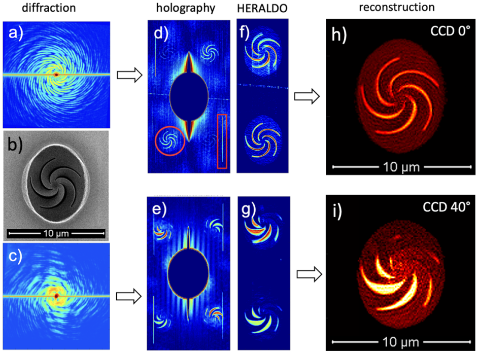
The experiment has been carried out at the DiProI beamline located at the FERMI FEL facility (Trieste, Italy) [28, 29], using a system previously employed for FEL-based transient grating experiments and thoroughly described elsewhere [30, 31]. This setup permitted to generate two FEL pulses and recombine them at the sample, precisely setting the crossing angle ( = 40∘) and ensuring time-coincidence. The spot size at the sample for each crossed FEL beam was about 60 × 120 full width at half-maximum (FWHM), the FEL wavelength was 20.8 nm, the pulse duration 50 fs (FWHM) and the typical intensity at the sample of about 1.1 J per shot, while the polarization was circular. We used as a sample different kinds of 3D structures, like the five-fold helicoidal structure shown in Fig. 1b). The sample was oriented in order to have the surface orthogonal to one of the two FEL beams. Close to each sample there were properly-oriented holographic pin-holes and an extended HERALDO reference [32, 33]. Two CCD detectors were placed on-axis with respect to either of the two FEL beams at a distance of 100 mm from the sample. Hereafter we will refer to these two CCDs as CCD 40∘ and CCD 0∘, where the latter is the one placed on-axis with the FEL beam orthogonal to the sample surface. Further details on the setup and on the composition of the samples can be found in the Supplementary Material (SM).
3 Results
Figs. 1a) and 1c) display an example of diffraction patterns after post processing corrections (see section 2 of the SM) collected, respectively, by CCD 0∘ and CCD 40∘ from the sample shown in Figure 1b). For each pair of such simultaneously collected diffraction patterns, we extract two holographic projections using the reference pinhole (Figs. 1d) and 1e)) and the HERALDO line (Figs. 1f) and 1g)). These holographic views are further refined using CDI reconstruction algorithms [34, 35]. In such a process, we use the holograms as input for the phase retrieval code and to define the support, which sets the spatial position of the unknown object during the CDI reconstruction of the diffracted phase. The 2D images obtained via the CDI reconstruction are presented in Fig. 1h) and 1i), for sample surface normal and tilted views respectively. In order to determine an absolute value of the sample transmission, during the CDI reconstruction, for each iteration the 2D images are renormalized by the average value of the clearance aperture in the sample (brighter regions in Figs 1h) and 1i)), i.e., the area where photons are not absorbed by the sample. The normal incidence view clearly resolves the five-fold geometry of the helicoidal structure, while the tilted view shows almost fully open structures, where the impinging photons does not interact with the sample (bottom left part of the image) and obscured areas (top right part of the image); here the lamellae forming the object overlap with the below substrate along the view direction due to the large view angle. The 3D reconstruction algorithm uses the information contained in the two 2D CDI views, as illustrated in Fig. 2a). These information are essentially the EUV transmission () and phase (), as shown in Fig. 2b) for the sample displayed in SM Fig. S1b). In the reconstruction process, the values and of the set of voxels along the directions defined by the two views must match the experimental data. The final solution is selected based on this matching, as evaluated by comparing both and of the experimental views with those computed from the 3D reconstructed sample. This evaluation is performed using a ray tracing approach, that assumes an aprioristic knowledge of sample thickness and composition, recently developed for the 3D stereographic reconstruction starting from 2D X-ray fluorescence images [36, 37, 38]. More information on the 2D and 3D reconstruction algorithms, as well as on their relation, can be found in the SM.
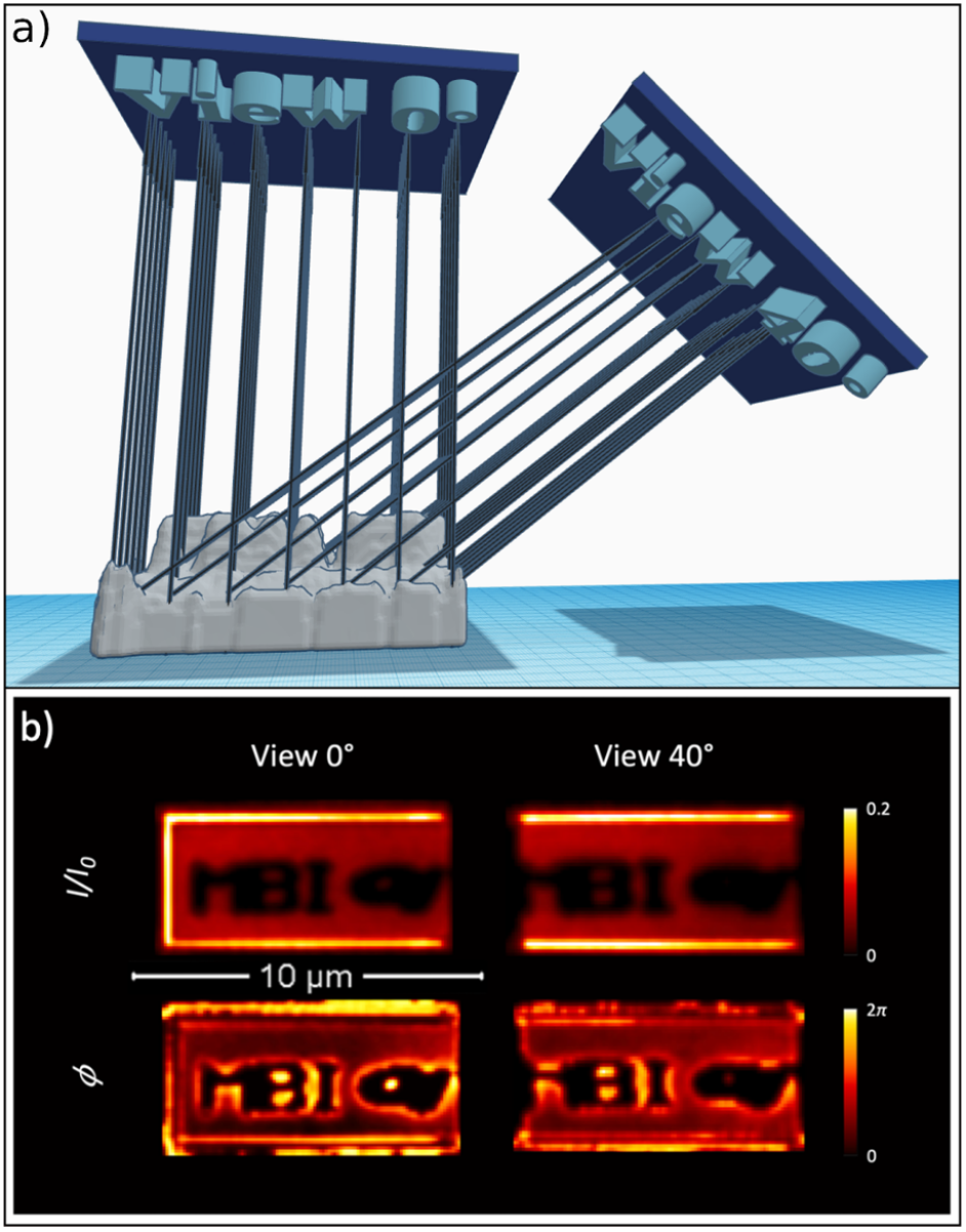
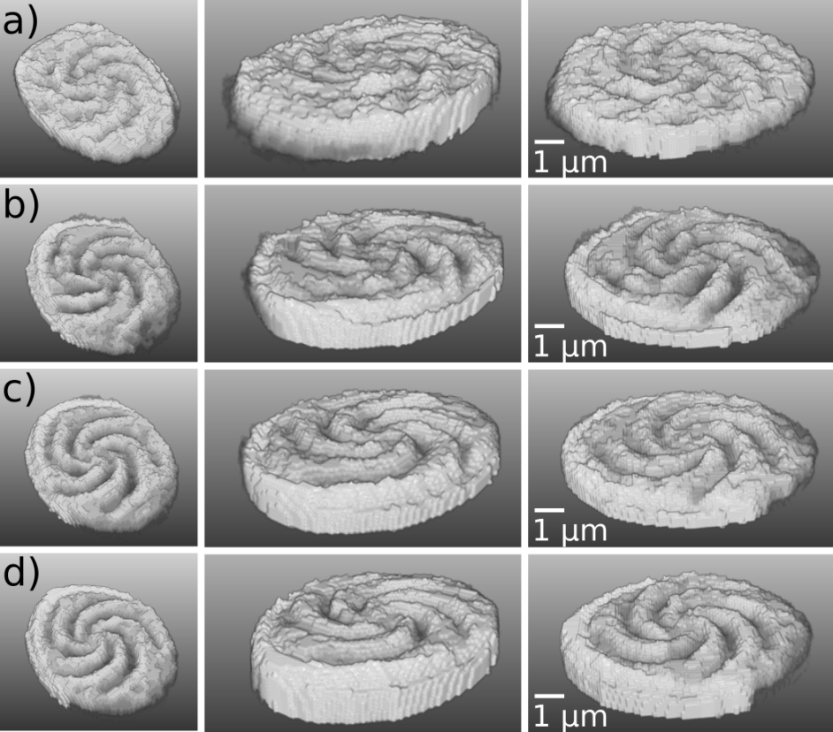
Fig. 3 shows an example of 3D reconstruction of the five-fold helicoidal structure shown in Fig. 1b). These reconstructions have been obtained using different datasets collected by exposing the sample to a different number of FEL shots, from one single-shot up to 750 shots. Other 3D reconstructed objects, including the sample displayed in Fig. 2b), are shown in the SM. In all cases the combination of holographically-guided CDI and our 3D ray tracing projection algorithms is able to retrieve a realistic stereographic solution of the sample. As quantitatively discussed further below, the image quality does not substantially degrade on reducing the number of exposures, highlighting the computational robustness of the stereographic retrieval procedure.
4 Discussion
In order to provide a quantitative estimate of the lateral resolution (), we computed the phase retrieval transfer function (PRTF) in the 1060 iterations of the CDI code. Fig. 4a) shows the comparison of the PRFT for the five-fold helicoidal structure displayed in Fig. 1b). In this analysis we consider reconstructions under the different exposure conditions shown in Fig. 3, namely: (i) single-shot, (ii) averaging two images each one corresponding to a single-shot acquisition, (iii) a 25-shot acquisition and (iv) averaging 30 images each one corresponding to a 25 shots acquisition. The value of the PRFT is well above the 1/e threshold suggested by Chapman et al. [14] for all the range in the exchanged momentum |q| recorded by the detector (details on the |q|-range are in the SM). We ascribe this to the more uniform coverage of the detector plane by the diffraction pattern originating by our circularly symmetric object compared to the lower-symmetry one considered by Chapman, resulting in a larger number of pixels for which the phase can be retrieved accurately. Therefore, we set the threshold for defining the resolution at PRFT = 0.7, roughly matching the high-|q| asymptotic value of the curves reported in Figs. 4a), which is likely to be limited by the SNR in such a high-|q| range. Different samples show different asymptotic values, as discussed in the SM. The PRTF function is larger than 0.7 in almost the entire |q|-range, for reconstructions obtained from datasets corresponding to a different number of shots. This indicates that the actual resolution does not substantially decrease from the multi-shot to the single-shot regime. According to the aforementioned empiric PRTF = 0.7 level, the lateral resolution ranges from about 90 10 nm for multi-shot averaged images to about 190 20 nm for the single-shot.
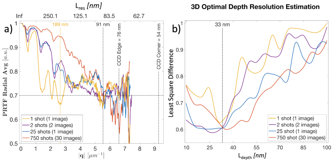
The depth resolution () along the z-coordinate (sample depth) for the 3D reconstructed sample shown in Fig. 3 was evaluated by varying the sampling step along z, from = 10 nm to 100 nm, and generating an ensemble of associated solutions. These are then evaluated through the structural similarity index measure (SSIM) [39], as described in section 5 of the SM. The SSIM analysis isolates the solutions that give the most similar output to the input 2D views, in terms of the least squares difference between the simulated and the reconstructed and maps. We defined the resolution in the z-axis as the value of in correspondence of which the least squares differences reach a minimum, meaning that the two CDI reconstructed views (both in amplitude and in phase) do not provide additional information to the ray tracing stereographic code. Fig. 4b) shows the dependence on of the least squares difference of the likelihood reconstruction selected by SSIM analysis for the four 3D reconstructions reported in Fig. 3, which are associated to different numbers of FEL shots. The SSIM analysis shows a similar value of = 33 5 nm, nearly independent from the number of shots. This suggests that 3D ray tracing algorithms based on the aprioristic knowledge of material properties (absorption length, layer thickness) are less sensitive to the natural degradation of the signal to noise ratio due to the reduced number of exposures. This observation is consistent with recent numerical simulations, where the 3D reconstruction process recovers the correct topography even if the input thickness maps have spatially homogeneous white noise [38].
5 Conclusions
We combined a non-collinear EUV split-recombination setup (compact, conceptually simple and easily reproducible) with a data processing protocol based on both CDI phase retrieval and ray tracing algorithms, to experimentally demonstrate the capability to determine the 3D structure of an object from a single-flash of EUV light (20.8 nm wavelength) at moderate fluence, with a resolution in the order of 100 nm. This allowed us to perform the experiment well below the typical damage threshold of most materials. Though the structures used in this pilot experiment have been realized ad hoc, they are quite representative of possible samples, e.g., magnetic nanostructures or biological samples, in terms of size of structural details (10’s-100’s nm) and overall dimensions (a few µm), indicating how this approach can be readily used to investigate such a class of samples. This approach can be profitably employed in time-resolved experiments, where high efficiency is crucial, and, in case of non-reproducible dynamics, inherently necessary. The introduction of an optical pulse (for pump-probe experiments) in this setup is straightforward, since it is routinely employed in EUV transient grating experiments [30, 31]. Such experiments were performed up to about 100 eV photon energy and showed how the system is compatible with spectroscopic approaches, where the EUV wavelength is scanned across specific resonances of the material [40], thus enabling element-selective 3D imaging. Extension to higher photon energies, up to the water window (300–500 eV) and beyond, towards the L-edges of 3d transition metals (600–1000 eV), is technically possible by exploiting state-of-the-art multilayer coatings [41]. Advances in the fabrication of transmissive X-ray diffraction optics [42, 43] enable us to envision split-recombination systems based on such optics, making this approach compatible even with hard X-rays. X-ray diffractive optics may allow handling several replicas of FEL pulses simultaneously impinging on the sample from more than two different angles [24, 25, 44]. This multiplex approach can greatly improve 3D reconstruction, that in the present version assumes that the sample composition (constituent materials and density) is known a priori, and the absence of gaps along the photon propagation direction. These assumptions can be reasonably met in many practical cases. However, multiple views could overcome the need for these assumptions to be met. For instance, for a pure absorbing sample, if the composition is uncertain it would be hard to determine from a limited number of views whether the light is traversing a shorter path of high absorbing material or a longer path through a lower absorbing one. Since both phase and amplitude are derived in CDI reconstruction the complex scattering quotient, i.e., the ratio of reconstructed phase and the natural logarithm of the reconstructed amplitude, can be used to determine the local material content [45]. This uncertainty in the sample composition could also account for the small differences observed between the real 3D samples used in the present work and the reconstructed models, since a given density and composition was assumed. The ray tracing approach presented here is a promising way to reconstruct 3D samples as it has already shown robustness and flexibility with the minimal (two) viewing angles involved in this experiment and enables large angular separation between views. The system can be readily exploited in time-domain studies, extended to include EUV spectroscopy and can surely be improved by exploiting more views, either by using multiple time and space coincident beams or, if single shot is not required, by rotating the sample.
6 Backmatter
Funding Content in the funding section will be generated entirely from details submitted to Prism. Authors may add placeholder text in the manuscript to assess length, but any text added to this section in the manuscript will be replaced during production and will display official funder names along with any grant numbers provided. If additional details about a funder are required, they may be added to the Acknowledgments, even if this duplicates information in the funding section. See the example below in Acknowledgements. For preprint submissions, please include funder names and grant numbers in the manuscript.
Acknowledgments Authors acknowledge the FERMI team for invaluable support and Hamed Merdji and his team for the fruitful discussions on stereo imaging based on disparity map.
Disclosures The authors declare no conflicts of interest.
Data availability Data underlying the results presented in this paper are not publicly available at this time but may be obtained from the authors upon reasonable request.
Supplemental document See Supplement 1 for supporting content.
References
- [1] Z. Rymuza, “Control tribological and mechanical properties of mems surfaces. part 1: critical review,” \JournalTitleMicrosystem Technologies 5, 173–180 (1999).
- [2] B.-F. Cui, X.-P. Han, and W.-B. Hu, “Micronanostructured design of dendrite-free zinc anodes and their applications in aqueous zinc-based rechargeable batteries,” \JournalTitleSmall Structures 2, 2000128 (2021).
- [3] D. A. Shapiro, Y.-S. Yu, T. Tyliszczak, J. Cabana, R. Celestre, W. Chao, K. Kaznatcheev, A. D. Kilcoyne, F. Maia, S. Marchesini, Y. S. Meng, T. Warwick, L. L. Yang, and H. A. Padmore, “Chemical composition mapping with nanometre resolution by soft x-ray microscopy,” \JournalTitleNature Photonics 8, 765–769 (2014).
- [4] H. Jiang, R. Xu, C.-C. Chen, W. Yang, J. Fan, X. Tao, C. Song, Y. Kohmura, T. Xiao, Y. Wang, Y. Fei, T. Ishikawa, W. L. Mao, and J. Miao, “Three-dimensional coherent x-ray diffraction imaging of molten iron in mantle olivine at nanoscale resolution,” \JournalTitlePhysical Review Letters 110, 205501 (2013).
- [5] A. Tripathi, J. Mohanty, S. H. Dietze, O. G. Shpyrko, E. Shipton, E. E. Fullerton, S. S. Kim, and I. McNulty, “Dichroic coherent diffractive imaging,” \JournalTitleProceedings of the National Academy of Sciences 108, 13393–13398 (2011).
- [6] M. Kördel, A. Dehlinger, C. Seim, U. Vogt, E. Fogelqvist, J. A. Sellberg, H. Stiel, and H. M. Hertz, “Laboratory water-window x-ray microscopy,” \JournalTitleOptica 7, 658–674 (2020).
- [7] F. Büttner, C. Moutafis, A. Bisig, P. Wohlhüter, C. M. Günther, J. Mohanty, J. Geilhufe, M. Schneider, C. v. K. Schmising, S. Schaffert, B. Pfau, M. Hantschmann, M. Riemeier, M. Emmel, S. Finizio, G. Jakob, M. Weigand, J. Rhensius, j. H. Franken, R. Lavrijsen, H. J. M. Swagten, H. Stoll, S. Eisebitt, and M. Kläui, “Magnetic states in low-pinning high-anisotropy material nanostructures suitable for dynamic imaging,” \JournalTitlePhysical Review B 87, 134422 (2013).
- [8] C. von Korff Schmising, B. Pfau, M. Schneider, C. Günther, M. Giovannella, J. Perron, B. Vodungbo, L. Müller, F. Capotondi, E. Pedersoli, N. Mahne, J. Lüning, and S. Eisebitt, “Imaging ultrafast demagnetization dynamics after a spatially localized optical excitation,” \JournalTitlePhysical review letters 112, 217203 (2014).
- [9] I. K. Robinson, I. A. Vartanyants, G. Williams, M. Pfeifer, and J. Pitney, “Reconstruction of the shapes of gold nanocrystals using coherent x-ray diffraction,” \JournalTitlePhysical review letters 87, 195505 (2001).
- [10] Y. Gao, R. Harder, S. H. Southworth, J. R. Guest, X. Huang, Z. Yan, L. E. Ocola, Y. Yifat, N. Sule, P. J. Ho, M. Pelton, N. F. Scherer, and L. Young, “Three-dimensional optical trapping and orientation of microparticles for coherent x-ray diffraction imaging,” \JournalTitleProceedings of the National Academy of Sciences 116, 4018–4024 (2019).
- [11] Y. Nishino, Y. Takahashi, N. Imamoto, T. Ishikawa, and K. Maeshima, “Three-dimensional visualization of a human chromosome using coherent x-ray diffraction,” \JournalTitlePhysical review letters 102, 018101 (2009).
- [12] D. Shapiro, P. Thibault, T. Beetz, V. Elser, M. Howells, C. Jacobsen, J. Kirz, E. Lima, H. Miao, A. M. Neiman, and D. Sayre, “Biological imaging by soft x-ray diffraction microscopy,” \JournalTitleProceedings of the National Academy of Sciences 102, 15343–15346 (2005).
- [13] R. Neutze, R. Wouts, D. Van Der Spoel, E. Weckert, and J. Hajdu, “Potential for biomolecular imaging with femtosecond x-ray pulses,” \JournalTitleNature 406, 752–757 (2000).
- [14] H. N. Chapman, A. Barty, M. J. Bogan, S. Boutet, M. Frank, S. P. Hau-Riege, S. Marchesini, B. W. Woods, S. Bajt, W. H. Benner, R. A. London, E. Plönjes, M. Kuhlmann, R. Treusch, S. Düsterer, T. Tschentscher, J. R. Schneider, E. Spiller, T. Möller, C. Bostedt, T. Hoener, D. A. Shapiro, K. O. Hodgson, D. Van Der Spoel, F. Burmeister, M. Bergh, C. Caleman, G. Huldt, M. M. Seibert, F. R. N. C. Maia, R. W. Lee, A. Szöke, N. Timneanu, and J. Hajdu, “Femtosecond diffractive imaging with a soft-x-ray free-electron laser,” \JournalTitleNature Physics 2, 839–843 (2006).
- [15] S. Marchesini, H. Chapman, S. Hau-Riege, R. London, A. Szoke, H. He, M. Howells, H. Padmore, R. Rosen, J. Spence, and U. Weierstall, “Coherent x-ray diffractive imaging: applications and limitations,” \JournalTitleOptics Express 11, 2344–2353 (2003).
- [16] J. Bielecki, F. R. Maia, and A. P. Mancuso, “Perspectives on single particle imaging with x rays at the advent of high repetition rate x-ray free electron laser sources,” \JournalTitleStructural Dynamics 7, 040901 (2020).
- [17] Z. Sun, J. Fan, H. Li, and H. Jiang, “Current status of single particle imaging with x-ray lasers,” \JournalTitleApplied Sciences 8, 132 (2018).
- [18] A. Barty, S. Boutet, M. J. Bogan, S. Hau-Riege, S. Marchesini, K. Sokolowski-Tinten, N. Stojanovic, R. Tobey, H. Ehrke, A. Cavalleri, S. Düsterer, M. Frank, S. Bajt, B. W. Woods, M. M. Seibert, J. Hajdu, R. Treusch, and H. N. Chapman, “Ultrafast single-shot diffraction imaging of nanoscale dynamics,” \JournalTitleNature photonics 2, 415–419 (2008).
- [19] M. M. Seibert, T. Ekeberg, F. R. Maia, M. Svenda, J. Andreasson, O. Jönsson, D. Odić, B. Iwan, A. Rocker, D. Westphal, M. Hantke, D. P. DePonte, A. Barty, J. Schulz, L. Gumprecht, N. Coppola, A. Aquila, M. Liang, T. A. White, A. Martin, C. Caleman, S. Stern, C. Abergel, V. Seltzer, J. M. Claverie, C. Bostedt, J. D. Bozek, D. Boutet, A. A. Miahnahri, M. Messerschmidt, J. Krzywinski, G. Williams, K. O. Hodgson, M. J. Bogan, C. Y. Hampton, R. G. Sierra, D. Starodub, I. Andersson, S. Bajt, M. Barthelmess, J. C. H. Spence, P. Fromme, U. Weierstall, R. Kirian, M. Hunter, R. B. Doak, R. Marchesini, S. P. Hau-Riege, M. Frank, R. L. Shoeman, L. Lomb, S. W. Epp, R. Hartmann, D. Rolles, A. Rudenko, C. Schmidt, L. Foucar, N. Kimmel, P. Holl, B. Rudek, B. Erk, A. Hömke, A. Reich, D. Pietschner, G. Weidenspointner, L. Strüder, G. Hauser, H. Gorke, J. Ullrich, I. Schlichting, S. Herrmann, G. Schaller, F. Schopper, H. Soltau, K. U. Kühnel, R. Andritschke, C. D. Schröter, F. Krasniqi, M. Bott, S. Schorb, D. Rupp, M. Adolph, T. Gorkhover, H. Hirsemann, G. Potdevin, H. Graafsma, B. Nilsson, H. N. Chapman, and J. Hajdu, “Single mimivirus particles intercepted and imaged with an x-ray laser,” \JournalTitleNature 470, 78–81 (2011).
- [20] N. Loh, M. J. Bogan, V. Elser, A. Barty, S. Boutet, S. Bajt, J. Hajdu, T. Ekeberg, F. R. Maia, J. Schulz, M. M. Seibert, B. Iwan, N. Timneanu, S. Marchesini, I. Schlichting, R. L. Shoeman, L. Lomb, M. Frank, M. Liang, and H. N. Chapman, “Cryptotomography: reconstructing 3d fourier intensities from randomly oriented single-shot diffraction patterns,” \JournalTitlePhysical review letters 104, 225501 (2010).
- [21] T. Ekeberg, M. Svenda, C. Abergel, F. R. Maia, V. Seltzer, J.-M. Claverie, M. Hantke, O. Jönsson, C. Nettelblad, G. Van Der Schot, M. Liang, D. P. DePonte, A. Barty, M. M. Seibert, B. Iwan, I. Andersson, N. D. Loh, A. V. Martin, H. N. Chapman, C. Bostedt, J. D. Bozek, k. R. Ferguson, S. W. Krzywinski, J andEpp, D. Rolles, A. Rudenko, R. K. Hartmann, N. Kimmel, and J. Hajdu, “Three-dimensional reconstruction of the giant mimivirus particle with an x-ray free-electron laser,” \JournalTitlePhysical review letters 114, 098102 (2015).
- [22] P. R. Kommera, V. B. Ramakrishnaiah, and C. M. Sweeney, “Accelerate m-tip on gpus and deploy to summit and nersc-9 (against simulated data) wbs 2.2. 4.05 exafel, milestone adse13-199,” Tech. rep., Los Alamos National Lab.(LANL), Los Alamos, NM (United States) (2021).
- [23] J. Geilhufe, C. Tieg, B. Pfau, C. Günther, E. Guehrs, S. Schaffert, and S. Eisebitt, “Extracting depth information of 3-dimensional structures from a single-view x-ray fourier-transform hologram,” \JournalTitleOptics Express 22, 24959–24969 (2014).
- [24] C. Chang, X. Pan, H. Tao, C. Liu, S. P. Veetil, and J. Zhu, “3d single-shot ptychography with highly tilted illuminations,” \JournalTitleOptics Express 29, 30878–30891 (2021).
- [25] D. Goldberger, J. Barolak, C. G. Durfee, and D. E. Adams, “Three-dimensional single-shot ptychography,” \JournalTitleOptics Express 28, 18887–18898 (2020).
- [26] K. Schmidt, J. Spence, U. Weierstall, R. Kirian, X. Wang, D. Starodub, H. Chapman, M. Howells, and R. Doak, “Tomographic femtosecond x-ray diffractive imaging,” \JournalTitlePhysical review letters 101, 115507 (2008).
- [27] J. Duarte, R. Cassin, J. Huijts, B. Iwan, F. Fortuna, L. Delbecq, H. Chapman, M. Fajardo, M. Kovacev, W. Boutu, and H. Merdji, “Computed stereo lensless x-ray imaging,” \JournalTitleNature Photonics 13, 449–453 (2019).
- [28] F. Capotondi, E. Pedersoli, N. Mahne, R. Menk, G. Passos, L. Raimondi, C. Svetina, G. Sandrin, M. Zangrando, M. Kiskinova, S. Bajt, M. Barthelmess, H. Fleckenstein, H. N. Chapman, J. Schulz, J. Bach, R. Frömter, S. Schleitzer, L. Müller, C. Gutt, and G. Grübel, “Invited article: Coherent imaging using seeded free-electron laser pulses with variable polarization: First results and research opportunities,” \JournalTitleReview of scientific instruments 84, 051301 (2013).
- [29] E. Allaria, R. Appio, L. Badano, W. Barletta, S. Bassanese, S. Biedron, A. Borga, E. Busetto, D. Castronovo, P. Cinquegrana, S. Cleva, D. Cocco, M. Cornacchia, P. Craievich, I. Cudin, G. D’Auria, M. Dal Forno, m. B. Danailov, R. De Monte, G. De Ninno, P. Delgiusto, A. Demidovich, S. Di Mitri, B. Diviacco, A. Fabris, R. Fabris, W. Fawley, M. Ferianis, E. Ferrari, S. Ferry, L. Froehlich, P. Furlan, G. Gaio, F. Gelmetti, L. Giannessi, M. Giannini, R. Gobessi, R. Ivanov, E. Karantzoulis, M. Lonza, A. Lutman, B. Mahieu, M. Milloch, S. V. Milton, M. Musardo, I. Nikolov, S. Noe, G. Parmigiani, F ang Penco, M. Petronio, L. Pivetta, M. Predonzani, F. Rossi, L. Rumiz, A. Salom, C. Scafuri, C. Serpico, P. Sigalotti, S. Spampinati, C. Spezzani, M. Svandrlik, C. Svetina, S. Tazzari, M. Trovo, R. Umer, A. Vascotto, M. Veronese, R. Visintini, M. Zaccaria, D. Zangrando, and M. Zangrando, “Highly coherent and stable pulses from the fermi seeded free-electron laser in the extreme ultraviolet,” \JournalTitleNature Photonics 6, 699–704 (2012).
- [30] F. Bencivenga, R. Cucini, F. Capotondi, A. Battistoni, R. Mincigrucci, E. Giangrisostomi, A. Gessini, M. Manfredda, I. Nikolov, E. Pedersoli, E. Principi, C. Svetina, P. Parisse, F. Casolari, M. B. Danailov, M. Kiskinova, and C. Masciovecchio, “Four-wave mixing experiments with extreme ultraviolet transient gratings,” \JournalTitleNature 520, 205–208 (2015).
- [31] R. Mincigrucci, L. Foglia, D. Naumenko, E. Pedersoli, A. Simoncig, R. Cucini, A. Gessini, M. Kiskinova, G. Kurdi, N. Mahne, M. Manfredda, I. P. Nikolov, E. Principi, L. Raimondi, M. Zangrando, C. Masciovecchio, F. Capotondi, and F. Bencivenga, “Advances in instrumentation for fel-based four-wave-mixing experiments,” \JournalTitleNuclear Instruments and Methods in Physics Research Section A: Accelerators, Spectrometers, Detectors and Associated Equipment 907, 132–148 (2018).
- [32] M. Guizar-Sicairos and J. R. Fienup, “Direct image reconstruction from a fourier intensity pattern using heraldo,” \JournalTitleOptics letters 33, 2668–2670 (2008).
- [33] M. Guizar-Sicairos and J. R. Fienup, “Holography with extended reference by autocorrelation linear differential operation,” \JournalTitleOptics express 15, 17592–17612 (2007).
- [34] J. R. Fienup, “Phase retrieval algorithms: a comparison,” \JournalTitleApplied optics 21, 2758–2769 (1982).
- [35] F. Capotondi, E. Pedersoli, M. Kiskinova, A. Martin, M. Barthelmess, and H. Chapman, “A scheme for lensless x-ray microscopy combining coherent diffraction imaging and differential corner holography,” \JournalTitleOptics express 20, 25152–25160 (2012).
- [36] F. Billè, G. Kourousias, E. Luchinat, M. Kiskinova, and A. Gianoncelli, “X-ray fluorescence microscopy artefacts in elemental maps of topologically complex samples: Analytical observations, simulation and a map correction method,” \JournalTitleSpectrochimica Acta Part B: Atomic Spectroscopy 122, 23–30 (2016).
- [37] G. Kourousias, F. Billè, G. Cautero, J. Bufon, A. Rachevski, S. Schillani, D. Cirrincione, M. Altissimo, R. Menk, G. Zampa, N. Zampa, I. Rashevskaya, R. Borghes, M. Gandola, A. Picciotto, G. Borghi, F. Ficorella, N. Zorzi, P. Bellutti, G. Bertuccio, A. Vacchi, and A. Gianoncelli, “Xrf topography information: Simulations and data from a novel silicon drift detector system,” \JournalTitleNuclear Instruments and Methods in Physics Research Section A: Accelerators, Spectrometers, Detectors and Associated Equipment 936, 80–81 (2019).
- [38] M. Ippoliti, F. Billè, A. G. Karydas, A. Gianoncelli, and G. Kourousias, “Reconstruction of 3d topographic landscape in soft x-ray fluorescence microscopy through an inverse x-ray-tracing approach based on multiple detectors,” \JournalTitleScientific Reports 12, 20145 (2022).
- [39] Z. Wang, A. C. Bovik, H. R. Sheikh, and E. P. Simoncelli, “Image quality assessment: from error visibility to structural similarity,” \JournalTitleIEEE transactions on image processing 13, 600–612 (2004).
- [40] R. Bohinc, G. Pamfilidis, J. Rehault, P. Radi, C. Milne, J. Szlachetko, F. Bencivenga, F. Capotondi, R. Cucini, L. Foglia, C. CMasciovecchio, R. Mincigrucci, E. Pedersoli, A. Simoncig, N. Mahne, A. Cannizzo, H. M. Frey, A. Ollmann, T. Feurer, A. A. Maznev, K. Nelson, and G. Knopp, “Nonlinear xuv-optical transient grating spectroscopy at the si l2, 3–edge,” \JournalTitleApplied physics letters 114, 181101 (2019).
- [41] S. Yulin, M. Trost, S. Schwinde, and S. Schröder, “Coatings with barrier layers for extreme-short wavelengths: Euv lithography for the semiconductor industry and beyond,” \JournalTitleVakuum in Forschung und Praxis 33, 24–29 (2021).
- [42] N. Kujala, M. Makita, J. Liu, A. Zozulya, M. Sprung, C. David, and J. Grünert, “Characterizing transmissive diamond gratings as beam splitters for the hard x-ray single-shot spectrometer of the european xfel,” \JournalTitleJournal of synchrotron radiation 26, 708–713 (2019).
- [43] B. Rösner, F. Koch, F. Döring, J. Bosgra, V. A. Guzenko, E. Kirk, M. Meyer, J. L. Ornelas, R. H. Fink, S. Stanescu, S. Swaraj, R. Belkhou, B. Watts, J. Raabe, and C. David, “Exploiting atomic layer deposition for fabricating sub-10 nm x-ray lenses,” \JournalTitleMicroelectronic Engineering 191, 91–96 (2018).
- [44] K. Kharitonov, M. Mehrjoo, M. Ruiz-Lopez, B. Keitel, S. Kreis, S.-g. Gang, R. Pan, A. Marras, J. Correa, C. B. Wunderer, and E. Plönjes, “Single-shot ptychography at a soft x-ray free-electron laser,” \JournalTitleScientific Reports 12, 14430 (2022).
- [45] W. Eschen, L. Loetgering, V. Schuster, R. Klas, A. Kirsche, L. Berthold, M. Steinert, T. Pertsch, H. Gross, M. Krause, J. Limpert, and j. Rothhard, “Material-specific high-resolution table-top extreme ultraviolet microscopy,” \JournalTitleLight: Science & Applications 11, 117 (2022).