Generation of biaxially accelerating static Airy light-sheets with 3D-printed freeform micro-optics
Abstract
One-dimensional Airy beams allow the generation of thin light-sheets without scanning, simplifying the complex optical arrangements of light-sheet microscopes (LSM) with an extended field-of-view (FOV). However, their uniaxial acceleration limits the maximum numerical aperture of the detection objective in order to keep both the active and inactive axes within the depth-of-field. This problem is particularly pronounced in miniaturized LSM implementations, such as those for endomicroscopy or multi-photon neural imaging in freely-moving animals using head-mounted miniscopes. We propose a new method to generate a static Airy light-sheet with biaxial acceleration, based on a novel phase profile. This light-sheet has the geometry of a spherical shell whose radius of curvature can be designed to match the field curvature of the micro-objective. We present an analytical model for the analysis of the light-sheet parameters and verify it by numerical simulations in the paraxial regime. We also discuss a micro-optical experimental implementation combining gradient-index optics with a 3D-nano-printed, fully refractive phase plate. The results confirm that we are able to match detection curvatures with radii in the 1.5 to 2 mm range.
Keywords light-sheet microscopy, Airy beam, accelerating beams, field curvature, two-photon polymerization
1 Introduction
Light-sheet fluorescence microscopy (LSM) has become an essential imaging modality in the life sciences, providing rapid volumetric imaging with minimal photodamage [1]. Although this makes it an ideal candidate for in-vivo imaging such as endomicroscopy[2, 3] and neural imaging in freely moving animals[4, 5], translation to miniaturized optical systems has been limited to date[6, 7]. A critical technology for incorporating LSM into these applications is high performance micro-objectives that can match their macroscopic counterparts in numerical aperture (NA) and field-of-view (FOV)[8]. Their performance comes at the cost of field curvature[9, 10], which presents an additional challenge for LSM: With the generally planar geometry of a Gaussian beam, only a limited portion of the illuminated area is brought into perfect focus in the image plane, limiting the effective FOV, imposing limits on the detection NA, or leading to loss in image contrast. Although the incorporation of scanning micromirrors could address this challenge to some extent[11], it further increases the system complexity and limits the achievable degree of miniaturization. Using Airy beams [12] has been proposed to match field curvature due to its uniaxial acceleration when implemented as a static light-sheet.[13]. An Airy light-sheet can be generated by imposing a one-dimensional cubic phase profile on a Gaussian beam, which is then focused by a cylindrical lens. Similar to other configurations using propagation-invariant beams[14, 15], it has been used to excite a larger FOV compared to a Gaussian sheet, while maintaining the same sectioning ability[16, 17]. However, incorporating the static Airy light-sheet only partially solves the curvature problem, as the planar Gaussian profile along its inactive axis still introduces a FOV reduction, limiting the performance required for micro-optical applications.
We have recently shown that this challenge can be addressed by an additional phase modulation on top of the cubic profile needed for the static Airy light-sheet.[18] This phase profile depends linearly on the active coordinate and quadratically on the inactive coordinate, and modifies the Gaussian profile of the light-sheet to achieve the same radius of curvature as the Airy beam. The result is a static, biaxially accelerating light-sheet that is unrestricted in its ability to section, thus increasing the effective FOV of the micro-objective. In this paper, we provide a detailed analytical derivation of the phase-plate parameterization using typical micro-objective specifications, allowing the light-sheet geometry to be tailored for maximized in-focus excitation. To verify the validity of our analysis, we perform simulations in the paraxial regime using ray tracing and beam propagation methods. By designing, implementing, and characterizing a micro-optical excitation system that incorporates a 3D-printed millimeter-scale phase plate, we experimentally demonstrate our ability to generate such a light-sheet in a fully miniaturized fashion.
2 Theory
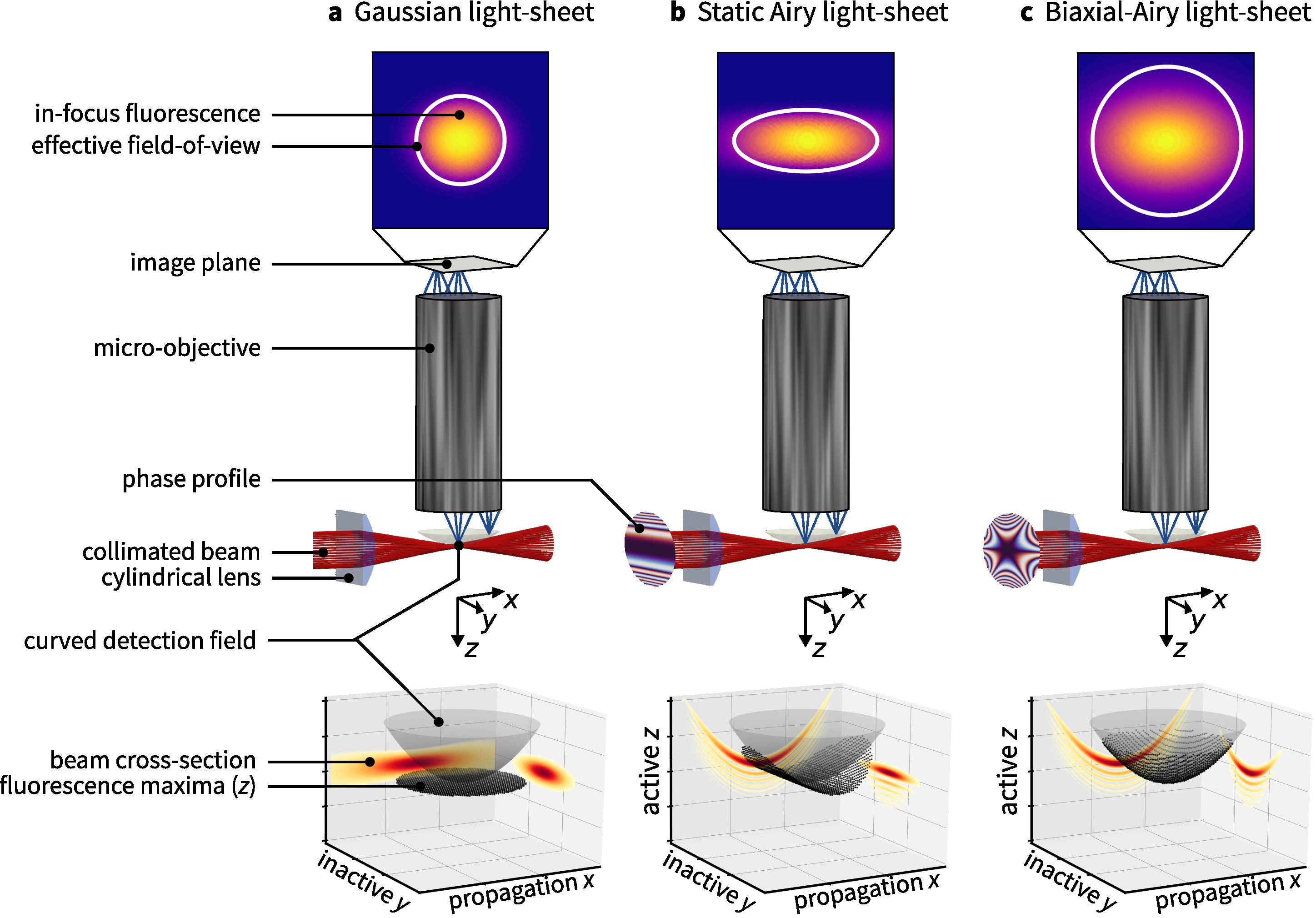
Figure 1 illustrates the optical architecture of a miniaturized light-sheet microscope with a high NA micro-objective. The illumination arm consists of a collimator (not shown) and a cylindrical lens to form a Gaussian light-sheet within the FOV of the micro-objective which is imaging the excited fluorescence onto a flat image sensor. The field curvature of the detection objective can be characterized by a radius so that the detection field follows
| (1) |
As common in LSM we use the coordinate system of the detection objective for the light-sheet, in which
-
–
is the micro-objective’s optical axis, corresponding to the cylindrical lens’ active axis,
-
–
is the optical axis of the cylindrical lens corresponding to the beam propagating direction, and
-
–
is the inactive coordinate of the cylindrical lens.
Figure 1 extends the concept to the generation of a static Airy light-sheet, resulting from a one-dimensional cubic phase modulation of the collimated beam incident on the cylindrical lens, which allows field curvature matching in . This already increases the effective FOV in this coordinate compared to the Gaussian light-sheet, and improves the sectioning capability due to a thinner light-sheet. A secondary phase modulation of the proposed phase profile aims at bending the Airy light-sheet along its inactive axis, thus generating a static and yet biaxially accelerating sheet (Fig. 1). In the next two sections we will discuss the design of the active and inactive phase profiles to achieve biaxial field curvature matching.


2.1 Acceleration along the propagation direction
An Airy light-sheet can be generated by modulating the collimated Gaussian beam with a beam waist by a one-dimensional cubic phase at the back-focal plane (BFP) of the cylindrical lens [13] along its active direction , such that the electric field follows
| (2) |
where is the amplitude of cubic modulation. A schematic depiction of this configuration is shown in Fig. 2, where the approximate intensity profile around the focal plane can be obtained in the paraxial domain as [19, 20]
| (3) |
where denotes the Airy function, is the axial coordinate, is a characteristic length, is an apodization factor and is the wave vector in a medium of refractive index [21]. It has been demonstrated by numerous groups[19, 22, 23, 24, 25] that this function is highly asymmetrical with prominent side lobes, and its main lobe follows a parabola with a radius of curvature of
| (4) |
Therefore, one must solve Eq. 4 for with to obtain an Airy light-sheet with an acceleration matching the field curvature of the detection objective. Using Eq. 2 we obtain
| (5) |
To finalize the geometry of the light-sheet,the beam needs to be matched to the FOV of the micro-objective, which we define as the area around the focal point within which the intensity remains larger than the half of the maximum value.[21] Since Eq. 5 does not depend on , we can control the geometry of the light-sheet in the propagation coordinate and in the inactive coordinate independently of each other via the focal length and the beam waist. Explicitly, the FOV along the active direction is obtained from
| (6) |
and along the inactive direction from
| (7) |
To obtain a symmetric FOV, the condition must be satisfied, which means that the focal length in the imaging medium must be equal to the radius of curvature.
2.2 Acceleration along the inactive direction
To induce a parabolic axial shift of the focal point along the inactive coordinate as well, we modify Eq. 3 by convolving it with a delta distribution with the required shift in the detection coordinate. This operation can be expressed as
| (8) | ||||
| (9) |
where denotes the convolution operation. By taking the Fourier transform of the electric field at the focal point along , we can again obtain the required electric field at the BFP, which can be approximated by
| (10) |
Details of this derivation can be found in Appendix A. The arguments of the first two exponential functions in Eq. 10 represent the conventional profile to generate an Airy beam as per Eq. 2, while the last term is an additional, phase-only term that we define as
| (11) |
In ray optical terms, this phase profile adds an additional launch angle from the phase plate to the cylindrical lens in its active coordinate, the magnitude of which varies as the square of the inactive coordinate. Because of the introduction of this additional angle in the BFP, each one-dimensional Airy ray will be shifted in at the focal plane of the cylindrical lens.
2.3 Combined phase profile
A combined phase plate with a sag height of , which generates both the one-dimensional Airy beam along the propagation coordinate, as well as the focus-shifted Gaussian beam along the inactive coordinate of the beam, can be obtained by
| (12) | ||||
| (13) |
where is the refractive index of the phase plate. The individual profiles are plotted alongside the combined one in Fig. 3. An important point about Eq. 13 is that, the Airy-related modulation scales linearly with , while the modulation related to the inactive profile is inversely proportional to it. This imposes a limit on the range of that can be realized: For lower values of , the parameter is smaller, and the assumptions made in the derivation of the apodized Airy beam become invalid.[19, 25] On the other hand, the modulation generated by the inactive profile dominates the phase plate. This leads to larger launch angles, which may violate the paraxial approximation of the optical element. At the upper end of the limit, it is useful to evaluate whether field curvature remains a problem as the beam becomes flatter and thicker, bypassing the initial problem.

3 Methods
3.1 Simulations
To assess that curvature-matching in the paraxial regime, we performed ray tracing simulations using a commercial software (OpticStudio 22.1, Zemax LLC). The simulation model implemented the configuration shown in Fig. 2 with the phase profile described in Section 2.3. We calculated the curvature of the beam in the active direction by evaluating the caustic[26] of 51 rays displaced in the active -coordinate (XZ-section in Fig. 2). For the inactive direction, we obtained the the displacement of the rays in the image plane by tracing 51 rays that were initially displaced along the -direction. To further investigate the validity of Eq. 9, we propagated the field defined in Eq. 10 using a two-dimensional FFT-based beam propagation method (BPM) in the Fresnel regime [27]. Propagation was performed to match the ray tracing simulations, which include the same beam waist, phase plate profile, and focal length of the cylindrical lens. 3-dimensional stacks were calculated within a range of around the focal point with a step size of , and a grid size of pixels with a width of . We set the input parameters with an excitation wavelength , an input beam waist , an effective focal length of the cylindrical lens and a refractive index of the phase plate [28] to match the experimental setup described below. Two radii of curvature were chosen in a typical range for micro-objectives of and . [8, 9, 29]
3.2 Fabrication and characterization
Phase plates with a diameter of were fabricated using a commercial two-photon polymerization based 3D printer (PPGT+, Nanoscribe GmbH & Co KG) with a commercial resin (IPS, NanoScribe GmbH & Co KG). The height profiles were pre-processed and subsequently fabricated on ITO-coated glass substrates using an in-house developed slicing and printing strategy previously described [30]. After development, the height profiles of the phase plates were measured by white light interferometry (WLI; NewView 9000, Zygo Corporation). A 0.3-NA objective (I100384, Zygo Cooperation) was used to obtain the surface profile of the entire plate and then fitted with standard Zernike polynomials to quantify the shape deviation. Since the tilt of the printing field in this system can only be controlled to within a few microns from print to print, the tilt Zernike coefficients were not included in the deviation analysis. The surface roughness was estimated by measuring a area of the phase plate with a 0.8-NA objective (I190053, Zygo Cooperation). A high-pass Gaussian spline filter with a cutoff frequency of was applied and the resulting root mean square error (RMSE) was calculated. [31]
3.3 Experimental setup and analysis of the beam profiles
A cylindrical gradient-index (GRIN) lens with an effective focal length of (Grintech GmbH) was bonded to the back of the substrate after fabrication so that the phase plate was located at its back focal plane. The entire assembly was illuminated with a fiber-based collimator (Grintech GmbH) using a SLED with a wavelength of as the light source (Fig. 4). The beam waist of the illumination beam after the collimator was . In order to obtain the 3D-dimensional beam profile in the vicinity of the focal plane of the cylindrical lens, the beam was imaged onto a CCD (UI-1240SE-NIR, IDS GmbH) using a 0.28-NA objective (Plan Apo 10x, Mitutoyo AC) and a relay lens, while the entire system was translated along the propagation direction of the light-sheet. To compare the analytical analysis, simulations and experiments, two-dimensional cross sections of the beam intensity along its propagation direction at () and along the inactive direction at the focal plane () were measured. The thickness and the were determined by finding the full-width at half-maximum of the beam intensity in the respective direction. The radius of curvature in and was determined by finding the location of the maximum intensity in the respective coordinate and fitting Eq. 1 to these data points using a least-squares algorithm.[32]

4 Results
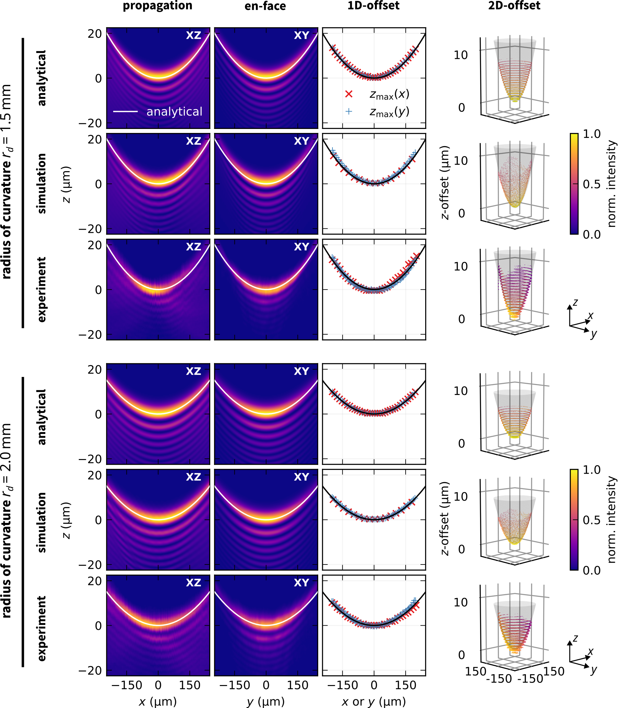
Fig. 5 shows the characterization of the surface profile obtained with the WLI. The surface errors become more pronounced towards the edge, where lift-off from the substrate is expected, but were found to be less than over a diameter of . With an RMS surface roughness below , the phase plate can be considered an optical quality surface.
The ray tracing simulations yielded radii of curvature that matched the design radii within numerical accuracy. Furthermore, the simulated beam profiles shown in Fig. 6 agree with those generated by evaluating Eq. 9. Deviations can be narrowed down to an asymmetric intensity distribution around the focus below , and predicted offsets in are consistent with both design and numerical predictions. The radii of curvature obtained from measured data matched the design values in most cases: For , the radii in the propagation coordinate and in the inactive coordinate were found to be and smaller than the design value, respectively. For , the radius of the active coordinate is equal to the designed radius, while the inactive coordinate is smaller than the designed radius. The beam profiles themselves show the typical Airy profile, although a reduced side lobe intensity is observed with an underestimation of the FOV in propagation direction of and in the inactive coordinate.

| design radius | active coordinate | inactive coordinate | |
|---|---|---|---|
| analytical | |||
| simulation (ray-tracing) | |||
| simulation (BPM) | |||
| experiment | |||
| analytical | |||
| simulation (ray-tracing) | |||
| simulation (BPM) | |||
| experiment |

5 Discussion and conclusions
The excellent agreement between the results of our analytical treatment and the ray traced simulations is a good indication of the validity of the design approach within the paraxial regime. Limitations of this approximation may arise in different scenarios: As discussed in Section 2.3, lower values of will lead to a reduced modulation of the Airy profile. Therefore, we expect the radius to be underestimated in the propagation coordinate, as observed in the experimental data. In the inactive direction, we also expect to see an underestimated radius, but for a different reason: The higher additional deflection leads to a gradual violation of the paraxial approximation, resulting in aberrations and thus lower radii. On the other hand, we observed the Airy radius to be the same as the design radius for larger values of , while the radius along the inactive direction is slightly smaller. We attribute this to a manufacturing effect as for smaller values of , the sag height decreases and therefore requires more accurate manufacturing capabilities. Both effects can lead to the deviations reflected in the experimental results. Nevertheless, we have been able to adjust both radii to a degree that does not affect the improvement in effective FOV, which was the underlying design goal.
In summary, we have derived a phase profile to generate an Airy light-sheet that follows the curved image plane of a typical micro-objective, while incorporating the advantages of using an Airy beam to provide a larger FOV and better sectioning. Ray tracing and BPM simulations in the paraxial regime validated the analytical analysis, with deviations between the two within numerical accuracy. A micro-optical implementation with mm-scale 3D-printed refractive phase plate was developed and combined with GRIN optics, and experimentally demonstrated to achieve curvature matching in the design range.
Acknowledgements
We express our gratitude to Anders Kragh Hansen (Technical University of Denmark) for having the initial idea of incorporating a quadratic phase profile for curvature, as well as to Anja Borre, Madhu Veettikazhy and Peter Andersen for discussions on the phase distribution. This project has received funding from the European Union’s Horizon 2020 research and innovation program under grant agreement No 871212.
Data availability
The simulation and evaluation code used to generate the results presented is available upon request.
Disclosures
Sophia Laura Schulz and Bernhard Messerschmidt are full-time employees of the company GRINTECH GmbH.
References
- [1] J. M. Girkin and M. T. Carvalho, “The light-sheet microscopy revolution,” Journal of Optics (United Kingdom) 20(5) (2018).
- [2] B. Glover, J. Teare, and N. Patel, “The Status of Advanced Imaging Techniques for Optical Biopsy of Colonic Polyps,” Clinical and translational gastroenterology 11(3), e00130 (2020).
- [3] H. Li, Z. Hao, J. Huang, et al., “500 m Field-of-View Probe-Based Confocal Microendoscope for Large-Area Visualization in the Gastrointestinal Tract,” Photonics Research 9(9), 1829 (2021).
- [4] B. A. Flusberg, A. Nimmerjahn, E. D. Cocker, et al., “High-speed, miniaturized fluorescence microscopy in freely moving mice,” Nature Methods 5(11), 935–938 (2008).
- [5] W. Zong, H. A. Obenhaus, E. R. Skytøen, et al., “Large-scale two-photon calcium imaging in freely moving mice,” Cell 185, 1240–1256.e30 (2022).
- [6] C. J. Engelbrecht, F. Voigt, and F. Helmchen, “Miniaturized selective plane illumination microscopy for high-contrast in vivo fluorescence imaging,” Optics Letters 35(9), 1413 (2010).
- [7] F.-D. Chen, H. Moradi-Chameh, P. Shah, et al., “Implantable photonic neural probes for light-sheet fluorescence brain imaging,” Neurophotonics 8, 1–31 (2021).
- [8] G. Matz, B. Messerschmidt, and H. Gross, “Design and evaluation of new color-corrected rigid endomicroscopic high NA GRIN-objectives with a sub-micron resolution and large field of view,” Optics Express 24, 10987 (2016).
- [9] G. Matz, B. Messerschmidt, W. Göbel, et al., “Chip-on-the-tip compact flexible endoscopic epifluorescence video-microscope for in-vivo imaging in medicine and biomedical research,” Biomedical Optics Express 8, 3329 (2017).
- [10] E. Pshenay-Severin, H. Bae, K. Reichwald, et al., “Multimodal nonlinear endomicroscopic imaging probe using a double-core double-clad fiber and focus-combining micro-optical concept,” Light: Science & Applications 10(1) (2021).
- [11] S. Bakas, D. Uttamchandani, H. Toshiyoshi, et al., “MEMS enabled miniaturized light-sheet microscopy with all optical control,” Scientific Reports 11(1), 1–11 (2021).
- [12] T. Vettenburg, H. I. Dalgarno, J. Nylk, et al., “Light-sheet microscopy using an Airy beam,” Nature Methods 11(5), 541–544 (2014).
- [13] L. Niu, C. Liu, Q. Wu, et al., “Generation of One-Dimensional Terahertz Airy Beam by Three-Dimensional Printed Cubic-Phase Plate,” IEEE Photonics Journal 9(4), 1–7 (2017).
- [14] F. O. Fahrbach, P. Simon, and A. Rohrbach, “Microscopy with self-reconstructing beams,” Nature Photonics 4, 780–785 (2010).
- [15] H. Kafian, M. Lalenejad, S. Moradi-Mehr, et al., “Light-Sheet Fluorescence Microscopy with Scanning Non-diffracting Beams,” Scientific Reports 10(1), 1–12 (2020).
- [16] J. Nylk, K. McCluskey, S. Aggarwal, et al., “Enhancement of image quality and imaging depth with Airy light-sheet microscopy in cleared and non-cleared neural tissue,” Biomedical Optics Express 7(10), 4021 (2016).
- [17] P. Piksarv, D. Marti, T. Le, et al., “Integrated single- and two-photon light sheet microscopy using accelerating beams,” Scientific Reports 7(1), 1–8 (2017).
- [18] Y. Taege, T. S. Winter, and Ç. Ataman, “A bi-axially accelerating Airy beam for miniaturized light-sheet microscopy,” in SPIE 12433, Advanced Fabrication Technologies for Micro/Nano Optics and Photonics XVI, G. von Freymann, E. Blasco, and D. Chanda, Eds., 124330A, SPIE, (San Francisco, CA, USA) (2023).
- [19] G. A. Siviloglou and D. N. Christodoulides, “Accelerating finite energy Airy beams,” Optics Letters 32(8), 979 (2007).
- [20] G. A. Siviloglou, J. Broky, A. Dogariu, et al., “Observation of accelerating airy beams,” Physical Review Letters 99(21), 23–26 (2007).
- [21] Y. Taege, A. Borre, M. Veettikazhy, et al., “Design parameters for Airy beams in light-sheet microscopy,” Applied Optics 61(17) (2022).
- [22] J. Baumgartl, M. Mazilu, and K. Dholakia, “Optically mediated particle clearing using Airy wavepackets,” Nature Photonics 2(11), 675–678 (2008).
- [23] N. K. Efremidis, Z. Chen, M. Segev, et al., “Airy beams and accelerating waves: an overview of recent advances,” Optica 6(5), 686 (2019).
- [24] R. P. Chen and C. F. Ying, “Beam propagation factor of an Airy beam,” Journal of Optics 13(8) (2011).
- [25] Y. Hu, G. A. Siviloglou, P. Zhang, et al., “Self-accelerating airy beams: Generation, control, and applications,” Springer Series in Optical Sciences 170, 1–46 (2012).
- [26] Y. Wen, Y. Chen, Y. Zhang, et al., “Tailoring accelerating beams in phase space,” Physical Review A 95(2), 1–8 (2017).
- [27] Y. Taege, A. L. Borre, M. Veettikazhy, et al., “Data underpinning: Design parameters for airy beams in light-sheet microscopy.”
- [28] M. Schmid, D. Ludescher, and H. Giessen, “Optical properties of photoresists for femtosecond 3d printing: refractive index, extinction, luminescence-dose dependence, aging, heat treatment and comparison between 1-photon and 2-photon exposure,” Optical Materials Express 9, 4564 (2019).
- [29] S. L. Schulz, H. Gross, C. Eggeling, et al., “Design and evaluation of miniaturized high numerical aperture and large field-of-view microscope objectives with reduced field curvature,” in Endoscopic Microscopy XVIII, M. J. Suter, G. J. Tearney, and T. D. Wang, Eds., 1235604, 14, SPIE, (San Francisco, USA) (2023).
- [30] Y. Taege, S. L. Schulz, B. Messerschmidt, et al., “A Miniaturized Illumination Unit for Airy Light-Sheet Microscopy using 3D-printed Freeform Optics,” in Imaging and Applied Optics Congress 2022 (3D, AOA, COSI, ISA, pcAOP), IW3C.1, Optica Publishing Group, (Vancouver, BC, Canada) (2022).
- [31] “Geometrical product specifications (gps) — surface texture: Arealpart 3: Specification operators(iso 25178-3:2012),” ISO 25178, British Standards Institution (2012).
- [32] P. Virtanen, R. Gommers, T. E. Oliphant, et al., “SciPy 1.0: Fundamental algorithms for scientific computing in Python,” Nature Methods 17, 261–272 (2020).
Appendix A Derivation of the phase profile
To obtain the required phase profile, we need an expression of the biaxial electric field at the focal plane , which we can approximate from the apodized Airy beam convoluted by the delta distribution as in Eq. 9 such that
| (14) |
The electric field at the backfocal plane can be obtained by taking the Fourier transform along the active direction of the lens , i.e.
| (15) |
Since the Gaussian component is only dependent on , it is neither affected by , nor by the delta distribution and can thus be separated.
| (16) |
Using the convolution theorem we can also simplify the convolution operation to a product, such that operates individually on the apodized Airy function, whose identity is known from Eq. 2. The Fourier transform of a linear delta distribution is an exponential function
| (17) |
where is the wavevector in the active coordinate. Inserting those identities into Eq. 16 leaves
| (18) | ||||
| (19) | ||||
| (20) |
which is a Gaussian beam (first exponential function), on which the cubic Airy phase (second exponential argument), as well as the biaxial tilt (third exponential argument) is imposed.
A.1 Approximations
We would like to underline the fact that several assumptions were made in the derivation of Eq. 20 which can be summarized by the following points
-
–
The Rayleigh range of the input Gaussian beam is much larger than the propagation distance from the backfocal to the focal plane of the lens, i.e. . This ensures that the light-sheet does not expand in .
-
–
All approximations made are strictly in the paraxial regime, such that the Abbe sine condition is fulfilled, i.e. .
-
–
There are several approximations made for the expression of the Airy beam, of which one is that the apodization must be sufficiently strong, i.e. . For our application, this condidition is reasonably fulfilled if .
-
–
All calculations are strictly proportionalities (e.g. constants like the impedance or scaling factors like are not mentioned explicitly).
Any violation of those assumptions may result in aberrations of the Airy beam, which lead to a variation in length and thickness of the light-sheet, and in mismatch of the field curvature as discussed in Section 5.
Appendix B Other inactive geometries
In general, the argument of the function in Eq. 20 can be modified to resemble an arbitrary focal shift in depending on a function , i.e.
| (21) |
An example for a benchtop application is the combination of a spatial light modulator (SLM) with a cylindrical lens to generate a 1D-Airy light-sheet. If the imaging arm is rolled with respect to the detection arm the light-sheet will intersect the focal plane at an angle. By setting to match this tilt one can compensate this misalignment without moving any mechanical components by employing a phase of
| (22) |
onto the SLM.
Appendix C Further detailed results
| active | active | |||||
| design radius | () | () | () | () | () | |
| analytical | 1.5 | 354 | 1.5 | 299 | 4.0 | |
| ray-traced | 1.5 | - | 1.5 | - | - | |
| BPM | 362 | 301 | 4.0 | |||
| experiment | 216 | 291 | 3.7 | |||
| analytical | 2.0 | 462 | 2.0 | 301 | 4.4 | |
| ray-traced | 1.5 | - | 1.5 | - | - | |
| BPM | 482 | 301 | 4.4 | |||
| experiment | 361 | 270 | 3.9 | |||