Label-Efficient Deep Learning in Medical Image Analysis: Challenges and Future Directions
Abstract
Deep learning has seen rapid growth in recent years and achieved state-of-the-art performance in a wide range of applications. However, training models typically requires expensive and time-consuming collection of large quantities of labeled data. This is particularly true within the scope of medical imaging analysis (MIA), where data are limited and labels are expensive to acquire. Thus, label-efficient deep learning methods are developed to make comprehensive use of the labeled data as well as the abundance of unlabeled and weak-labeled data. In this survey, we extensively investigated over 300 recent papers to provide a comprehensive overview of recent progress on label-efficient learning strategies in MIA. We first present the background of label-efficient learning and categorize the approaches into different schemes. Next, we examine the current state-of-the-art methods in detail through each scheme. Specifically, we provide an in-depth investigation, covering not only canonical semi-supervised, self-supervised, and multi-instance learning schemes but also recently emerged active and annotation-efficient learning strategies. Moreover, as a comprehensive contribution to the field, this survey not only elucidates the commonalities and unique features of the surveyed methods but also presents a detailed analysis of the current challenges in the field and suggests potential avenues for future research.
keywords:
\KWDMedical Image Analysis , Label-Efficient Learning , Annotation-Efficient Learning , Weakly-Supervised Learning1 Introduction
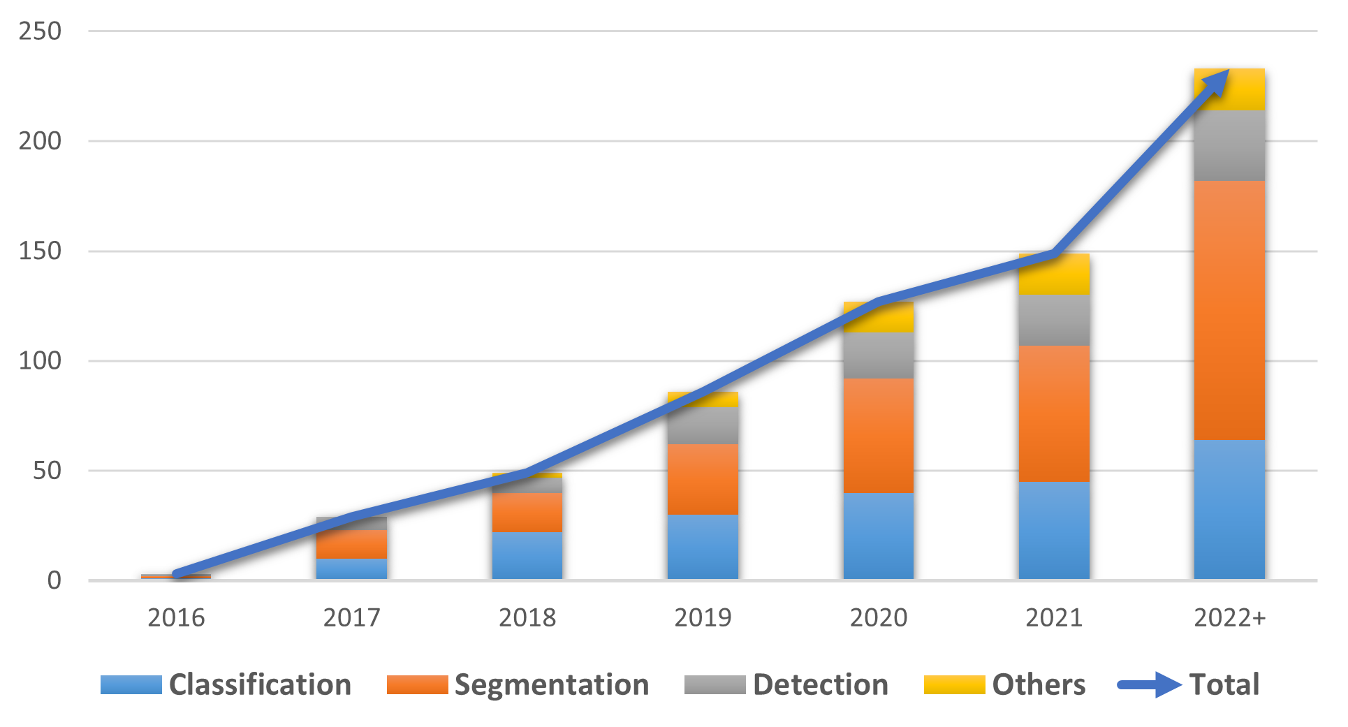
Computer-aided medical image analysis (MIA) plays a more and more critical role in achieving efficiency and accuracy in the early detection, diagnosis, and treatment of diseases. In recent years, MIA systems powered by deep learning (DL) have provided a more objective approach to learning from large and heterogeneous medical image datasets and improved disease diagnosis accuracy. However, DL models require abundant precisely annotated data to effectively capture anatomical heterogeneity and disease-specific traits [341] due to their data-driven nature. Unfortunately, due to a shortage of available annotators [187], there is a significant gap between the demand for annotation and the available annotated datasets. Hence, the urgency to curtail annotation expenses, expedite the annotation procedure, and alleviate the load on annotators has emerged as a crucial hurdle in DL-based MIA tasks. Traditional fully-supervised DL methods, on the other hand, depend solely on comprehensively annotated datasets. Recently, strategies based on semi-supervised, self-supervised, and multi-instance learning have been widely utilized to maximize the utility of existing medical data that may be only partially annotated by point, scribble, box, pixel-wise, etc. or even completely unannotated data. In this paper, we dub these methods as label-efficient learning. As seen in Fig. 1, label-efficient learning methods have significantly proliferated in recent years. Meanwhile, label-efficient learning methods excelling in other MIA tasks like denoising, image registration, and super-resolution have also been rising beyond common classification, segmentation, and detection.
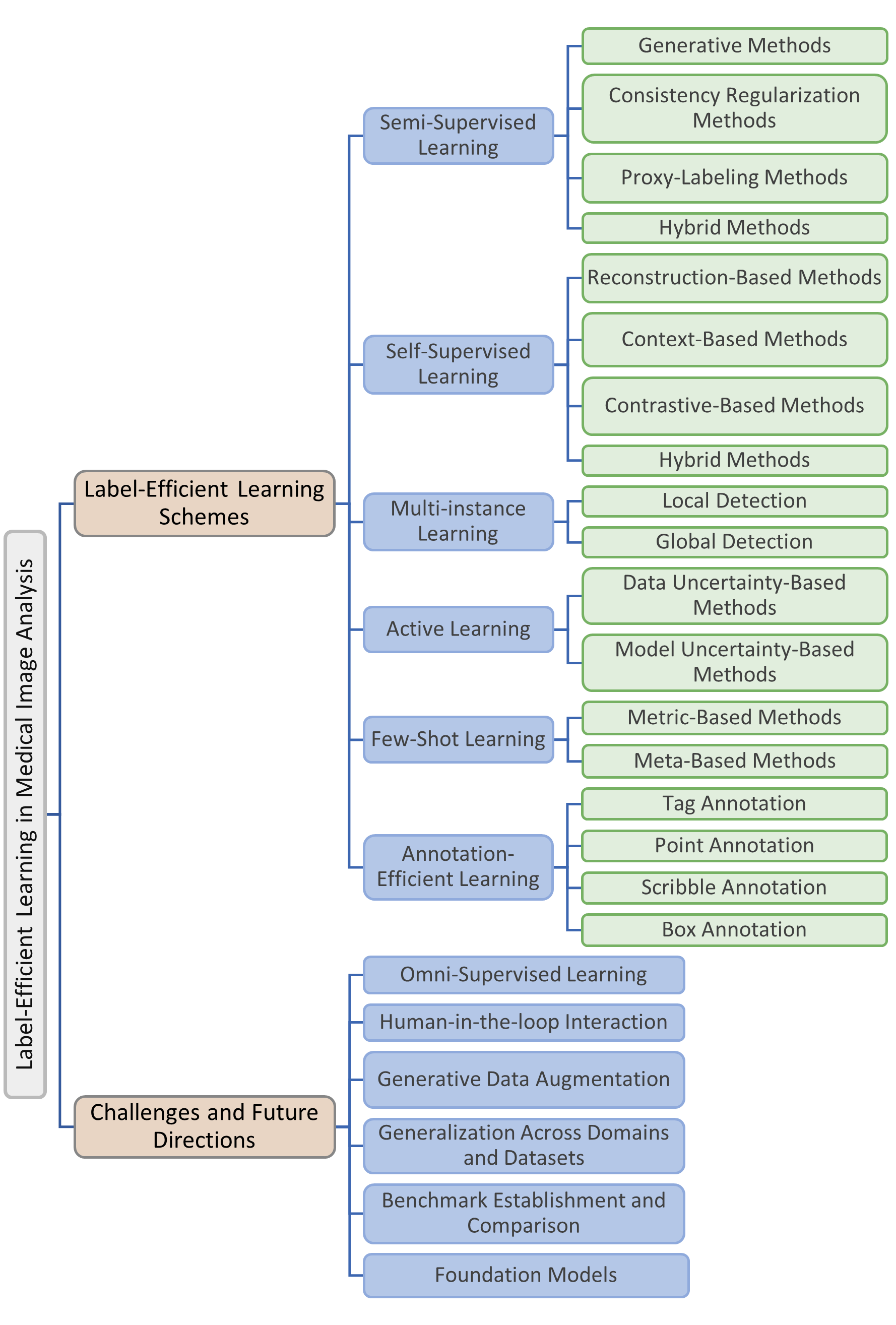
Several surveys related to label-efficient learning in medical image analysis have been published in recent years. Cheplygina et al. [47] categorized methods under supervised, semi-supervised, multi-instance, and transfer learning and named them “not-so-supervised” learning, while Budd et al. [24] surveyed human-in-the-loop strategies for MIA tasks. However, methods in these surveys are either limited in scope or lag behind the current trends. While Kumari and Singh [138] present a contemporary review focused on data- and label-efficient learning in the medical domain, the taxonomy they present lacks sufficient clarity and directness, which may lead to interpretational difficulties for readers. Conversely, our taxonomy is based on learning schemes and provides distinct and straightforward guidance. Furthermore, the above reviews fall short in addressing several crucial questions of significant interest to researchers. In contrast, our paper comprehensively addresses these topics, providing an in-depth exploration of these critical aspects and the outline is illustrated in Fig. 2.
Aiming to provide a comprehensive overview and future challenges of label-efficient learning methods in MIA, we review more than 300 quality-assured and recent label-efficient learning methods based on semi-supervised, multi-instance, self-supervised, active, and annotation-efficient learning strategies. To pinpoint pertinent contributions, Google Scholar was employed to search for papers with related topics. ArXiv was combined through for papers citing one of a set of terms related to label-efficient medical imaging. Additionally, conference proceedings like CVPR, ICCV, ECCV, NIPS, AAAI, and MICCAI were scrutinized based on the titles of the papers, as well as journals such as MIA, IEEE TMI, and Nature Bioengineering. References in all chosen papers were examined. When overlapping work had been reported in multiple publications, only the publication(s) considered most significant were incorporated.
To the best of our knowledge, this is the first comprehensive review in the field of label-efficient MIA. In each learning scheme, we formulate the fundamental problem, offer the necessary background, and display the experimental results case by case. With the challenges proposed at the end of the survey, we explore feasible future directions in several branches to potentially enlighten the follow-up research on label-efficient learning.
The remainder of this paper is organized as follows. In Section 2, the necessary background and categorization is presented. In Sections 3–8, we introduce the primary label-efficient learning schemes in MIA, including semi-supervised learning in Section 3, self-supervised learning in Section 4, multi-instance learning in Section 5, active learning in Section 6, few-shot learning in Section 7, and annotation-efficient learning in Section 8. We discuss the existing challenges in label-efficient learning and present several heuristic solutions for these open problems in Section 9, where promising future research directions are proposed as well. Finally, we conclude the paper in Section 10.
2 Background and Categorization
In this section, we review the background of the learning schemes covering label-efficient learning. In addition, we present the categorization of each learning scheme in MIA.
2.1 Semi-Supervised Learning
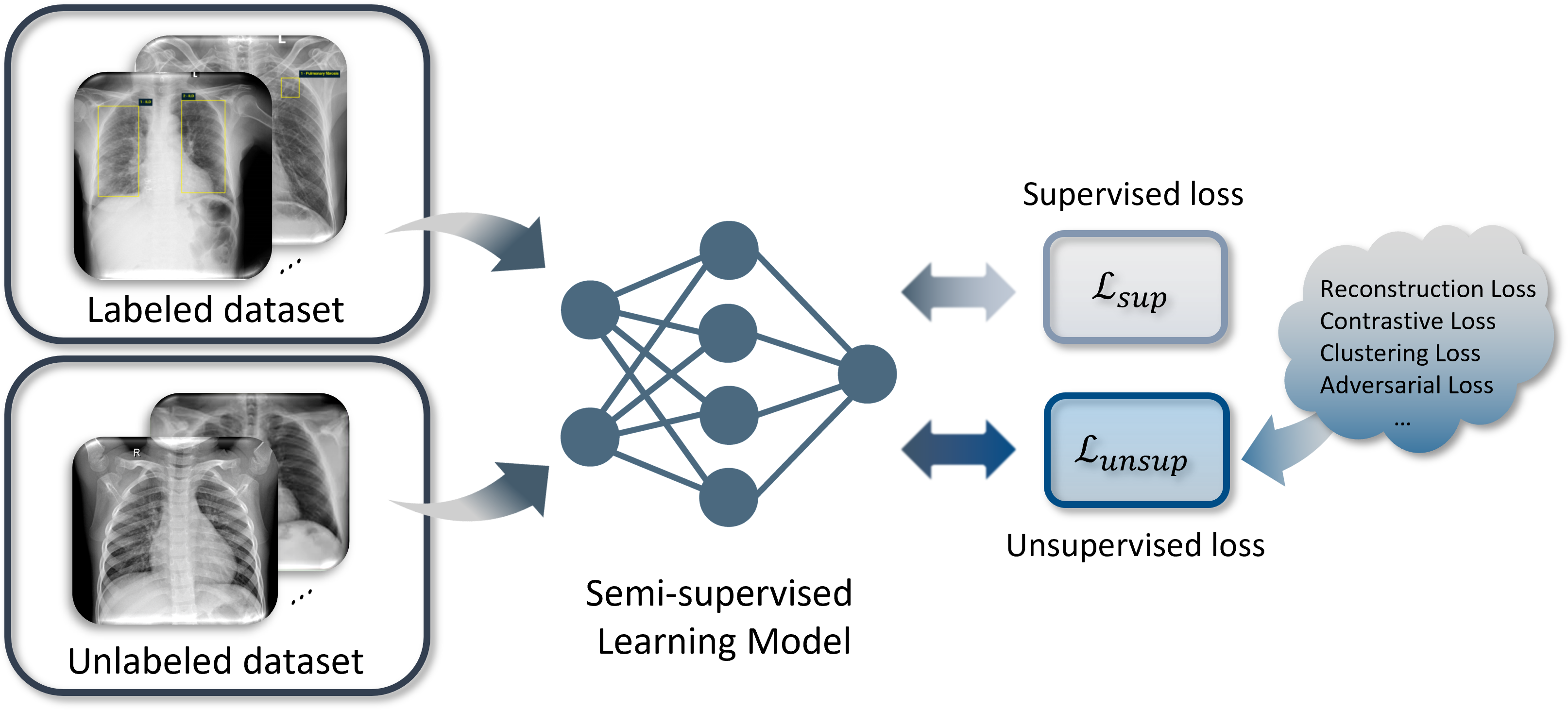
As illustrated in Fig. 3, Semi-supervised learning (Semi-SL) introduces an additional unlabeled dataset to help the model learn task-related invariant features and aim to achieve better performance than supervised learning. Concretely, one has a set of labeled data points , in which represents the raw data sample from the given input space and is the corresponding label. In the meantime, an unlabeled dataset with a much larger scale is involved, i.e., . And denotes the entire dataset. During the training process, the optimization problem333Several assumptions and prior knowledge of Semi-SL can be referred to Appendix A.1. that Semi-SL intends to solve is defined as:
| (1) |
where represents the model parameters, is the supervised loss function, represents the unsupervised loss function, and is a regularization term. In addition, control the trade-off between unsupervised loss and regularization term .
Based on how the model incorporates and leverages unlabeled data, we will discuss the categories of Semi-SL methods and their applications in MIA starting from proxy-labeling methods, followed by generative methods, consistency regularization methods, and finally hybrid methods. Meanwhile, we present a brief summary of the representative publications in Tab. 1
2.2 Self-Supervised Learning
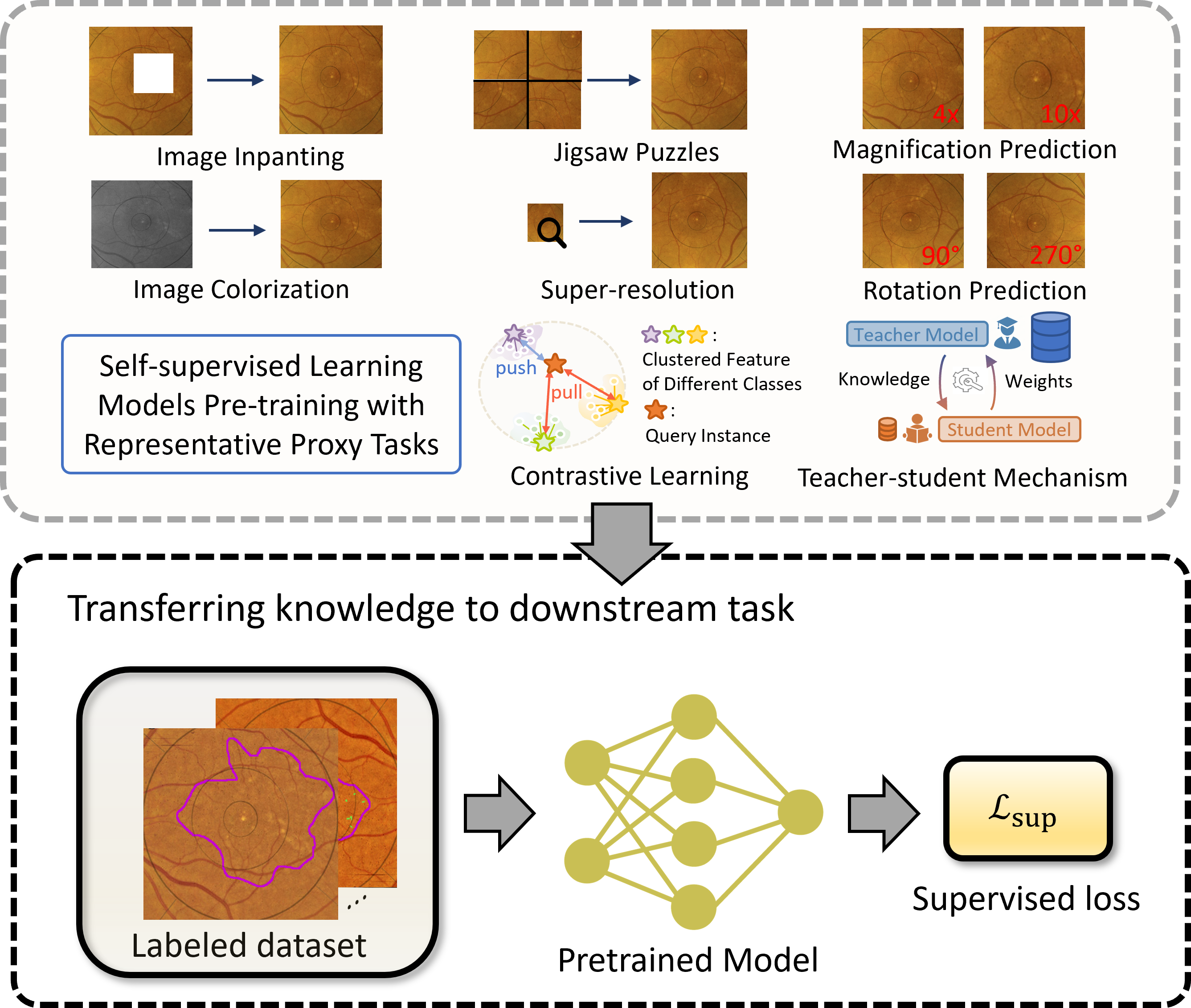
Self-supervised learning (Self-SL) was proposed to extract and learn the underlying features of a large-scale unlabeled dataset without human annotation. Generally, Self-SL methods build proxy tasks for the model to learn the latent features and representations from a massive amount of unlabeled data, thus facilitating the performance on downstream tasks, as shown in Fig. 4. Concretely, the training procedure of Self-SL can be divided into two stages: pre-training with proxy tasks and fine-tuning on different downstream tasks. During the pre-training phase, researchers design proxy tasks that satisfy the following two properties [116]: (1) The label of the input data for the proxy task can be generated automatically by the data itself; (2) the neural network can learn related representations or features of the input data by solving the proxy task.
After the pre-training with proxy tasks, the learned representations will be utilized to solve the main task. The advantages of utilizing proxy tasks are two-fold: on the one hand, by defining particular tasks, the model can be targeted to learn features or representations of the specific studied data; on the other hand, by using a large amount of unlabeled data for pre-training, the model can significantly avoid overfitting during fine-tuning compared to supervised learning, especially for small datasets, in downstream training.
Based on the characteristics of the proxy tasks, we group the mainstream Self-SL methods in MIA into the following four general categories: Reconstruction-Based Methods, Context-Based Methods, Contrastive-Based Methods, and Hybrid Methods with a summary of the representative publications in Tab. 2.
2.3 Multi-instance Learning
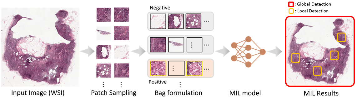
In multi-instance learning (MIL), the concept of a bag is introduced. A bag is composed of instances: , where denotes an instance in bag , and the training dataset consists of bags: . Next, suppose and are the labels of bag and the instance inside it, respectively, in which denotes positive and denotes negative for the binary classification scenario. Two common assumptions can be made based on this basic definition of MIL:
-
1.
If bag is positive, then there exists at least one positive instance and is unknown. This assumption can be summarized as: if , then .
-
2.
If bag is negative, then all the instances in are negative, namely, if , then .
Based on the assumptions, MIL methods can perform both bag-level and instance-level tasks (illustrated in Fig. 5), with the latter often used in weakly-supervised learning. Concretely, MIL algorithms leverage the instances to identify positive or negative bags, which contributes not only to the image-level diagnosis but also to precise abnormal region detection and localization. This great interpretability of the MIL algorithm fits well in MIA, as both the global structure and local details are crucial components for solving such problems.
In this survey, we categorize MIL methods that aim at detecting all the particular target patterns in the data, such as every patch with a special disease manifestation in a large histopathology image, as local detection; and methods that aim at simply detecting whether or not the particular target patterns exist in the given sample as global detection. Note that taxonomy is in line with the methodology of MIL, i.e., to classify bag-level label (global detection) or to classify instance-level label (local detection). Tab. 3 presents an overview of the representative publications of each method.
2.4 Active Learning
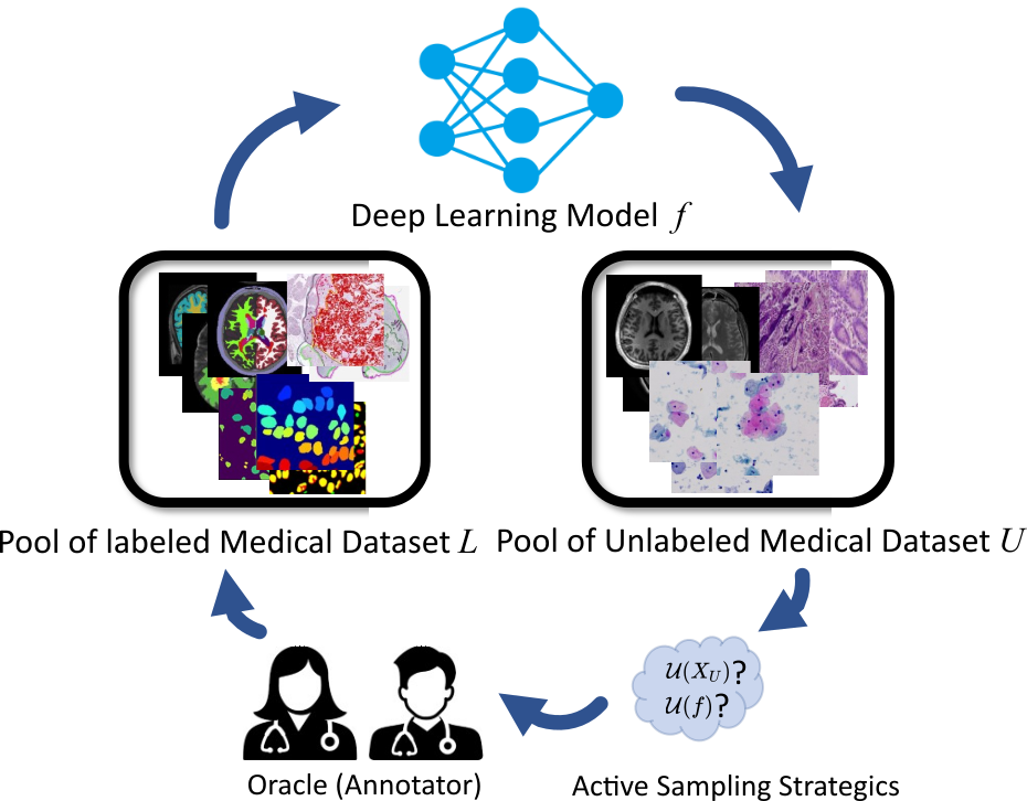
Active learning (AL) is a relatively understudied area in the MIA field. It attempts to maintain the performance of a deep learning model while annotating the fewest data with the help of an oracle, which resonates with the philosophy of label-efficient learning, i.e., how to effectively use noisy, limited, and unannotated data throughout the deep learning process. More specifically, its goal is to select the most valuable samples and forward them to the oracle (e.g., human annotator) for labeling to improve the generalization capability of the model. In active learning (AL) practice, the measurement of annotation uncertainty using various strategies is often considered as the metric for sample value. Meanwhile, in order to preserve the network’s generalization capability, different mechanisms have been developed to ensure that the sampled images are distributed diversely.
As Fig. 7 illustrates, before the start of the data selection process, a deep learning model is initialized or pre-trained from a labeled dataset with its corresponding parameter . After that, AL sampling algorithms construct an uncertainty metric for each item of unlabeled dataset . This metric determines whether an oracle is required for annotation, and we denote this newly annotated dataset as . Then the network model will either use the combined labeled data to train from scratch or only use them to fine-tune the model. Denoting the fully labeled version of as , the goal of AL is to build a model with to perform equivalently or better than .
Based on how the uncertainty is obtained, we categorize AL methods into data uncertainty-based methods and model uncertainty-based methods. Data uncertainty-based methods attempt to get a sample with the greatest uncertainty from a batched dataset, while model uncertainty-based methods tend to sample the samples that cause the greatest uncertainty of the deep learning model’s performance. A brief summary of surveyed AL papers is presented in Tab. 4.
2.5 Few-Shot Learning
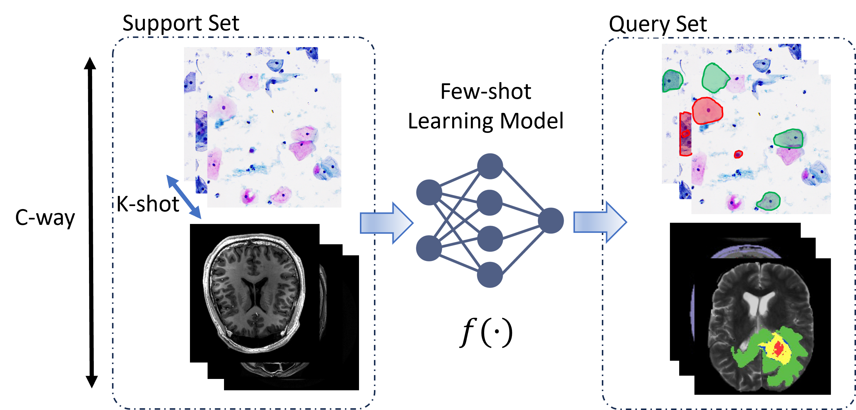
Few-shot learning (FSL) is the problem of building a deep learning model to make predictions based on a limited number of samples. This limited sample size restricts the model’s generalization ability in conventional learning schemes. In FSL literature, the terms support set and query set represent the training and testing sets, respectively. Each support set comprises distinct categories, each containing training samples, thus establishing a C-way K-shot configuration. In this section, we categorize FSL methods based on mainstream MIA literature into two categories: metric-based methods, meta-based methods444To aid the understanding of each subfield, a detailed description is provided in Appendix A.3.. For a comprehensive review of general FSL methods, please refer to Song et al. [261].
2.6 Annotation-Efficient Learning

Annotation-efficient learning is a technique that utilizes deep learning methods with partially labeled data for dense predictions to improve labeling efficiency. The intuitive approach to increase annotation efficiency is to provide markings other than fully dense annotations. While there may be overlapping techniques with the aforementioned categories, annotation-efficient learning methods specifically focus on leveraging the specific characteristics of the different forms of annotation to enhance the annotation efficiency and hence minimize the granularity difference between the annotation and the prediction. Fig. 8 shows different forms of annotation, and we will separately review the annotation-efficient learning methods that address the “not exact label” through a coarse-to-fine way. Specifically, we will discuss the techniques related to Tag, Point, Scribble, and Box annotations. Tab. LABEL:tab:anno provides an overview of representative publications in this category.
3 Semi-supervised Learning in MIA
| Reference (Year) | Organ | Semi-SL Algorithm Design | Dataset | Result | |
| Classification | Madani et al. [195] | Lung | Semi-superivsed GAN | NIH PLCO; NIH Chest X-Ray | Acc (Accuracy): 0.851 |
| Diaz-Pinto et al. [58] | Retina | Semi-supervised DCGAN | ORIGA-light; DRISHTI-GS; RIM-ONE; HRF; DRD; | AUC: 0.9017 | |
| sjchoi86-HRF; ACRIMA; DRIVE; Messidor | |||||
| Shi et al. [250] | Lung; Breast | Graph Temporal Ensembling | TCGA-Lung; TCGA-Breast | TCGA-Lung: F1: 0.893; TCGA-Breast: F1: 0.930 | |
| Yu et al. [340] | Colon | Mean Teacher | Private Dataset: 13,111 Images | Patch-level AUC: 0.980; Patient-level AUC: 0.974 | |
| Wang et al. [302] | Breast; Retina | Virtual Adversarial Training + Self-training | RetinalOCT; Private Dataset: 39,904 Images | Acc: 0.9513; Macro-R (Macro-Recall): 0.9330 | |
| Liu et al. [172] | Lung; Skin | Anti-curriculum Self-training | ChestX-Ray14; ISIC 2018 | ChestX-ray14: AUC: 0.8177; ISIC: AUC: 0.9436 | |
| Zhang et al. [353] | Spinal cord | Consistency Regularization + Pseudo-labeling + Active Learning | Private Dataset: 7,295 Images; | Acc: 0.9582; Macro-P (Macro-Precision): 0.8609 | |
| Gao et al. [78] | Multi-Organ | Dual-task Consistency | TGCA-RCC; TCGA-BR; TGCA-LU | AUC: 0.972 | |
| Zeng et al. [345] | Colon; Skin; Chest | Self-training + Feature Adversarial Training | NCT-CRC-HE; ISIC 2018; Chest X-Ray14 | NCT-CRC-HE: Acc: 0.9029; AUC: 0.9908 (With 200 labeled data) | |
| ISIC 2018: Acc: 0.9368; AUC: 0.9487 (With 20% labeled data) | |||||
| Chest X-Ray14: AUC: 0.7506 (With 2% labeled data) | |||||
| Xie et al. [322] | Retina | Semi-supervised GAN | iChallenge; ODIR | iChallenge: Acc: 0.7731; AUC: 0.9382 | |
| ODIR: Acc: 0.6514; AUC: 0.9221 | |||||
| Yang et al. [333] | Multi-Organ | Self-training | KC Dataset; ISIC 2018; RSNA Dataset | KC: AUC: 0.8471 (With 5% labeled data) | |
| ISIC 2018: AUC: 0.8439 (With 5% labeled data) | |||||
| RSNA: AUC: 0.8193 (With 5% labeled data) | |||||
| Segmentation | Bai et al. [14]2017 | Heart | CRF-based Self-training | Private Dataset: 8050 Images | DSC: 0.920 |
| Li et al. [160] | Skin | -model | ISIC 2017 | DSC: 0.874; Acc: 0.943 | |
| Nie et al. [218] | Prostate | Self-training | Private Dataset: 70 Images | DSC: 0.970; ASD (Average Surface Distance): 1.401 | |
| Yu et al. [342] | Heart | Uncertainty-aware Mean Teacher | ASG | DSC: 0.8888; 95HD: 7.32; JI: 0.8021 | |
| Zhou et al. [370] | Multi-Organ | Multi-planar Co-training | Private Dataset: 310 Volumes | DSC: 0.7794 | |
| Li et al. [161] | Liver; Retina; Skin | Transformation-consistent Mean Teacher | ISIC 2017; REFUGE; LiTS | ISIC: DSC: 0.8344; REFUGE: DSC: 0.9543; LiTS: DSC: 0.9427 | |
| Liu et al. [179] | Skin; Lung | Mean Teacher + Sample Relation Consistency | ISIC 2018; ChestX-ray14 | ISIC: AUC: 0.9358; ChestX-ray8: AUC: 0.7923 | |
| Li et al. [155] | Heart | Shape-aware Consistency Regularization | ASG | DSC: 0.8954; JI (Jaccard Index): 0.8124 | |
| Fan et al. [66] | Lung | Attention Self-training | IS-COVID | DSC: 0.739 | |
| Chaitanya et al. [34] | Heart; Prostate; Pancreas | Semi-supervised GAN + Deformation and Additive Intensity Field | ACDC; DECATHLON | ACDC: DSC (Dice coefficient): 0.834; DECATHLON: DSC: 0.529 | |
| Luo et al. [193] | Nasopharynx | Uncertainty Rectified Pyramid Consistency | Private Dataset: 258 MR Images | DSC: 0.8076 | |
| Luo et al. [191] | Heart; | Dual-task Consistency | ASG; NIH PCT | ASG: DSC: 0.8942; NIH PCT: DSC: 0.7827; | |
| Li et al. [149] | Lung; Skin; Liver | StyleGAN2 | ChestX-ray14; JSRT Database; ISIC 2018; LiTS; CHAOS | DSC: Lung: 0.9668; ISIC: 0.8329; LiTS: 0.9169 | |
| You et al. [339] | Heart; Pancreas | Mean Teacher + Contrastive Learning | ASG; NIH PCT | ASG: DSC: 0.9085; NIH PCT: DSC: 0.7539 | |
| Wang et al. [297] | Heart; Prostate | Mean Teacher + Contrastive Learning | ACDC; ProMRI | ACDC: DSC: 0.914; ProMRI: DSC: 0.704 | |
| Wu et al. [316] | Heart; Pancreas | Uncertainty-based Mutual Consistency | ASG; NIH PCT; ACDC | DSC: ASG: 0.9107; NIH PCT: 0.8059; ACDC: 0.8851 | |
| Luo et al. [192] | Heart | Co-training Variant | ACDC | DSC: 0.864 | |
| Shi et al. [251] | Multi-Organ | Consistency Regularization + Teacher-student Model | CVC; ETIS-Larib Polyp; Private Dataset: 1,100 images | AP50: 0.917 (With 5% supervised dataset) | |
| Xu et al. [326] | Multi-Organ | Mean Teacher | Left Atrium (LA) Dataset; BraTS 2019 | DSC: 0.9031; JI: 0.8243; ASD: 1.76 | |
| Bashir et al. [18] | Multi-Organ | Consistency Regularization | MoNuSeg; BCSS | MoNuSeg: mIoU: 0.7172; DSC: 0.8260; Acc: 0.8886 (With 1/32 data being labeled) | |
| BCSS: mIoU: 0.4709; DSC: 0.6184; Acc: 0.7320 (With 1/8 data being labeled) | |||||
| Bai et al. [15] | Multi-Organ | Mean Teacher | Left Atrium (LA) Dataset; Pancreas-NIH; ACDC | LA: DSC: 0.8962; JI: 0.8131; ASD: 1.76; | |
| Pancreas-NIH: DSC:0.8291; JI: 0.7097; ASD:2.25; | |||||
| ACDC: DSC:0.8884; JI: 0.8062; ASD:1.17; | |||||
| Miao et al. [210] | Multi-Organ | Causality Co-training | ACDC; Pancreas-CT; BraTS 2019 | ACDC: DSC: 0.8966 JI: 0.8234; ASD: 0.88 (With 10% labeled data) | |
| Pancreas-CT: DSC: 0.7289 JI: 0.5806; ASD: 4.37 (With 6/62 volumes having annotations) | |||||
| BRaTs 2019: DSC: 0.8354 JI: 0.7346; ASD: 1.98 (With 10% labeled data) | |||||
| Chaitanya et al. [33] | Heart; Prostate | Self-training | ACDC; MICCAI 2019; MMWHS | ACDC: DSC: 0.759 (With 1 labeled data) | |
| MICCAI 2019: DSC: 0.578 (With 1 labeled data) | |||||
| MMWHS: DSC: 0.572 (With 1 labeled data) | |||||
| Wang et al. [309] | Heart; Pancreas | Co-training | Left Atrial (LA) Dataset; NIH-Pancreas | LA: DSC: 0.8871; JI: 0.8041; ASD: 1.90 | |
| NIH-Pancreas: DSC: 0.7500; JI: 0.6127; ASD: 3.27 | |||||
| Basak and Yin [17] | Heart; Kidney; Gland | Contrastive Self-training | ACDC; KiTS19; CRAG | ACDS: DSC: 0.912; ASD: 1.49 (With 20% labeled data) | |
| KiTS19: DSC: 0.919; ASD: 1.51 (With 10% labeled data) | |||||
| CRAG: DSC: 0.891; ASD: 2.01 (With 20% labeled data) | |||||
| Zhang et al. [352] | Heart; Pancreas | Co-training | Left Atrial (LA) Dataset; NIH-Pancreas | LA: DSC: 0.8871; JI: 0.8041; ASD: 1.90 | |
| NIH-Pancreas: DSC: 0.7500; JI: 0.6127; ASD: 3.27 | |||||
| Lei et al. [145] | Liver; Skin | Adversarial Consistency + Dynamic Convolution Network | LiTS; ISIC 2018 | LiTS: DSC: 0.9412; ASD: 3.51 | |
| Chen et al. [41] | Heart; Brain | Task-specific Consistency Regularization | MICCAI 2018; DECATHLON | MICCAI 2018: DSC: 0.8775; JI: 0.7880; ASD: 2.04 | |
| DECATHLON: DSC: 0.8775 | |||||
| Meng et al. [206] | Multi-Organ | Consistency Regularization + Adaptive Graph Neural Network | SEG (Combined by Refuge; Drishti-GS; ORIGA; RIGA; | SEG: DSC: 0.882 | |
| RIMONE Datasets); UKBB | UKBB: MAE (Mean Absolute Error): 0.097 | ||||
| Detection | Wang et al. [289] | Lung | MixMatch + Focal Loss | LUNA; NLST | LUNA: CPM: 0.872 |
| Zhou et al. [367] | Multi-Organ | Teacher-student Model + Adaptive Consistency Loss | DSB; DeepLesion | DSB: mAP: 0.694; DeepLesion: Sens (Sensitivity): 0.779 |
3.1 Proxy-labeling Methods
Proxy-labeling methods provide proxy labels for unlabeled data samples in . They include those data samples with high confidence proxy labels in the training dataset, training in an iterative manner. Proxy-labeling methods can be mainly categorized into two sub-categories: Self-training methods and multi-view learning methods.
3.1.1 Self-training Methods
Self-training methods aim to learn a prediction function with parameters by using a fraction of labeled data samples . After that, the trained prediction function is utilized to provide proxy labels of unlabeled data samples . Normally, a threshold is manually set and the sample-label pair will be added to the labeled dataset if the highest prediction probability in the output of outweighs . The updated labeled dataset will be consequently used to train the prediction function , and this process is conducted iteratively until cannot make predictions with enough confidence.
Entropy minimization [86] is a method that regularizes the model based on the low-density assumption, encouraging the model to generate low-entropy prediction for the unlabeled data. Pseudo-label [141] is a simple yet effective self-training mechanism which inherits the concept of entropy minimization in the prediction space. The labeled samples are trained in a supervised way, and unlabeled data are assigned with the most confident predictions. In MIA, pseudo-label is employed as an auxiliary component to enhance model performance [33, 66, 353]. In fact, proxy labels are normally noisy and may not reflect the ground truth. Therefore, various quality measurements such as uncertainty-aware confidence evaluation [291], conditional random field-based proxy label refinement [14], and adversarial training-based method [369] have been developed to ensure that reliable supervision signals can be generated based on pseudo labels. Pseudo-label has also been used in MIA to refine a given annotation with the assistance of unlabeled data. Qu et al. [230] introduce pseudo-label into nuclei segmentation and design an iterative learning algorithm to refine the background of weakly labeled images where only nuclei are annotated, leaving large areas ignored. Similar ideas can also be seen in Nie et al. [218].
3.1.2 Multi-view learning methods
Multi-view learning methods assume that each sample has two or multiple complementary views and features of the same sample extracted with different views are supposed to be consistent. Therefore, the key idea of multi-view learning methods is to train the model with multiple views of the sample or train multiple learners and minimize the disagreement between them, thus learning the underlying features of the data from multiple aspects. Co-training is a method that falls into this category. It assumes that data sample can be represented by two views, and , and each of them are capable of solely training a good learner, respectively. Consequently, the two learners are set to make predictions of each view’s unlabeled data, and iteratively choose the candidates with the highest confidence for the other model [335]. Another variation of multi-view learning methods is Tri-training [372], which is proposed to tackle the lack of multiple view data and mistaken labels of unlabeled data produced by self-training methods. Tri-training aims to learn three models from three different training sets obtained with bootstrap sampling. Recently a deep learning version of Tri-training, i.e. Tri-Net, has been proposed in Chen et al. [39].
Co-training, or deep co-training, is dominant in multi-view learning in MIA, with a steady flow of publications[69, 298, 318, 345, 360, 370]. To conduct whole brain segmentation, Zhao et al. [360] implement co-training by obtaining different views of data with data augmentation. A similar idea can be seen for 3D medical image segmentation in [318] and [370]. These two works both utilize co-training by learning individual models from different views of 3D volumes such as the sagittal, coronal, and axial planes. Further works have been proposed to refine co-training. To produce reliable and confident predictions, Wang et al. [298] develop a self-paced learning strategy for co-training, forcing the network to start with the easier-to-segment regions and transition to the difficult areas gradually. Rather than discarding samples with low-quality pseudo-labels, Zeng et al. [345] introduce a novel regularization approach, which focuses on extracting discriminative information from such samples by injecting adversarial noise at the feature level, thereby smoothing the decision boundary. Meanwhile, to avoid the errors of different model components accumulating and causing deviation, Fang and Li [69] develop an end-to-end model called difference minimization network for medical image segmentation by conducting co-training with an encoder shared by two decoders.
3.2 Generative Methods
Generative Semi-SL assumes that the entire dataset is generated from the same latent distribution. In this sense, the key point of generative methods is to learn and simulate the latent distribution with the help of unlabeled data. Then the model with a well learned latent distribution aims to improve performance by combining supervised information.
Generative adversarial network (GAN) is a widely used model leveraging both labeled and unlabeled data. The standard GAN is composed of a generator and a discriminator , trying to satisfy the Nash equilibrium [84]. Typically, a generator is trained aiming to generate plausible images and a discriminator is trained to distinguish the generated image and the real one. The unlabeled data can be involved during the adversarial training process, in which the discriminator aims to distinguish the generated fake input and real unlabeled data. By solving the two-player minimax game, GAN can learn the underlying distribution with the help of unlabeled data. The MIA filed has seen publications with respect to generative Semi-SL methods based on GAN [34, 58, 108, 118, 195, 369]. Chaitanya et al. [34] directly incorporate the unlabeled data during the adversarial training of GAN to train a better generator for boosting medical data augmentation, arguing that utilizing unlabeled samples allows more variations in shape and intensity so as to make the model robust and guide the optimization. A similar idea can be seen in [108]. While Zhou et al. [369] develop a generator network to predict the pseudo lesion masks for unlabeled data and utilize the discriminator to facilitate the quality of generated lesion mask. Other researchers have designed quite a number of methods modifying the discriminator . Instead of only distinguishing real or fake images, Odena [219] seek to learn the category information by predicting classes and an additional real or fake class. In this way, the unlabeled data can contribute to the model during the classification of the categories. In the context of MIA, the architecture proposed in [219] has produced fruitful results in various fields, such as retinal image synthesis [58, 118, 322], glaucoma assessment [58], chest X-ray classification [195], and so on [108].
Variational autoencoder (VAE) is also useful and prospective in utilizing unlabeled data. It is an autoencoder rooted in Bayesian inference theory [129]. A typical VAE encodes a data sample into a latent variable and decodes it into the reconstruction of input by maximizing the variational lower bound. Our review of related literature shows that VAEs in MIA scenarios are mostly utilized for learning the inherent feature similarity from a large unlabeled dataset, thus contributing to a well-constrained latent space which can consequently avoid the need of numerous labeled data for training [243, 293]. Sedai et al. [243] propose a dual-VAE framework to conduct semi-supervised segmentation of the optic cup in retinal fundus images, in which one VAE learns the data distribution with unlabeled data and transfers the prior knowledge to the other VAE which conducts segmentation with the labeled data. Instead of using a mean vector and a variance vector for the latent representation, Wang and Lukasiewicz [293] adapts the VAE architecture into 3D medical image segmentation by introducing a mean vector and a covariance matrix to involve the correlation of different slices of an input volume.
3.3 Consistency Regularization Methods
Based on the smoothness or manifold assumption, Consistency regularization methods follow the idea that the perturbation of data points does not change the prediction of the model. Meanwhile, this process does not require label information, which is proved an effective constraint for learning the unlabeled data.
-model [240] is a simple yet effective implementation of the above idea. This method uses a shared encoder to obtain different views of the input sample through augmentation and force the classifier to produce the same prediction for different augmentations of . Meanwhile, label information is included in the training process to improve the performance of the classifier. By designing a -model-based semi-supervised algorithm, Li et al. [160] set a new record for skin lesion segmentation with only 300 labeled images, surpassing the state-of-the-art which was fully-supervised and used a set of 2,000 labeled images. Similar idea can be seen in [23, 207], where Bortsova et al. [23] conduct semi-supervised chest X-ray segmentation by learning prediction consistency given a set of transformations, and Meng et al. [207] utilize graph convolution networks to constrain the regional consistency and marginal consistency for Semi-SL optic disc and cup Segmentation. Temporal ensembling [139] was developed to improve the prediction stability of the -model by adding exponentially moving average module for updating prediction. And a number of researchers have implemented this module to address MIA-related problems [31, 95, 190, 250]. To conduct accurate breast mass segmentation, Cao et al. [31] introduce uncertainty into the temporal ensembling model by using uncertainty maps as guidance for the neural network to ensure the reliability of generated predictions. Similarly, Luo et al. [190] propose an uncertainty-aware temporal ensembling to learn from external partially labeled data for chest X-ray screening. Instead of directly feeding the augmented version of sample into neural networks, Gyawali et al. [95] employ a VAE model to firstly extract the disentangled latent space and use it as stochastic embedding for the model input, leading to improved temporal ensembling in chest X-ray classification. During the training process of temporal ensembling, the activation of each training sample is only updated once in one epoch. By implementing exponentially moving average on model parameters rather than network activations, Mean teacher [275] overcomes this disadvantage and has been applied in the MIA field as well [3, 161, 310, 326, 342]. [161] is a typical application of the mean teacher model in MIA, which utilizes this model to conduct transformation-consistent medical image segmentation. However, with no ground-truth given for unlabeled training data, the output of the teacher model can be inaccurate and noisy. Yu et al. [342] incorporate an uncertainty map with the mean teacher model to ensure the reliability of targets generated by the teacher. Similar idea can be found in [3]. Wang et al. [310] further improve uncertainty-aware methods for segmentation of the left atrium and kidney by proposing a double-uncertainty-weighted method, which extends segmentation uncertainty to feature level uncertainty. While Xu et al. [326] put emphasis on boosting the performance of Mean teacher model via selecting productive unsupervised consistency targets. In their work, a simple-yet-effective ambiguity-consensus mean-teacher model is proposed to better exploit the complementary informative clues from unlabeled data.
3.4 Hybrid Methods
A burgeoning Semi-SL research direction is to combine the aforementioned types of methods together and unify them into a holistic framework for better performance [289, 302, 353]. These are called hybrid methods in this survey. For example, Wang et al. [302] and Zhang et al. [353] combine consistency regularization with self-training to solve medical image classification problems. Besides, Mixup [348] has been utilized frequently as an effective data augmentation strategy in hybrid methods. In [94], the authors implement Mixup on both input and latent space to create more sample-label pairs based on both labeled and unlabeled data to facilitate medical image classification. By leveraging Mixup and focal loss, Wang et al. [289] improve MixMatch [21], which is a combination of consistency regularization and pseudo-labeling, in the filed of 3D medical image detection. By leveraging multiple Semi-SL methods, the model is able to learn the underlying invariant features and meanwhile empowered with a strong predictive capability.
3.5 Discussion
Various unlabeled data inclusion and regularization approach lead to numerous Semi-SL methods. Many research efforts are devoted to generating pseudo labels for unlabeled data to enrich the training dataset, during which the measurement of pseudo labels’ quality and confidence plays an essential role. In addition, other researchers aim to leverage the unlabeled data to learn the distribution of real data such as generative methods or learn a model with robust prediction ability such as consistency regularization methods. Establishing a theoretical foundation for this process is also a critical area of study, albeit with limited research efforts to date, as highlighted in Miao et al. [210]. Further, an open problem for Semi-SL is how to ensure the model performs well when input unlabeled data are noisy, i.e., to learn task-specific and perturbation-invariant features. Besides, a burgeoning research direction is to combine various Semi-SL methods to maximize the exploitation and utilization of unlabeled data and boost MIA tasks.
4 Self-supervised Learning in MIA
| Reference (Year) | Organ | Proxy Task Design | Dataset | Result | |
| Classification | Li et al. [159] | Retina | Multi-modal Contrastive Learning | ADAM; PALM | ADAM: AUC: 0.7458; PALM: AUC: 0.9855; |
| Koohbanani et al. [133] | Breast; | Magnification Prediction; | CAMELYON 2016; | CAMELYON 2016: AUC: 0.937; | |
| Cervix; | Solving Magnification Puzzle; | KATHER; | KATHER: AUC: 0.951; | ||
| Colon | Hematoxylin Channel Prediction | Private Dataset: 217 Images | Private Dataset: AUC: 0.974 | ||
| Azizi et al. [12] | Skin; Lung | Multi-Instance Contrastive Learning | Priavte Dermatology Dataset; CheXpert | Private: Top-1 Acc: 0.7002; CheXpert: AUC: 0.7729 | |
| Tiu et al. [280] | Lung | Contrastive Learning | CheXpert | AUC: 0.889 | |
| Chen et al. [43] | Breast; Lung; Kidney | Contrastive Learning | TCGA-BRCA; TCGA-NSCLS; TCGA-RCC | AUC: TCGA-Breast: 0.874; TCGA-NSCLS: 0.952; TCGA-RCC: 0.980 | |
| Mahapatra et al. [198] | Lymph; Lung; | Contrastive Learning Variant | CAMELYON 2017; DRD; GGC | Acc: CAMELYON 2017: 0.929; DRD: 0.951; GGC: 0.916; | |
| Retina; Prostate | ChestX-ray14; CheXpert; | ChestX-ray14: Acc: 0.861; ChestXpert: Acc: 0.913 | |||
| Wang et al. [308] | Skin | Self-supervised Knowledge Distillation | ISIC 2019 | AUC: 0.977; ACC: 0.846; mAP: 0.796 | |
| Segmentation | Hervella et al. [105] | Retina | Multi-modal Reconstruction | Isfahan MISP | AUC: 0.8183 |
| Spitzer et al. [264] | Brain | Patch Distance Prediction | BigBrain | DSC: 0.80 | |
| Bai et al. [13] | Heart | Anatomical Position Prediction | Private Dataset: 3825 Subjects | DSC: 0.934 | |
| Sahasrabudhe et al. [239] | Multi-Organ | WSI Patch Magnification Identification | MoNuSeg | AJI: 0.5354; AHD (Average Hausdorff Distance): 7.7502 | |
| Tao et al. [274] | Pancreas | Rubik’s Cube Recovery | NIH PCT; MRBrainS18 | NIH PCT: DSC: 0.8408; MRBrainS18: DSC: 0.7756 | |
| Lu et al. [185] | Brain | Fiber Streamlines Density Map Prediction; | dHCP | DSC: 0.822; | |
| Registration-based Segmentation Imitation | |||||
| Tang et al. [273] | Abdomen; Liver; | Contrastive Learning; Masked Volume Inpainting; | DECATHLON; | DECATHLON: DSC: 0.787; | |
| Prostate | 3D Rotation Prediction | BTCV | BTCV: DSC: 0.918 | ||
| Jiang et al. [115] | Multi-organ | Anatomical-invariant Contrastive Learning | FLARE 2022; BTCV | FLARE 2022: DSC: 0.869; NSD: 0.913; BTCV: DSC: 0.886 | |
| He et al. [104] | Heart; Artery; Brain | Geometric Visual Similarity Learning | MM-WHS-CT; ASOCA; CANDI; STOIC | DSC: Heart: 0.912; Artery: 0.813; Brain: 0.900 | |
| Liu et al. [181] | Tooth | Hierarchical Global-local Contrastive Learning | Private Dataset: 13,000 Scans | DSC: 0.949; mIoU: 0.931 | |
| Zheng et al. [362] | Multi-Organ | Multi-scale Visual Representation Self-supervised Learning | BCV; MSD; KiTS | DSC: 0.836; MSD: 0.962; KiTS: 0.852 | |
| Regression | Abbet et al. [1] | Gland | Image Colorization | Private Dataset: 660 Images | Brier Score: 0.2725; C-Index: 0.6943 |
| Srinidhi et al. [265] | Breast; | WSI Patch Resolution Sequence | BreastPathQ; | BreastPathQ: ICC Coefficient: 0.907; | |
| Colon | Prediction | CAMELYON 2016; | CAMELYON 2016: AUC: 0.882; | ||
| KATHER | KATHER: Acc: 0.986; F1: 0.934 | ||||
| Fan et al. [67] | Brain; Lung | Image Colorization; Cross-channel | GBM; TCGA-LUSC; NLST | C-Index: GBM: 0.670; LUSC: 0.679; NLST: 0.711 | |
| Others | Zhuang et al. [376] | Brain | Rubik’s Cube Recovery | BraTS 2018; Private Dataset: 1,486 Images | BraTS 2018: mIoU: 0.773; Private: Acc: 0.838 |
| Chen et al. [42] | Multi-Organ | Disturbed Image Context Restoration | Private Fetus Dataset: 2,694 Images; | Private Fetus Dataset: F1: 0.8942; | |
| Private Multi-organ Dataset: 150 Images; | Private Multi-organ Dataset: Mean Distance: 2.90 | ||||
| BraTS 2017 | BraTS 2017: DSC: 0.8557 | ||||
| Zhao et al. [355] | Brain | Super-resolution Reconstruction | Private Dataset: 47 Images | S3 Sharpness: 0.5482 | |
| Li et al. [154] | Abdomen | CT Reconstruction | LDCTGC | PSNR: 22.1758; SSIM: 0.7800 | |
| Cao et al. [30] | Brain | Missing Modality Synthesis | BraTS 2015; ADNI | ADNI: IS (Inception Score): 2.15; FID: 64.29 | |
| Haghighi et al. [96] | Lung | Self-Discovery + Self-Classification | LUNA; LiTS; CAD-PE; BraTS 2018; | Classification: LUNA: AUC: 0.9847; | |
| +Self-Restoration | ChestX-ray14; LIDC-IDRI; SIIM-ACR | Segmentation: IoU: LiTS: 0.8560; BraTS 2018: 0.6882 | |||
| Taleb et al. [270] | Brain; Retina; | 3D Contrastive Predictive Coding; 3D Jigsaw Puzzles; | BraTS 2018; | BraTS 2018: DSC: 0.9080; | |
| Pancreas | 3D Rotation Prediction; 3D Exemplar Networks | DECATHLON; | DECATHLON: DSC 0.635; | ||
| Relative 3D Patch Location; | DRD | DRD: DSC 0.80 | |||
| Li et al. [146] | Breast; Pancreas; Kidney | Super-resolution Reconstruction; Color Normalization | WTS; Private Dataset: 533 Images | PSNR: 28.32; SSIM: 0.8304 | |
| Wang et al. [306] | Multi-Organ | Contrastive Learning | TCGA; KATHER; MHIST | MHIST: F1: 0.0.8993; KATHER: F1: 0.9582; | |
| PAIP; PatchCAMELYON | PatchCAMELYON: F1: 0.8983; AUC: 0.9779 | ||||
| Zhou et al. [366] | Lung; Brain; Liver | Contrastive Learning + Image Reconstruction | ChestX-ray14; CheXpert; LUNA | AUC: Chest: 0.831; LUNA: 0.922; | |
| BraTS 2018; LiTS; | DSC: LiTS: 0.937; BraTS 2018: 0.85 | ||||
| Yan et al. [327] | Multi-Organ | Global and Local Contrastive Learning | DeepLesion; NIH LN; Private Dataset: 94 Patients | Mean Radial Error: 4.3; Maximum Radial Error: 16.4 | |
| Cai et al. [26] | Lung; Brain; Retina | Dual-Distribution Reconstruction | RSNA-Lung; LAG; VinDr-CXR; Brain Tumor MRI; | AUC: RSNA-Lung: 0.913; Brain Tumor MRI: 0.972; | |
| Private Lung Dataset: 5,000 Images | VinDr-CXR: 0.859; LAG: 0.931; Private Lung Dataset: 0.710; | ||||
| Haghighi et al. [97] | Lung | Contrastive Learning + Reconstruction + | ChestX-ray14; CheXpert; | AUC: ChestX-ray14: 0.8112; CheXpert: 0.8759; | |
| Adversarial Learning | Montgomery | Montgomery: DSC: 0.9824 | |||
| Li et al. [151] | Retina | Frequency-boosted Image Enhancement | EyePACS; | EyePACS: FIQA: 0.81; | |
| Private Dataset: more than 10,000 Images | Private Dataset: SSIM: 0.879 |
-
1
For the sake of brevity, we denote references that contain more than one task in the following abbreviations: C: Classification, S:Segmentation, D:Detection, SR: Super-resolution, DN: Denoising, IT: Image Translation, RE: Registration.
4.1 Reconstruction-Based Methods
Reconstruction-based methods in Self-SL focus on exploring the inherent structures of data without the help of human annotations. These methods are conducted on several tasks including super-resolution [146, 355], inpainting [356], colorization [1], and the emerging MIA-specific application, multi-modal reconstruction [30, 105].
A straightforward way to establish the reconstruction task is proposed by Li et al. [154], who adopt an auto-encoder network to encode and reconstruct normal-dose computed tomography (CT) images for learning the latent features by minimizing the mean squared error (MSE) loss. After self-supervised pre-training, the encoder is utilized for feature extraction, and a supervised loss is computed with the encoded latent features. However, the self-supervised pre-training based on the minimization of reconstruction loss might neglect the basic structure of the input image and capture the color space distribution instead [1]. More proxy tasks have been motivated to solve this challenge.
The super-resolution reconstruction task is to generate fine-grained and realistic high-resolution images by utilizing low-resolution input images. In this proxy task, the targeted model can learn the underlying semantic features and structures of data. Zhao et al. [355] propose an anti-aliasing algorithm based on super-resolution reconstruction to reduce aliasing and restore the quality of magnetic resonance images (MRIs). While Li et al. [151] utilize the frequency information in fundus image as guidance to conduct image enhancement. In the meantime, super-resolution reconstruction is also an appropriate proxy task for gigapixel histopathology whole-slide images (WSIs) because low-resolution WSIs are rather easy to store and process. From this application, Li et al. [146] conduct single image super-resolution for WSIs using GAN.
The image colorization task is to predict the RGB version of the gray-scale images. During this process, the network is trained to capture the contour and shape of different tissues in the sample and fill them with respective colors [1, 67, 167]. Abbet et al. [1] introduce the image colorization task into survival analysis of colorectal cancer. They train a convolutional auto-encoder to convert the original input image into a two-channel image, namely, hematoxylin and eosin. Then, MSE loss is applied to measure the difference between the original input image and its converted counterpart. Moreover, in the context of survival analysis, Fan et al. [67] extend their methodology beyond image colorization to include a cross-channel pre-text task. This additional task challenges the model to restore the lightness channel in image patches, utilizing the information from their color channels.
The image inpainting task aims to predict and fill in missing parts based on the remaining regions of the input image. This proxy task allows the model to recognize the common features of identical objects, such as color and structure, and thus to predict the missing parts consistently with the rest of the image. Zhao et al. [356] propose a restoration module based on Self-SL to facilitate the anomaly detection of optical coherence tomography (OCT) and chest X-ray. It demonstrates that the restoration of missing regions facilitates the model’s learning of the anatomic information.
In recent years, the multi-modal reconstruction task has emerged [30, 105]. In this task, the model uses the aligned multi-modal images of a patient to reconstruct an image in one modality by taking another modality as the input. Hervella et al. [105] propose this proxy task to enrich the model with joint representations of different modalities, arguing that each modality offers a complementary aspect of the object. Therefore, they take retinography and fluorescein angiography into consideration to facilitate retinal image understanding. Meanwhile, Cao et al. [30] develop a self-supervised collaborative learning algorithm, aiming at learning modality-invariant features for medical image synthesis by generating the missing modality with auto-encoder and GAN.
4.2 Context-Based Methods
Context-based methods utilize the inherent context information of the input image. Recent years have witnessed attempts to design novel predictive tasks for specific MIA tasks by training the network for prediction of the output class or localization of objects with the original image as the supervision signal [13, 264, 265]. Bai et al. [13] propose a proxy task to predict the anatomical positions from cardiac chamber view planes by applying an encoder-decoder structure. This proxy task properly employs the chamber view plane information, which is available from cardiac MR scans easily. While Zheng et al. [362] aims to perform finer-grained representation and deal with different target scales by designing a multi-scale consistency objective to boost medical image segmentation. Further advancements in proxy tasks for 3D medical images are presented by He et al. [104]. They propose a novel paradigm, termed Geometric Visual Similarity Learning, which integrates a topological invariance prior into the assessment of inter-image similarity. This approach aims to ensure consistent representation of semantic regions. In addition, Srinidhi et al. [265] propose an MIA-specific proxy task, Resolution Sequence Prediction, which utilizes the multi-resolution information contained in the pyramid structure of WSIs. A neural network is employed to predict the order of multi-resolution image patches out of all possible sequences that can be generated from these patches. In this way, both contextual structure and local details can be captured by the network at lower and higher magnifications, respectively.
Other efforts have been made to explore the spatial context structure of input data, such as the order of different patches constituting an image, or the relative position of several patches in the same image, which can provide useful semantic features for the network. Chen et al. [42] focus on the proxy task, dubbed context restoration, of randomly switching the position of two patches in a given image iteratively and restoring the original image. During this process, semantic features can be learned in a straightforward way. Instead of concentrating on the inherent intensity distribution of an image, Li et al. [158] aims to improve the performance of a network with rotation angle prediction as the proxy task. The input retinal images are first augmented, generating several views, then randomly rotated. The model is encouraged to predict the rotation angle and cluster the representations with similar features. More advanced proxy tasks such as Jigsaw Puzzles [73] and Rubik’s Cube [135] are also attracting an increasing number of researchers. Taleb et al. [269] improve the Jigsaw Puzzle task with multi-modal data. Concretely, an input image is constituted of out-of-order patches of different modalities and the model is expected to restore the original image. Rubik’s Cube is a task set for 3-dimensional data. Zhuang et al. [376] and Tao et al. [274] introduce Rubik’s Cube into the MIA area, and significantly boost the performance of a deep learning model on 3D data. In this method, the 3D volume will first be cut into a grid of cubes and a random rotation operation will be conducted on these cubes. The aim of this proxy task is to recover the original volume.
However, for histopathology images, common proxy tasks such as prediction of the rotation or relative position of objects may only provide minor improvements to the model in histopathology due to the lack of a sense of global orientation in WSIs [85, 133]. Therefore, Koohbanani et al. [133] propose proxy tasks targeted at histopathology, namely, magnification prediction, solving magnification puzzle, and hematoxylin channel prediction. In this way, their model can significantly integrate and learn the contextual, multi-resolution, and semantic features inside the WSIs.
4.3 Contrastive-Based Methods
Contrastive-based methods are based on the idea that the learned representations of different views of the same image should be similar and those of different images should be clearly distinguishable. Intriguingly, the ideas behind several high-performance algorithms such as SimCLR [45] and BYOL [87] have been incorporated into the MIA field [12, 306]. Multi-Instance Contrastive Learning (MICLe), is proposed by Azizi et al. [12], is a refinement and improvement of SimCLR. Instead of using one input to generate augmented views for contrastive learning, they propose to minimize the disagreement of several views from multiple input images of the same patient, creating of more positive pairs. Meanwhile, Wang et al. [306] adopt the BYOL architecture to facilitate histopathology image classification. A contribution of their work was to collect the currently largest WSI dataset for Self-SL pre-training. It includes 2.7 million patches cropped from 32,529 WSIs covering over 25 anatomic sites and 32 classes of cancer subtypes. Similarly, Ghesu et al. [79] develop a contrastive learning and online clustering algorithm based on over 100 million radiography, CT, MRI, and ultrasound images. By leveraging this large unlabeled dataset for pre-training, the performance and convergence rate of the proposed model show a significant improvement over the state-of-the-art. Another line of work that utilizes large-scale unsupervised dataset is [215], in which over 1.3 million multi-modal data from 55 publicly available datasets are integrated. In addition to considering different perspectives of the same input, Jiang et al. [115] introduce a contrastive objective for the learning of anatomically invariant features. This approach is designed to fully exploit the inherent similarities in anatomical structures across diverse medical imaging volumes.
Further studies take into account the global and local contrast for better representation learning. Their methods usually minimize the InfoNCE loss [221] to capture the global and local level information. In [327], the authors implement the InfoNCE by encoding each pixel of the input image. Their goal is to generate embeddings that can precisely describe the anatomical location of that pixel. To achieve this, they develop a pixel-level contrastive learning framework to generate embeddings at both the global and local level. Further, Liu et al. [181] propose a hierarchical contrastive learning objective to capture the unsupervised representation of intra-oral mesh scans from point-level, region-level, and cross-level.
4.4 Hybrid Methods
Studies have made efforts to combine some or all of the different types of Sefl-SL methods into a universal framework to learn latent representations from multiple perspectives, such as semantic features and structure information inside unlabeled data [96, 273, 332, 366]. For instance, Tang et al. [273] combine masked volume inpainting, contrastive coding, and image rotation tasks into a Swin Transformer encoder architecture for medical image segmentation.
4.5 Discussion
Self-SL methods aim to learn and obtain a model with prior knowledge by manipulating and exploiting unlabeled data. The key to the superior performance of Self-SL models is the design of proxy tasks. Numerous existing Self-SL methods directly adopt proxy tasks prevailing in natural image processing into the MIA field. However, the unique properties of medical images, such as CT, WSI, and MRI, should be exclusively considered and injected into the design process of proxy tasks. The medical field has witnessed pioneering research efforts, exemplified by Zhang et al. [346], that aim to establish guidelines for the design of Self-SL proxy tasks. Further, proxy task design based on the combination of different medical image modalities is a prospective research direction, during which the model can capture disentangled features of each modality, leading to a robust pre-trained network. For example, large vision-language pre-trained models [225, 364, 365] are emerging in chest X-ray and obtaining ever-increasing research interests.
5 Multi-instance Learning in MIA
| Reference (Year) | Organ | MIL Algorithm Design | Dataset | Result | |
| Classification | Manivannan et al. [202] | Retina; | Discriminative Subspace Transformation + | Messidor; TMA-UCSB; | Messidor: Acc: 0.728; TMA-UCSB: AUC: 0.967; |
| Breast | Margin-based Loss | DR Dataset; Private Dataset: 884 Images | DR Dataset: Acc: 0.8793; Private: Kappa: 0.7212 | ||
| Ilse et al. [113] | Breast; Colon | Attention-based MIL | TMA-UCSB; CRCHistoPhenotypes | TMA-UCSB: Acc: 0.755; CRCHistoPhenotypes: Acc: 0.898 | |
| Couture et al. [52] | Breast | Quantile Function-based MIL | CBCS3 | Acc: 0.952 | |
| Liu et al. [174] | Brain | Landmark-based MIL | ADNI; MIRIAD | ADNI: AUC: 0.9586; MIRIAD: AUC: 0.9716 | |
| Campanella et al. [27] | Prostate; Skin; Lymph | MIL + RNN | Private Dataset: 44,732 Images | AUC: Prostate: 0.986; Skin: 0.986; Lymph: 0.965 | |
| Wang et al. [301] | Breast | Instance Features Recalibration | Private Dataset: 608 Images | Acc: 0.865 | |
| Yao et al. [336] | Lung; Brain | Multiple Instance FCN | NLST; TCGA | NLST: C-Index: 0.678; TCGA: C-Index: 0.657 | |
| Wang et al. [305] | Retina | Uncertainty-aware MIL + RNN Aggregation | Duke-AMD; Private Dataset: 4,644 Volumes | Acc: Duke-AMD: 0.979; Private Dataset: 0.951 | |
| Zhao et al. [359] | Colon | VAE-GAN Feature Extraction + | TCGA-COAD | Acc: 0.6761; F1: 0.6667; | |
| GNN Bag-level Representation Learning | AUC: 0.7102 | ||||
| Chikontwe et al. [48] | Colon | Jointly Learning of Instance- and Bag-level Feature | Private Dataset: 366 Images | F1: 0.9236; P (Precision): 0.9254; R (Recall): 0.9231; Acc: 0.9231 | |
| Raju et al. [232] | Colon | Graph Attention MIL | MCO | Acc: 0.811; F1: 0.798 | |
| Han et al. [100] | Lung | Automatic Instance Generation | Private Dataset: 460 Examples | AUC: 0.99 | |
| Yao et al. [337] | Lung; Colon | Siamese Multi-instance FCN + Attention MIL | NLST; MCO | NLST: AUC: 0.7143; MCO: AUC: 0.644 | |
| Hashimoto et al. [101] | Lymph | Domain Adversarial + Multi-scale MIL | Private Dataset: 196 Images | Acc: 0.871 | |
| Shao et al. [246] | Breast; Lung; Kidney | Transformer-based MIL | CAMELYON 2016; TCGA-NSCLC; TCGA-RCC | Acc: CAMELYON: 0.8837; TCGA-NSCLC: 0.8835; TCGA-RCC: 0.9466 | |
| Li et al. [147] | Breast; Lung | Dual-stream MIL + Contrastive Learning | CAMELYON 2016; TCGA Lung Cancer | CAMELYON 2016: AUC: 0.9165; TCGA: AUC: 0.9815 | |
| Li et al. [164] | Lung | Virtual Bags + Self-SL Location Prediction | Private Dataset: 460 Examples | AUC: 0.981; Acc: 0.958; F1: 0.895; Sens: 0.936 | |
| Lu et al. [184] | Kidney; Lung; | Attention-based MIL + Clustering | TCGA-RCC + Private Dataset: 135 WSIs; | Kidney: AUC: 0.972; | |
| Lymph node | CPTAC-NSCLC + Private Dataset: 131 WSIs; | Lung: AUC: 0.975; | |||
| CAMELYON 2016,17 + Private Dataset: 133 WSIs | Lymph node: AUC: 0.940 | ||||
| Wang et al. [312] | Thyroid | Transformer-based MIL + Knowledge Distillation | Private Dataset: 595 Images | AUC: 0.9835; P: 0.9482; R: 0.9151; F1: 0.9297 | |
| Zhang et al. [349] | Breast; Lung | Double-Tier Feature Distillation MIL | CAMELYON 2016; TCGA-Lung | CAMELYON 2016: AUC: 0.946; TCGA-Lung: AUC: 0.961 | |
| Schirris et al. [241] | Breast; Colon | Heterogeneity-aware MIL + Contrastive Learning | TCGA-CRCk; TCGA-BC | TCGA-CRCk: AUC: 0.87; TCGA-BC: AUC: 0.81 | |
| Su et al. [266] | Breast; Kidney | Intelligent Sampling Method + Attention MIL | CAMELYON 2016; Private Dataset: 112 Images | CAMELYON 2016: AUC: 0.891; Private: AUC: 0.974 | |
| Zhu et al. [374] | Breast; Lung; Kidney | Reinforcement Learning + Contrastive Learning + MIL | CAMELYON 2016; TCGA-Lung; TCGA-Kidney | AUC: CAMELYON: 0.9452; TCGA-Lung: 0.9637; TCGA-Kidney: 0.9573 | |
| Yang et al. [331] | Colon; Muscle | Curriculum Learning + MIL | CRCHistoPhenotypes; Private Muscle Dataset: 266 Images | CRCHistoPhenotypes: AUC: 0.898; Private: AUC: 0.907 | |
| Shi et al. [249] | Stomach; Bladder | Multi-scale Graph MIL | TCGA-STAD; TCGA-BLCA; Private Stomach Dataset: 574 Images | AUC: TCGA-STAD: 0.829; TCGA-BLCA: 0.886; Private: 0.907 | |
| Yan et al. [329] | Bladder | Hierarchical Deep MIL | TCGA-Bladder | TCGA-Bladder: AUC: 0.92 | |
| Shi et al. [248] | Breast; Kidney | Multi-scale Transformer + MIL | BRIGHT; TCGA-BRCA; TCGA-RCC | AUC: BRIGHT: 0.848; TCGA-BRCA: 0.921; TCGA-RCC: 0.990 | |
| Liu et al. [175] | Lung; Breast; Brain | GAN + MIL | NLST; TCGA-BRCA; TCGA-LGG | C-Index: NLST: 0.672; TCGA-BRCA: 0.566; TCGA-LGG: 0.642 | |
| Segmentation | Jia et al. [114] | Colon | Multi-scale MIL + Area Constraint Regularization | Private TMA/Colon Dataset: 60 Images/910 Images | F1: TMA: 0.622; Colon: 0.836 |
| Xu et al. [324] | Breast | Instance-level and Pixel-level Label Generation | CAMELYON 2016 | Image-level Acc: 0.929; Pixel-level IoU: 0.847 | |
| Dov et al. [62] | Thyroid | Maximum Likelihood Estimation-based MIL | Private Dataset: 908 Images | AUC: 0.87 | |
| Others | Schwab et al. [242] | Lung | Jointly Classification and Localization | RSNA-Lung; MIMIC-CXR; Private Dataset: 1,003 Images | AUC: 0.93 |
| Wang et al. [307] | Pancreas | Jointly Global-level Classification and | Private Dataset: 800 Images | DSC: 0.6029; | |
| Local-level Segmentation | Sens: 0.9975 |
-
1
For the sake of brevity, we denote references that contain more than one task in the following abbreviations: C: Classification, S:Segmentation, D:Detection.
5.1 Local Detection
Since we define local detection as detecting or localizing all the particular disease patterns of an input image, papers with the purpose of segmentation or localization can be classified into this category. Most researchers design their local detection model to infer every patch label and thereby obtain both the local annotations and the global labels. Thus, the local detection methods often include global detection methods since inferring image-level labels after obtaining the local annotations.
Schwab et al. [242], apply the basic MIL algorithm to conduct the localization and classification of chest X-rays. They input every patch of the original sample into a CNN, and the model outputs a score for the patch representing its probability of containing a critical finding. Once the patch-level classifier is trained, the most straightforward way to perform slide-level classification is to integrate the patch-level predictions with max-pooling or average-pooling. The design for the pooling function plays an important role in the performance improvement of the MIL algorithm. For instance, in [52], the authors design a more general MIL aggregation method by utilizing a quantile function as the pooling function. By doing so, a more thorough description of the heterogeneity of each sample can be provided, enhancing the quality of global classification. Other studies [113, 301] propose learning-based aggregation operators to provide insight into the contribution of each instance to the bag. Among them, several are based on the attention-based MIL developed by Ilse et al. [113]. By introducing the attention mechanism into MIL, their model can better capture the key features of regions of interest with interpretation. For the pancreatic ductal adenocarcinoma (PDAC) prediction problem, Wang et al. [307] design an inductive attention guidance network for both classification and segmentation. The attention mechanism works as a connection between the global classifier and local (instance) segmenter by guiding the location of PDAC regions.
Other intriguing improvements in local detection are springing up as well. Researchers have tried many different ways to facilitate instance prediction [62, 114, 202, 324]. Dov et al. [62] demonstrate that the general MIL methods perform poorly on cytopathology data for two reasons: instances that contain key information are located sparsely in a gigapixel pathology image, and the informative instances have various characteristics of abnormality. Thus, they propose a MIL structure involving maximum likelihood estimation to predict multiple labels, i.e., bag-level labels and diagnostic scores; instance-level labels and informativeness, simultaneously. Similarly, when studying the classification of the retinal nerve fiber layer (RNFL), Manivannan et al. [202] have observed that regions that contain the RNFL generally have strong intra-class variation, making them difficult to distinguish from other regions. Therefore, they map the instances into a discriminative subspace to increase the discrepancy for disentangled instance feature learning. Jia et al. [114] incorporate the multi-scale image feature into the learning process to obtain more latent information on histopathology images. Finally, to address the problem that only image-level labels are provided in MIL, Xu et al. [324] design an automatic instance-level label generation method. Their work has led to an interesting MIL algorithm design direction and may shed light on how to improve the performance of local detection algorithms.
In parallel, there has been significant progress in related domains such as phenotype categorization [101, 336, 337] and multi-label classification [209]. These investigations have further exemplified the versatility and potential of the MIL algorithm in addressing complex challenges across various subfields.
5.2 Global Detection
Global detection refers to methods that simply aim to find out whether or not target patterns exist. For example, for the COVID-19 screening problem, researchers [164] have designed MIL algorithms to classify an input sample as severe or not instead of locating every abnormal patch.
To facilitate the prediction of image-level labels (e.g. WSI-level label), researchers normally start from one of two aspects, namely instance- and bag-level. Most existing MIL algorithms [101, 184, 212, 281] are based on the basic assumption that instances of the same bag are independent and identically distributed. Consequently, the correlations among instances are neglected, which is not realistic. Recently several works have taken the correlation among instances or tissues into consideration [100, 232, 246, 301, 312]. In [246], the authors introduce vision transformer (ViT) into MIL for gigapixel WSIs due to its great advantage in capturing the long-distance information and correlation among instances in a sequence. Meanwhile, to conduct precise lymph node metastasis prediction, Wang et al. [312] not only incorporate a pruned Transformer into MIL but also develop a knowledge distillation mechanism based on other similar datasets, such as a papillary thyroid carcinoma dataset, effectively avoiding the overfitting problem caused by the insufficient number of samples in the original dataset. Similarly, Raju et al. [232] design a graph attention MIL algorithm for colorectal cancer staging, which utilizes different tissues as nodes to construct graphs for instance relation learning. Further, in order to utilize the multi-resolution characteristics of WSIs, Shi et al. [249] consider WSIs as multi-scale graphs and utilize attention mechanism to integrate their information for primary tumor stage prediction. Similar idea can be found in [248, 319, 329]. Besides, Liu et al. [175] firstly propose an integration of GAN with MIL mechanism for robust and interpretable WSI survival analysis by more accurately estimating target distribution.
For bag-level improvement, recent years have witnessed two feasible approaches, namely, improved pooling methods and pseudo bags. On the one hand, in order to aggregate the instances with the most information, some researchers have developed novel aggregation methods in MIL algorithms instead of the traditional max pooling [54, 48]. For example, in [48], the authors design a pyramid feature aggregation method to directly obtain a bag-level feature vector. On the other hand, however, there is an inherent problem for MIA, especially for histopathology — the number of WSIs (bags) is usually small, while in contrast, one WSI has numerous patches, leading to an imbalance in the number of bags and instances. To address this problem, Zhang et al. [349] randomly split the instances of a bag into several smaller bags, called ”pseudo bags”, with labels that are consistent with the original bag. A similar idea can also be seen in [164].
Other improvements in MIL algorithms are also worth mentioning [266, 276, 305]. In [266], an intelligent sampling method is developed to collect instances with high confidence. This method excludes patches shared among different classes and tends to select the patches that match with the bag-level label. In [276], the authors utilize the extreme value theory to measure the maximum feature deviations and consequently leverage them to recognize the positive instances, while in [305], the authors introduce an uncertainty evaluation mechanism into MIL for the first time, and train a robust classifier based on this mechanism to cope with OCT image classification problem.
5.3 Discussion
Multi-instance learning in MIA is mainly applied to whole slide image analysis, which can be described as “a needle in a haystack” problem, making bag-level decisions out of thousands of instances. MIL methods are developed to locate the discriminative patches as a basis for diagnosis. To achieve this goal, MIL research can be divided into several focuses. For the bag-instance correlation, a WSI is represented as a bag containing selected patches during training, which leads to the question of how the patches should be selected to make the bag representative of the WSI. Further, how to handle and leverage the imbalance of positive and negative samples could have a significant impact on model performance. For the instance-instance correlation, the proper modeling and utilization of instance relations can boost the performance of MIL algorithms and advance the interpretability of the model.
6 Active Learning in MIA
| Reference (Year) | Organ | Sampling Method | Dataset | Result | |
| Classification | Gal et al. [77] | Skin | BALD + KL-divergence | ISIC 2016 | 22% image input: AUC: 0.75 |
| Wu et al. [315] | Lung | Loss Prediction Network | CC-CCII Dataset | 42% Chest X-Ray input: Acc: 86.6% | |
| Li et al. [156] | Prostate | CurriculumNet + O2U-Net | ISIC 2017; PANDA Dataset | 60% input: QWK: 0.895 | |
| Segmentation | Yang et al. [330] | Gland; Lymph | Cosine Similarity + Bootstrapping + FCN | GlaS 2015; Private Dataset: 80 US images | MICCAI 2015: 50% input: F1: 0.921; Private Dataset: 50% input: F1: 0.871 |
| Konyushkova et al. [132] | Brain (Striatum; Hippocampus) | Geometric Priors + Boosted Trees | BraTS 2012; EFPL EM Dataset | MRI Data: 60% input: DSC0.76; EM Data: 40% input: DSC0.60 | |
| Nath et al. [213] | Brain | Entropy + SVGD Optimization | MSD 2018 Dataset | 22.69% Hippocampus MRI input: DSC: 0.7241 | |
| Ozdemir et al. [224] | Shoulder | BNN + MMD Divergence | Private Dataset: 36 Volume of MRIs | 48% MRI input: DSC0.85 | |
| Zhao et al. [361] | Hand; Skin | U-Net | RSNA-Bone; ISIC 2017 | 9 AL Iteration: DSC: 0.834 | |
| Others | Mahapatra et al. [197] | Chest | Bayesian Neural Network + | JSRT Database; | Classification: 35% input: AUC: 0.953; |
| cGAN Data Augmentation | ChestX-ray8 | Segmentation: 35% input: DSC: 0.910 | |||
| Zhou et al. [371] | Colon | Traditional Data Augmentation | Private Dataset: 6 colonoscopy videos | Classification: 4% input: AUC: 0.9204; | |
| Entropy + Diversity | 38 polyp videos + 121 CTPA datasets | Detection: 2.04% input: AUC: 0.9615 |
-
1
For the sake of brevity, we denote references that contain more than one task in the following abbreviations: C: Classification, S: Segmentation, D: Detection.
6.1 Data Uncertainty-Based Methods
Developed from the conventional entropy uncertainty metrics555To aid the understanding of these metrics, a detailed description of the prior knowledge is provided in Appendix A.2., Konyushkova et al. [132] defined geometric smoothness priors with boosted trees to classify the formed graph representation of electron microscopy images. Here, they flatten 3D images into supervoxels with the SLIC algorithm [2] to conduct graph representations. Yang et al. [330] use cosine similarity and a bootstrapping technique to evaluate the uncertainty and representativeness of the output feature with a DCAN [40]-like network. Zhou et al. [371] propose the concept of “active selection” policies, which is the highest confidence based on the entropy and diversity results from sampled data in the mean prediction results.
Aside from leveraging conventional metrics, utilizing metrics from the deep learning model is another trend. Intuitively, Wu et al. [315] utilize network loss as well as the diversity condition as the uncertainty metric for sampling from a loss prediction network, and conduct the COVID-19 classification task from another classification network. Nath et al. [213] leverage marginal probabilities between the query images and the labeled ones, they build a mutual information metric as the diversity metric to serve as a regularizer. Moreover, they adopt Dice log-likelihood instead of its original entropy-based log-likelihood for Stein variational gradient descent optimizer [176] to solve the label imbalance problem. Zhao et al. [361] utilize Dice’s coefficient of the predicted mask calculated between the middle layer and the final layer of the model as the uncertainty metric for the image segmentation task. They use their DS-UNet with a denseCRF [136] refiner to annotate low uncertainty samples and oracle annotators for the others. Li et al. [156] use k-means clustering and curriculum classification (CC) based on the CurriculumNet [90] for uncertainty and representativeness estimation. Furthermore, they consider the condition under which noisy medical labels are present and accomplish their automatic exclusion using O2U-Net [111].
6.2 Model Uncertainty-Based Methods
Bayesian neural networks have attracted increasing attention for their ability to represent and propagate the probability of the DL model. Gal et al. [77] employ Bayesian CNNs for skin cancer classification with Bayesian active learning by disagreement (BALD) [109]. Ozdemir et al. [224] form a Bayesian network and employ Monte Carlo dropout [76] to obtain the variance information as the model uncertainty. They also construct a representativeness metric produced by infoVAE [357] for maximum likelihood sampling in the latent space. Mahapatra et al. [197] also uses a Bayesian neural network to sample the training data. Meanwhile, they use conditional GAN to generate realistic medical images for data augmentation.
6.3 Discussion
Whether from the data or from the model, uncertainty measurement is a critical task throughout the whole AL process. The current research directions regarding label-efficient AL methods in MIA focus primarily on the improvement of AL query strategies and the optimization of training methods. For the future, researchers could i) delve into hybrid AL query strategies together with diversity assessment, ii) concentrate on hybrid training schemes (i.e., combined Semi-SL, Self-SL schemes) to yield an intermediate feature representation to further guide the training process, iii) mitigate the degradation of annotation quality when encountering noisy labels.
7 Few-Shot Learning in MIA
| Reference (Year) | Organ | Prior Knowledge Source | Dataset | Result | |
| Classification | Medela et al. [205] | Colon; Breast; | Siamese Network | [121]; Private Dataset | 15-shot Balanced Acc93% |
| Lung | |||||
| Chao and Belanger [35] | Liver; Kidney | MAML | TGCA Dataset | 8-shot AUC: 0.6944 | |
| Colon; Breast | |||||
| Singh et al. [256] | Breast; Skin | Reptile | BreakHis dataset [263]; ISIC 2018 | BreakHis (with CutMix): 10-shot Acc: 0.8612; | |
| ISIC 2018 (with MixUp): 10-shot Acc: 0.8425 | |||||
| Deuschel et al. [57] | Colon | Prototype Network | Private Dataset: 356 Annotated WSIs | 20-shot (with data augmentation): | |
| Acc: 0.489; F1 Score: 0.728 | |||||
| Tang et al. [272] | Brain | Prototype Network | ABD-110 Dataset [271]; ABD-30 Dataset [140] | ADB-110: One-shot Dice Score: 0.8191 | |
| ABD-MR Dataset [122] | ABD-30 One-shot: Dice Score: 0.7248 | ||||
| ABD-MR One-shot: Dice Score: 0.7926 | |||||
| Segmentation | Cui et al. [53] | Brain; Liver | Prototype Network | MRBrainS18; | MRBrainS18: Three-shot Dice Score: 0.8198 |
| BTCV Abdomen Dataset [80] | BTCV: One-shot Dice Score: 0.6913 | ||||
| Roy et al. [238] | Liver; Spleen; | U-Net + | VISCERAL Dataset [282] | One-shot: Dice Score: 0.485; ASD: 10.48 | |
| Kidney; Psoas | Channel Squeeze-and-Excitation Blocks | ||||
| Khandelwal and Yushkevich [128] | Spine; Vertebrae | Meta-Learning Domain Generalization | MICCAI 2014; xVertSeg Dataset [134]; | 5-shot Dice Score: 0.8052 | |
| Versatile Dataset [245] | |||||
| Wang et al. [300] | Spleen; Kidney; Liver; | Siamese Network | CANDI Dataset [125]; Multi-organ Dataset | 4-shot: Dice Score: 0.890; Jaccard: 0.804 | |
| Stomach; Pancreas; | [81, 235, 49, 325] | ||||
| Duodenum; Esophagus | |||||
| Yu et al. [343] | Liver; Spleen; | Prototype Network | VISCERAL Dataset [282] | One-Shot Dice Score: 0.703 | |
| Kidney; Psoas | |||||
| Guo et al. [91] | Heart | Multi-level Semantic Adaptation | MICCAI 2018; Private Dataset: 3,000 | 5-shot: Dice Score: 0.9564; IoU: 0.9185 | |
| CT Images and 13,500 Echo Images | |||||
| Others | He et al. [103] | Heart; Vertebrae | Perception CNN | MM-WHS 2017 [377]; | 10-shot: Dice Score: 0.867; ASD: 0.41 |
| LBPA40 Dataset [82] |
-
1
For the sake of brevity, we denote references in Others class in the following abbreviations: R: Registration.
7.1 Metric-Based Methods
Due to the high-dimensionality of input images, it is natural to design feature extractor specifically targets at the sparse data to obtain better embedding for the inputs. Roy et al. [238] utilized their channel squeeze & spatial excitation (sSE) Blocks [237] to import the feature extracted by U-Net-like architecture from the support set. In addition, they offer an effective technique for volumetric segmentation by optimally matching a small number of support volume slices with all query volume slices. Multi-scale information is another source for extracting feature for FSL. Guo et al. [91] proposed their multi-level semantic adaption (MSA) mechanism that can self-adaptively handle sequence-level, frame-level and pixel-level features, thus the MSA can process the hierarchical attention metric. Particularly, they utilize LSTMs to consider the temporal correlation among each frame of the sequence data.
Exploiting existing metric-learning architectures targeting general FSL tasks is another common approach in this subfield. prototype networks [260] computes prototype representations for each base class by averaging the feature vectors. They then measure the distances between these prototype representations and each query image. Deuschel et al. [57] broadened the prototypes into a latent space and designed a COREL loss to discriminate the prototypes and features. Tang et al. [272] utilized a recurrent mask fashion to progressively learn the correlations between mask and features, and incorporated PANet [296], an extended version of the Prototype Networks for few-shot medical image segmentation. Cui et al. [53] discovers a multi-modal mixed prototype for each category and makes dense predictions based on cosine distances between the deep embeddings of the pixels and the category prototypes. Yu et al. [343] present a location-sensitive local prototype network that exploits spatial priors to perform few-shot medical image segmentation. The method reduces the challenging problem of segmenting the entire image into easily solvable sub-problems of segmenting particular regions using local grid information.
Other than prototype networks, siamese networks [131] are another type of prior feature extraction method for FSL. In this architecture, two identical subnetworks, that share the same architecture and weights. Each subnetwork takes one input from a pair and independently processes it. The outputs of the subnetworks are then combined, usually through a similarity metric, to determine the similarity or dissimilarity of the input pair. Medela et al. [205] incorporates a siamese network with a pre-trained VGG16 [254] backbone with triplet loss to classify tumor types. Wang et al. [300] exploit anatomical similarities to actively learn dense correspondences between the support and query images. The core principles are inspired by the traditional practice of multi-atlas segmentation, in which registration, label propagation, and label fusion, are combined into a single framework in their work.
7.2 Meta-Based Methods
Most of the advancements in meta-based algorithms are focused on gradient update policy. Khandelwal and Yushkevich [128] adapted meta-learning domain generalization (MLDG) [150] method to minimize the loss on the meta-training domains. Based on the 3D-UNet architecture, their hand-crafted gradient update policy aims to integrate the knowledge from the meta-training phase into the meta-testing stage and is stated as: , where denotes the meta-training data, denotes the meta-testing data, denote two losses from meta-training phase as well as meta-testing phase respectively, denotes the network parameters, and denote the learning rate-like hyper parameters. The modification brings accurate and generalized few-shot segmentation outcomes in three datasets by up to 10% Dice score improvement compared to the human oracle.
Chao and Belanger [35] integrated the well-known model-agnostic meta-learning (MAML) [72] into the classification task, which can fast adapt over insufficient samples. The framework iteratively samples a large number of meta-training tasks from the support set to obtain a strong enough generalization ability, so that when faced with a new task, it can be fitted quickly. The framework generally contains two loops: One is the outer loop that updates the parameters of the whole framework using the gradient information from the inner loop. In the inner loop, it samples the tasks from the support set, tests them in the query set, and updates the parameters individually. The parameter update policy is as: , where denotes the ResNet18 [102] backbone used in the work, denotes the sampled batch, denotes the cancer type distribution of the sampled batch, denotes the parameters updated in the sample batch, denotes the loss function, and denotes the hyper-parameter. The loop of sampling performs meta-learning using the cumulative test error of the backbone model and obtains the parameters as output.
Singh et al. [256] integrated another popular method in meta-learning field, named Reptile [217], which is a similar framework structure as the MAML. The only difference is that it iterates several times in the inner loop and takes the final gradient message back to the outer loop. The parameter update rule goes in: , where denotes the model parameters in the outer loop, denotes the parameters of the -th sample in the inner sampling loop, denotes the number of sampled data, and denotes the hyper-parameter. The use of the Reptile algorithm reduces the computational cost due to the gradient calculations are fewer than the MAML method. To ensure the performance of the method, the authors combined random augmentation strategies to enhance the generalization capability of the model.
Moreover, methods mixing the above-mentioned types are rising recently, Mahajan et al. [196] separately take metric-learning and gradient-learning type methods for their MetaDermDiagnosis Network to solve the skin lesion classification problem. In addition, they implement Group Equivalent convolutions (G-convolutions) [51] to improve disease identification performance, as these images typically lack any prevailing global orientation/canonical structure, and G-convolutions make the network equivalent to discrete transformations.
7.3 Discussions
While FSL advancements centered around metric-based and meta-based methods and shown promising results. However, metric-based models often struggle with highly heterogeneous medical datasets, while meta-based approaches can be computationally intensive and complex to implement [261]. As we look to the future, the integration of other learning schemes could address these shortcomings. This includes, but is not limited to semi-supervised learning [200], transfer learning [186], and ensemble learning [226], to name a few [103]. Such a holistic approach promises the development of models that are not only more robust but also exhibit greater versatility.
8 Annotation-Efficient Learning in MIA
8.1 Tag Annotation
Tag annotation, which is a text/binary label for each image, is the most efficient form. Most of such are based on the concept of class activation mapping (CAM) [363]. Several works propose to use of CAM to generate object localization proposals or even to perform whole-object pixel-wise segmentation. For the detection task, Hwang and Kim [112] propose a two-branch network that jointly optimizes the classification and lesion detection tasks. In this approach, the CAM-based lesion detection network is supervised with only image-level annotations, and the two branches are mutually guided by the weight-sharing technique, where a weighting parameter is adopted to control the focus of learning from the classification task to the detection task. For lesion detection, Dubost et al. [63] propose a weakly-supervised regression network. The proposed method is validated on both 2D and 3D medical images. For the segmentation task, Li et al. [162] propose a breast tumor segmentation method with only image-level annotations based on CAM and deep-level set (CAM-DLS). It integrates domain-specific anatomical information from breast ultrasound to reduce the search space for breast tumor segmentation. Meanwhile, Chen et al. [46] proposes a causal CAM method for organ segmentation, which is based on the idea of causal inference with a category-causality chain and an anatomy-causality chain. In addition, several studies [169, 166] demonstrate that bridging the classification task and dense prediction task (e.g., detection and segmentation) via CAM-based methods is beneficial for both tasks. Compared to natural images, medical images are usually from low contrast, limited texture, and varying acquisition protocols [347], which makes directly applying CAM-based methods less effective. Fortunately, incorporating the clinical priors (e.g., objects’ size [74]) into the weakly supervised detection task is promising to improve the performance.
8.2 Point Annotation
Point annotation refers to the annotation of a single point of an object. Several studies [61, 127, 236] focus on using extreme points as the annotation to perform pixel-level segmentation. These methods typically consist of three steps: 1) extreme point selection; 2) initial segmentation with a random walk algorithm; 3) training of the segmentation model with the initial segmentation results. The last two steps can be iterated until the segmentation results are stable. However, these methods require the annotators to locate the boundary of the objects, which is still laborious in practice. In contrast, other studies [19, 167, 229, 230, 278, 286, 338, 358] use center point annotation to perform pixel-level segmentation for the task of cell/nuclear segmentation. These methods typically adopt the Vorinor [130] and cluster algorithms to perform coarse segmentation. Then different methods are used to refine the segmentation results, such as iterative optimization [229, 230], self-training [358], and co-training [167].
Compared with full annotation, point annotation can reduce the annotation time by around 80% [230]. However, some issues have not been addressed. First, existing methods typically derived pseudo labels from the point annotation, which are based on strong constraints or assumptions (e.g., Voronoi) from the data, restricting the generalization of these methods to other datasets [167].
| Reference (Year) | Application | Organ | Method | Dataset | Results | |
| Tag | Hwang and Kim [112] | Detection | Lung; Breast | CAM + Self-Transfer Learning | Private Dataset: 11K X-rays; | AP Shenzhen set: 0.872; |
| DDSM; MIAS | MC set: 0.892; MIAS set: 0.326 | |||||
| Gondal et al. [83] | Detection | Eye | CAM | DRD; DiaretDB1 | Hemorrhages SE: 0.91; FP s/I 1.5; Hard Exudates SE: 0.87; FPs/I 1.5; | |
| Soft Exudates SE: 0.89; FPs/I: 1.5; RSD SE: 0.52; FPs/I: 1.5 | ||||||
| Wang et al. [299] | Detection | Eye | Expectation-Maximization + | DRD; Messidor | mAP Kaggle: 0.8394; Messidor: 0.9091 | |
| Low-Rank Subspace Learning | ||||||
| Nguyen et al. [216] | Segmentation | Eye | CAM + CRF + Active Shape Model | Private Dataset: 40 MRI Images | DSC: T1w: 0.845±0.056; T2w: 0.839±0.049 | |
| Wang et al. [303] | Detection | Lung | CAM + Unsupervised Segmentation | Private Dataset: 540 CT Images | Hit Rate: 0.865 | |
| Shen et al. [247] | Detection | Breast | Globally-aware Multiple Instance | NYUBCS; CBIS-DDSM | DSC malignant: 0.325 ± 0.231; DSC Benign: 0.240 ± 0.175; | |
| Classifier | AP malignant: 0.396 ± 0.275; AP Benign: 0.283 ± 0.24 | |||||
| Chen et al. [46] | Segmentation | prostate; Cardiac; | Causal Inference; CAM | ACDC; ProMRI; CHAOS | ProMRI DSC: 0.8640.004; ASD: 3.861.20; MSD: 3.851.33 | |
| Abdominal Organ | ACDC DSC: 0.8750.008; ASD: 1.620.41; MSD: 1.170.24 | |||||
| CHAOS DSC: 0.781 | ||||||
| Liu et al. [180] | detection | Eye | contrastive learning; knowledge distillation | Private: 7,000 OCT | AUC: 98.05; Dice: 50.95 | |
| Point | Khan et al. [127] | Segmentation | Multi-organ | Confidence Map Supervision | SegTHOR | DSC Aorta: 0.9441 0.0187; Esophagus 0.8983 0.0416; |
| Zhao and Yin [358] | Segmentation | Cell | Self-/Co-/Hybrid-Training | PHC; Phase100 | DSC PHC: 0.871; Phase 100: 0.811 | |
| Dorent et al. [61] | Segmentation | Brain | CNN + CRF | Vestibular-Schwannoma-SEG | DSC: 0.8190.080; HD95: 3.77.4; P: 0.9290.059 | |
| Guo et al. [89] | Segmentation | Multi-organ | Superpixel; Confident learning | MoNuSeg | Dice: 79.42; IoU: 65.15 | |
| Xia et al. [317] | Segmentation | Multi-organ | Multi-task | MoNuSeg | Dice: 75.39; AJI: 58.19 | |
| Scribble | Wang et al. [290] | Segmentation | Body | Image-Specific Fine-Tuning | Private Dataset: 18 MRI Images; BRATS | Private DSC: 0.89370.0231; BRATS DSC: 0.88110.0609 |
| Lee and Jeong [142] | Segmentation | Cell | Exponential Moving Average | MoNuSeg | DSC: 0.6408; mIoU: 0.5811 | |
| Zhang and Zhuang [351] | Segmentation | Heart | Mixup + Consistency | ACDC; MSCMRseg | ACDC DSC: 0.848; MSCMRseg DSC: 0.800 | |
| Box | Rajchl et al. [231] | Segmentation | Brain; Lung | DenseCRF | Private Dataset: 55 MRI Images | Brain DSC: 0.9410.041; Lung DSC: 0.8290.100 |
| Wang et al. [292] | Segmentation | lymph; Lung; Skin | RECIST measurement propagation algorithm; | TCIA; | TCIA ASSD: 0.866; HD95: 3.263; DSC: 0.785 | |
| RECIST Loss; RECIST3D Loss | LIDC–IDRI; | TCIA ASSD: 0.990; HD95: 3.628; DSC: 0.753 | ||||
| HAM10000; | HAM10000 ASSD: 0.314; HD95: 1.299; DSC: 0.832 | |||||
| Zhu et al. [373] | Segmentation | Prostate | Annotation calibration; Gradient de-conflicting | PROMISE12 | Dice: 81.01; IoU: 68.77 |
Second, due to the lack of explicit boundary supervision, there is a non-negligible performance gap between the weakly supervised methods with points and the fully supervised methods.
8.3 Scribble Annotation
Scribble annotation, a set of scribbles drawn on an image by the annotators, has been recognized as a user-friendly alternative to bounding box annotation [268]. Compared with point annotation, it provides the rough shape and size information of the objects, which is promising to improve the segmentation performance, especially for objects with complex shapes. Wang et al. [290] propose a self-training framework with differences in model predictions and user-provided scribbles. Can et al. [29] develop a random walk algorithm that incrementally performs region growing method around the scribble ground truth, while Lee and Jeong [143] introduce Scribble2Label, a method that integrates the supervision signals from both scribble annotations and pseudo labels with the exponential moving average. Furthermore, Dorent et al. [60] extend the Scribble-Pixel method to the domain adaptation scenario, where a new formulation of domain adaptation is proposed based on CRF and co-segmentation with the scribble annotation. In a recent work, Zhang and Zhuang [351] adopt mix augmentation and cycle consistency for the Scribble-Pixel method, demonstrating the improvement of both weakly and fully supervised segmentation methods.
8.4 Box Annotation
Box annotation encloses the segmented region within a rectangle, and various recent studies have focused on this Box-Pixel scenario. Rajchl et al. [231] employ a densely-connected random field (DCRF) with an iterative optimization method for MRI segmentation. Wang and Xia [294, 295] adopt MIL and smooth maximum approximation based on the bounding box tightness prior [110], that is, an object instance should touch all four sides of its bounding box. Thus, a vertical or horizontal crossing line within a box yields a positive bag because it covers at least one foreground pixel. Studies [295] demonstrate that the Box-Pixel method yields promising performance, being only 1–2% inferior to the fully supervised methods.
8.5 Discussion
Points are most suitable for objects with uniform shapes and sizes, particularly when there is a large number of objects present. These points indicate the location of the objects. Scribbles, on the other hand, are used to label different semantic elements by marking them and are best suited for objects with uniform shapes but varied sizes. Boxes can provide an approximation of the shape and size information of objects, making them ideal for tasks such as segmentation or detection where objects have high variations in their shape and size. Out of all these annotation types, image tagging is the most efficient, requiring the least amount of annotation cost. Several studies have aimed to reduce the performance gap between different annotation-efficient methods based on various annotations. Future work could explore the following topics: i) integrating multiple supervision signals into a unified learning framework, such as multi-task learning and omni-supervised learning; ii) actively reducing the annotation cost through human-in-the-loop techniques, such as active learning; and iii) mining inherent knowledge from multi-modality data.
9 Challenges and Future Directions
Our comprehensive discussion of label-efficient learning schemes in MIA raises several challenges that should be taken into account to improve the performance of the DL model. In this section, we describe the crucial challenges and shed light on potential future directions for solving these challenges.
9.1 Omni-Supervised Learning
Although the methods we have presented have achieved promising performance, many of them are targeted at addressing ad hoc label shortage problems, i.e., these methods do not utilize as much supervision as possible. Served as a special regime of Semi-SL, Omni-supervised learning is a crucial trend for label-efficient learning in MIA for the simultaneous utilization of different forms of supervision. Studies [32, 189] have demonstrated the feasibility of omni-supervised learning under teacher-student [275] and the dynamic label assignment [32] pipeline, respectively. In the teacher-student training approach, the model trained on fully annotated datasets serves as the teacher model, and features extracted from the weakly-/un-annotated datasets serve as guidance to refine the model. Through designated mechanisms, the student model utilizes the teacher model with the provided guidance to further improve performance. Meanwhile, the dynamic label assignment approach forms the crafted metrics from different types of labels in the training process and dynamically gives the final predicted labels.
During the process of omni-supervised learning, however, centralizing or releasing different supervision health data raises multiple ethical, legal, regulatory, and technological issues [233]. On the one hand, collecting and maintaining a high-quality medical dataset consumes a large amount of expense, time, and effort. On the other hand, the privacy of patients may be compromised during the centralization or release of health datasets, even with techniques such as anonymization and safe transfer. To address the privacy preservation problem during model development, researchers proposed federated learning (FL) to conduct training in a data-decentralized manner. This approach has yielded fruitful results in The field of MIA [55, 157, 183]. However, current FL algorithms are primarily trained in a supervised manner. When applying the FL to real-world scenarios in MIA, a crucial problem, namely, label deficiency, may appear in local health datasets. Labels may be missing to varying degrees between medical centers, or the granularity of the labels will vary. A promising research direction is to design label-efficient federated learning methods to address this significant problem. For example, semi-supervised learning [178], active learning, and self-supervised learning [59] are suitable to be incorporated into this setting.
9.2 Human-in-the-loop Interaction
The application of expert knowledge to refine the output of the model is often carried out in practice, and there have been various efforts to investigate this field, known as human-in-the-loop (HITL). The AL scheme can be considered a part of HITL as it involves the introduction of expert knowledge to refine data supervision. However, AL focuses on efficiently using limited labeled data to improve a model’s performance, often involving human annotators. HITL, on the other hand, involves training models based on feedback or rewards provided by humans, often to shape the model’s behavior or outputs in ways that align with human preferences or judgments. In HITL, expert knowledge is introduced as action supervision under the reinforcement learning (RL) schemes to improve the performance of the DL model [165, 194]. Under the RL scheme, a set of “agents” is formulated to learn expert behaviors in an interactive environment via trial and error. In MIA tasks, RL methods mainly treat the interactive refinement process as the Markov decision process (MDP) and give the solution by the RL process. RL-based interventional model training brings the potential for dealing with rare cases in MIA, since the expert-provided interactions can refine the prediction result at the final stage to hinge samples that failed to process by the DL model. In addition, recent developments in diverse learning methodologies, including but not limited to few-shot learning [4, 71] and interpretability-guided learning [199], have contributed to improved efficacy of human-in-the-loop workflows, thereby reducing labor costs in MIA. This indicates a positive trend towards increased cost-effectiveness in this field.
9.3 Generative Data Augmentation
Data augmentation with synthesized images produced by generative-based methods is regarded as a way to unlock additional information from the dataset and leads the way in computation speed and quality of results in the scope of generative methods [253]. In the field of MIA, numerous studies [168, 311] have investigated data augmentation with the original GAN [84] and its variations. However, the unique adversarial training procedure of GANs may suffer from training instability [88] and mode collapse [170], yielding “Copy GAN”, which only generates a limited set of samples [334]. Thus, synthesizing augmented data with high visual realism and diversity is the key challenge of GAN. Meanwhile, the probabilistic diffusion model [106], has recently sparked much interest in MIA applications [16, 123]. This model establishes a forward diffusion stage in which the input data is gradually disrupted by adding Gaussian noise over multiple stages and then learns to reverse the diffusion process to obtain the required noise-free data from noisy data samples. Despite their recognized computational overhead [320], diffusion models are generally praised for their high mode coverage and sample quality, and various efforts have been made to ease the computational cost and further improve their generalization capability.
9.4 Generalization Across Domains and Datasets
From semi-supervised learning to annotation-efficient learning, we have introduced a considerable number of methods that address the problem of the low-quantity and/or -quality of labels. Nevertheless, recent results reveal that these novel methods may encounter significant performance degradation when shifting to different domains or datasets. The generalization problem in the MIA field arises due to multiple causes, such as variance among scanner manufacturers, scanning parameters, and subject cohorts. And various current deep learning algorithms cannot be robustly deployed in various real scenarios. To address this practical problem, the concept of domain generalization has been introduced, of which the key idea is to learn a trained model that encapsulates general knowledge so as to adapt to unseen domains and new datasets with little effort and cost. A plethora of methods have been developed to tackle the domain generalization problem [368], such as domain alignment [152], meta-learning [163], data augmentation [227], and so on. MIA has also seen some publications with respect to domain generalization [37, 38, 153, 177]. Further, another challenge for generalization across domains and datasets is that the proposed methods may require numerous labeled multi-source data to extract domain-invariant features. For example, Yuan et al. [344] have made a successful attempt to achieve model generalization in source domains with limited annotations by leveraging active learning and semi-supervised domain generalization, eliminating the dilemma between domain generalization and expensive annotations.
9.5 Benchmark Establishment and Comparison
Label-efficient learning in MIA spans multiple tasks, such as classification, segmentation, and detection, as well as multiple organs, such as the retina, lung, and kidney. Differences and variances in tasks and target organs lead to confounding experiment settings and unfair performance comparisons. Meanwhile, a lack of sufficient public health datasets also contributes to this dilemma. For example, many researchers can only conduct experiments to measure the performance of their proposed algorithms based on their own private datasets due to reasons such as privacy. Moreover, a number of medical image datasets does not contain standard train-test split and most of the algorithms evaluate the performance on different test-split data. In this regard, the results in different papers are not directly comparable, and they can only provide an overall indication about the performance of the models. However, few publications have emerged [92] to address the problem, especially for label-efficient learning. Thus, benchmarking remains a pressing problem for model evaluation. On the one hand, the public should urge for the availability of large datasets. On the other hand, a clearly defined set of benchmarking tasks and the corresponding evaluation procedures should be established. Further, specific experimental details should be stipulated to facilitate the comparability of different label-efficient learning algorithms.
9.6 Foundation Models
The inherent distinctions among MIA tasks, such as classification, segmentation and detection, coupled with label scarcity hinder the progress and applicability of label-efficient algorithms. Recent advancements in foundation model [11, 228] have ushered in a new era in this domain, marking a significant turning point. Foundation model undergoes training using extensive and heterogeneous datasets, often employing large-scale self-supervision techniques. It’s important to understand that these models stand out due to their scale, versatility, and ability to perform multiple tasks in label-efficient learning compared to the conventional Self-SL models.
Foundation models in medical imaging are typically developed either from scratch or through fine-tuning existing models. Training from scratch involves building models using massive, diverse medical datasets from the ground up, allowing for highly specialized adaptation to medical contexts [44, 79]. Alternatively, fine-tuning involves adjusting pretrained foundation models, often developed for general computer vision field, to suit specific MIA downstream tasks, leveraging their pretrained knowledge for improved efficiency and effectiveness in label-efficient learning [204]. Recent developments in segmentation and detection tasks [148, 173, 314] showcase the remarkable adaptability of these models. Building upon the adaptability of foundation models in medical imaging, their application in label-efficient fine-tuning, zero-shot learning, and generalizability across modalities heralds new research directions in label-efficient MIA, with limited research efforts to date [182]. These research directions strive to address challenges such as improving diagnostic efficiency with limited labels, achieving accurate predictions in unfamiliar scenarios, and leveraging the capabilities of the foundation models across diverse data modalities.
10 Conclusion
Despite significant advances in computer-aided MIA, the question of how to endow deep learning models with enormous data remains a daunting challenge. Deep learning models under label-efficient schemes have shown significant flexibility and superiority in dealing with high degree of quality- and quantity-variant data. To that end, we have presented the first comprehensive label-efficient learning survey in MIA. A variety of learning schemes, including semi-supervised, self-supervised, multi-instance, active and annotation-efficient learning in the general field are classified and analyze thoroughly. We hope that by systematically sorting out the methodologies for each learning schemes, this survey will shed light on more progress in the future.
Acknowledgments
This work was supported by National Natural Science Foundation of China (No. 62202403), Hong Kong Innovation and Technology Fund (No. PRP/034/22FX), and the Project of Hetao Shenzhen-Hong Kong Science and Technology Innovation Cooperation Zone (HZQB-KCZYB-2020083).
Appendix A Concepts of Prior Knowledge
A.1 Assumptions and Detail in Semi-supervised Learning
A.1.1 Assumptions in Semi-supervised Learning
Semi-SL is not universally effective, as stated in [65, 321], a necessary condition for Semi-SL algorithms to work is that the marginal data distribution contains underlying information about the posterior distribution , where and represent the data sample over input space and the associated label, respectively. Otherwise the additional unlabeled data will be useless to infer information about , which means the Semi-SL algorithms may achieve similar or even worse performance compared with supervised learning algorithms. Therefore, several assumptions over the input data distribution have been proposed to constrain the data structure and ensure the algorithms can be generalized from a limited labeled dataset to a large-scale unlabeled dataset. Following [65, 223], the assumptions in Semi-SL are introduced as follows:
Smoothness assumption. Suppose are two input data samples over input space . If the distance between and is very close, i.e., , where is an artificially set threshold, then the associated labels and should also be the same. Note that sometimes there is an additional constraint in the smoothness assumption. In [223], and are required to belong to the same high-density region, so as to avoid the situation that these two samples reside on the brink of different high-density regions and are misclassified as one category.
Cluster assumption. In this assumption, we assume that data points with similar underlying information are likely to form high-density regions, i.e., clusters. If the two data points and lie in the same cluster, then they are expected to have the same label. In fact, the cluster assumption can be considered as a special case of the smoothness assumption. According to [36], if the two data points and can be connected with a line that does not pass through any low-density area, they belong to the same cluster.
Low-density assumption. The decision boundary of the classifier is assumed to lie in the low-density areas instead of high-density ones, which can be derived from the cluster assumption and smoothness assumption. On the one hand, if the decision boundary resides in the high-density regions, the two data points and located in the same cluster but opposite sided of the decision boundary will be categorized as different classes, which obviously violates the cluster assumption and smoothness assumption. On the other hand, following the cluster and smoothness assumption, data points in any high-density areas are expected to be assigned the same label, which means the decision boundary of the model can only lie in the low-density areas, thus satisfying the low-density assumption.
Manifold assumption. A manifold is a concept in geometry, that represents a geometric structure in a high-dimensional space, i.e., a collection of data points in the input space . For example, a curve in 2-dimensional space can be thought of as a 1-dimensional manifold, and a surface in 3-dimensional space can be seen as a 2-dimensional manifold. The manifold assumption states that there is a certain geometry of the data distribution in the high-dimensional space, namely that the data are concentrated around a certain low-dimensional manifold. Due to the fact that high-dimensional data not only poses a challenge to machine learning algorithms, but also leads to a large computational load and the problem of dimensional catastrophe, it will be much more effective to estimate the data distribution if they lie in a low-dimensional manifold.
A.1.2 Detail of Key Generative Methods
Researchers can obtain various generative methods according to different assumptions on the latent distribution. On the one hand, it can be easy to formulate a generative method once an assumption on the distribution is made, whereas on the other, the hypothetical generative model must match the real data distribution to avoid the unlabeled data in turn degrading the generalization performance. One can formulate the modeling process of generative methods as follows:
| (2) | ||||
where the generative methods models the joint distribution . Eq. (2) indicates that if the correct assumption on prior and conditional distribution is made, the input data can be expected to come from the latent distribution.
Definition of Generative Adversarial Network (GAN). The aim of generator is to iteratively learn the latent distribution from real data starting from generating data with random noise distribution . Meanwhile, the goal of discriminator is to correctly distinguish the fake input generated by and real data . Formally, we can formulate the optimization problem of a GAN as follows:
where represents the loss function of generator and discriminator . Concretely, aims to minimize the objective function by confusing with generated data , while aims at maximizing the objective function by making correct predictions.
Definition of Variational Autoencoder (VAE). The typical VAE consists of two objectives: one is to minimize the discrepancy between input data and its reconstruction version produced by the decoder, and the other is to model a latent space following a simple distribution, such as a standard multivariate Gaussian distribution. Thus, the loss function for training a VAE can be formulated as follows:
where represents the mean square error; is the reconstruction version of input data generated by the decoder given parameters ; represents the Kullback-Leibler divergence which measures the distance between two distributions; and denotes the posterior distribution produced by the encoder given parameters .
A.2 Conventional Uncertainty Metrics in Active Learning
The uncertainty measure reflects the degree of dispersion of a random input. There are many ways to measure the uncertainty of inputs. Starting with simple metrics like standard deviation and variance, current studies in MIA mainly focus on margin sampling [28] and entropy sampling [107]. Denote the probability as , we give the definition of these metrics as follows.
Margin sampling [28] estimates the probability difference between the first and second most likely labels according to the deep model parameter and expect the least residual value by the following notation:
Entropy sampling [107] is another conventional metric for sampling. In a binary or multi-classification scenario, the sampled data with higher entropy can be selected as the expected annotation data. For a -class task, entropy sampling metric can be denoted as follows:
A.3 Detail of Key Few-shot Methods
Definition of Metric-Learning. The objective of a metric-based strategy is to measure the distance across limited data samples and attempt to generalize the measurement on more data. Consider two embedded image-label pairs and and a distance function parameterized by neural networks to determine the separation between them. For any image to be predicted, this strategy calculates the two distances between the embeddings and and assigns the class label to the output with fewer values.
Definition of Meta-Learning. Meta-learning, or learning to learn [277] technique divides the learning scheme together with the samples into the meta-training phase and the meta-testing phase. In each phase, it samples the divided data, forming several meta-tasks. Here we denote the corresponding tasks as and . In the meta-training phase, the meta-learner would learn the common representation from the support set. Then, the learned parameters are fed into a regular learner to test the performance of the query set from the same meta-task and optimize the model parameters through the designated loss . After meta-training, the trained model would have the ability to deal with the assigned problem with very few samples. Denote the support set and the corresponding query set pair as , the whole process can be depicted in the following notation:
Appendix B Datasets
As a supplement to the main text, we summarize representative publicly available datasets across 16 different organs such as the brain, chest, prostate, etc. in Tab. B1. These publicly available MIA datasets can be leveraged to construct label-efficient learning algorithms for numerous purposes, including classification, detection, and segmentation.
*Summary of publicly available databases for label-efficient learning in MIA (continued) Domain Dataset (Year) Task Link Gland GlaS [258] Segmentation https://warwick.ac.uk/fac/cross_fac/tia/data/glascontest/download/ CRAG (2017) Segmentation https://warwick.ac.uk/fac/sci/dcs/research/tia/data/mildnet Prostate Prostate-MRI-US-Biopsy [262] Segmentation https://wiki.cancerimagingarchive.net/pages/viewpage.action?pageId=68550661 PANDA [25] Classification https://www.kaggle.com/c/prostate-cancer-grade-assessment/data/ ProMRI [171, 279] Segmentation https://promise12.grand-challenge.org/ TMA-Zurich [9] Classification https://www.nature.com/articles/s41598-018-30535-1?source=app#data-availability GGC (2019) Classification https://gleason2019.grand-challenge.org/Register/ Heart MSCMRseg [375] Segmentation https://zmiclab.github.io/zxh/0/mscmrseg19/ MM-WHS (2017) Segmentation https://zmiclab.github.io/zxh/0/mmwhs/ Endocardium-MRI [6] Segmentation https://www.sciencedirect.com/science/article/pii/S1361841508000029#aep-e-component-id41 M&Ms (2020) Segmentation https://www.ub.edu/mnms/ ASG [323] Segmentation http://atriaseg2018.cardiacatlas.org/ Eye DRISHTI-GS [259] Segmentation https://www.kaggle.com/datasets/lokeshsaipureddi/drishtigs-retina-dataset-for-onh-segmentation REFUGE [222] Segmentation https://ieee-dataport.org/documents/refuge-retinal-fundus- glaucoma-challenge DRD [64] Detection https://www.kaggle.com/competitions/diabetic-retinopathy-detection RetinalOCT [126] Classification https://www.kaggle.com/datasets/paultimothymooney/kermany2018 ReTOUCH [22] Classification https://retouch.grand-challenge.org/ ORIGA-light [354] Classification https://www.medicmind.tech/retinal-image-databases sjchoi86-HRF (2017) Classification https://github.com/cvblab/retina_dataset DRIVE (2004) Classification https://drive.grand-challenge.org/ Isfahan MISP (2017) Segmentation https://www.ncbi.nlm.nih.gov/pmc/articles/PMC5394805/ Duke-AMD [70] Classification https://people.duke.edu/~sf59/RPEDC_Ophth_2013_dataset.htm ADAM [68] Segmentation https://amd.grand-challenge.org/ PALM [75] Segmentation https://palm.grand-challenge.org/ FFA [98] Classification http://misp.mui.ac.ir/data/eye-images.html OCTA-500 (2022) Classification https://ieee-dataport.org/open-access/octa-500 GAMMA (2021) Classification https://aistudio.baidu.com/aistudio/competition/detail/119/0/introduction Messidor [56] Classification https://www.adcis.net/en/third-party/messidor/ Kidney KiTS (2019) Segmentation https://kits21.kits-challenge.org/ Skin HAM10000 [285] Segmentation https://dataverse.harvard.edu/dataset.xhtml?persistentId=doi:10.7910/DVN/DBW86T ISIC [93] Classification https://challenge.isic-archive.com/data/#2016 ISIC [50] Segmentation https://challenge.isic-archive.com/data/#2017/ ISIC [211] Segmentation https://challenge.isic-archive.com/landing/2018/ Hand RSNA-Bone [99] Segmentation https://www.rsna.org/education/ai-resources-and-training/ai-image-challenge/rsna-pediatric-bone-age-challenge-2017/ Colon KATHER [120] Classification https://zenodo.org/record/1214456#.Y8fgV-zP1hE MHIST (2021) Classification https://bmirds.github.io/MHIST/ CRCHistoPhenotypes [257] Classification https://warwick.ac.uk/fac/cross_fac/tia/data/crchistolabelednucleihe Abdomen ACDC [20] Segmentation https://www.creatis.insa-lyon.fr/Challenge/acdc/databases.html CHAOS [122] Segmentation https://chaos.grand-challenge.org/
*Summary of publicly available databases for label-efficient learning in MIA (continued) Domain Dataset (Year) Task Link Breast BACH [7] Classification https://iciar2018-challenge.grand-challenge.org/Dataset/ NYUBCS [313] Segmentation https://datacatalog.med.nyu.edu/dataset/10518 CBIS-DDSM [144] Segmentation https://www.kaggle.com/datasets/awsaf49/cbis-ddsm-breast-cancer-image-dataset MIAS [267] Detection https://www.kaggle.com/datasets/kmader/mias-mammography TCGA-Breast Classification https://portal.gdc.cancer.gov/repository INBreast (2012) Classification https://biokeanos.com/source/INBreast BreastPathQ (2019) Classification https://breastpathq.grand-challenge.org/Overview/ CAMELYON (2016) Classification https://camelyon16.grand-challenge.org/Data/ CAMELYON (2017) Classification https://camelyon17.grand-challenge.org/Data/ BreakHis (2016) Classification https://web.inf.ufpr.br/vri/databases/breast-cancer- histopathological-database-breakhis/ CBCS3 [283] Classification https://unclineberger.org/cbcs/for-researchers/ TNBC [214] Segmentation https://ega-archive.org/datasets/EGAD00001000063 TUPAC [287] Segmentation https://github.com/CODAIT/deep-histopath MITOS12 [188] Segmentation http://ludo17.free.fr/mitos_2012/dataset.html MITOS14 Segmentation https://mitos-atypia-14.grand-challenge.org/Dataset/ TMA-UCSB [119] Classification https://bioimage.ucsb.edu/research/biosegmentation Cell PHC [203] Segmentation http://celltrackingchallenge.net/ CPM [288] Segmentation http://simbad.u-strasbg.fr/simbad/sim-id?Ident=CPM+17 Liver LiTS (2017) Segmentation https://competitions.codalab.org/competitions/17094 PAIP (2019) Segmentation https://paip2019.grand-challenge.org/Dataset/ Lymph Node PatchCAMELYON (2017) Classification https://patchcamelyon.grand-challenge.org/Download/ NIH LN (2016) Classification https://wiki.cancerimagingarchive.net/pages/viewpage.action?pageId=19726546 Pancreas NIH PCT Segmentation https://wiki.cancerimagingarchive.net/display/Public/Pancreas-CT Multi-organ DSB (2018) Segmentation https://www.kaggle.com/competitions/data-science-bowl-2018/data DeepLesion [328] Detection https://nihcc.app.box.com/v/DeepLesion WTS [124] Super-resolution https://www.nature.com/articles/s42003-020-01151-5 #data-availability DECATHLON [255] Segmentation http://medicaldecathlon.com/ MoNuSeg [137] Segmentation https://monuseg.grand-challenge.org/ MoCTSeg [81] Segmentation https://www.synapse.org/#!Synapse:syn3376386 BTCV [80] Segmentation https://zenodo.org/record/1169361#.Y8Ud-OxBwUE CT-ORG [235, 234] Segmentation https://wiki.cancerimagingarchive.net/pages/viewpage.action?pageId=61080890 NIH PLCO [220] Classification https://cdas.cancer.gov/datasets/plco/ BCV (2017) Segmentation https://www.synapse.org/#!Synapse:syn3193805/files/ MIDOG [10] Segmentation https://imig.science/midog/the-dataset/
References
- Abbet et al. [2020] Abbet, C., Zlobec, I., Bozorgtabar, B., Thiran, J.P., 2020. Divide-and-rule: self-supervised learning for survival analysis in colorectal cancer, in: Proc. Int. Conf. Med. Image Comput. Comput.-Assisted Intervention, Springer. pp. 480–489.
- Achanta et al. [2012] Achanta, R., Shaji, A., Smith, K., Lucchi, A., Fua, P., Süsstrunk, S., 2012. Slic superpixels compared to state-of-the-art superpixel methods. IEEE Trans. Pattern Anal. Mach. Intell. 34, 2274–2282.
- Adiga et al. [2023] Adiga, S., Dolz, J., Lombaert, H., 2023. Anatomically-aware uncertainty for semi-supervised image segmentation. Medical Image Analysis , 103011.
- Al Chanti et al. [2021] Al Chanti, D., Duque, V.G., Crouzier, M., Nordez, A., Lacourpaille, L., Mateus, D., 2021. Ifss-net: Interactive few-shot siamese network for faster muscle segmentation and propagation in volumetric ultrasound. IEEE Trans. Med. Imaging 40, 2615–2628.
- Amunts et al. [2013] Amunts, K., Lepage, C., Borgeat, L., Mohlberg, H., Dickscheid, T., Rousseau, M.É., Bludau, S., Bazin, P.L., Lewis, L.B., Oros-Peusquens, A.M., et al., 2013. Bigbrain: an ultrahigh-resolution 3d human brain model. Science 340, 1472–1475.
- Andreopoulos and Tsotsos [2008] Andreopoulos, A., Tsotsos, J.K., 2008. Efficient and generalizable statistical models of shape and appearance for analysis of cardiac mri. Med. Image Anal. 12, 335–357.
- Aresta et al. [2019] Aresta, G., Araújo, T., Kwok, S., Chennamsetty, S.S., Safwan, M., Alex, V., Marami, B., Prastawa, M., Chan, M., Donovan, M., et al., 2019. Bach: Grand challenge on breast cancer histology images. Med. Image Anal. 56, 122–139.
- Armato III et al. [2011] Armato III, S.G., McLennan, G., Bidaut, L., McNitt-Gray, M.F., Meyer, C.R., Reeves, A.P., Zhao, B., Aberle, D.R., Henschke, C.I., Hoffman, E.A., et al., 2011. The lung image database consortium (lidc) and image database resource initiative (idri): a completed reference database of lung nodules on ct scans. Med. Phys. 38, 915–931.
- Arvaniti et al. [2018] Arvaniti, E., Fricker, K.S., Moret, M., Rupp, N., Hermanns, T., Fankhauser, C., Wey, N., Wild, P.J., Rueschoff, J.H., Claassen, M., 2018. Automated gleason grading of prostate cancer tissue microarrays via deep learning. Scientific reports 8, 1–11.
- Aubreville et al. [2021] Aubreville, M., Bertram, C., Veta, M., Klopfleisch, R., Stathonikos, N., Breininger, K., ter Hoeve, N., Ciompi, F., Maier, A., 2021. Mitosis domain generalization challenge, in: Proc. Int. Conf. Med. Image Comput. Comput.-Assisted Intervention, pp. 1–15.
- Azad et al. [2023] Azad, B., Azad, R., Eskandari, S., Bozorgpour, A., Kazerouni, A., Rekik, I., Merhof, D., 2023. Foundational models in medical imaging: A comprehensive survey and future vision. arXiv preprint arXiv:2310.18689 .
- Azizi et al. [2021] Azizi, S., Mustafa, B., Ryan, F., Beaver, Z., Freyberg, J., Deaton, J., Loh, A., Karthikesalingam, A., Kornblith, S., Chen, T., et al., 2021. Big self-supervised models advance medical image classification, in: Proc. IEEE Conf. Comput. Vis. Pattern Recogn., pp. 3478–3488.
- Bai et al. [2019] Bai, W., Chen, C., Tarroni, G., Duan, J., Guitton, F., Petersen, S.E., Guo, Y., Matthews, P.M., Rueckert, D., 2019. Self-supervised learning for cardiac mr image segmentation by anatomical position prediction, in: Proc. Int. Conf. Med. Image Comput. Comput.-Assisted Intervention, Springer. pp. 541–549.
- Bai et al. [2017] Bai, W., Oktay, O., Sinclair, M., Suzuki, H., Rajchl, M., Tarroni, G., Glocker, B., King, A., Matthews, P.M., Rueckert, D., 2017. Semi-supervised learning for network-based cardiac mr image segmentation, in: Proc. Int. Conf. Med. Image Comput. Comput.-Assisted Intervention, Springer. pp. 253–260.
- Bai et al. [2023] Bai, Y., Chen, D., Li, Q., Shen, W., Wang, Y., 2023. Bidirectional copy-paste for semi-supervised medical image segmentation, in: Proceedings of the IEEE/CVF Conference on Computer Vision and Pattern Recognition, pp. 11514–11524.
- Baranchuk et al. [2021] Baranchuk, D., Rubachev, I., Voynov, A., Khrulkov, V., Babenko, A., 2021. Label-efficient semantic segmentation with diffusion models. arXiv:2112.03126.
- Basak and Yin [2023] Basak, H., Yin, Z., 2023. Pseudo-label guided contrastive learning for semi-supervised medical image segmentation, in: Proceedings of the IEEE/CVF Conference on Computer Vision and Pattern Recognition, pp. 19786--19797.
- Bashir et al. [2024] Bashir, R.M.S., Qaiser, T., Raza, S.E.A., Rajpoot, N.M., 2024. Consistency regularisation in varying contexts and feature perturbations for semi-supervised semantic segmentation of histology images. Medical Image Analysis 91, 102997.
- Belharbi et al. [2021] Belharbi, S., Ben Ayed, I., McCaffrey, L., Granger, E., 2021. Deep active learning for joint classification & segmentation with weak annotator, in: Proc. IEEE Winter Conf. App. Comput. Vis., pp. 3338--3347.
- Bernard et al. [2018] Bernard, O., Lalande, A., Zotti, C., Cervenansky, F., Yang, X., Heng, P.A., Cetin, I., Lekadir, K., Camara, O., Ballester, M.A.G., et al., 2018. Deep learning techniques for automatic mri cardiac multi-structures segmentation and diagnosis: is the problem solved? IEEE Trans. Med. Imaging 37, 2514--2525.
- Berthelot et al. [2019] Berthelot, D., Carlini, N., Goodfellow, I., Papernot, N., Oliver, A., Raffel, C.A., 2019. Mixmatch: A holistic approach to semi-supervised learning. Proc. Adv. Neural Inf. Process. Syst. 32.
- Bogunović et al. [2019] Bogunović, H., Venhuizen, F., Klimscha, S., Apostolopoulos, S., Bab-Hadiashar, A., Bagci, U., Beg, M.F., Bekalo, L., Chen, Q., Ciller, C., et al., 2019. Retouch: the retinal oct fluid detection and segmentation benchmark and challenge. IEEE Trans. Med. Imaging 38, 1858--1874.
- Bortsova et al. [2019] Bortsova, G., Dubost, F., Hogeweg, L., Katramados, I., Bruijne, M.d., 2019. Semi-supervised medical image segmentation via learning consistency under transformations, in: Proc. Int. Conf. Med. Image Comput. Comput.-Assisted Intervention, Springer. pp. 810--818.
- Budd et al. [2021] Budd, S., Robinson, E.C., Kainz, B., 2021. A survey on active learning and human-in-the-loop deep learning for medical image analysis. Med. Image Anal. 71, 102062.
- Bulten et al. [2020] Bulten, W., Litjens, G., Pinckaers, H., Ström, P., Eklund, M., Kartasalo, K., Demkin, M., Dane, S., 2020. The panda challenge: Prostate cancer grade assessment using the gleason grading system. MICCAI challenge .
- Cai et al. [2022] Cai, Y., Chen, H., Yang, X., Zhou, Y., Cheng, K.T., 2022. Dual-distribution discrepancy with self-supervised refinement for anomaly detection in medical images. arXiv preprint arXiv:2210.04227 .
- Campanella et al. [2019] Campanella, G., Hanna, M.G., Geneslaw, L., Miraflor, A., Werneck Krauss Silva, V., Busam, K.J., Brogi, E., Reuter, V.E., Klimstra, D.S., Fuchs, T.J., 2019. Clinical-grade computational pathology using weakly supervised deep learning on whole slide images. Nat. Med. 25, 1301--1309.
- Campbell et al. [2000] Campbell, C., Cristianini, N., Smola, A., et al., 2000. Query learning with large margin classifiers, in: ICML, p. 0.
- Can et al. [2018] Can, Y.B., Chaitanya, K., Mustafa, B., Koch, L.M., Konukoglu, E., Baumgartner, C.F., 2018. Learning to segment medical images with scribble-supervision alone, in: Deep Learning in Medical Image Analysis and Multimodal Learning for Clinical Decision Support. Springer, pp. 236--244.
- Cao et al. [2020a] Cao, B., Zhang, H., Wang, N., Gao, X., Shen, D., 2020a. Auto-gan: self-supervised collaborative learning for medical image synthesis, in: AAAI Conf. Artif. Intell., pp. 10486--10493.
- Cao et al. [2020b] Cao, X., Chen, H., Li, Y., Peng, Y., Wang, S., Cheng, L., 2020b. Uncertainty aware temporal-ensembling model for semi-supervised abus mass segmentation. IEEE Trans. Med. Imaging 40, 431--443.
- Chai et al. [2022] Chai, Z., Lin, H., Luo, L., Heng, P.A., Chen, H., 2022. Orf-net: Deep omni-supervised rib fracture detection from chest ct scans, in: Proc. Int. Conf. Med. Image Comput. Comput.-Assisted Intervention, Springer. pp. 238--248.
- Chaitanya et al. [2023] Chaitanya, K., Erdil, E., Karani, N., Konukoglu, E., 2023. Local contrastive loss with pseudo-label based self-training for semi-supervised medical image segmentation. Medical Image Analysis 87, 102792.
- Chaitanya et al. [2021] Chaitanya, K., Karani, N., Baumgartner, C.F., Erdil, E., Becker, A., Donati, O., Konukoglu, E., 2021. Semi-supervised task-driven data augmentation for medical image segmentation. Med. Image Anal. 68, 101934.
- Chao and Belanger [2021] Chao, S., Belanger, D., 2021. Generalizing few-shot classification of whole-genome doubling across cancer types, in: Proc. IEEE Conf. Comput. Vis. Pattern Recogn., pp. 3382--3392.
- Chapelle et al. [2009] Chapelle, O., Scholkopf, B., Zien, A., 2009. Semi-supervised learning (chapelle, o. et al., eds.; 2006)[book reviews]. IEEE Transactions on Neural Networks 20, 542--542.
- Che et al. [2023a] Che, H., Chen, S., Chen, H., 2023a. Image quality-aware diagnosis via meta-knowledge co-embedding, in: Proceedings of the IEEE/CVF Conference on Computer Vision and Pattern Recognition, pp. 19819--19829.
- Che et al. [2023b] Che, H., Cheng, Y., Jin, H., Chen, H., 2023b. Towards generalizable diabetic retinopathy grading in unseen domains, in: International Conference on Medical Image Computing and Computer-Assisted Intervention, Springer. pp. 430--440.
- Chen et al. [2018] Chen, D., Wang, W., Gao, W., Zhou, Z., 2018. Tri-net for semi-supervised deep learning, in: Proceedings of twenty-seventh international joint conference on artificial intelligence, pp. 2014--2020.
- Chen et al. [2016] Chen, H., Qi, X., Yu, L., Heng, P.A., 2016. Dcan: deep contour-aware networks for accurate gland segmentation, in: Proc. IEEE Conf. Comput. Vis. Pattern Recogn., pp. 2487--2496.
- Chen et al. [2022a] Chen, J., Zhang, J., Debattista, K., Han, J., 2022a. Semi-supervised unpaired medical image segmentation through task-affinity consistency. IEEE Transactions on Medical Imaging 42, 594--605.
- Chen et al. [2019] Chen, L., Bentley, P., Mori, K., Misawa, K., Fujiwara, M., Rueckert, D., 2019. Self-supervised learning for medical image analysis using image context restoration. Med. Image Anal. 58, 101539.
- Chen et al. [2022b] Chen, R.J., Chen, C., Li, Y., Chen, T.Y., Trister, A.D., Krishnan, R.G., Mahmood, F., 2022b. Scaling vision transformers to gigapixel images via hierarchical self-supervised learning, in: Proc. IEEE Conf. Comput. Vis. Pattern Recogn., pp. 16144--16155.
- Chen et al. [2023] Chen, R.J., Ding, T., Lu, M.Y., Williamson, D.F., Jaume, G., Chen, B., Zhang, A., Shao, D., Song, A.H., Shaban, M., et al., 2023. A general-purpose self-supervised model for computational pathology. arXiv preprint arXiv:2308.15474 .
- Chen et al. [2020] Chen, T., Kornblith, S., Norouzi, M., Hinton, G., 2020. A simple framework for contrastive learning of visual representations, in: Proc. Int. Conf. Mach. Learn., PMLR. pp. 1597--1607.
- Chen et al. [2022c] Chen, Z., Tian, Z., Zhu, J., Li, C., Du, S., 2022c. C-cam: Causal cam for weakly supervised semantic segmentation on medical image, in: Proc. IEEE Conf. Comput. Vis. Pattern Recogn., pp. 11676--11685.
- Cheplygina et al. [2019] Cheplygina, V., de Bruijne, M., Pluim, J.P., 2019. Not-so-supervised: a survey of semi-supervised, multi-instance, and transfer learning in medical image analysis. Med. Image Anal. 54, 280--296.
- Chikontwe et al. [2020] Chikontwe, P., Kim, M., Nam, S.J., Go, H., Park, S.H., 2020. Multiple instance learning with center embeddings for histopathology classification, in: Proc. Int. Conf. Med. Image Comput. Comput.-Assisted Intervention, Springer. pp. 519--528.
- Clark et al. [2013] Clark, K., Vendt, B., Smith, K., Freymann, J., Kirby, J., Koppel, P., Moore, S., Phillips, S., Maffitt, D., Pringle, M., et al., 2013. The cancer imaging archive (tcia): maintaining and operating a public information repository. Journal of digital imaging 26, 1045--1057.
- Codella et al. [2018] Codella, N.C., Gutman, D., Celebi, M.E., Helba, B., Marchetti, M.A., Dusza, S.W., Kalloo, A., Liopyris, K., Mishra, N., Kittler, H., et al., 2018. Skin lesion analysis toward melanoma detection: A challenge at the 2017 international symposium on biomedical imaging (isbi), hosted by the international skin imaging collaboration (isic), in: 2018 IEEE 15th International Symposium on Biomedical Imaging (ISBI 2018), IEEE. pp. 168--172.
- Cohen and Welling [2016] Cohen, T., Welling, M., 2016. Group equivariant convolutional networks, in: Proc. Int. Conf. Mach. Learn., PMLR. pp. 2990--2999.
- Couture et al. [2018] Couture, H.D., Marron, J.S., Perou, C.M., Troester, M.A., Niethammer, M., 2018. Multiple instance learning for heterogeneous images: Training a cnn for histopathology, in: Proc. Int. Conf. Med. Image Comput. Comput.-Assisted Intervention, Springer. pp. 254--262.
- Cui et al. [2020] Cui, H., Wei, D., Ma, K., Gu, S., Zheng, Y., 2020. A unified framework for generalized low-shot medical image segmentation with scarce data. IEEE Trans. Med. Imaging 40, 2656--2671.
- Das et al. [2018] Das, K., Conjeti, S., Roy, A.G., Chatterjee, J., Sheet, D., 2018. Multiple instance learning of deep convolutional neural networks for breast histopathology whole slide classification, in: Proc. IEEE Int. Symp. Biomed. Imaging, IEEE. pp. 578--581.
- Dayan et al. [2021] Dayan, I., Roth, H.R., Zhong, A., Harouni, A., Gentili, A., Abidin, A.Z., Liu, A., Costa, A.B., Wood, B.J., Tsai, C.S., et al., 2021. Federated learning for predicting clinical outcomes in patients with covid-19. Nat. Med. 27, 1735--1743.
- Decencière et al. [2014] Decencière, E., Zhang, X., Cazuguel, G., Lay, B., Cochener, B., Trone, C., Gain, P., Ordonez, R., Massin, P., Erginay, A., et al., 2014. Feedback on a publicly distributed image database: the messidor database. Image Anal. Stereol. 33, 231--234.
- Deuschel et al. [2021] Deuschel, J., Firmbach, D., Geppert, C.I., Eckstein, M., Hartmann, A., Bruns, V., Kuritcyn, P., Dexl, J., Hartmann, D., Perrin, D., et al., 2021. Multi-prototype few-shot learning in histopathology, in: Proc. IEEE Conf. Comput. Vis. Pattern Recogn., pp. 620--628.
- Diaz-Pinto et al. [2019] Diaz-Pinto, A., Colomer, A., Naranjo, V., Morales, S., Xu, Y., Frangi, A.F., 2019. Retinal image synthesis and semi-supervised learning for glaucoma assessment. IEEE Trans. Med. Imaging 38, 2211--2218.
- Dong and Voiculescu [2021] Dong, N., Voiculescu, I., 2021. Federated contrastive learning for decentralized unlabeled medical images, in: MICCAI, Springer. pp. 378--387.
- Dorent et al. [2020] Dorent, R., Joutard, S., Shapey, J., Bisdas, S., Kitchen, N., Bradford, R., Saeed, S., Modat, M., Ourselin, S., Vercauteren, T., 2020. Scribble-based domain adaptation via co-segmentation, in: Proc. Int. Conf. Med. Image Comput. Comput.-Assisted Intervention, Springer. pp. 479--489.
- Dorent et al. [2021] Dorent, R., Joutard, S., Shapey, J., Kujawa, A., Modat, M., Ourselin, S., Vercauteren, T., 2021. Inter extreme points geodesics for end-to-end weakly supervised image segmentation, in: Proc. Int. Conf. Med. Image Comput. Comput.-Assisted Intervention, Springer. pp. 615--624.
- Dov et al. [2021] Dov, D., Kovalsky, S.Z., Assaad, S., Cohen, J., Range, D.E., Pendse, A.A., Henao, R., Carin, L., 2021. Weakly supervised instance learning for thyroid malignancy prediction from whole slide cytopathology images. Med. Image Anal. 67, 101814.
- Dubost et al. [2020] Dubost, F., Adams, H., Yilmaz, P., Bortsova, G., van Tulder, G., Ikram, M.A., Niessen, W., Vernooij, M.W., de Bruijne, M., 2020. Weakly supervised object detection with 2d and 3d regression neural networks. Med. Image Anal. 65, 101767.
- Dugas et al. [2015] Dugas, E., Jorge, J., Cukierski, W., 2015. Diabetic retinopathy detection challenge. https://www.kaggle.com/c/diabetic-retinopathy-detection.
- Engelen and Hoos [2020] Engelen, J., Hoos, H., 2020. A survey on semi-supervised learning. Machine Learning 109, 373--440.
- Fan et al. [2020] Fan, D.P., Zhou, T., Ji, G.P., Zhou, Y., Chen, G., Fu, H., Shen, J., Shao, L., 2020. Inf-net: Automatic covid-19 lung infection segmentation from ct images. IEEE Trans. Med. Imaging 39, 2626--2637.
- Fan et al. [2023] Fan, L., Sowmya, A., Meijering, E., Song, Y., 2023. Cancer survival prediction from whole slide images with self-supervised learning and slide consistency. IEEE Transactions on Medical Imaging .
- Fang et al. [2022] Fang, H., Li, F., Fu, H., Sun, X., Cao, X., Lin, F., Son, J., Kim, S., Quellec, G., Matta, S., et al., 2022. Adam challenge: Detecting age-related macular degeneration from fundus images. IEEE Transactions on Medical Imaging .
- Fang and Li [2020] Fang, K., Li, W.J., 2020. Dmnet: difference minimization network for semi-supervised segmentation in medical images, in: Proc. Int. Conf. Med. Image Comput. Comput.-Assisted Intervention, Springer. pp. 532--541.
- Farsiu et al. [2014] Farsiu, S., Chiu, S.J., O’Connell, R.V., Folgar, F.A., Yuan, E., Izatt, J.A., Toth, C.A., Group, A.R.E.D.S..A.S.D.O.C.T.S., et al., 2014. Quantitative classification of eyes with and without intermediate age-related macular degeneration using optical coherence tomography. Ophthalmology 121, 162--172.
- Feng et al. [2021] Feng, R., Zheng, X., Gao, T., Chen, J., Wang, W., Chen, D.Z., Wu, J., 2021. Interactive few-shot learning: Limited supervision, better medical image segmentation. IEEE Trans. Med. Imaging 40, 2575--2588.
- Finn et al. [2017] Finn, C., Abbeel, P., Levine, S., 2017. Model-agnostic meta-learning for fast adaptation of deep networks, in: Proc. Int. Conf. Mach. Learn., PMLR. pp. 1126--1135.
- Freeman and Garder [1964] Freeman, H., Garder, L., 1964. Apictorial jigsaw puzzles: The computer solution of a problem in pattern recognition. IEEE Transactions on Electronic Computers , 118--127.
- Früh et al. [2021] Früh, M., Fischer, M., Schilling, A., Gatidis, S., Hepp, T., 2021. Weakly supervised segmentation of tumor lesions in pet-ct hybrid imaging. J. Med. Imaging 8, 054003.
- Fu et al. [2019] Fu, H., Li, F., Orlando, J.I., Bogunovic, H., Sun, X., Liao, J., Xu, Y., Zhang, S., Zhang, X., 2019. Palm: Pathologic myopia challenge. IEEE Dataport .
- Gal and Ghahramani [2016] Gal, Y., Ghahramani, Z., 2016. Dropout as a bayesian approximation: Representing model uncertainty in deep learning, in: Proc. Int. Conf. Mach. Learn., PMLR. pp. 1050--1059.
- Gal et al. [2017] Gal, Y., Islam, R., Ghahramani, Z., 2017. Deep bayesian active learning with image data, in: Proc. Int. Conf. Mach. Learn., PMLR. pp. 1183--1192.
- Gao et al. [2023] Gao, Z., Hong, B., Li, Y., Zhang, X., Wu, J., Wang, C., Zhang, X., Gong, T., Zheng, Y., Meng, D., et al., 2023. A semi-supervised multi-task learning framework for cancer classification with weak annotation in whole-slide images. Medical Image Analysis 83, 102652.
- Ghesu et al. [2022] Ghesu, F.C., Georgescu, B., Mansoor, A., Yoo, Y., Neumann, D., Patel, P., Vishwanath, R., Balter, J.M., Cao, Y., Grbic, S., et al., 2022. Self-supervised learning from 100 million medical images. arXiv preprint arXiv:2201.01283 .
- Gibson et al. [2018a] Gibson, E., Giganti, F., Hu, Y., Bon-Mati, E., Bandula, S., Gurusamy, K., Davidson, B., Pereira, S.P., Clarkson, M.J., Barratt, D.C., 2018a. Multi-organ abdominal ct reference standard segmentations. This data set was developed as part of independent research supported by Cancer Research UK (Multidisciplinary C28070/A19985) and the National Institute for Health Research UCL/UCL Hospitals Biomedical Research Centre .
- Gibson et al. [2018b] Gibson, E., Giganti, F., Hu, Y., Bonmati, E., Bandula, S., Gurusamy, K., Davidson, B., Pereira, S.P., Clarkson, M.J., Barratt, D.C., 2018b. Automatic multi-organ segmentation on abdominal ct with dense v-networks. IEEE Trans. Med. Imaging 37, 1822--1834.
- Gibson et al. [2018c] Gibson, E., Li, W., Sudre, C., Fidon, L., Shakir, D., Wang, G., 2018c. & whyntie, t., 2018. niftynet: a deep-learning platform for medical imaging. Comput. Methods Programs Biomed. 158, 113--122.
- Gondal et al. [2017] Gondal, W.M., Köhler, J.M., Grzeszick, R., Fink, G.A., Hirsch, M., 2017. Weakly-supervised localization of diabetic retinopathy lesions in retinal fundus images, in: Proc. IEEE Int. Conf. Image Process., IEEE. pp. 2069--2073.
- Goodfellow et al. [2014] Goodfellow, I., Pouget-Abadie, J., Mirza, M., Xu, B., Warde-Farley, D., Ozair, S., Courville, A., Bengio, Y., 2014. Generative adversarial nets. Proc. Adv. Neural Inf. Process. Syst. 27.
- Graham et al. [2020] Graham, S., Epstein, D., Rajpoot, N., 2020. Dense steerable filter cnns for exploiting rotational symmetry in histology images. IEEE Trans. Med. Imaging 39, 4124--4136.
- Grandvalet and Bengio [2004] Grandvalet, Y., Bengio, Y., 2004. Semi-supervised learning by entropy minimization. Proc. Adv. Neural Inf. Process. Syst. 17.
- Grill et al. [2020] Grill, J.B., Strub, F., Altché, F., Tallec, C., Richemond, P., Buchatskaya, E., Doersch, C., Avila Pires, B., Guo, Z., Gheshlaghi Azar, M., et al., 2020. Bootstrap your own latent-a new approach to self-supervised learning. Proc. Adv. Neural Inf. Process. Syst. 33, 21271--21284.
- Gulrajani et al. [2017] Gulrajani, I., Ahmed, F., Arjovsky, M., Dumoulin, V., Courville, A.C., 2017. Improved training of wasserstein gans. Proc. Adv. Neural Inf. Process. Syst. 30.
- Guo et al. [2023] Guo, R., Xie, K., Pagnucco, M., Song, Y., 2023. Sac-net: Learning with weak and noisy labels in histopathology image segmentation. Medical Image Analysis 86, 102790.
- Guo et al. [2018] Guo, S., Huang, W., Zhang, H., Zhuang, C., Dong, D., Scott, M.R., Huang, D., 2018. Curriculumnet: Weakly supervised learning from large-scale web images, in: Proc. Eur. Conf. Comput. Vis., pp. 135--150.
- Guo et al. [2021] Guo, S., Xu, L., Feng, C., Xiong, H., Gao, Z., Zhang, H., 2021. Multi-level semantic adaptation for few-shot segmentation on cardiac image sequences. Med. Image Anal. 73, 102170.
- Gut et al. [2022] Gut, D., Tabor, Z., Szymkowski, M., Rozynek, M., Kucybała, I., Wojciechowski, W., 2022. Benchmarking of deep architectures for segmentation of medical images. IEEE Trans. Med. Imaging 41, 3231--3241.
- Gutman et al. [2016] Gutman, D., Codella, N.C., Celebi, E., Helba, B., Marchetti, M., Mishra, N., Halpern, A., 2016. Skin lesion analysis toward melanoma detection: A challenge at the international symposium on biomedical imaging (isbi) 2016, hosted by the international skin imaging collaboration (isic). arXiv preprint arXiv:1605.01397 .
- Gyawali et al. [2020] Gyawali, P.K., Ghimire, S., Bajracharya, P., Li, Z., Wang, L., 2020. Semi-supervised medical image classification with global latent mixing, in: Proc. Int. Conf. Med. Image Comput. Comput.-Assisted Intervention, Springer. pp. 604--613.
- Gyawali et al. [2019] Gyawali, P.K., Li, Z., Ghimire, S., Wang, L., 2019. Semi-supervised learning by disentangling and self-ensembling over stochastic latent space, in: Proc. Int. Conf. Med. Image Comput. Comput.-Assisted Intervention, Springer. pp. 766--774.
- Haghighi et al. [2020] Haghighi, F., Hosseinzadeh Taher, M.R., Zhou, Z., Gotway, M.B., Liang, J., 2020. Learning semantics-enriched representation via self-discovery, self-classification, and self-restoration, in: Proc. Int. Conf. Med. Image Comput. Comput.-Assisted Intervention, Springer. pp. 137--147.
- Haghighi et al. [2022] Haghighi, F., Taher, M.R.H., Gotway, M.B., Liang, J., 2022. Dira: Discriminative, restorative, and adversarial learning for self-supervised medical image analysis, in: Proc. IEEE Conf. Comput. Vis. Pattern Recogn., pp. 20824--20834.
- Hajeb Mohammad Alipour et al. [2012] Hajeb Mohammad Alipour, S., Rabbani, H., Akhlaghi, M.R., 2012. Diabetic retinopathy grading by digital curvelet transform. Comput. Math. Methods Med. 2012.
- Halabi et al. [2019] Halabi, S.S., Prevedello, L.M., Kalpathy-Cramer, J., Mamonov, A.B., Bilbily, A., Cicero, M., Pan, I., Pereira, L.A., Sousa, R.T., Abdala, N., et al., 2019. The rsna pediatric bone age machine learning challenge. Radiology 290, 498.
- Han et al. [2020] Han, Z., Wei, B., Hong, Y., Li, T., Cong, J., Zhu, X., Wei, H., Zhang, W., 2020. Accurate screening of covid-19 using attention-based deep 3d multiple instance learning. IEEE Trans. Med. Imaging 39, 2584--2594.
- Hashimoto et al. [2020] Hashimoto, N., Fukushima, D., Koga, R., Takagi, Y., Ko, K., Kohno, K., Nakaguro, M., Nakamura, S., Hontani, H., Takeuchi, I., 2020. Multi-scale domain-adversarial multiple-instance cnn for cancer subtype classification with unannotated histopathological images, in: Proc. IEEE Conf. Comput. Vis. Pattern Recogn., pp. 3852--3861.
- He et al. [2016] He, K., Zhang, X., Ren, S., Sun, J., 2016. Deep residual learning for image recognition, in: Proc. IEEE Conf. Comput. Vis. Pattern Recogn., pp. 770--778.
- He et al. [2021] He, Y., Li, T., Ge, R., Yang, J., Kong, Y., Zhu, J., Shu, H., Yang, G., Li, S., 2021. Few-shot learning for deformable medical image registration with perception-correspondence decoupling and reverse teaching. IEEE J. Biomed. Health. Inf. .
- He et al. [2023] He, Y., Yang, G., Ge, R., Chen, Y., Coatrieux, J.L., Wang, B., Li, S., 2023. Geometric visual similarity learning in 3d medical image self-supervised pre-training, in: Proceedings of the IEEE/CVF Conference on Computer Vision and Pattern Recognition, pp. 9538--9547.
- Hervella et al. [2018] Hervella, Á.S., Rouco, J., Novo, J., Ortega, M., 2018. Retinal image understanding emerges from self-supervised multimodal reconstruction, in: Proc. Int. Conf. Med. Image Comput. Comput.-Assisted Intervention, Springer. pp. 321--328.
- Ho et al. [2020] Ho, J., Jain, A., Abbeel, P., 2020. Denoising diffusion probabilistic models. Adv. Neural Inf. Process Syst. 33, 6840--6851.
- Holub et al. [2008] Holub, A., Perona, P., Burl, M.C., 2008. Entropy-based active learning for object recognition, in: 2008 IEEE Computer Society Conference on Computer Vision and Pattern Recognition Workshops, IEEE. pp. 1--8.
- Hou et al. [2022] Hou, J., Ding, X., Deng, J.D., 2022. Semi-supervised semantic segmentation of vessel images using leaking perturbations, in: Proc. IEEE Winter Conf. App. Comput. Vis., pp. 2625--2634.
- Houlsby et al. [2011] Houlsby, N., Huszár, F., Ghahramani, Z., Lengyel, M., 2011. Bayesian active learning for classification and preference learning. arXiv preprint arXiv:1112.5745 .
- Hsu et al. [2019] Hsu, C.C., Hsu, K.J., Tsai, C.C., Lin, Y.Y., Chuang, Y.Y., 2019. Weakly supervised instance segmentation using the bounding box tightness prior. Proc. Adv. Neural Inf. Process. Syst. 32.
- Huang et al. [2019] Huang, J., Qu, L., Jia, R., Zhao, B., 2019. O2u-net: A simple noisy label detection approach for deep neural networks, in: Proc. IEEE Conf. Comput. Vis. Pattern Recogn., pp. 3326--3334.
- Hwang and Kim [2016] Hwang, S., Kim, H.E., 2016. Self-transfer learning for weakly supervised lesion localization, in: Proc. Int. Conf. Med. Image Comput. Comput.-Assisted Intervention, Springer. pp. 239--246.
- Ilse et al. [2018] Ilse, M., Tomczak, J., Welling, M., 2018. Attention-based deep multiple instance learning, in: Proc. Int. Conf. Mach. Learn., PMLR. pp. 2127--2136.
- Jia et al. [2017] Jia, Z., Huang, X., Eric, I., Chang, C., Xu, Y., 2017. Constrained deep weak supervision for histopathology image segmentation. IEEE Trans. Med. Imaging 36, 2376--2388.
- Jiang et al. [2023] Jiang, Y., Sun, M., Guo, H., Bai, X., Yan, K., Lu, L., Xu, M., 2023. Anatomical invariance modeling and semantic alignment for self-supervised learning in 3d medical image analysis, in: Proceedings of the IEEE/CVF International Conference on Computer Vision, pp. 15859--15869.
- Jing and Tian [2020] Jing, L., Tian, Y., 2020. Self-supervised visual feature learning with deep neural networks: A survey. IEEE Trans. Pattern Anal. Mach. Intell. 43, 4037--4058.
- Johnson et al. [2019] Johnson, A.E., Pollard, T.J., Berkowitz, S.J., Greenbaum, N.R., Lungren, M.P., Deng, C.y., Mark, R.G., Horng, S., 2019. Mimic-cxr, a de-identified publicly available database of chest radiographs with free-text reports. Sci. Data 6, 1--8.
- Kamran et al. [2021] Kamran, S.A., Hossain, K.F., Tavakkoli, A., Zuckerbrod, S.L., Baker, S.A., 2021. Vtgan: Semi-supervised retinal image synthesis and disease prediction using vision transformers, in: Proc. IEEE Conf. Comput. Vis. Pattern Recogn., pp. 3235--3245.
- Kandemir et al. [2014] Kandemir, M., Zhang, C., Hamprecht, F.A., 2014. Empowering multiple instance histopathology cancer diagnosis by cell graphs, in: Proc. Int. Conf. Med. Image Comput. Comput.-Assisted Intervention, Springer. pp. 228--235.
- Kather et al. [2019] Kather, J.N., Krisam, J., Charoentong, P., Luedde, T., Herpel, E., Weis, C.A., Gaiser, T., Marx, A., Valous, N.A., Ferber, D., et al., 2019. Predicting survival from colorectal cancer histology slides using deep learning: A retrospective multicenter study. PLoS Med. 16, e1002730.
- Kather et al. [2016] Kather, J.N., Weis, C.A., Bianconi, F., Melchers, S.M., Schad, L.R., Gaiser, T., Marx, A., Zöllner, F.G., 2016. Multi-class texture analysis in colorectal cancer histology. Scientific reports 6, 1--11.
- Kavur et al. [2021] Kavur, A.E., Gezer, N.S., Barış, M., Aslan, S., Conze, P.H., Groza, V., Pham, D.D., Chatterjee, S., Ernst, P., Özkan, S., et al., 2021. Chaos challenge-combined (ct-mr) healthy abdominal organ segmentation. Med. Image Anal. 69, 101950.
- Kazerouni et al. [2022] Kazerouni, A., Aghdam, E.K., Heidari, M., Azad, R., Fayyaz, M., Hacihaliloglu, I., Merhof, D., 2022. Diffusion models for medical image analysis: A comprehensive survey. arXiv preprint arXiv:2211.07804 .
- Keikhosravi et al. [2020] Keikhosravi, A., Li, B., Liu, Y., Conklin, M.W., Loeffler, A.G., Eliceiri, K.W., 2020. Non-disruptive collagen characterization in clinical histopathology using cross-modality image synthesis. Communications biology 3, 1--12.
- Kennedy et al. [2012] Kennedy, D.N., Haselgrove, C., Hodge, S.M., Rane, P.S., Makris, N., Frazier, J.A., 2012. Candishare: a resource for pediatric neuroimaging data. Neuroinformatics 10, 319--322.
- Kermany et al. [2018] Kermany, D.S., Goldbaum, M., Cai, W., Valentim, C.C., Liang, H., Baxter, S.L., McKeown, A., Yang, G., Wu, X., Yan, F., et al., 2018. Identifying medical diagnoses and treatable diseases by image-based deep learning. Cell 172, 1122--1131.
- Khan et al. [2019] Khan, S., Shahin, A.H., Villafruela, J., Shen, J., Shao, L., 2019. Extreme points derived confidence map as a cue for class-agnostic interactive segmentation using deep neural network, in: Proc. Int. Conf. Med. Image Comput. Comput.-Assisted Intervention, Springer. pp. 66--73.
- Khandelwal and Yushkevich [2020] Khandelwal, P., Yushkevich, P., 2020. Domain generalizer: A few-shot meta learning framework for domain generalization in medical imaging, in: Domain Adaptation and Representation Transfer, and Distributed and Collaborative Learning. Springer, pp. 73--84.
- Kingma and Welling [2013] Kingma, D.P., Welling, M., 2013. Auto-encoding variational bayes. arXiv preprint arXiv:1312.6114 .
- Kise et al. [1998] Kise, K., Sato, A., Iwata, M., 1998. Segmentation of page images using the area voronoi diagram. Comput. Vis. Image Underst. 70, 370--382.
- Koch et al. [2015] Koch, G., Zemel, R., Salakhutdinov, R., et al., 2015. Siamese neural networks for one-shot image recognition, in: ICML deep learning workshop, Lille. p. 0.
- Konyushkova et al. [2019] Konyushkova, K., Sznitman, R., Fua, P., 2019. Geometry in active learning for binary and multi-class image segmentation. Comput. Vis. Image Underst. 182, 1--16.
- Koohbanani et al. [2021] Koohbanani, N.A., Unnikrishnan, B., Khurram, S.A., Krishnaswamy, P., Rajpoot, N., 2021. Self-path: Self-supervision for classification of pathology images with limited annotations. IEEE Trans. Med. Imaging 40, 2845--2856.
- Korez et al. [2015] Korez, R., Ibragimov, B., Likar, B., Pernuš, F., Vrtovec, T., 2015. A framework for automated spine and vertebrae interpolation-based detection and model-based segmentation. IEEE Trans. Med. Imaging 34, 1649--1662.
- Korf [1985] Korf, R.E., 1985. Macro-operators: A weak method for learning. Artif. Intell. 26, 35--77.
- Krähenbühl and Koltun [2011] Krähenbühl, P., Koltun, V., 2011. Efficient inference in fully connected crfs with gaussian edge potentials. Proc. Adv. Neural Inf. Process. Syst. 24.
- Kumar et al. [2017] Kumar, N., Verma, R., Sharma, S., Bhargava, S., Vahadane, A., Sethi, A., 2017. A dataset and a technique for generalized nuclear segmentation for computational pathology. IEEE Trans. Med. Imaging 36, 1550--1560.
- Kumari and Singh [2023] Kumari, S., Singh, P., 2023. Data efficient deep learning for medical image analysis: A survey. arXiv preprint arXiv:2310.06557 .
- Laine and Aila [2016] Laine, S., Aila, T., 2016. Temporal ensembling for semi-supervised learning. arXiv preprint arXiv:1610.02242 .
- Landman et al. [2015] Landman, B., Xu, Z., Igelsias, J., Styner, M., Langerak, T., Klein, A., 2015. Miccai multi-atlas labeling beyond the cranial vault--workshop and challenge, in: Proc. MICCAI Multi-Atlas Labeling Beyond Cranial Vault—Workshop Challenge, p. 12.
- Lee et al. [2013] Lee, D.H., et al., 2013. Pseudo-label: The simple and efficient semi-supervised learning method for deep neural networks, in: Workshop on challenges in representation learning, ICML, p. 896.
- Lee and Jeong [2020a] Lee, H., Jeong, W.K., 2020a. Scribble2label: Scribble-supervised cell segmentation via self-generating pseudo-labels with consistency, in: Proc. Int. Conf. Med. Image Comput. Comput.-Assisted Intervention, Springer. pp. 14--23.
- Lee and Jeong [2020b] Lee, H., Jeong, W.K., 2020b. Scribble2label: Scribble-supervised cell segmentation via self-generating pseudo-labels with consistency, in: Proc. Int. Conf. Med. Image Comput. Comput.-Assisted Intervention, Springer. pp. 14--23.
- Lee et al. [2017] Lee, R.S., Gimenez, F., Hoogi, A., Miyake, K.K., Gorovoy, M., Rubin, D.L., 2017. A curated mammography data set for use in computer-aided detection and diagnosis research. Sci. Data 4, 1--9.
- Lei et al. [2022] Lei, T., Zhang, D., Du, X., Wang, X., Wan, Y., Nandi, A.K., 2022. Semi-supervised medical image segmentation using adversarial consistency learning and dynamic convolution network. IEEE Transactions on Medical Imaging .
- Li et al. [2021a] Li, B., Keikhosravi, A., Loeffler, A.G., Eliceiri, K.W., 2021a. Single image super-resolution for whole slide image using convolutional neural networks and self-supervised color normalization. Med. Image Anal. 68, 101938.
- Li et al. [2021b] Li, B., Li, Y., Eliceiri, K.W., 2021b. Dual-stream multiple instance learning network for whole slide image classification with self-supervised contrastive learning, in: Proc. IEEE Conf. Comput. Vis. Pattern Recogn., pp. 14318--14328.
- Li et al. [2023a] Li, C., Wong, C., Zhang, S., Usuyama, N., Liu, H., Yang, J., Naumann, T., Poon, H., Gao, J., 2023a. Llava-med: Training a large language-and-vision assistant for biomedicine in one day. arXiv preprint arXiv:2306.00890 .
- Li et al. [2021c] Li, D., Yang, J., Kreis, K., Torralba, A., Fidler, S., 2021c. Semantic segmentation with generative models: Semi-supervised learning and strong out-of-domain generalization, in: Proc. IEEE Conf. Comput. Vis. Pattern Recogn., pp. 8300--8311.
- Li et al. [2018a] Li, D., Yang, Y., Song, Y.Z., Hospedales, T.M., 2018a. Learning to generalize: Meta-learning for domain generalization, in: AAAI Conf. Artif. Intell.
- Li et al. [2023b] Li, H., Liu, H., Fu, H., Xu, Y., Shu, H., Niu, K., Hu, Y., Liu, J., 2023b. A generic fundus image enhancement network boosted by frequency self-supervised representation learning. Medical Image Analysis 90, 102945.
- Li et al. [2018b] Li, H., Pan, S.J., Wang, S., Kot, A.C., 2018b. Domain generalization with adversarial feature learning, in: Proc. IEEE Conf. Comput. Vis. Pattern Recogn., pp. 5400--5409.
- Li et al. [2020a] Li, H., Wang, Y., Wan, R., Wang, S., Li, T.Q., Kot, A., 2020a. Domain generalization for medical imaging classification with linear-dependency regularization. NIPS 33, 3118--3129.
- Li et al. [2020b] Li, M., Hsu, W., Xie, X., Cong, J., Gao, W., 2020b. Sacnn: Self-attention convolutional neural network for low-dose ct denoising with self-supervised perceptual loss network. IEEE Trans. Med. Imaging 39, 2289--2301.
- Li et al. [2020c] Li, S., Zhang, C., He, X., 2020c. Shape-aware semi-supervised 3d semantic segmentation for medical images, in: Proc. Int. Conf. Med. Image Comput. Comput.-Assisted Intervention, Springer. pp. 552--561.
- Li et al. [2021d] Li, W., Li, J., Wang, Z., Polson, J., Sisk, A.E., Sajed, D.P., Speier, W., Arnold, C.W., 2021d. Pathal: An active learning framework for histopathology image analysis. IEEE Trans. Med. Imaging .
- Li et al. [2020d] Li, X., Gu, Y., Dvornek, N., Staib, L.H., Ventola, P., Duncan, J.S., 2020d. Multi-site fmri analysis using privacy-preserving federated learning and domain adaptation: Abide results. Med. Image Anal. 65, 101765.
- Li et al. [2021e] Li, X., Hu, X., Qi, X., Yu, L., Zhao, W., Heng, P.A., Xing, L., 2021e. Rotation-oriented collaborative self-supervised learning for retinal disease diagnosis. IEEE Trans. Med. Imaging 40, 2284--2294.
- Li et al. [2020e] Li, X., Jia, M., Islam, M.T., Yu, L., Xing, L., 2020e. Self-supervised feature learning via exploiting multi-modal data for retinal disease diagnosis. IEEE Trans. Med. Imaging 39, 4023--4033.
- Li et al. [2018c] Li, X., Yu, L., Chen, H., Fu, C.W., Heng, P.A., 2018c. Semi-supervised skin lesion segmentation via transformation consistent self-ensembling model. arXiv preprint arXiv:1808.03887 .
- Li et al. [2020f] Li, X., Yu, L., Chen, H., Fu, C.W., Xing, L., Heng, P.A., 2020f. Transformation-consistent self-ensembling model for semisupervised medical image segmentation. IEEE Trans. Neural Networks Learn. Syst. 32, 523--534.
- Li et al. [2022] Li, Y., Liu, Y., Huang, L., Wang, Z., Luo, J., 2022. Deep weakly-supervised breast tumor segmentation in ultrasound images with explicit anatomical constraints. Med. Image Anal. 76, 102315.
- Li et al. [2019] Li, Y., Yang, Y., Zhou, W., Hospedales, T., 2019. Feature-critic networks for heterogeneous domain generalization, in: Proc. Int. Conf. Mach. Learn., PMLR. pp. 3915--3924.
- Li et al. [2021f] Li, Z., Zhao, W., Shi, F., Qi, L., Xie, X., Wei, Y., Ding, Z., Gao, Y., Wu, S., Liu, J., et al., 2021f. A novel multiple instance learning framework for covid-19 severity assessment via data augmentation and self-supervised learning. Med. Image Anal. 69, 101978.
- Liao et al. [2020] Liao, X., Li, W., Xu, Q., Wang, X., Jin, B., Zhang, X., Wang, Y., Zhang, Y., 2020. Iteratively-refined interactive 3d medical image segmentation with multi-agent reinforcement learning, in: Proc. IEEE Conf. Comput. Vis. Pattern Recogn., pp. 9394--9402.
- Lin et al. [2021] Lin, Y., Liu, L., Ma, K., Zheng, Y., 2021. Seg4reg+: Consistency learning between spine segmentation and cobb angle regression, in: Proc. Int. Conf. Med. Image Comput. Comput.-Assisted Intervention, Springer. pp. 490--499.
- Lin et al. [2023] Lin, Y., Qu, Z., Chen, H., Gao, Z., Li, Y., Xia, L., Ma, K., Zheng, Y., Cheng, K.T., 2023. Nuclei segmentation with point annotations from pathology images via self-supervised learning and co-training. Medical Image Analysis 89, 102933.
- Lin et al. [2022] Lin, Y., Wang, Z., Cheng, K.T., Chen, H., 2022. Insmix: Towards realistic generative data augmentation for nuclei instance segmentation, in: Proc. Int. Conf. Med. Image Comput. Comput.-Assisted Intervention, Springer. pp. 140--149.
- Lin et al. [2019] Lin, Y., Zhou, H.Y., Ma, K., Yang, X., Zheng, Y., 2019. Seg4reg networks for automated spinal curvature estimation, in: workshop of Proc. Int. Conf. Med. Image Comput. Comput.-Assisted Intervention, Springer. pp. 69--74.
- Lin et al. [2018] Lin, Z., Khetan, A., Fanti, G., Oh, S., 2018. Pacgan: The power of two samples in generative adversarial networks. Proc. Adv. Neural Inf. Process. Syst. 31.
- Litjens et al. [2014] Litjens, G., Toth, R., van de Ven, W., Hoeks, C., Kerkstra, S., van Ginneken, B., Vincent, G., Guillard, G., Birbeck, N., Zhang, J., et al., 2014. Evaluation of prostate segmentation algorithms for mri: the promise12 challenge. Med. Image Anal. 18, 359--373.
- Liu et al. [2022] Liu, F., Tian, Y., Chen, Y., Liu, Y., Belagiannis, V., Carneiro, G., 2022. Acpl: Anti-curriculum pseudo-labelling for semi-supervised medical image classification, in: Proc. IEEE Conf. Comput. Vis. Pattern Recogn., pp. 20697--20706.
- Liu et al. [2023a] Liu, J., Zhang, Y., Chen, J.N., Xiao, J., Lu, Y., A Landman, B., Yuan, Y., Yuille, A., Tang, Y., Zhou, Z., 2023a. Clip-driven universal model for organ segmentation and tumor detection, in: Proc. IEEE Conf. Comput. Vis. Pattern Recogn., pp. 21152--21164.
- Liu et al. [2018] Liu, M., Zhang, J., Adeli, E., Shen, D., 2018. Landmark-based deep multi-instance learning for brain disease diagnosis. Med. Image Anal. 43, 157--168.
- Liu et al. [2024] Liu, P., Ji, L., Ye, F., Fu, B., 2024. Advmil: Adversarial multiple instance learning for the survival analysis on whole-slide images. Medical Image Analysis 91, 103020.
- Liu [2017] Liu, Q., 2017. Stein variational gradient descent as gradient flow. Proc. Adv. Neural Inf. Process. Syst. 30.
- Liu et al. [2021a] Liu, Q., Chen, C., Qin, J., Dou, Q., Heng, P.A., 2021a. Feddg: Federated domain generalization on medical image segmentation via episodic learning in continuous frequency space, in: CVPR, pp. 1013--1023.
- Liu et al. [2021b] Liu, Q., Yang, H., Dou, Q., Heng, P.A., 2021b. Federated semi-supervised medical image classification via inter-client relation matching, in: MICCAI, Springer. pp. 325--335.
- Liu et al. [2020] Liu, Q., Yu, L., Luo, L., Dou, Q., Heng, P.A., 2020. Semi-supervised medical image classification with relation-driven self-ensembling model. IEEE Trans. Med. Imaging 39, 3429--3440.
- Liu et al. [2023b] Liu, X., Liu, Q., Zhang, Y., Wang, M., Tang, J., 2023b. TSSK-Net: Weakly supervised biomarker localization and segmentation with image-level annotation in retinal OCT images. Computers in Biology and Medicine , 106467.
- Liu et al. [2023c] Liu, Z., He, X., Wang, H., Xiong, H., Zhang, Y., Wang, G., Hao, J., Feng, Y., Zhu, F., Hu, H., 2023c. Hierarchical self-supervised learning for 3d tooth segmentation in intra-oral mesh scans. IEEE Transactions on Medical Imaging 42, 467--480.
- Lu et al. [2023] Lu, M.Y., Chen, B., Zhang, A., Williamson, D.F., Chen, R.J., Ding, T., Le, L.P., Chuang, Y.S., Mahmood, F., 2023. Visual language pretrained multiple instance zero-shot transfer for histopathology images, in: Proceedings of the IEEE/CVF Conference on Computer Vision and Pattern Recognition, pp. 19764--19775.
- Lu et al. [2022a] Lu, M.Y., Chen, R.J., Kong, D., Lipkova, J., Singh, R., Williamson, D.F., Chen, T.Y., Mahmood, F., 2022a. Federated learning for computational pathology on gigapixel whole slide images. Med. Image Anal. 76, 102298.
- Lu et al. [2021a] Lu, M.Y., Williamson, D.F., Chen, T.Y., Chen, R.J., Barbieri, M., Mahmood, F., 2021a. Data-efficient and weakly supervised computational pathology on whole-slide images. Nat. Biomed. Eng. 5, 555--570.
- Lu et al. [2021b] Lu, Q., Li, Y., Ye, C., 2021b. Volumetric white matter tract segmentation with nested self-supervised learning using sequential pretext tasks. Med. Image Anal. 72, 102094.
- Lu et al. [2022b] Lu, Q., Liu, W., Zhuo, Z., Li, Y., Duan, Y., Yu, P., Qu, L., Ye, C., Liu, Y., 2022b. A transfer learning approach to few-shot segmentation of novel white matter tracts. Med. Image Anal. 79, 102454.
- Lu and Chen [2020] Lu, Z., Chen, J., 2020. National pathology quality report in 2019. Chinese J. Pathol. 49, 667--669.
- Ludovic et al. [2013] Ludovic, R., Daniel, R., Nicolas, L., Maria, K., Humayun, I., Jacques, K., Frédérique, C., Catherine, G., et al., 2013. Mitosis detection in breast cancer histological images an icpr 2012 contest. J. Pathol. Inform. 4, 8.
- Luo et al. [2021a] Luo, L., Chen, H., Zhou, Y., Lin, H., Heng, P.A., 2021a. Oxnet: Deep omni-supervised thoracic disease detection from chest x-rays, in: Proc. Int. Conf. Med. Image Comput. Comput.-Assisted Intervention, Springer. pp. 537--548.
- Luo et al. [2020] Luo, L., Yu, L., Chen, H., Liu, Q., Wang, X., Xu, J., Heng, P.A., 2020. Deep mining external imperfect data for chest x-ray disease screening. TMI 39, 3583--3594.
- Luo et al. [2021b] Luo, X., Chen, J., Song, T., Wang, G., 2021b. Semi-supervised medical image segmentation through dual-task consistency, in: AAAI Conf. Artif. Intell., pp. 8801--8809.
- Luo et al. [2022] Luo, X., Hu, M., Song, T., Wang, G., Zhang, S., 2022. Semi-supervised medical image segmentation via cross teaching between cnn and transformer, in: Proc. Int. Conf. Medical Imaging Deep Learn., PMLR. pp. 820--833.
- Luo et al. [2021c] Luo, X., Liao, W., Chen, J., Song, T., Chen, Y., Zhang, S., Chen, N., Wang, G., Zhang, S., 2021c. Efficient semi-supervised gross target volume of nasopharyngeal carcinoma segmentation via uncertainty rectified pyramid consistency, in: Proc. Int. Conf. Med. Image Comput. Comput.-Assisted Intervention, Springer. pp. 318--329.
- Ma et al. [2020] Ma, C., Xu, Q., Wang, X., Jin, B., Zhang, X., Wang, Y., Zhang, Y., 2020. Boundary-aware supervoxel-level iteratively refined interactive 3d image segmentation with multi-agent reinforcement learning. IEEE Trans. Med. Imaging 40, 2563--2574.
- Madani et al. [2018] Madani, A., Moradi, M., Karargyris, A., Syeda-Mahmood, T., 2018. Semi-supervised learning with generative adversarial networks for chest x-ray classification with ability of data domain adaptation, in: Proc. IEEE Int. Symp. Biomed. Imaging, IEEE. pp. 1038--1042.
- Mahajan et al. [2020] Mahajan, K., Sharma, M., Vig, L., 2020. Meta-dermdiagnosis: Few-shot skin disease identification using meta-learning, in: Proc. IEEE Conf. Comput. Vis. Pattern Recogn. Workshops, pp. 730--731.
- Mahapatra et al. [2018] Mahapatra, D., Bozorgtabar, B., Thiran, J.P., Reyes, M., 2018. Efficient active learning for image classification and segmentation using a sample selection and conditional generative adversarial network, in: Proc. Int. Conf. Med. Image Comput. Comput.-Assisted Intervention, Springer. pp. 580--588.
- Mahapatra et al. [2022] Mahapatra, D., Ge, Z., Reyes, M., 2022. Self-supervised generalized zero shot learning for medical image classification using novel interpretable saliency maps. IEEE Trans. Med. Imaging .
- Mahapatra et al. [2021] Mahapatra, D., Poellinger, A., Shao, L., Reyes, M., 2021. Interpretability-driven sample selection using self supervised learning for disease classification and segmentation. IEEE Trans. Med. Imaging 40, 2548--2562.
- Mai et al. [2021] Mai, S., Li, Q., Zhao, Q., Gao, M., 2021. Few-shot transfer learning for hereditary retinal diseases recognition, in: Proc. Int. Conf. Med. Image Comput. Comput.-Assisted Intervention, Springer. pp. 97--107.
- Makropoulos et al. [2018] Makropoulos, A., Robinson, E.C., Schuh, A., Wright, R., Fitzgibbon, S., Bozek, J., Counsell, S.J., Steinweg, J., Vecchiato, K., Passerat-Palmbach, J., et al., 2018. The developing human connectome project: A minimal processing pipeline for neonatal cortical surface reconstruction. Neuroimage 173, 88--112.
- Manivannan et al. [2017] Manivannan, S., Cobb, C., Burgess, S., Trucco, E., 2017. Subcategory classifiers for multiple-instance learning and its application to retinal nerve fiber layer visibility classification. IEEE Trans. Med. Imaging 36, 1140--1150.
- Maška et al. [2014] Maška, M., Ulman, V., Svoboda, D., Matula, P., Matula, P., Ederra, C., Urbiola, A., España, T., Venkatesan, S., Balak, D.M., et al., 2014. A benchmark for comparison of cell tracking algorithms. Bioinformatics 30, 1609--1617.
- Mazurowski et al. [2023] Mazurowski, M.A., Dong, H., Gu, H., Yang, J., Konz, N., Zhang, Y., 2023. Segment anything model for medical image analysis: an experimental study. Med. Image Anal. 89, 102918.
- Medela et al. [2019] Medela, A., Picon, A., Saratxaga, C.L., Belar, O., Cabezón, V., Cicchi, R., Bilbao, R., Glover, B., 2019. Few shot learning in histopathological images: reducing the need of labeled data on biological datasets, in: Proc. IEEE Int. Symp. Biomed. Imaging, IEEE. pp. 1860--1864.
- Meng et al. [2022] Meng, Y., Zhang, H., Zhao, Y., Gao, D., Hamill, B., Patri, G., Peto, T., Madhusudhan, S., Zheng, Y., 2022. Dual consistency enabled weakly and semi-supervised optic disc and cup segmentation with dual adaptive graph convolutional networks. IEEE transactions on medical imaging 42, 416--429.
- Meng et al. [2023] Meng, Y., Zhang, H., Zhao, Y., Gao, D., Hamill, B., Patri, G., Peto, T., Madhusudhan, S., Zheng, Y., 2023. Dual consistency enabled weakly and semi-supervised optic disc and cup segmentation with dual adaptive graph convolutional networks. IEEE transactions on medical imaging 42, 416--429.
- Menze et al. [2014] Menze, B.H., Jakab, A., Bauer, S., Kalpathy-Cramer, J., Farahani, K., Kirby, J., Burren, Y., Porz, N., Slotboom, J., Wiest, R., et al., 2014. The multimodal brain tumor image segmentation benchmark (brats). IEEE Trans. Med. Imaging 34, 1993--2024.
- Mercan et al. [2017] Mercan, C., Aksoy, S., Mercan, E., Shapiro, L.G., Weaver, D.L., Elmore, J.G., 2017. Multi-instance multi-label learning for multi-class classification of whole slide breast histopathology images. IEEE Trans. Med. Imaging 37, 316--325.
- Miao et al. [2023] Miao, J., Chen, C., Liu, F., Wei, H., Heng, P.A., 2023. Caussl: Causality-inspired semi-supervised learning for medical image segmentation, in: Proceedings of the IEEE/CVF International Conference on Computer Vision, pp. 21426--21437.
- Milton [2019] Milton, M.A.A., 2019. Automated skin lesion classification using ensemble of deep neural networks in isic 2018: Skin lesion analysis towards melanoma detection challenge. arXiv preprint arXiv:1901.10802 .
- Naik et al. [2020] Naik, N., Madani, A., Esteva, A., Keskar, N.S., Press, M.F., Ruderman, D., Agus, D.B., Socher, R., 2020. Deep learning-enabled breast cancer hormonal receptor status determination from base-level h&e stains. Nat. Commun. 11, 1--8.
- Nath et al. [2020] Nath, V., Yang, D., Landman, B.A., Xu, D., Roth, H.R., 2020. Diminishing uncertainty within the training pool: Active learning for medical image segmentation. IEEE Trans. Med. Imaging 40, 2534--2547.
- Naylor et al. [2018] Naylor, P., Laé, M., Reyal, F., Walter, T., 2018. Segmentation of nuclei in histopathology images by deep regression of the distance map. IEEE Trans. Med. Imaging 38, 448--459.
- Nguyen et al. [2023] Nguyen, D.M., Nguyen, H., Diep, N.T., Pham, T.N., Cao, T., Nguyen, B.T., Swoboda, P., Ho, N., Albarqouni, S., Xie, P., et al., 2023. Lvm-med: Learning large-scale self-supervised vision models for medical imaging via second-order graph matching. arXiv preprint arXiv:2306.11925 .
- Nguyen et al. [2019] Nguyen, H.G., Pica, A., Hrbacek, J., Weber, D.C., La Rosa, F., Schalenbourg, A., Sznitman, R., Cuadra, M.B., 2019. A novel segmentation framework for uveal melanoma in magnetic resonance imaging based on class activation maps, in: Proc. Int. Conf. Mach. Learn., PMLR. pp. 370--379.
- Nichol and Schulman [2018] Nichol, A., Schulman, J., 2018. Reptile: a scalable metalearning algorithm. arXiv preprint arXiv:1803.02999 2, 4.
- Nie et al. [2018] Nie, D., Gao, Y., Wang, L., Shen, D., 2018. Asdnet: attention based semi-supervised deep networks for medical image segmentation, in: Proc. Int. Conf. Med. Image Comput. Comput.-Assisted Intervention, Springer. pp. 370--378.
- Odena [2016] Odena, A., 2016. Semi-supervised learning with generative adversarial networks. arXiv preprint arXiv:1606.01583 .
- Oken et al. [2011] Oken, M.M., Hocking, W.G., Kvale, P.A., Andriole, G.L., Buys, S.S., Church, T.R., Crawford, E.D., Fouad, M.N., Isaacs, C., Reding, D.J., et al., 2011. Screening by chest radiograph and lung cancer mortality: the prostate, lung, colorectal, and ovarian (plco) randomized trial. Jama 306, 1865--1873.
- Oord et al. [2018] Oord, A.v.d., Li, Y., Vinyals, O., 2018. Representation learning with contrastive predictive coding. arXiv preprint arXiv:1807.03748 .
- Orlando et al. [2020] Orlando, J.I., Fu, H., Breda, J.B., van Keer, K., Bathula, D.R., Diaz-Pinto, A., Fang, R., Heng, P.A., Kim, J., Lee, J., et al., 2020. Refuge challenge: A unified framework for evaluating automated methods for glaucoma assessment from fundus photographs. Med. Image Anal. 59, 101570.
- Ouali et al. [2020] Ouali, Y., Hudelot, C., Tami, M., 2020. An overview of deep semi-supervised learning. arXiv preprint arXiv:2006.05278 .
- Ozdemir et al. [2021] Ozdemir, F., Peng, Z., Fuernstahl, P., Tanner, C., Goksel, O., 2021. Active learning for segmentation based on bayesian sample queries. Knowledge-Based Systems 214, 106531.
- Park et al. [2023] Park, S., Lee, E.S., Shin, K.S., Lee, J.E., Ye, J.C., 2023. Self-supervised multi-modal training from uncurated images and reports enables monitoring ai in radiology. Medical Image Analysis , 103021.
- Paul et al. [2021] Paul, A., Tang, Y.X., Shen, T.C., Summers, R.M., 2021. Discriminative ensemble learning for few-shot chest x-ray diagnosis. Med. Image Anal. 68, 101911.
- Qiao et al. [2020] Qiao, F., Zhao, L., Peng, X., 2020. Learning to learn single domain generalization, in: Proc. IEEE Conf. Comput. Vis. Pattern Recogn., pp. 12556--12565.
- Qiu et al. [2023] Qiu, J., Li, L., Sun, J., Peng, J., Shi, P., Zhang, R., Dong, Y., Lam, K., Lo, F.P.W., Xiao, B., et al., 2023. Large ai models in health informatics: Applications, challenges, and the future. IEEE J. Biomed. Health. Inf. .
- Qu et al. [2019] Qu, H., Wu, P., Huang, Q., Yi, J., Riedlinger, G.M., De, S., Metaxas, D.N., 2019. Weakly supervised deep nuclei segmentation using points annotation in histopathology images, in: Proc. Int. Conf. Mach. Learn., PMLR. pp. 390--400.
- Qu et al. [2020] Qu, H., Wu, P., Huang, Q., Yi, J., Yan, Z., Li, K., Riedlinger, G.M., De, S., Zhang, S., Metaxas, D.N., 2020. Weakly supervised deep nuclei segmentation using partial points annotation in histopathology images. IEEE Trans. Med. Imaging 39, 3655--3666.
- Rajchl et al. [2016] Rajchl, M., Lee, M.C., Oktay, O., Kamnitsas, K., Passerat-Palmbach, J., Bai, W., Damodaram, M., Rutherford, M.A., Hajnal, J.V., Kainz, B., et al., 2016. Deepcut: Object segmentation from bounding box annotations using convolutional neural networks. IEEE Trans. Med. Imaging 36, 674--683.
- Raju et al. [2020] Raju, A., Yao, J., Haq, M.M., Jonnagaddala, J., Huang, J., 2020. Graph attention multi-instance learning for accurate colorectal cancer staging, in: Proc. Int. Conf. Med. Image Comput. Comput.-Assisted Intervention, Springer. pp. 529--539.
- Rieke et al. [2020] Rieke, N., Hancox, J., Li, W., Milletari, F., Roth, H.R., Albarqouni, S., Bakas, S., Galtier, M.N., Landman, B.A., Maier-Hein, K., et al., 2020. The future of digital health with federated learning. NPJ Digit. Med. 3, 1--7.
- Rister et al. [2020] Rister, B., Yi, D., Shivakumar, K., Nobashi, T., Rubin, D.L., 2020. Ct-org, a new dataset for multiple organ segmentation in computed tomography. Sci. Data 7, 1--9.
- Roth et al. [2015] Roth, H.R., Lu, L., Farag, A., Shin, H.C., Liu, J., Turkbey, E.B., Summers, R.M., 2015. Deeporgan: Multi-level deep convolutional networks for automated pancreas segmentation, in: Proc. Int. Conf. Med. Image Comput. Comput.-Assisted Intervention, Springer. pp. 556--564.
- Roth et al. [2021] Roth, H.R., Yang, D., Xu, Z., Wang, X., Xu, D., 2021. Going to extremes: weakly supervised medical image segmentation. Mach. Learn. Knowl. Extr. 3, 507--524.
- Roy et al. [2018] Roy, A.G., Navab, N., Wachinger, C., 2018. Concurrent spatial and channel ‘squeeze & excitation’in fully convolutional networks, in: Proc. Int. Conf. Med. Image Comput. Comput.-Assisted Intervention, Springer. pp. 421--429.
- Roy et al. [2020] Roy, A.G., Siddiqui, S., Pölsterl, S., Navab, N., Wachinger, C., 2020. ‘squeeze & excite’guided few-shot segmentation of volumetric images. Med. Image Anal. 59, 101587.
- Sahasrabudhe et al. [2020] Sahasrabudhe, M., Christodoulidis, S., Salgado, R., Michiels, S., Loi, S., André, F., Paragios, N., Vakalopoulou, M., 2020. Self-supervised nuclei segmentation in histopathological images using attention, in: Proc. Int. Conf. Med. Image Comput. Comput.-Assisted Intervention, Springer. pp. 393--402.
- Sajjadi et al. [2016] Sajjadi, M., Javanmardi, M., Tasdizen, T., 2016. Regularization with stochastic transformations and perturbations for deep semi-supervised learning. Proc. Adv. Neural Inf. Process. Syst. 29.
- Schirris et al. [2022] Schirris, Y., Gavves, E., Nederlof, I., Horlings, H.M., Teuwen, J., 2022. Deepsmile: Contrastive self-supervised pre-training benefits msi and hrd classification directly from h&e whole-slide images in colorectal and breast cancer. Med. Image Anal. 79, 102464.
- Schwab et al. [2020] Schwab, E., Gooßen, A., Deshpande, H., Saalbach, A., 2020. Localization of critical findings in chest x-ray without local annotations using multi-instance learning, in: Proc. IEEE Int. Symp. Biomed. Imaging, IEEE. pp. 1879--1882.
- Sedai et al. [2017] Sedai, S., Mahapatra, D., Hewavitharanage, S., Maetschke, S., Garnavi, R., 2017. Semi-supervised segmentation of optic cup in retinal fundus images using variational autoencoder, in: Proc. Int. Conf. Med. Image Comput. Comput.-Assisted Intervention, Springer. pp. 75--82.
- Seff et al. [2015] Seff, A., Lu, L., Barbu, A., Roth, H., Shin, H.C., Summers, R.M., 2015. Leveraging mid-level semantic boundary cues for automated lymph node detection, in: Proc. Int. Conf. Med. Image Comput. Comput.-Assisted Intervention, Springer. pp. 53--61.
- Sekuboyina et al. [2021] Sekuboyina, A., Husseini, M.E., Bayat, A., Löffler, M., Liebl, H., Li, H., Tetteh, G., Kukačka, J., Payer, C., Štern, D., et al., 2021. Verse: A vertebrae labelling and segmentation benchmark for multi-detector ct images. Med. Image Anal. 73, 102166.
- Shao et al. [2021] Shao, Z., Bian, H., Chen, Y., Wang, Y., Zhang, J., Ji, X., et al., 2021. Transmil: Transformer based correlated multiple instance learning for whole slide image classification. Proc. Adv. Neural Inf. Process. Syst. 34, 2136--2147.
- Shen et al. [2021] Shen, Y., Wu, N., Phang, J., Park, J., Liu, K., Tyagi, S., Heacock, L., Kim, S.G., Moy, L., Cho, K., et al., 2021. An interpretable classifier for high-resolution breast cancer screening images utilizing weakly supervised localization. Med. Image Anal. 68, 101908.
- Shi et al. [2023a] Shi, J., Tang, L., Gao, Z., Li, Y., Wang, C., Gong, T., Li, C., Fu, H., 2023a. Mg-trans: Multi-scale graph transformer with information bottleneck for whole slide image classification. IEEE Transactions on Medical Imaging .
- Shi et al. [2023b] Shi, J., Tang, L., Li, Y., Zhang, X., Gao, Z., Zheng, Y., Wang, C., Gong, T., Li, C., 2023b. A structure-aware hierarchical graph-based multiple instance learning framework for pt staging in histopathological image. IEEE Transactions on Medical Imaging .
- Shi et al. [2020] Shi, X., Su, H., Xing, F., Liang, Y., Qu, G., Yang, L., 2020. Graph temporal ensembling based semi-supervised convolutional neural network with noisy labels for histopathology image analysis. Med. Image Anal. 60, 101624.
- Shi et al. [2023c] Shi, Y., Wang, H., Ji, H., Liu, H., Li, Y., He, N., Wei, D., Huang, Y., Dai, Q., Wu, J., et al., 2023c. A deep weakly semi-supervised framework for endoscopic lesion segmentation. Medical Image Analysis 90, 102973.
- Shiraishi et al. [2000] Shiraishi, J., Katsuragawa, S., Ikezoe, J., Matsumoto, T., Kobayashi, T., Komatsu, K.i., Matsui, M., Fujita, H., Kodera, Y., Doi, K., 2000. Development of a digital image database for chest radiographs with and without a lung nodule: receiver operating characteristic analysis of radiologists’ detection of pulmonary nodules. American Journal of Roentgenology 174, 71--74.
- Shorten and Khoshgoftaar [2019] Shorten, C., Khoshgoftaar, T.M., 2019. A survey on image data augmentation for deep learning. J. Big Data 6, 1--48.
- Simonyan and Zisserman [2014] Simonyan, K., Zisserman, A., 2014. Very deep convolutional networks for large-scale image recognition. arXiv preprint arXiv:1409.1556 .
- Simpson et al. [2019] Simpson, A.L., Antonelli, M., Bakas, S., Bilello, M., Farahani, K., Van Ginneken, B., Kopp-Schneider, A., Landman, B.A., Litjens, G., Menze, B., et al., 2019. A large annotated medical image dataset for the development and evaluation of segmentation algorithms. arXiv preprint arXiv:1902.09063 .
- Singh et al. [2021] Singh, R., Bharti, V., Purohit, V., Kumar, A., Singh, A.K., Singh, S.K., 2021. Metamed: Few-shot medical image classification using gradient-based meta-learning. Pattern Recognit. 120, 108111.
- Sirinukunwattana et al. [2016] Sirinukunwattana, K., Raza, S.E.A., Tsang, Y.W., Snead, D.R., Cree, I.A., Rajpoot, N.M., 2016. Locality sensitive deep learning for detection and classification of nuclei in routine colon cancer histology images. IEEE Trans. Med. Imaging 35, 1196--1206.
- Sirinukunwattana et al. [2015] Sirinukunwattana, K., Snead, D.R., Rajpoot, N.M., 2015. A stochastic polygons model for glandular structures in colon histology images. IEEE Transactions on Medical Imaging 34, 2366--2378.
- Sivaswamy et al. [2014] Sivaswamy, J., Krishnadas, S., Joshi, G.D., Jain, M., Tabish, A.U.S., 2014. Drishti-gs: Retinal image dataset for optic nerve head (onh) segmentation, in: Proc. IEEE Int. Symp. Biomed. Imaging, IEEE. pp. 53--56.
- Snell et al. [2017] Snell, J., Swersky, K., Zemel, R., 2017. Prototypical networks for few-shot learning. Proc. Adv. Neural Inf. Process. Syst. 30.
- Song et al. [2023] Song, Y., Wang, T., Cai, P., Mondal, S.K., Sahoo, J.P., 2023. A comprehensive survey of few-shot learning: Evolution, applications, challenges, and opportunities. ACM Comput. Surv. .
- Sonn et al. [2013] Sonn, G.A., Natarajan, S., Margolis, D.J., MacAiran, M., Lieu, P., Huang, J., Dorey, F.J., Marks, L.S., 2013. Targeted biopsy in the detection of prostate cancer using an office based magnetic resonance ultrasound fusion device. J. Urol. 189, 86--92.
- Spanhol et al. [2015] Spanhol, F.A., Oliveira, L.S., Petitjean, C., Heutte, L., 2015. A dataset for breast cancer histopathological image classification. IEEE Trans. Biomed. Eng. 63, 1455--1462.
- Spitzer et al. [2018] Spitzer, H., Kiwitz, K., Amunts, K., Harmeling, S., Dickscheid, T., 2018. Improving cytoarchitectonic segmentation of human brain areas with self-supervised siamese networks, in: Proc. Int. Conf. Med. Image Comput. Comput.-Assisted Intervention, Springer. pp. 663--671.
- Srinidhi et al. [2022] Srinidhi, C.L., Kim, S.W., Chen, F.D., Martel, A.L., 2022. Self-supervised driven consistency training for annotation efficient histopathology image analysis. Med. Image Anal. 75, 102256.
- Su et al. [2022] Su, Z., Tavolara, T.E., Carreno-Galeano, G., Lee, S.J., Gurcan, M.N., Niazi, M., 2022. Attention2majority: Weak multiple instance learning for regenerative kidney grading on whole slide images. Med. Image Anal. 79, 102462.
- Suckling et al. [2015] Suckling, J., Parker, J., Dance, D., Astley, S., Hutt, I., Boggis, C., Ricketts, I., Stamatakis, E., Cerneaz, N., Kok, S., et al., 2015. Mammographic image analysis society (mias) database v1. 21.
- Tajbakhsh et al. [2020] Tajbakhsh, N., Jeyaseelan, L., Li, Q., Chiang, J.N., Wu, Z., Ding, X., 2020. Embracing imperfect datasets: A review of deep learning solutions for medical image segmentation. Med. Image Anal. 63, 101693.
- Taleb et al. [2021] Taleb, A., Lippert, C., Klein, T., Nabi, M., 2021. Multimodal self-supervised learning for medical image analysis, in: International Conference on Information Processing in Medical Imaging, Springer. pp. 661--673.
- Taleb et al. [2020] Taleb, A., Loetzsch, W., Danz, N., Severin, J., Gaertner, T., Bergner, B., Lippert, C., 2020. 3d self-supervised methods for medical imaging. Proc. Adv. Neural Inf. Process. Syst. 33, 18158--18172.
- Tang et al. [2021a] Tang, H., Liu, X., Han, K., Xie, X., Chen, X., Qian, H., Liu, Y., Sun, S., Bai, N., 2021a. Spatial context-aware self-attention model for multi-organ segmentation, in: Proc. IEEE Winter Conf. App. Comput. Vis., pp. 939--949.
- Tang et al. [2021b] Tang, H., Liu, X., Sun, S., Yan, X., Xie, X., 2021b. Recurrent mask refinement for few-shot medical image segmentation, in: Proc. IEEE Conf. Comput. Vis. Pattern Recogn., pp. 3918--3928.
- Tang et al. [2022] Tang, Y., Yang, D., Li, W., Roth, H.R., Landman, B., Xu, D., Nath, V., Hatamizadeh, A., 2022. Self-supervised pre-training of swin transformers for 3d med. image anal., in: Proc. IEEE Conf. Comput. Vis. Pattern Recogn., pp. 20730--20740.
- Tao et al. [2020] Tao, X., Li, Y., Zhou, W., Ma, K., Zheng, Y., 2020. Revisiting rubik’s cube: self-supervised learning with volume-wise transformation for 3d medical image segmentation, in: Proc. Int. Conf. Med. Image Comput. Comput.-Assisted Intervention, Springer. pp. 238--248.
- Tarvainen and Valpola [2017] Tarvainen, A., Valpola, H., 2017. Mean teachers are better role models: Weight-averaged consistency targets improve semi-supervised deep learning results. Proc. Adv. Neural Inf. Process. Syst. 30.
- Tennakoon et al. [2019] Tennakoon, R., Bortsova, G., Ørting, S., Gostar, A.K., Wille, M.M., Saghir, Z., Hoseinnezhad, R., de Bruijne, M., Bab-Hadiashar, A., 2019. Classification of volumetric images using multi-instance learning and extreme value theorem. IEEE Trans. Med. Imaging 39, 854--865.
- Thrun and Pratt [1998] Thrun, S., Pratt, L., 1998. Learning to learn: Introduction and overview, in: Learning to learn. Springer, pp. 3--17.
- Tian et al. [2020] Tian, K., Zhang, J., Shen, H., Yan, K., Dong, P., Yao, J., Che, S., Luo, P., Han, X., 2020. Weakly-supervised nucleus segmentation based on point annotations: A coarse-to-fine self-stimulated learning strategy, in: Proc. Int. Conf. Med. Image Comput. Comput.-Assisted Intervention, Springer. pp. 299--308.
- Tian et al. [2015] Tian, Z., Liu, L., Zhang, Z., Fei, B., 2015. Superpixel-based segmentation for 3d prostate mr images. IEEE Trans. Med. Imaging 35, 791--801.
- Tiu et al. [2022] Tiu, E., Talius, E., Patel, P., Langlotz, C.P., Ng, A.Y., Rajpurkar, P., 2022. Expert-level detection of pathologies from unannotated chest x-ray images via self-supervised learning. Nat. Biomed. Eng. , 1--8.
- Tomita et al. [2019] Tomita, N., Abdollahi, B., Wei, J., Ren, B., Suriawinata, A., Hassanpour, S., 2019. Attention-based deep neural networks for detection of cancerous and precancerous esophagus tissue on histopathological slides. JAMA network open 2, e1914645--e1914645.
- Jimenez-del Toro et al. [2016] Jimenez-del Toro, O., Müller, H., Krenn, M., Gruenberg, K., Taha, A.A., Winterstein, M., Eggel, I., Foncubierta-Rodríguez, A., Goksel, O., Jakab, A., et al., 2016. Cloud-based evaluation of anatomical structure segmentation and landmark detection algorithms: Visceral anatomy benchmarks. IEEE Trans. Med. Imaging 35, 2459--2475.
- Troester et al. [2018] Troester, M.A., Sun, X., Allott, E.H., Geradts, J., Cohen, S.M., Tse, C.K., Kirk, E.L., Thorne, L.B., Mathews, M., Li, Y., et al., 2018. Racial differences in pam50 subtypes in the carolina breast cancer study. J. Natl. Cancer Inst. 110, 176--182.
- Trullo et al. [2017] Trullo, R., Petitjean, C., Ruan, S., Dubray, B., Nie, D., Shen, D., 2017. Segmentation of organs at risk in thoracic ct images using a sharpmask architecture and conditional random fields, in: Proc. IEEE Int. Symp. Biomed. Imaging, IEEE. pp. 1003--1006.
- Tschandl et al. [2018] Tschandl, P., Rosendahl, C., Kittler, H., 2018. The ham10000 dataset, a large collection of multi-source dermatoscopic images of common pigmented skin lesions. Sci. Data 5, 1--9.
- Valvano et al. [2021] Valvano, G., Leo, A., Tsaftaris, S.A., 2021. Learning to segment from scribbles using multi-scale adversarial attention gates. IEEE Trans. Med. Imaging 40, 1990--2001.
- Veta et al. [2019] Veta, M., Heng, Y.J., Stathonikos, N., Bejnordi, B.E., Beca, F., Wollmann, T., Rohr, K., Shah, M.A., Wang, D., Rousson, M., et al., 2019. Predicting breast tumor proliferation from whole-slide images: the tupac16 challenge. Med. Image Anal. 54, 111--121.
- Vu et al. [2019] Vu, Q.D., Graham, S., Kurc, T., To, M.N.N., Shaban, M., Qaiser, T., Koohbanani, N.A., Khurram, S.A., Kalpathy-Cramer, J., Zhao, T., et al., 2019. Methods for segmentation and classification of digital microscopy tissue images. Front. Bioeng. Biotechnol. , 53.
- Wang et al. [2020a] Wang, D., Zhang, Y., Zhang, K., Wang, L., 2020a. Focalmix: Semi-supervised learning for 3d medical image detection, in: Proc. IEEE Conf. Comput. Vis. Pattern Recogn., pp. 3951--3960.
- Wang et al. [2018a] Wang, G., Li, W., Zuluaga, M.A., Pratt, R., Patel, P.A., Aertsen, M., Doel, T., David, A.L., Deprest, J., Ourselin, S., et al., 2018a. Interactive medical image segmentation using deep learning with image-specific fine tuning. IEEE Trans. Med. Imaging 37, 1562--1573.
- Wang et al. [2021a] Wang, G., Zhai, S., Lasio, G., Zhang, B., Yi, B., Chen, S., Macvittie, T.J., Metaxas, D., Zhou, J., Zhang, S., 2021a. Semi-supervised segmentation of radiation-induced pulmonary fibrosis from lung ct scans with multi-scale guided dense attention. IEEE Trans. Med. Imaging 41, 531--542.
- Wang et al. [2022a] Wang, H., Yi, F., Wang, J., Yi, Z., Zhang, H., 2022a. Recistsup: Weakly-supervised lesion volume segmentation using recist measurement. IEEE Trans. Med. Imaging .
- Wang and Lukasiewicz [2022] Wang, J., Lukasiewicz, T., 2022. Rethinking bayesian deep learning methods for semi-supervised volumetric medical image segmentation, in: Proc. IEEE Conf. Comput. Vis. Pattern Recogn., pp. 182--190.
- Wang and Xia [2021a] Wang, J., Xia, B., 2021a. Accurate cup-to-disc ratio measurement with tight bounding box supervision in fundus photography. arXiv preprint arXiv:2110.00943 .
- Wang and Xia [2021b] Wang, J., Xia, B., 2021b. Bounding box tightness prior for weakly supervised image segmentation, in: Proc. Int. Conf. Med. Image Comput. Comput.-Assisted Intervention, Springer. pp. 526--536.
- Wang et al. [2019a] Wang, K., Liew, J.H., Zou, Y., Zhou, D., Feng, J., 2019a. Panet: Few-shot image semantic segmentation with prototype alignment, in: Proc. IEEE Conf. Comput. Vis. Pattern Recogn., pp. 9197--9206.
- Wang et al. [2022b] Wang, K., Zhan, B., Zu, C., Wu, X., Zhou, J., Zhou, L., Wang, Y., 2022b. Semi-supervised medical image segmentation via a tripled-uncertainty guided mean teacher model with contrastive learning. Med. Image Anal. 79, 102447.
- Wang et al. [2021b] Wang, P., Peng, J., Pedersoli, M., Zhou, Y., Zhang, C., Desrosiers, C., 2021b. Self-paced and self-consistent co-training for semi-supervised image segmentation. Med. Image Anal. 73, 102146.
- Wang et al. [2018b] Wang, R., Chen, B., Meng, D., Wang, L., 2018b. Weakly supervised lesion detection from fundus images. IEEE Trans. Med. Imaging 38, 1501--1512.
- Wang et al. [2021c] Wang, S., Cao, S., Wei, D., Xie, C., Ma, K., Wang, L., Meng, D., Zheng, Y., 2021c. Alternative baselines for low-shot 3d medical image segmentation---an atlas perspective, in: AAAI Conf. Artif. Intell., pp. 634--642.
- Wang et al. [2019b] Wang, S., Zhu, Y., Yu, L., Chen, H., Lin, H., Wan, X., Fan, X., Heng, P.A., 2019b. Rmdl: Recalibrated multi-instance deep learning for whole slide gastric image classification. Med. Image Anal. 58, 101549.
- Wang et al. [2021d] Wang, X., Chen, H., Xiang, H., Lin, H., Lin, X., Heng, P.A., 2021d. Deep virtual adversarial self-training with consistency regularization for semi-supervised medical image classification. Med. Image Anal. 70, 102010.
- Wang et al. [2020b] Wang, X., Deng, X., Fu, Q., Zhou, Q., Feng, J., Ma, H., Liu, W., Zheng, C., 2020b. A weakly-supervised framework for covid-19 classification and lesion localization from chest ct. IEEE Trans. Med. Imaging 39, 2615--2625.
- Wang et al. [2017] Wang, X., Peng, Y., Lu, L., Lu, Z., Bagheri, M., Summers, R.M., 2017. Chestx-ray8: Hospital-scale chest x-ray database and benchmarks on weakly-supervised classification and localization of common thorax diseases, in: Proc. IEEE Conf. Comput. Vis. Pattern Recogn., pp. 2097--2106.
- Wang et al. [2020c] Wang, X., Tang, F., Chen, H., Luo, L., Tang, Z., Ran, A.R., Cheung, C.Y., Heng, P.A., 2020c. Ud-mil: uncertainty-driven deep multiple instance learning for oct image classification. IEEE J. Biomed. Health. Inf. 24, 3431--3442.
- Wang et al. [2021e] Wang, X., Yang, S., Zhang, J., Wang, M., Zhang, J., Huang, J., Yang, W., Han, X., 2021e. Transpath: Transformer-based self-supervised learning for histopathological image classification, in: Proc. Int. Conf. Med. Image Comput. Comput.-Assisted Intervention, Springer. pp. 186--195.
- Wang et al. [2021f] Wang, Y., Tang, P., Zhou, Y., Shen, W., Fishman, E.K., Yuille, A.L., 2021f. Learning inductive attention guidance for partially supervised pancreatic ductal adenocarcinoma prediction. IEEE Trans. Med. Imaging 40, 2723--2735.
- Wang et al. [2023a] Wang, Y., Wang, Y., Cai, J., Lee, T.K., Miao, C., Wang, Z.J., 2023a. Ssd-kd: A self-supervised diverse knowledge distillation method for lightweight skin lesion classification using dermoscopic images. Medical Image Analysis 84, 102693.
- Wang et al. [2023b] Wang, Y., Xiao, B., Bi, X., Li, W., Gao, X., 2023b. Mcf: Mutual correction framework for semi-supervised medical image segmentation, in: Proceedings of the IEEE/CVF Conference on Computer Vision and Pattern Recognition, pp. 15651--15660.
- Wang et al. [2020d] Wang, Y., Zhang, Y., Tian, J., Zhong, C., Shi, Z., Zhang, Y., He, Z., 2020d. Double-uncertainty weighted method for semi-supervised learning, in: Proc. Int. Conf. Med. Image Comput. Comput.-Assisted Intervention, Springer. pp. 542--551.
- Wang et al. [2020e] Wang, Z., Lin, Y., Cheng, K.T.T., Yang, X., 2020e. Semi-supervised mp-mri data synthesis with stitchlayer and auxiliary distance maximization. Med. Image Anal. 59, 101565.
- Wang et al. [2022c] Wang, Z., Yu, L., Ding, X., Liao, X., Wang, L., 2022c. Lymph node metastasis prediction from whole slide images with transformer-guided multi-instance learning and knowledge transfer. IEEE Trans. Med. Imaging .
- Wu et al. [2019] Wu, N., Phang, J., Park, J., Shen, Y., Kim, S.G., Heacock, L., Moy, L., Cho, K., Geras, K.J., 2019. The nyu breast cancer screening dataset v1. 0. New York Univ., New York, NY, USA, Tech. Rep .
- Wu et al. [2023] Wu, Q., Zhang, Y., Elbatel, M., 2023. Self-prompting large vision models for few-shot medical image segmentation, in: MICCAI Workshop on Domain Adaptation and Representation Transfer, Springer. pp. 156--167.
- Wu et al. [2021] Wu, X., Chen, C., Zhong, M., Wang, J., Shi, J., 2021. Covid-al: The diagnosis of covid-19 with deep active learning. Med. Image Anal. 68, 101913.
- Wu et al. [2022] Wu, Y., Ge, Z., Zhang, D., Xu, M., Zhang, L., Xia, Y., Cai, J., 2022. Mutual consistency learning for semi-supervised medical image segmentation. Med. Image Anal. 81, 102530.
- Xia et al. [2023] Xia, L., Qu, Z., An, J., Gao, Z., 2023. A Weakly Supervised Method With Colorization for Nuclei Segmentation Using Point Annotations. IEEE Transactions on Instrumentation and Measurement .
- Xia et al. [2020] Xia, Y., Yang, D., Yu, Z., Liu, F., Cai, J., Yu, L., Zhu, Z., Xu, D., Yuille, A., Roth, H., 2020. Uncertainty-aware multi-view co-training for semi-supervised medical image segmentation and domain adaptation. Med. Image Anal. 65, 101766.
- Xiang et al. [2023] Xiang, H., Shen, J., Yan, Q., Xu, M., Shi, X., Zhu, X., 2023. Multi-scale representation attention based deep multiple instance learning for gigapixel whole slide image analysis. Medical Image Analysis 89, 102890.
- Xiao et al. [2022] Xiao, Z., Kreis, K., Vahdat, A., 2022. Tackling the generative learning trilemma with denoising diffusion gans, in: Proc. Int. Conf. Learn. Represent.
- Xiaojin [2008] Xiaojin, Z., 2008. Semi-supervised learning literature survey. Computer Sciences TR 1530.
- Xie et al. [2023] Xie, Y., Wan, Q., Xie, H., Xu, Y., Wang, T., Wang, S., Lei, B., 2023. Fundus image-label pairs synthesis and retinopathy screening via gans with class-imbalanced semi-supervised learning. IEEE Transactions on Medical Imaging .
- Xiong et al. [2021] Xiong, Z., Xia, Q., Hu, Z., Huang, N., Bian, C., Zheng, Y., Vesal, S., Ravikumar, N., Maier, A., Yang, X., et al., 2021. A global benchmark of algorithms for segmenting the left atrium from late gadolinium-enhanced cardiac magnetic resonance imaging. Med. Image Anal. 67, 101832.
- Xu et al. [2019] Xu, G., Song, Z., Sun, Z., Ku, C., Yang, Z., Liu, C., Wang, S., Ma, J., Xu, W., 2019. Camel: A weakly supervised learning framework for histopathology image segmentation, in: Proc. IEEE Conf. Comput. Vis. Pattern Recogn., pp. 10682--10691.
- Xu et al. [2016] Xu, Z., Lee, C.P., Heinrich, M.P., Modat, M., Rueckert, D., Ourselin, S., Abramson, R.G., Landman, B.A., 2016. Evaluation of six registration methods for the human abdomen on clinically acquired ct. IEEE Trans. Biomed. Eng. 63, 1563--1572.
- Xu et al. [2023] Xu, Z., Wang, Y., Lu, D., Luo, X., Yan, J., Zheng, Y., Tong, R.K.y., 2023. Ambiguity-selective consistency regularization for mean-teacher semi-supervised medical image segmentation. Medical Image Analysis 88, 102880.
- Yan et al. [2022] Yan, K., Cai, J., Jin, D., Miao, S., Guo, D., Harrison, A.P., Tang, Y., Xiao, J., Lu, J., Lu, L., 2022. Sam: Self-supervised learning of pixel-wise anatomical embeddings in radiological images. IEEE Trans. Med. Imaging .
- Yan et al. [2018] Yan, K., Wang, X., Lu, L., Summers, R.M., 2018. Deeplesion: automated mining of large-scale lesion annotations and universal lesion detection with deep learning. J. Med. Imaging 5, 036501.
- Yan et al. [2023] Yan, R., Shen, Y., Zhang, X., Xu, P., Wang, J., Li, J., Ren, F., Ye, D., Zhou, S.K., 2023. Histopathological bladder cancer gene mutation prediction with hierarchical deep multiple-instance learning. Medical Image Analysis 87, 102824.
- Yang et al. [2017] Yang, L., Zhang, Y., Chen, J., Zhang, S., Chen, D.Z., 2017. Suggestive annotation: A deep active learning framework for biomedical image segmentation, in: Proc. Int. Conf. Med. Image Comput. Comput.-Assisted Intervention, Springer. pp. 399--407.
- Yang et al. [2022a] Yang, M., Xie, Z., Wang, Z., Yuan, Y., Zhang, J., 2022a. Su-micl: Severity-guided multiple instance curriculum learning for histopathology image interpretable classification. IEEE Trans. Med. Imaging 41, 3533--3543.
- Yang et al. [2022b] Yang, P., Yin, X., Lu, H., Hu, Z., Zhang, X., Jiang, R., Lv, H., 2022b. Cs-co: A hybrid self-supervised visual representation learning method for h&e-stained histopathological images. Med. Image Anal. 81, 102539.
- Yang et al. [2023] Yang, Q., Chen, Z., Yuan, Y., 2023. Hierarchical bias mitigation for semi-supervised medical image classification. IEEE Transactions on Medical Imaging .
- Yang et al. [2019] Yang, X., Lin, Y., Wang, Z., Li, X., Cheng, K.T., 2019. Bi-modality medical image synthesis using semi-supervised sequential generative adversarial networks. IEEE J. Biomed. Health. Inf. 24, 855--865.
- Yang et al. [2021] Yang, X., Song, Z., King, I., Xu, Z., 2021. A survey on deep semi-supervised learning. arXiv preprint arXiv:2103.00550 .
- Yao et al. [2019] Yao, J., Zhu, X., Huang, J., 2019. Deep multi-instance learning for survival prediction from whole slide images, in: Proc. Int. Conf. Med. Image Comput. Comput.-Assisted Intervention, Springer. pp. 496--504.
- Yao et al. [2020] Yao, J., Zhu, X., Jonnagaddala, J., Hawkins, N., Huang, J., 2020. Whole slide images based cancer survival prediction using attention guided deep multiple instance learning networks. Med. Image Anal. 65, 101789.
- Yoo et al. [2019] Yoo, I., Yoo, D., Paeng, K., 2019. Pseudoedgenet: Nuclei segmentation only with point annotations, in: Proc. Int. Conf. Med. Image Comput. Comput.-Assisted Intervention, Springer. pp. 731--739.
- You et al. [2022] You, C., Zhou, Y., Zhao, R., Staib, L., Duncan, J.S., 2022. Simcvd: Simple contrastive voxel-wise representation distillation for semi-supervised medical image segmentation. IEEE Trans. Med. Imaging .
- Yu et al. [2021a] Yu, G., Sun, K., Xu, C., Shi, X.H., Wu, C., Xie, T., Meng, R.Q., Meng, X.H., Wang, K.S., Xiao, H.M., et al., 2021a. Accurate recognition of colorectal cancer with semi-supervised deep learning on pathological images. Nat. Commun. 12, 1--13.
- Yu et al. [2021b] Yu, H., Yang, L.T., Zhang, Q., Armstrong, D., Deen, M.J., 2021b. Convolutional neural networks for medical image analysis: state-of-the-art, comparisons, improvement and perspectives. Neurocomputing 444, 92--110.
- Yu et al. [2019] Yu, L., Wang, S., Li, X., Fu, C.W., Heng, P.A., 2019. Uncertainty-aware self-ensembling model for semi-supervised 3d left atrium segmentation, in: Proc. Int. Conf. Med. Image Comput. Comput.-Assisted Intervention, Springer. pp. 605--613.
- Yu et al. [2021c] Yu, Q., Dang, K., Tajbakhsh, N., Terzopoulos, D., Ding, X., 2021c. A location-sensitive local prototype network for few-shot medical image segmentation, in: Proc. IEEE Int. Symp. Biomed. Imaging, IEEE. pp. 262--266.
- Yuan et al. [2022] Yuan, J., Ma, X., Chen, D., Kuang, K., Wu, F., Lin, L., 2022. Label-efficient domain generalization via collaborative exploration and generalization, in: Proc. ACM Int. Conf. Multimedia, pp. 2361--2370.
- Zeng et al. [2023] Zeng, Q., Xie, Y., Lu, Z., Xia, Y., 2023. Pefat: Boosting semi-supervised medical image classification via pseudo-loss estimation and feature adversarial training, in: Proceedings of the IEEE/CVF Conference on Computer Vision and Pattern Recognition, pp. 15671--15680.
- Zhang et al. [2023a] Zhang, C., Zheng, H., Gu, Y., 2023a. Dive into the details of self-supervised learning for medical image analysis. Medical Image Analysis 89, 102879.
- Zhang et al. [2021] Zhang, D., Chen, B., Chong, J., Li, S., 2021. Weakly-supervised teacher-student network for liver tumor segmentation from non-enhanced images. Med. Image Anal. 70, 102005.
- Zhang et al. [2017] Zhang, H., Cisse, M., Dauphin, Y.N., Lopez-Paz, D., 2017. mixup: Beyond empirical risk minimization. arXiv preprint arXiv:1710.09412 .
- Zhang et al. [2022a] Zhang, H., Meng, Y., Zhao, Y., Qiao, Y., Yang, X., Coupland, S.E., Zheng, Y., 2022a. Dtfd-mil: Double-tier feature distillation multiple instance learning for histopathology whole slide image classification, in: Proc. IEEE Conf. Comput. Vis. Pattern Recogn., pp. 18802--18812.
- Zhang et al. [2020] Zhang, K., Liu, X., Shen, J., Li, Z., Sang, Y., Wu, X., Zha, Y., Liang, W., Wang, C., Wang, K., et al., 2020. Clinically applicable ai system for accurate diagnosis, quantitative measurements, and prognosis of covid-19 pneumonia using computed tomography. Cell 181, 1423--1433.
- Zhang and Zhuang [2022] Zhang, K., Zhuang, X., 2022. Cyclemix: A holistic strategy for medical image segmentation from scribble supervision, in: Proc. IEEE Conf. Comput. Vis. Pattern Recogn., pp. 11656--11665.
- Zhang et al. [2023b] Zhang, S., Zhang, J., Tian, B., Lukasiewicz, T., Xu, Z., 2023b. Multi-modal contrastive mutual learning and pseudo-label re-learning for semi-supervised medical image segmentation. Medical Image Analysis 83, 102656.
- Zhang et al. [2022b] Zhang, W., Zhu, L., Hallinan, J., Zhang, S., Makmur, A., Cai, Q., Ooi, B.C., 2022b. Boostmis: Boosting medical image semi-supervised learning with adaptive pseudo labeling and informative active annotation, in: Proc. IEEE Conf. Comput. Vis. Pattern Recogn., pp. 20666--20676.
- Zhang et al. [2010] Zhang, Z., Yin, F.S., Liu, J., Wong, W.K., Tan, N.M., Lee, B.H., Cheng, J., Wong, T.Y., 2010. Origa-light: An online retinal fundus image database for glaucoma analysis and research, in: 2010 Annual international conference of the IEEE engineering in medicine and biology, IEEE. pp. 3065--3068.
- Zhao et al. [2020a] Zhao, C., Dewey, B.E., Pham, D.L., Calabresi, P.A., Reich, D.S., Prince, J.L., 2020a. Smore: a self-supervised anti-aliasing and super-resolution algorithm for mri using deep learning. IEEE Trans. Med. Imaging 40, 805--817.
- Zhao et al. [2021a] Zhao, H., Li, Y., He, N., Ma, K., Fang, L., Li, H., Zheng, Y., 2021a. Anomaly detection for medical images using self-supervised and translation-consistent features. IEEE Trans. Med. Imaging 40, 3641--3651.
- Zhao et al. [2017] Zhao, S., Song, J., Ermon, S., 2017. Infovae: Information maximizing variational autoencoders. arXiv preprint arXiv:1706.02262 .
- Zhao and Yin [2020] Zhao, T., Yin, Z., 2020. Weakly supervised cell segmentation by point annotation. IEEE Trans. Med. Imaging 40, 2736--2747.
- Zhao et al. [2020b] Zhao, Y., Yang, F., Fang, Y., Liu, H., Zhou, N., Zhang, J., Sun, J., Yang, S., Menze, B., Fan, X., et al., 2020b. Predicting lymph node metastasis using histopathological images based on multiple instance learning with deep graph convolution, in: Proc. IEEE Conf. Comput. Vis. Pattern Recogn., pp. 4837--4846.
- Zhao et al. [2019] Zhao, Y.X., Zhang, Y.M., Song, M., Liu, C.L., 2019. Multi-view semi-supervised 3d whole brain segmentation with a self-ensemble network, in: Proc. Int. Conf. Med. Image Comput. Comput.-Assisted Intervention, Springer. pp. 256--265.
- Zhao et al. [2021b] Zhao, Z., Zeng, Z., Xu, K., Chen, C., Guan, C., 2021b. Dsal: Deeply supervised active learning from strong and weak labelers for biomedical image segmentation. IEEE J. Biomed. Health. Inf. 25, 3744--3751.
- Zheng et al. [2023] Zheng, R., Zhong, Y., Yan, S., Sun, H., Shen, H., Huang, K., 2023. Msvrl: Self-supervised multiscale visual representation learning via cross-level consistency for medical image segmentation. IEEE Transactions on Medical Imaging 42, 91--102.
- Zhou et al. [2016] Zhou, B., Khosla, A., Lapedriza, A., Oliva, A., Torralba, A., 2016. Learning deep features for discriminative localization, in: Proc. IEEE Conf. Comput. Vis. Pattern Recogn., pp. 2921--2929.
- Zhou et al. [2022a] Zhou, H.Y., Chen, X., Zhang, Y., Luo, R., Wang, L., Yu, Y., 2022a. Generalized radiograph representation learning via cross-supervision between images and free-text radiology reports. Nat. Mach. Intell. 4, 32--40.
- Zhou et al. [2023] Zhou, H.Y., Lian, C., Wang, L., Yu, Y., 2023. Advancing radiograph representation learning with masked record modeling. arXiv preprint arXiv:2301.13155 .
- Zhou et al. [2021a] Zhou, H.Y., Lu, C., Yang, S., Han, X., Yu, Y., 2021a. Preservational learning improves self-supervised medical image models by reconstructing diverse contexts, in: Proc. IEEE Conf. Comput. Vis. Pattern Recogn., pp. 3499--3509.
- Zhou et al. [2021b] Zhou, H.Y., Wang, C., Li, H., Wang, G., Zhang, S., Li, W., Yu, Y., 2021b. Ssmd: semi-supervised medical image detection with adaptive consistency and heterogeneous perturbation. Med. Image Anal. 72, 102117.
- Zhou et al. [2022b] Zhou, K., Liu, Z., Qiao, Y., Xiang, T., Loy, C.C., 2022b. Domain generalization: A survey. IEEE Trans. Pattern Anal. Mach. Intell. .
- Zhou et al. [2019a] Zhou, Y., He, X., Huang, L., Liu, L., Zhu, F., Cui, S., Shao, L., 2019a. Collaborative learning of semi-supervised segmentation and classification for medical images, in: Proc. IEEE Conf. Comput. Vis. Pattern Recogn., pp. 2079--2088.
- Zhou et al. [2019b] Zhou, Y., Wang, Y., Tang, P., Bai, S., Shen, W., Fishman, E., Yuille, A., 2019b. Semi-supervised 3d abdominal multi-organ segmentation via deep multi-planar co-training, in: IEEE Winter Conf. Appl. Comput., IEEE. pp. 121--140.
- Zhou et al. [2021c] Zhou, Z., Shin, J.Y., Gurudu, S.R., Gotway, M.B., Liang, J., 2021c. Active, continual fine tuning of convolutional neural networks for reducing annotation efforts. Med. Image Anal. 71, 101997.
- Zhou and Li [2005] Zhou, Z.H., Li, M., 2005. Tri-training: Exploiting unlabeled data using three classifiers. IEEE Transactions on knowledge and Data Engineering 17, 1529--1541.
- [373] Zhu, M., Chen, Z., Yuan, Y., . FedDM: Federated Weakly Supervised Segmentation via Annotation Calibration and Gradient De-Conflicting. IEEE Transactions on Medical Imaging , 1632--1643.
- Zhu et al. [2022] Zhu, Z., Yu, L., Wu, W., Yu, R., Zhang, D., Wang, L., 2022. Murcl: Multi-instance reinforcement contrastive learning for whole slide image classification. IEEE Trans. Med. Imaging .
- Zhuang [2016] Zhuang, X., 2016. Multivariate mixture model for cardiac segmentation from multi-sequence mri, in: Proc. Int. Conf. Med. Image Comput. Comput.-Assisted Intervention, Springer. pp. 581--588.
- Zhuang et al. [2019] Zhuang, X., Li, Y., Hu, Y., Ma, K., Yang, Y., Zheng, Y., 2019. Self-supervised feature learning for 3d medical images by playing a rubik’s cube, in: Proc. Int. Conf. Med. Image Comput. Comput.-Assisted Intervention, Springer. pp. 420--428.
- Zhuang and Shen [2016] Zhuang, X., Shen, J., 2016. Multi-scale patch and multi-modality atlases for whole heart segmentation of mri. Med. Image Anal. 31, 77--87.