Training a spiking neural network on an event-based label-free flow cytometry dataset
Abstract.
Imaging flow cytometry systems aim to analyze a huge number of cells or micro-particles based on their physical characteristics. The vast majority of current systems acquire a large amount of images which are used to train deep artificial neural networks. However, this approach increases both the latency and power consumption of the final apparatus. In this work-in-progress, we combine an event-based camera with a free-space optical setup to obtain spikes for each particle passing in a microfluidic channel. A spiking neural network is trained on the collected dataset, resulting in 97.7% mean training accuracy and 93.5% mean testing accuracy for the fully event-based classification pipeline.
1. Introduction
Flow cytometry is a technique used to classify different types of cells based on characteristics such as size, shape, or fluorescence. Often, the cells used in flow cytometry are labeled with different biomarkers. Such biomarkers can lead to chemical interactions with the cells that impair the experimental data (Doan et al., 2018). Label-free, or stain-free, imaging flow cytometry collect images from flowing cells which have not been labelled. These images are used to train a machine learning model for classification.
The goal of a flow cytometry setup is to classify with high accuracy and low latency (or, equivalently, high throughput). To achieve this, it is necessary to use a sophisticated high-speed camera and a lot of computing power to process large amounts of data.
Recent work has shown that a neuromorphic event-based camera can achieve low latency with ¿1,000 frames per second (He et al., 2022). Importantly, the event-based camera does not record full frames at each time step, but rather records only changes in the scene at each pixel. This operating principle inherently filters out temporally redundant information.
However, in order to use conventional computer vision algorithms on event-based data, it is usually necessary to convert the data into frames. A more natural approach is to process the event-based imaging data using event-based image processing algorithms. We propose the use of spiking neural networks (SNNs) to process the imaging data in a fully event-based and asynchronous way. In this work-in-progress, we show first results that we obtained by training a spiking neural network on event-based flow cytometry data on a GPU. To the best of our knowledge, this are the first results of event-based flow cytometry using spiking neural networks in the literature (Ref. (He et al., 2022) uses an artificial neural network, and Ref. (Zhang et al., 2022) does not report any performance results).
2. Methods
We proceed by describing the experimental setup in Section 2.1 and the spiking neural network we trained in Section 2.2.
2.1. Imaging flow cytometry
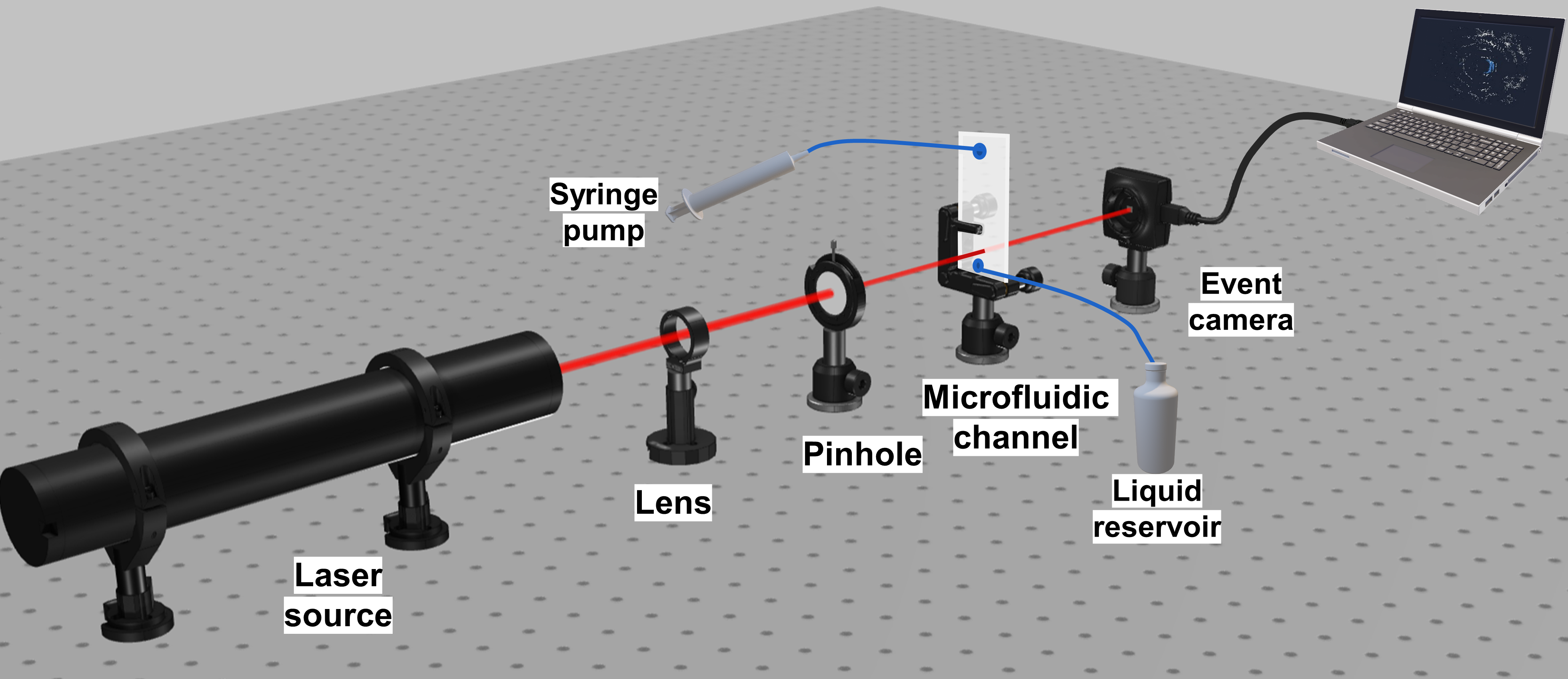
The experimental setup is shown in Figure 1. Here, we send artificial PMMA microbeads of different sizes to a narrow microfluidic channel (of width 200 ). The intensity of the light detected by the event-camera stays constant as long as no particle is moving across the field of view. This does not trigger any pixel to fire events. However, once particles enter the field of view,diffraction, scattering and interference of the light trigger firing events which we use later to train our spiking neural network. The fact that we obtain events only in the presence of particles significantly improves the memory efficiency of the overall system. The size of the dataset we get with our system is on the order of tens of gigabytes, whereas in a previous work using a traditional frame-based camera, the dataset was on the order of hundreds of gigabytes (Lugnan et al., 2020).
We used two different classes of spherical microparticles (class A of diameter 16 µm and class B with a diameter of 20 µm). We ran four separate experiments for each class of microparticles, where each experiment ran for for a total of 480 seconds of data. The accumulation time for a single particle is . Therefore, we have around 6,000 samples per experiment, or 24,000 samples in total per class. We train the network for four different train-test splits, each split using a different experiment as testing data, and the remaining experiments as training data.
2.2. Spiking neural network
We pre-process our event-based imaging data using the Tonic library (Lenz et al., 2021). The pre-processing involved a spatial downsampling from to (event polarity is left unchanged), and a temporal downsampling by passing the events for each neuron corresponding to a downsampled pixel of fixed polarity through a discretized leaky-integrate-and-fire (LIF) neuron:
where , , denote the membrane potential, binary spike output, and binary refractory period of neuron at timestep , respectively. Further, is the membrane decay rate, is the synaptic weight, is the threshold voltage, is the refractory period, equals unity if there is an input event from the event-based camera in the th patch of pixels at timestep (and zero otherwise).
For classification, we use a feedforward network of LIF neurons with an input layer of size , a hidden layer of size , and an output layer of size (using population coding with neurons per output class). The neuron parameters are constant for all neurons in the network, with the membrane decay rate and membrane threshold . We use the Adam optimizer to optimize the mean squared error on the output spike rate, with a desired population spike rate of (correct) and (incorrect). Backpropagation is applied through a shifted Heaviside function in the forward pass and a fast sigmoid
with slope in the backward pass (Zenke and Ganguli, 2018). We use snnTorch (Eshraghian et al., 2021) for our implementation.
3. Results
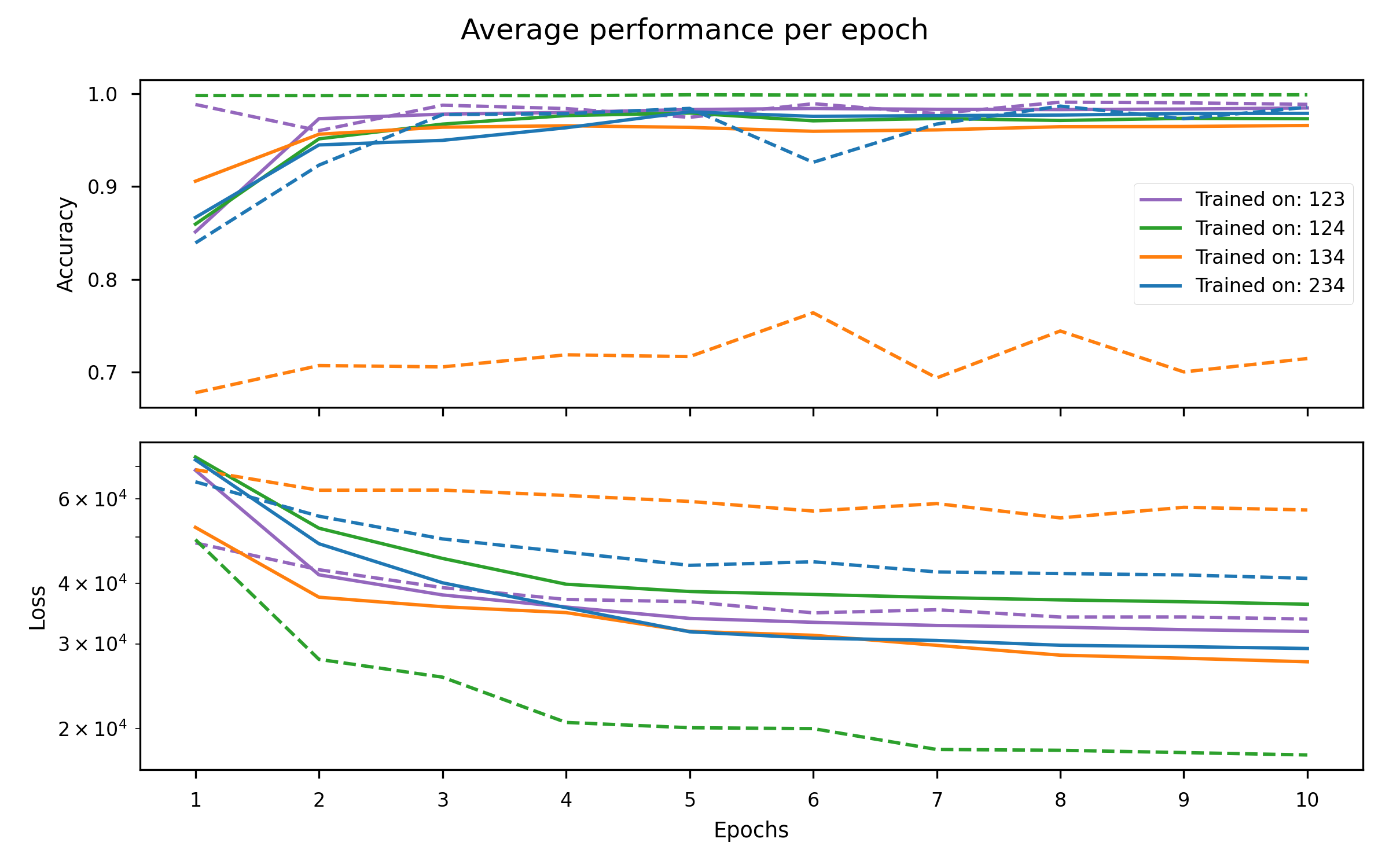
We trained the spiking neural network described in Section 2.2 for 10 epochs on a NVIDIA GeForce RTX 2080 Ti GPU with 11 GB of memory. Figure 2 show the training and testing performance for this experiment. The figure shows that the accuracy exceeds 98.5% for all experiments, with the exception of the testing accuracy for the network trained on experiments 1, 3, and 4. More work is needed to analyze the anomalous performance on this data split.
4. Outlook
As next steps, we are investigating the use of exact timing information for a time-to-first-spike classification that should yield even faster classification. In this work, we fixed the hyperparameters use to the values mentioned in Section 2.2. For further performance gains, we will optimize the hyperparameters and network architecture through a cross-validation procedure. Finally, we will transfer the SNN to a neuromorphic chip for a fully neuromorphic processing pipeline.
Acknowledgements.
This work was performed in the context of the European projects Neoteric (grant agreement 871330), Prometheus (grant agreement 101070195) and Post-Digital project (grant agreement 860360).References
- (1)
- Doan et al. (2018) Minh Doan, Ivan Vorobjev, Paul Rees, Andrew Filby, Olaf Wolkenhauer, Anne E Goldfeld, Judy Lieberman, Natasha Barteneva, Anne E Carpenter, and Holger Hennig. 2018. Diagnostic potential of imaging flow cytometry. Trends in Biotechnology 36, 7 (2018), 649–652.
- Eshraghian et al. (2021) Jason K. Eshraghian, Max Ward, Emre Neftci, Xinxin Wang, Gregor Lenz, Girish Dwivedi, Mohammed Bennamoun, Doo Seok Jeong, and Wei D. Lu. 2021. Training Spiking Neural Networks Using Lessons From Deep Learning. (2021). arXiv:2109.12894
- He et al. (2022) Weihua He, Yongxiang Feng, Junwen Zhu, Huichao Chai, and Wenhui Wang. 2022. Neuromorphic-Enabled Event-Based Deep Imaging Flow Cytometry. In 26th International Conference on Miniaturized Systems for Chemistry and Life Sciences.
- Lenz et al. (2021) Gregor Lenz, Kenneth Chaney, Sumit Bam Shrestha, Omar Oubari, Serge Picaud, and Guido Zarrella. 2021. Tonic: event-based datasets and transformations. Documentation available under https://tonic.readthedocs.io.
- Lugnan et al. (2020) Alessio Lugnan, Emmanuel Gooskens, Jeremy Vatin, Joni Dambre, and Peter Bienstman. 2020. Machine learning issues and opportunities in ultrafast particle classification for label-free microflow cytometry. Scientific Reports 10, 1 (2020), 1–13.
- Zenke and Ganguli (2018) Friedemann Zenke and Surya Ganguli. 2018. SuperSpike: Supervised Learning in Multilayer Spiking Neural Networks. Neural Computation 30, 6 (2018), 1514–1541.
- Zhang et al. (2022) Ziyao Zhang, Maria Sabrina Ma, Jason K. Eshraghian, Daniele Vigolo, Ken-Tye Yong, and Omid Kavehei. 2022. Work in Progress: Neuromorphic Cytometry, High-throughput Event-based flow Flow-Imaging. In 2022 8th International Conference on Event-Based Control, Communication, and Signal Processing (EBCCSP). IEEE.