Experimental determination of ruthenium L-shell fluorescence yields and Coster-Kronig transition probabilities
Abstract
The L-shell fluorescence yields and the Coster-Kronig factors of ruthenium (and the corresponding uncertainty) were determined for the first time experimentally by applying radiometrically calibrated instrumentation of the Physikalisch-Technische Bundesanstalt. The resulting fluorescence yields (, , ) and the Coster-Kronig factors (, , ) agree reasonable well with parts of the data from the literature.
Keywords ruthenium fluorescence yield Coster-Kronig fundamental parameter photon-in/photon-out experiment XRF
1 Introduction
Ruthenium is a versatile and widely used chemical element playing a crucial role in important areas of science and technology. Several applications in the area of semiconductor fabrication or catalysis can be identified, where ruthenium is essential. For extreme ultraviolet lithography masks [1, 2] or as interconnect metal [3, 4, 5] either ruthenium or ruthenium-containing materials are very relevant. In catalysis, ruthenium-based catalysts provide remarkable properties in several different applications [6]. In addition to this, ruthenium is also of relevance for emerging applications for energy storage [7, 8] and medicine [9, 10].
But, if X-ray fluorescence (XRF) based techniques are to be used for determining the ruthenium content in such materials, one quickly finds that the knowledge of the relevant atomic fundamental parameter (FP) data for ruthenium is very limited: For ruthenium, especially its L-shell FP data and namely the L-subshell fluorescence yields and Coster-Kronig factors (CK), no experimentally determined data seems to exist so far. Available data in the literature is either purely theoretically determined or perhaps even less favorable, only interpolated employing adjacent chemical elements. As these FPs quantitatively describe the process of X-ray fluorescence generation, they are very crucial for most quantification approaches in XRF. Thus, they have a direct influence on the accuracy of the XRF quantification results.
As this is a highly inadequate situation, we applied the PTB’s reference-free X-ray spectrometry toolset in order to experimentally determine the fluorescence yields and the Coster-Kronig factors of the -subshells of ruthenium for the first time. Based on transmission and fluorescence experiments on thin film samples, such FP data can be derived as already demonstrated for a wide range of chemical elements [11, 12, 13, 14, 15].
2 Experimental procedure
For an experimental determination of L-shell fluorescence yields and Coster-Kronig transition probabilities, both fluorescence- and transmission experiments with a selective excitation of the three L-subshells on either a free standing thin foil or a thin coating on a carrier foil are required [11, 12, 13, 15]. In the present work, these experiments were conducted on the four-crystal monochromator (FCM) beamline [16] of BESSY II using a vacuum chamber that is in-house developed [17]. This chamber was endowed with a silicon drift detector (SDD) of which the detection efficiency is radiometrically calibrated and the response functions are determined experimentally [18].
The employed sample was a highly homogeneous 150 nm ruthenium deposition on a 500 nm Si3N4 membrane. To be able to isolate the ruthenium contribution from the total sample transmission, also a blank membrane of nominally identical thickness was used. Any potential moderate variation in the Si3N4 membrane thickness is only a second-order contribution to the uncertainties. Both samples were positioned in the chamber’s center by using an x-y-scanning stage.
The angle between the incoming beam and the sample as well as the angle between sample surface and detector was set to 45°.
The transmission measurements were conducted in an energy range around the Ru-L absorption edges between 2.1 keV and 4 keV. Furthermore, X-ray fluorescence measurements were performed in the incident-energy domain between 2.8 keV and 3.4 keV. The established methodolgy [11, 19, 12, 20] to derive the relevant L-shell FPs from this exerimental dataset is described in the following.
According to the Sherman equation [21], the measured count rate of fluorescence photons of a one-elemental foil, which is irradiated under 45°, is the product of the fluorescence production cross section of the considered shell, the incoming as well as the fluorescence photon flux , the detection efficiency of the SDD, the mass deposition of that element, the attenuation correction factor and the solid angle of detection of the SDD.
The self-attenuation correction factor takes into account the attenuation of the incident radiation and of the fluorescence radiation on its way through the sample. The corresponding sample-specific attenuation correction factor is determined by transmission experiments taking advantage of the fact, that the knowledge of the ruthenium deposition thickness and its density is not needed since they appear only in a product with the mass absorption coefficient or with the subshell photoionization cross section . The product is derived from the transmittance data using the Lambert-Beer law.
For incident energies between the edge and the edge, the fluorescence production factor for the -subshell is
| (1) |
where is the ruthenium L3 fluorescence yield which should be determined. The sample-specific attenuation correction factor is defined as
| (2) |
Here, is the angle between the incident beam and the sample surface, is the angle between the sample surface and the SDD detctor.
Due to the so-called Coster-Kronig effect, the effective photoionization cross section for L3 and L2 is a linear combination with the higher bound shells since for photon energies above the excitation energy of the next subshell, created holes in can decay into by ejecting outer electrons. As a result, more than the directly created holes in exist. The CK-factor provides the probability for this to happen and similar transitions can occur between the and the and shells. So for an incident photon energy above the threshhold, the fluorescence production factors are defined as:
| (3) | ||||
| (4) | ||||
| (5) |
Here, the are the photoionization cross sections of the respective subshell[19], and are the Coster-Kronig factors. For incident energies below the subsequent subshell, the corresponding subshell photoionization cross section is zero ( for ). Therefore, the fluorescence yields are determined for energies above the excitation energy of the considered and below the subsequent subshell.
All relevant observables are accessible from the experimental data as are determined for the relevant energies by measuring the transmission of the ruthenium coating. is determined by spectral deconvolution of the recorded SDD spectra considering the relevant fluorescence lines and relevant background contributions such as bremsstrahlung. and are known because of PTB’s calibrated instrumentation [22].
Taking into account the theoretical ratio of scattering to ionization cross sections, which one can take from databases[23], the sample-specific total photoionization cross section can be derived. To isolate the subshell contributions of the different , Ebel polynomials [23] for each contribution as well as a total cross section for lower bound shells are scaled into the data (see figure 1). For this scaling process, only the datapoints slightly above each absorption edge are used to minimize the effect of the fine structure.
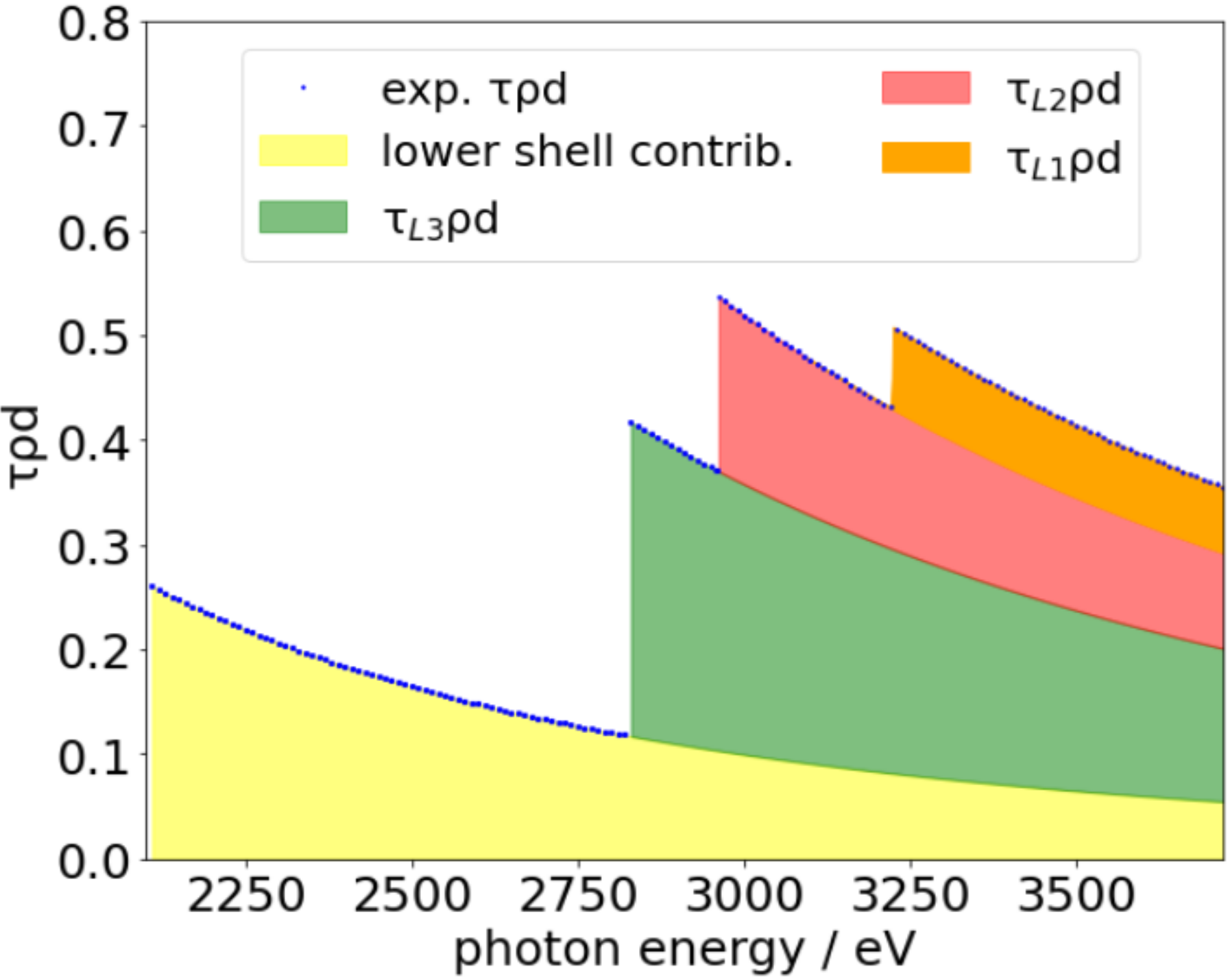
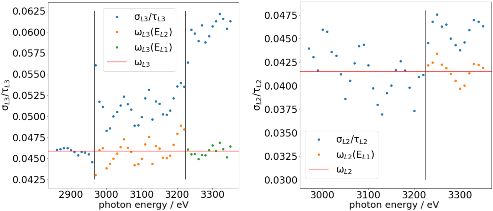
With these determined , the equations for the fluorescence production cross sections can be solved for . By replacing by the effective photoionization cross section according to equations 3-5, eqn 1 can be applied also for energies above the next subshell. Therefore, to determine , energies between and were considered, see figure 2: With the already determined , the modified version of eqn 1 can be solved for . is determined in the same way but applied for the fluorescence of the L2 shell and for with the already determined . With these determined and , from the fluorescence of the L3 shell for energies above , can be determined.
3 Results
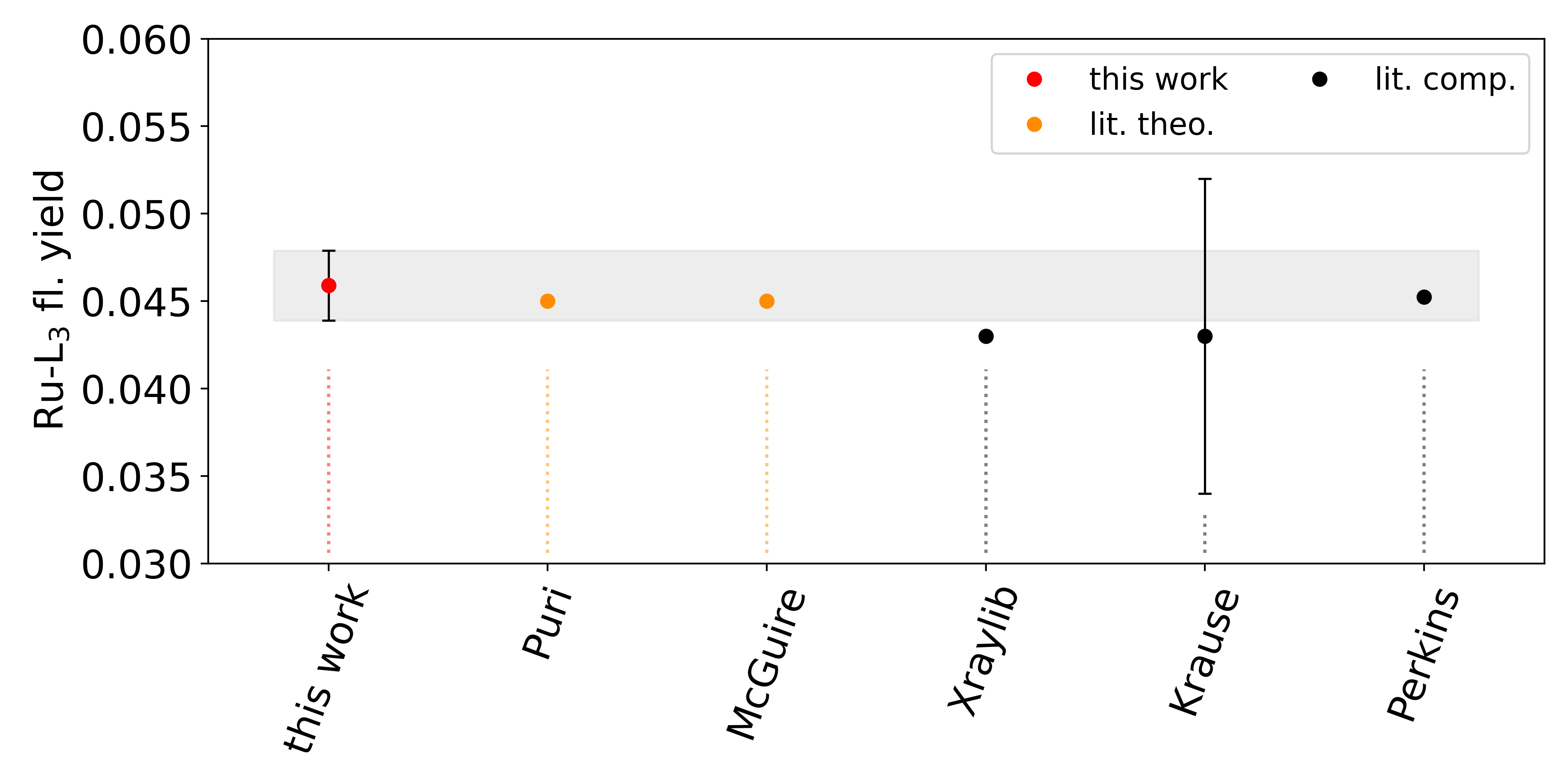
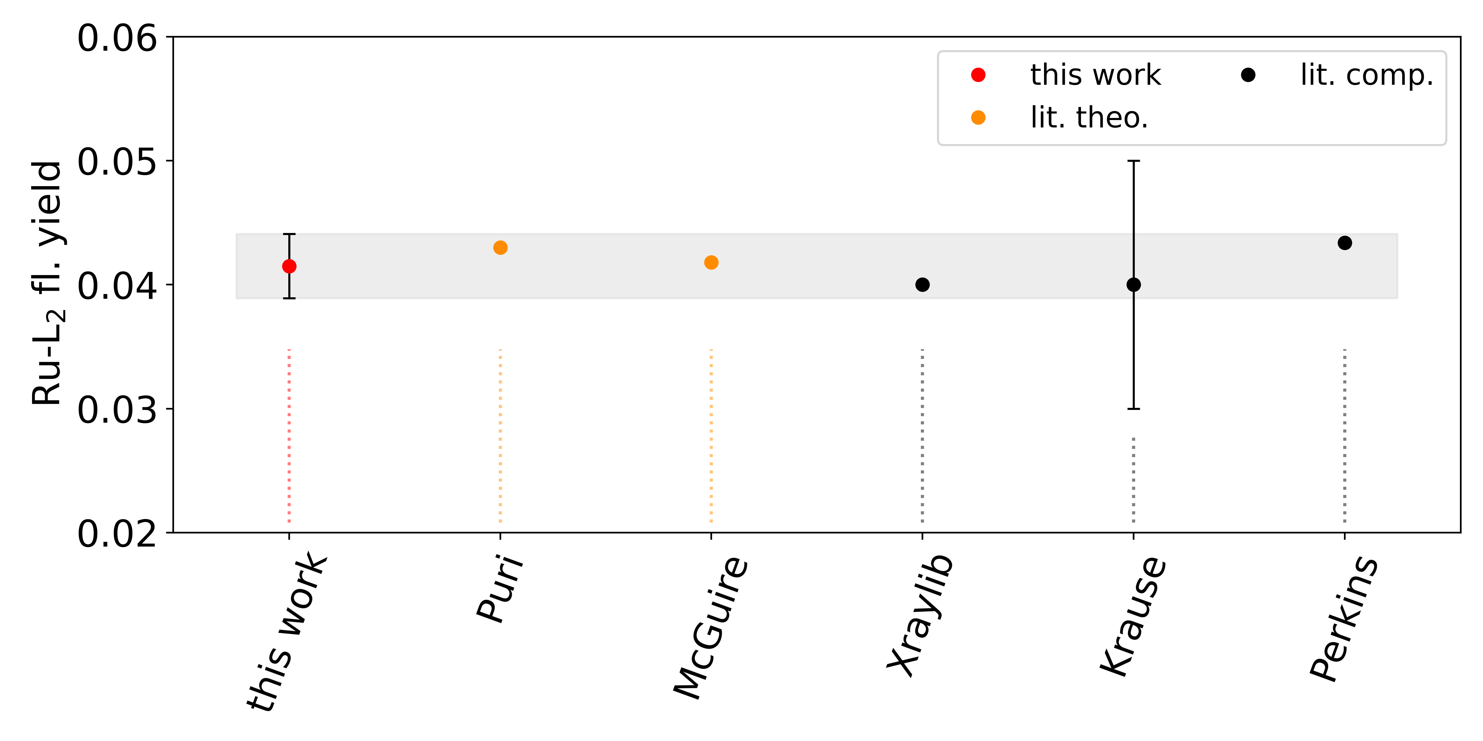
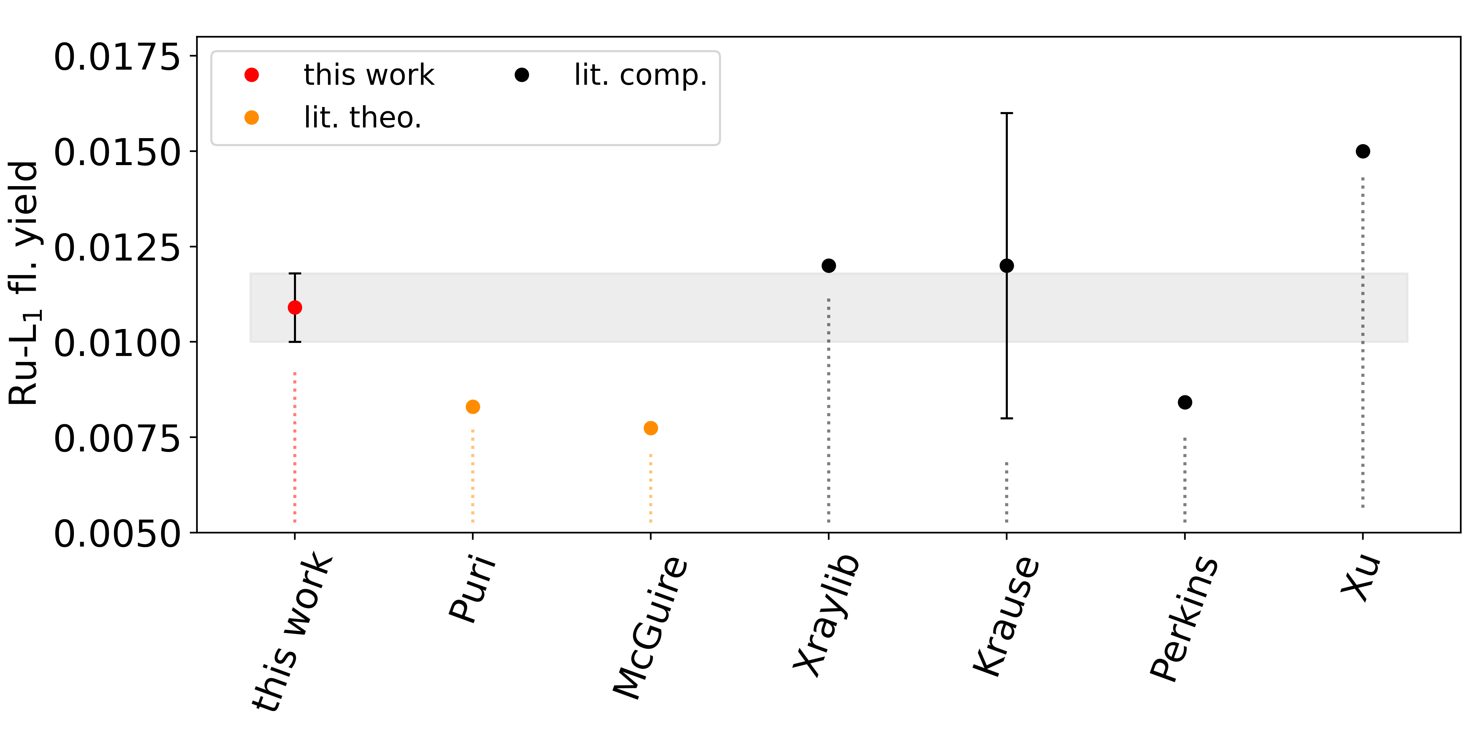
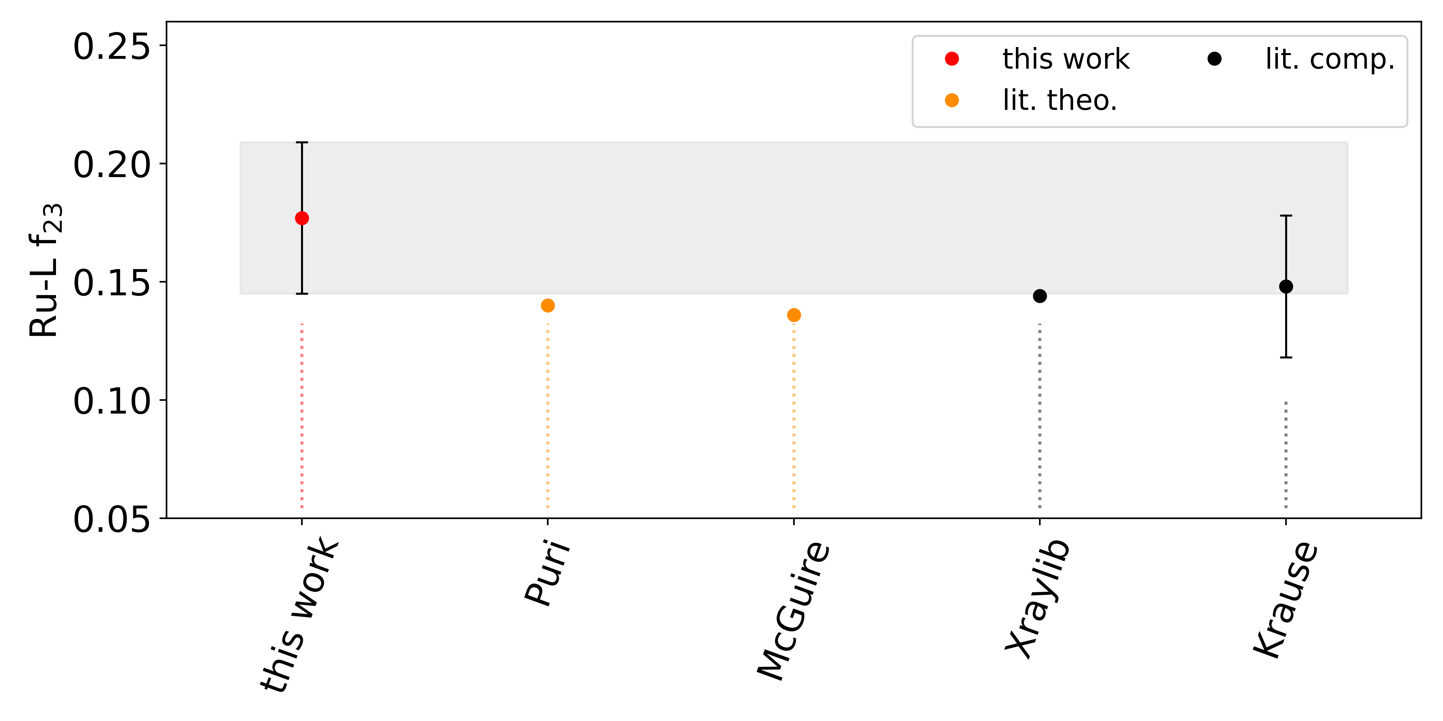
The determined fluorescence yields are , and . The resulting Coster-Kronig factors are , and . These values are compared with values from the literature in table 1 and figure 3 and 4. The respective uncertainties were calculated via error propagation. The main contributions to the total uncertainty budget of the fluorescence yields were arising from the spectral deconvolution (~2 %) and from the photoionization cross sections (~2 %). The uncertainty budget is calculated by applying the reference-free XRF approach for the FP determination, discussed in more detail in [24].
The X-raylib [25] and Krause[26] values of and are slightly outside of the error domain of the values determined in this work. The agreement with respect to the theoretically calculated data of Puri[27] and McGuire[28] is better in the case of but even worse for . The data of Perkins[29] as well as the data by Xu[30] behaves very similarly. For , all available data from the literature agrees well with the result obtained here. With respect to the Coster-Kronig factors, the tabulated data in X-raylib and the Krause compilation is in good agreement with our results. However, the results are on or slightly outside the boundary of our uncertainty budget for all three CK values. The data by McGuire and Puri is outside of our results considering their uncertainty budget.
| Ru | Ru | Ru | |
| this work (XRF) | 0.0459(20) | 0.0415(26) | 0.0109(9) |
| X-raylib [25] (comp.) | 0.043 | 0.040 | 0.012 |
| Krause [26] (comp.) | 0.043(9) | 0.040(10) | 0.012(4) |
| Perkins et. al. [29] (comp.) | 0.045231 | 0.043368 | 0.0084138 |
| McGuire [28] (theory) | 0.0450 | 0.0418 | 0.00774 |
| Puri et. al. [27] (theory) | 0.045 | 0.043 | 0.0083 |
| Xu et. al. [30] (comp.) | 0.015 | ||
| Ru | Ru | Ru | |
| this work (XRF) | 0.177(32) | 0.528(90) | 0.173(73) |
| X-raylib [25] (comp.) | 0.144 | 0.61 | 0.10 |
| Krause [26] (comp.) | 0.148(30) | 0.61(7) | 0.10(2) |
| McGuire [28] (theory) | 0.136 | 0.779 | 0.057 |
| Puri et. al. [27] (theory) | 0.140 | 0.766 | 0.057 |
4 Conclusion
The Coster-Kronig factors and the fluorescence yields of ruthenium are determined experimentally by applying PTB’s radiometrically calibrated instrumentation. The values determined are in reasonably good agreement with the values from the literature, although some literature values are slightly outside the uncertainty ranges of this work. The magnitude of the determined uncertainties of this work is much lower than the estimated uncertainties of Krause [26] in the case of the fluorescence yield values. With respect to the Coster-Kronig factors, similar uncertainties were achieved here. In summary, this uncertainty reduction will positively influence the total uncertainties of fundamental parameter-based quantitative X-ray fluorescence experiments.
As stated already in previous works of our group [14, 24, 31, 15], the X-raylib database is also in the case of the Ru-L shell fundamental parameters a reliable reference.
Conflict of interest
There are no conflicts to declare.
Acknowledgments
This project has received funding from the ECSEL Joint Undertaking (JU) IT2 under grant agreement No 875999. The JU receives support from the European Union’s Horizon 2020 research and innovation programme and the Netherlands, Belgium, Germany, France, Austria, Hungary, the United Kingdom, Romania and Israel.
References
- [1] Wu M, de Marneffe JF, Opsomer K, Detavernier C, Delabie A, Naujok P, et al. Characterization of Ru4-xTax (x = 1,2,3) alloy as material candidate for EUV low-n mask. Micro and Nano Engineering. 2021;12:100089. Available from: https://www.sciencedirect.com/science/article/pii/S2590007221000101.
- [2] Philipsen V, Luong KV, Opsomer K, Souriau L, Rip J, Detavernier C, et al. Mask absorber development to enable next-generation EUVL. In: Ando A, editor. Photomask Japan 2019: XXVI Symposium on Photomask and Next-Generation Lithography Mask Technology. vol. 11178. International Society for Optics and Photonics. SPIE; 2019. p. 111780F. Available from: https://doi.org/10.1117/12.2537967.
- [3] Wen LG, Adelmann C, Pedreira OV, Dutta S, Popovici M, Briggs B, et al. Ruthenium metallization for advanced interconnects. In: 2016 IEEE International Interconnect Technology Conference / Advanced Metallization Conference (IITC/AMC); 2016. p. 34–36.
- [4] Barmak K, Ezzat S, Gusley R, Jog A, Kerdsongpanya S, Khaniya A, et al. Epitaxial metals for interconnects beyond Cu. Journal of Vacuum Science & Technology A. 2020;38(3):033406. Available from: https://doi.org/10.1116/6.0000018.
- [5] Kondati Natarajan S, Nies CL, Nolan M. The role of Ru passivation and doping on the barrier and seed layer properties of Ru-modified TaN for copper interconnects. The Journal of Chemical Physics. 2020;152(14):144701. Available from: https://doi.org/10.1063/5.0003852.
- [6] Gramage-Doria R, Bruneau C. Ruthenium-catalyzed C–H bond functionalization in cascade and one-pot transformations. Coordination Chemistry Reviews. 2021;428:213602. Available from: https://www.sciencedirect.com/science/article/pii/S0010854520305865.
- [7] Hsu S, Rommel S, Eversfield P, Muller K, Klemm E, Thiel W, et al. A Rechargeable Hydrogen Battery Based on Ru Catalysis. Angewandte Chemie International Edition. 2014;53(27):7074–7078. Available from: https://onlinelibrary.wiley.com/doi/abs/10.1002/anie.201310972.
- [8] Liu T, Liu Z, Kim G, Frith JT, Garcia-Araez N, Grey CP. Understanding LiOH Chemistry in a Ruthenium-Catalyzed Li–O2 Battery. Angewandte Chemie International Edition. 2017;56(50):16057–16062. Available from: https://onlinelibrary.wiley.com/doi/abs/10.1002/anie.201709886.
- [9] Antonarakis E, Emadi A. Ruthenium-based chemotherapeutics: Are they ready for prime time? Cancer chemotherapy and pharmacology. 2010 03;66:1–9.
- [10] Allardyce CS, Dyson PJ. Ruthenium in medicine: current clinical uses and future prospects. Platinum Metals Reviews. 2001;45(ARTICLE):62.
- [11] Kolbe M, Hönicke P, Müller M, Beckhoff B. L-subshell fluorescence yields and Coster-Kronig transition probabilities with a reliable uncertainty budget for selected high- and medium-Z elements. Phys Rev A. 2012;86:042512.
- [12] Kolbe M, Hönicke P. Reliable determination of fundamental parameters of Zr and Ti for a reliable quantitative X-ray fluorescence analysis. X-Ray Spectrom. 2015;44(4):217–220.
- [13] Kayser Y, Hönicke P, Wansleben M, Wählisch A, Beckhoff B. Experimental determination of the gadolinium L subshells fluorescence yields and Coster-Kronig transition probabilities. X-Ray Spectrometry. 2022.
- [14] Hönicke P, Unterumsberger R, Wauschkuhn N, Krämer M, Beckhoff B, Indelicato P, et al. Experimental and theoretical approaches for determining the K-shell fluorescence yield of carbon. Radiat Phys Chem. 2022:110501.
- [15] Wauschkuhn N, Frenzel K, Beckhoff B, Hönicke P. Experimental determination of tantalum L-shell fluorescence yields and Coster–Kronig transition probabilities. J Anal At Spectrom. 2023;38:197–203. Available from: http://dx.doi.org/10.1039/D2JA00325B.
- [16] Krumrey M. Design of a Four-Crystal Monochromator Beamline for Radiometry at BESSY II. J Synchrotron Rad. 1998;5(1):6–9.
- [17] Kolbe M, Beckhoff B, Krumrey M, Ulm G. Thickness determination for Cu and Ni nanolayers: Comparison of reference-free fundamental-parameter based X-ray fluorescence analysis and X-ray reflectometry. Spectrochimica Acta B. 2005;60:505–510.
- [18] Scholze F, Procop M. Modelling the response function of energy dispersive X-ray spectrometers with silicon detectors. X-Ray Spectrom. 2009;38(4):312–321.
- [19] Hönicke P, Kolbe M, Müller M, Mantler M, Krämer M, Beckhoff B. Experimental verification of the individual energy dependencies of the partial L-shell photoionization cross sections of Pd and Mo. Phys Rev Lett. 2014;113(16):163001.
- [20] Ménesguen Y, Lépy MC, Sampaio JM, Marques JP, Parente F, Guerra M, et al. A combined experimental and theoretical approach to determine X-ray atomic fundamental quantities of Tin. X-Ray Spectrom. 2018;47(5):341–351.
- [21] Sherman J. The theoretical derivation of fluorescent X-ray intensities from mixtures. Spectrochim Acta. 1955;7:283–306.
- [22] Beckhoff B. Reference-free X-ray spectrometry based on metrology using synchrotron radiation. J Anal At Spectrom. 2008;23:845 – 853.
- [23] Ebel H, Svagera R, Ebel MF, Shaltout A, Hubbell JH. Numerical description of photoelectric absorption coefficients for fundamental parameter programs. X-Ray Spectrom. 2003;32:442–451.
- [24] Unterumsberger R, Hönicke P, Colaux J, Jeynes C, Wansleben M, Müller M, et al. Accurate experimental determination of Gallium K- and L3-shell XRF fundamental parameters. J Anal At Spectrom. 2018;33(6):1003–1013.
- [25] Schoonjans T, Brunetti A, Golosio B, del Rio MS, Solé VA, Ferrero C, et al. The xraylib library for X-ray–matter interactions. Recent developments. Spectrochim Acta B. 2011;66:776 – 784.
- [26] Krause MO. Atomic Radiative and Radiationless Yields for K and L shells. J Phys Chem Ref Data. 1979;8(2):307–327.
- [27] Puri S, Mehta D, Chand B, Singh N, Trehan PN. L shell fluorescence yields and Coster-Kronig transition probabilities for the elements with 25 < Z < 96. X-Ray Spectrom. 1993;22(5):358–361.
- [28] McGuire E. Atomic L-shell Coster-Kronig, Auger, and Radiative Rates and Fluorescence Yields for Na-Th. Phys Rev A. 1971;3(2):587.
- [29] Perkins ST, Cullen DE, Chen MH, Rathkopf J, Scofield J, Hubbell JH. Tables and graphs of atomic subshell and relaxation data derived from the LLNL Evaluated Atomic Data Library (EADL), Z = 1-100. UCRL 50400. 1991;30.
- [30] Xu J, Rosato E. Relative intensities of diagram and satellite L-X-rays for elements Z = 37 - 56. Journal de Physique. 1987;48(C-9):661–664.
- [31] Hönicke P, Kolbe M, Krumrey M, Unterumsberger R, Beckhoff B. Experimental determination of the oxygen K-shell fluorescence yield using thin SiO2 and Al2O3 foils. Spectrochim Acta B. 2016;124:94–98.