present address: ]Physik-Institut, Universität Zürich, Winterthurerstrasse 190, CH-8057 Zürich, Switzerland
Spin-orbit coupling induced Van Hove singularity in proximity to a Lifshitz transition in Sr4Ru3O10
Abstract
Van Hove singularities (VHss) in the vicinity of the Fermi energy often play a dramatic role in the physics of strongly correlated electron materials. The divergence of the density of states generated by VHss can trigger the emergence of new phases such as superconductivity, ferromagnetism, metamagnetism, and density wave orders. A detailed understanding of the electronic structure of these VHss is therefore essential for an accurate description of such instabilities. Here, we study the low-energy electronic structure of the trilayer strontium ruthenate \ceSr4Ru3O_10, identifying a rich hierarchy of VHss using angle-resolved photoemission spectroscopy and millikelvin scanning tunneling microscopy. Comparison of -resolved electron spectroscopy and quasiparticle interference allows us to determine the structure of the VHss and demonstrate the crucial role of spin-orbit coupling in shaping them. We use this to develop a minimal model from which we identify a new mechanism for driving a field-induced Lifshitz transition in ferromagnetic metals.
I Introduction
Van Hove singularities (VHss) in the density of states appear due to stationary points, , in the quasiparticle dispersion relation [1]. They are a direct consequence of the periodicity of the crystal lattice, appearing naturally at high symmetry points in the Brillouin zone (BZ), but also away from these points due to higher order hopping terms and band hybridizations. The consequences of a VHs for the properties of a material are intricately linked to the abrupt change or divergence in density of states associated with it. In two-dimensional systems, band minima or maxima lead to a step change in the density of states while saddle points result in logarithmic or higher order divergences dependent on their symmetry and parameters of the band structure [2]. Tuning such a density of states divergence through the Fermi energy can be expected to drive electronic instabilities, concomitant with the resulting Lifshitz transition where the topology of the Fermi surface changes. Indeed, many properties of strongly correlated electron materials have been associated with VHss and accompanying Lifshitz transitions, including, e.g. metamagnetic transitions in heavy fermion systems [3, 4, 5], the pseudogap phase [6, 7, 8] and high- superconductivity [9] in cuprates, the interplay between superconducting and Mott insulating states in twisted bilayer graphene [10, 11, 12, 13] and charge density wave formation and superconductivity in kagome materials [14, 15].
There is thus a need to identify model systems through which to develop a coherent understanding of the role of VHss in shaping the collective states of quantum materials. A particularly promising system is the Ruddlesden-Popper series of strontium ruthenates \ceSr_n+1Ru_n O_3n+1, which are known to exhibit multiple VHss close to the Fermi energy [16, 17]. While the member, \ceSrRuO3, is a ferromagnet [18], the first two members with and are non-magnetic. \ceSr2RuO4 () hosts unconventional superconductivity which can be markedly enhanced by uniaxial pressure [19], concomitant with a VHs being driven through the Fermi level [20]. \ceSr3Ru2O7 () is on the verge of magnetism, exhibiting strong ferromagnetic fluctuations close to criticality and a metamagnetic transition in applied magnetic field [21]. Unusual scaling relations in its thermodynamic properties have, in turn, been linked to the presence of higher-order VHss near the Fermi level [17]. The member, \ceSr4Ru3O_10, is the first member in which a bulk ferromagnetic ground state is realized.[22] Its crystal structure consists of trilayers of \ceSrRuO3 stacked along the -axis, connected by the apical oxygen of the \ceRuO6 octahedra (Fig. 1a). These \ceRuO6 octahedra are furthermore rotated around the -axis, leading to an orthorhombic unit cell.
Sr4Ru3O_10 undergoes a ferromagnetic transition with a Curie temperature K (Fig. 1b), with the magnetization aligned parallel to the -axis [22]. At K, a secondary peak in the magnetic susceptibility is observed which is associated with a metamagnetic transition triggered by in-plane magnetic field [22, 23], and accompanied by an increase in the volume of the unit cell[24, 25]. For magnetic fields applied in the -plane decreases, reaching K at T [26]. A link between the metamagnetic properties and VHss in proximity of the Fermi energy has been postulated [27], however direct experimental evidence for this is so far lacking [28, 29, 30].
Here, in a combined study of \ceSr4Ru3O_10 by angle-resolved photoemission spectroscopy (ARPES) and scanning tunneling microscopy and spectroscopy (STM/STS), we provide a comprehensive picture of the low-energy electronic structure, identifying multiple VHss in the vicinity of the Fermi level. Relating the spectral function measured by ARPES and quasiparticle interference (QPI) reveals that the VHs closest to the Fermi energy emerges due to a combined influence of octahedral rotations and spin-orbit coupling. Our results suggest a mechanism for the metamagnetic transition where the magnetization direction in conjunction with spin-orbit coupling drives a field-induced Lifshitz transition.
II Results
II.1 Surface electronic structure
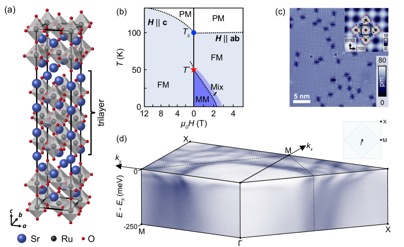
Due to the layered structure of the strontium ruthenates, cleavage results in atomically clean and flat \ceSrO-terminated surfaces[31, 32, 33, 34, 35, 29]. Fig. 1c shows such a \ceSrO surface of \ceSr4Ru3O_10. We find large terraces with only a few defects. Two types of defects centred in the hollow site between Sr atoms can be seen in Fig. 1c: defects with a symmetry that we associate with substitutional Ru site defects, and cross-like defects with symmetry. The substitutional Ru site defects occur with two different chiralities due to the octahedral rotations. The cross-like defects can be attributed to \ceCO2 complexes resulting from the chemisorption of CO molecules at the surface [36]. Both types of defects act as scattering centres, giving a strong signal for QPI which we discuss below. From topographies with lateral sizes larger than nm, we determine a density of surface defects of . The inset of Fig. 1c shows a magnified view, where the square atomic lattice centred on the Sr atoms is visible. Density functional theory (DFT) calculations suggest that the oxygen octahedra in the surface layer exhibit a larger octahedral rotation angle compared to the bulk, close to (Suppl. Fig. S2), although not resulting in additional periodicities. Consistent with the calculations, we do not see any evidence for a surface reconstruction.
An overview of the electronic structure, as obtained by ARPES, is shown in Fig. 1d. The data indicates a complex multi-band fermiology in this compound, consistent with other reports [28, 30]. Strong matrix element variations are found throughout the tetragonal Brillouin zone. Large nearly square-shaped electron pockets are visible around the Brillouin zone centre, whose corners reach approximately to the orthorhombic Brillouin zone boundary along the direction (). These are in good qualitative agreement with calculations of the expected spin-minority Fermi surfaces [29, 37]. Around the corners of the tetragonal Brillouin zone, we observe a complex set of additional intertwined Fermi pockets. These have small matrix elements close to the tetragonal Brillouin zone centre, although replicas of these states are also visible close to the Brillouin zone centre at selected photon energies (see, e.g., Suppl. Fig. S3).
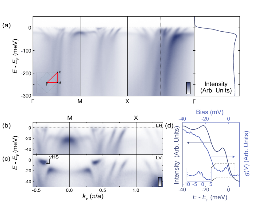
To investigate the electronic structure from which this complex Fermiology derives in detail, we show in Fig. 2a dispersions measured along the high-symmetry path of the tetragonal Brillouin zone. Consistent with the large number of Fermi pockets, we find a complex multi-band electronic structure. Sharp quasiparticles are visible within of the Fermi level, but the spectral features quickly broaden with increasing binding energy, and the measured states show kink-like features around this energy scale, with a reduction in quasiparticle velocity at the Fermi level. A similar phenomenology has been observed in the single-layer compound [38], where the associated deviations in linearity of the real part of the self-energy calculated by dynamical mean-field theory (DMFT) have been attributed to a crossover from a Fermi liquid to a more incoherent regime. In this respect, we note that \ceSr4Ru3O10 is known to host a Fermi liquid ground state, with a temperature dependence of its resistivity which persists up to comparable energy scales as in \ceSr2RuO4 [39].
II.2 Low energy electronic structure and hybridization gaps
Beyond this conceptual similarity, however, we note that there are significantly more low-energy states within meV of the Fermi level in \ceSr4Ru3O10 than in \ceSr2RuO4. Notably, at an energy scale of meV, we find several rather flat band features that contribute a high density of states near the Fermi level (Fig. 2a). The state at at a binding energy of has previously been observed, and assigned as part of the spin-majority states from spin-resolved photoemission [30]. In addition, our measurements reveal a rich hierarchy of VHss around the point at the Brillouin zone face, shown in more detail in Fig. 2b, c.
At at the M-point, the top of a hole band and bottom of a weakly-dispersing electron band intersect (Fig. 2b). This reflects the folding of two VHss onto each other due to the octahedral rotations in this structure, which render the and directions equivalent within the orthorhombic Brillouin zone. Interestingly, a clear hybridization gap develops along the electron-like part of its dispersion, located at a momentum of . This is particularly evident when measured using linear-vertical polarization (Fig. 2c).
These features are also evident in -integrated measurements of the ARPES intensity of Fig. 2c, plotted in Fig. 2d. Two peaks are resolved, one at below the Fermi level and one which is peaked at below , being cut off by the Fermi function. They correspond to the high-intensity hybridized/gapped states seen in Fig. 2c about . Comparison of the -integrated intensity to high-resolution tunneling spectra measured at , Fig. 2d, reveals that the lower binding energy feature is in fact composed of multiple contributions, with a well-defined two-peak structure observed within a bias voltage range of to , shown in the inset.
The gap seen in ARPES (Fig. 2c) can be conceptually understood as emerging from the crossing of two bands, which hybridize to form a gap as illustrated in Fig. 3a. This results in the formation of two new VHss in the band structure, one at the top and one at the bottom edge of the hybridisation gap (orange lines). The uppermost one is extremely close to the Fermi energy and should therefore be the most relevant VHs for the metamagnetic properties of \ceSr4Ru3O10. To identify the type of VHs which forms, we thus perform QPI measurements within mV around the Fermi level.
Fig. 3b shows the Fourier transform of a map in zero magnetic field measured just below the Fermi level at mV. Several characteristic scattering vectors are observed that are consistent with previous work [29]. Specifically, the three rings centered at with can be related to bands of minority-spin character[29, 37]. We extract the positions of two of these rings, red and green circles, corresponding to intra-band scattering, while the middle ring corresponds to inter-band scattering between the two. Converting these scattering vectors (indicated by arrows in Fig. 3b) into -space (Fig. 3c), we find excellent agreement with the large electron Fermi surfaces observed in our ARPES measurements. We also identify two additional sharp scattering vectors in Fig. 3b, whose positions are shown by blue and orange circles. When transforming their positions into -space, we find that they match the tips of the leaves of the clover-shaped Fermi pocket centered at X. The scattering vectors in -space corresponding to the QPI patterns identified in Fig. 3b are shown as arrows with the corresponding colors in Fig. 3c, and yield Fermi contours which are again in excellent agreement with our ARPES measurements.
Building on this agreement, we now turn to identifying signatures of the near- VHs evident in the ARPES measurements. Signatures of VHss in QPI are expected to appear as distinct scattering patterns near and close to the atomic peaks [40]. Indeed, we observe a set of QPI features close to the atomic peaks at and (yellow arcs in Fig. 3b). Close-ups around the atomic peak at (Fig. 3b1, b2) reveal a change in the orientation of the dominant scattering pattern between mV, Fig. 3b1, and , Fig. 3b2. Line cuts along the (red arrow) and (blue arrow) directions (Fig. 3d, e) exhibit a hole-like dispersion along with a maximum at , and an electron-like dispersion with a minimum at the same energy along . The change in sign of the curvature between the and directions allows us to identify with a saddle point VHs. Consistent with that assignment, the spatially-averaged spectrum (Fig. 3f) shows a peak at , the same energy at which the dispersions collapse onto the atomic peak. When applying an out-of-plane magnetic field of T, we find that the observed dispersion of the VHs moves to lower energies (Fig. 3g-i) by meV, behaviour that is indicative of the states being of spin-majority character.
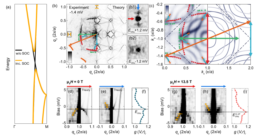
To connect the observation of the hybridization gap close to the Fermi level by ARPES with the saddle-point VHs identified in QPI, we develop a minimal model based on a ferromagnetic monolayer of \ceSr2RuO4. We start with a model for the band structure of \ceSr2RuO4 with octahedral rotation, as guided by our DFT calculations. We introduce ferromagnetism along the -axis and include spin-orbit coupling (SOC), matching the relevant parameters to the band structure obtained from ARPES and STM. This captures key features of the electronic structure close to , in particular reproducing the saddle point VHs at , and yielding a hybridization gap that opens away from the point when SOC is included (Fig. 3a) as well as a Fermi surface consistent with QPI and with many of the pockets observed by ARPES (dark solid lines in Fig. 3c). These features are also captured in DFT calculations of bulk ferromagnetic \ceSr4Ru3O_10,[37] which display qualitative similarities with our broad-scale measured dispersions in ARPES (Fig. 2). However, the location of the VHss depends sensitively on details of the DFT calculation, where exchange splitting is typically overestimated [30], and disentangling the correct low-energy electronic structure from such calculations is challenging in this complex, multi-band system. Our minimal model instead provides a simplified description of the relevant physics.
For comparison with experimental data, we calculate the QPI patterns using the continuum LDOS (cLDOS) method[41, 42] (see methods). The calculations reveal distinct QPI patterns for the SOC-induced VHs, showing excellent agreement with the experimental data. The calculated QPI map is shown in the right half of Figs. 3(b, d and e), where we clearly observe features related to the VHs that is situated just below the Fermi energy. Taken together, our spectroscopic measurements and model calculations thus allow us to identify that the VHs in the immediate vicinity of the Fermi energy arises as a result of a spin-orbit coupling induced gap in the band structure, rather than one of the VHss at the zone face of the underpinning non-relativistic band structure. A similar spin-orbit coupling induced gap is found in the surface layer of \ceSr2RuO4[43, 44], where octahedral rotations, similar to those in the bulk of \ceSr4Ru3O10, arise as a consequence of a surface reconstruction[45].
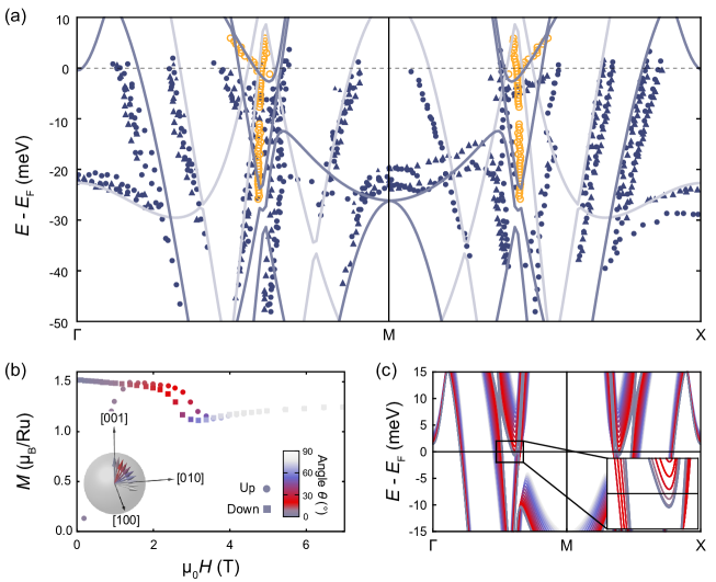
While the QPI data is very well described by a model of the band structure accounting only for a single layer of octahedra, such a minimal model (dark blue lines in Fig. 3c and Fig. 4(a)) only captures approximately half of the features seen in ARPES. This difference can be ascribed to the different probing depths of the two methods: while STM is sensitive only to the electronic states in the top-most layer, ARPES can probe electronic states arising within the first few layers, and thus can be expected to be sensitive to the trilayer-split electronic states expected for the multi-layer structure of \ceSr4Ru3O10. In fact, we find a generally good agreement with both the extracted dispersions (Fig. 4a) and our measured Fermi surfaces (Fig. 3c) if we take two copies of our minimal model, one as fit to the QPI data (dark blue lines), and another shifted by 40 meV (light blue lines) to account for the different octahedral rotation and splitting due to interlayer hopping. This reproduces the hole-like states with the smallest around the -point associated with another saddle-point VHs located above here, and also well reproduces the flat bands observed near .
III Discussion
From the ARPES and QPI data and comparison with the minimal tight-binding model for the band structure, we have shown that – from a hierarchy of VHss that are formed in the low-energy electronic structure of \ceSr4Ru3O10 – the most relevant is a saddle point VHs. This is created by spin-orbit coupling, resulting in the hybridization of a light spin-minority band with a heavy spin-majority band (Suppl. Fig. S6), and generating a VHs which lies extremely close to the Fermi energy. Given its proximity to , this is a prime candidate for driving the metamagnetic behaviour of \ceSr4Ru3O10.
How a VHs influences macroscopic properties, however, depends on the form of the density of states divergence associated with the VHs, which in turn is known to depend sensitively on the details of the band dispersion and its symmetry [17, 47, 2]. A full understanding of the origin of the VHs allows determining its impact on the density of states and potentially thermodynamic properties. From our study of the low-energy electronic structure by ARPES and QPI, we can deduce that the VHs closest to the Fermi energy is a saddle point VHs with two-fold symmetry originating from a hybridization gap induced by the interplay of spin-orbit coupling and the octahedral rotations. This is different from the case of \ceSr3Ru2O7, where it has been suggested that the VHs acquires four-fold symmetry and is of higher order due to the Brillouin zone folding[17]: here the VHs remains unaffected by the folding.
Our detailed understanding of the VHs here furthermore allows us to explore the effect of an applied magnetic field on the electronic structure using our minimal model. For an out-of-plane magnetic field, because the VHs is due to hybridization between a spin-majority and a spin-minority band, its energy shift with magnetic field does not behave simply as might be expected for a Zeeman-like behaviour, but is dominated by how the crossing point between the two bands changes in field. This can result in apparent -factors which acquire any value between zero and infinity even in the absence of correlation effects, depending on the relative slope of the two bands which hybridize. Here, due to the heavier nature of the spin-majority band, the VHs moves away from the Fermi energy with magnetic field. We can furthermore consider the effects of an in-plane magnetic field, where a rich phase diagram is known to result (Fig. 1b). We can model this by considering the effect of the magnetization rotating away from the -axis. We extract the magnetization direction from measurements performed in a field at an angle of from the -axis (Fig. 4b, data reproduced from ref. 46). In zero magnetic field, the magnetization is along the -axis, whereas an in-plane magnetic field tilts the magnetization from out-of-plane towards the in-plane direction by an angle . The metamagnetic transition in \ceSr4Ru3O10, detected in the magnetization, occurs when the magnetization exhibits an angle of with respect to the -axis, as indicated by the red colour in Fig. 4b.
We introduce the magnetization direction into our minimal model, resulting in the Hamiltonian[48, 49]
| (1) |
where is the paramagnetic tight-binding model, the exchange splitting, a vector of unit length pointing in the magnetization direction and the spin-orbit coupling constant. Fig. 4c shows the resulting change of the band structure between out-of-plane () and in-plane () direction of the magnetization. With increasing angle , the VHs at the top of the spin-orbit coupling induced gap is pushed across the Fermi energy. This change in energy is a direct consequence of the spin-orbit coupling term, , and the orbital character of the / VHs at the M point. For , the and terms result in hybridization between the / and the VHs of the same spin character, pushing the / VHs up in energy. This hybridization vanishes for . The spin-orbit coupling induced gap shifts with the / VHs as the magnetization is rotated. For the parameters of the tight-binding model determined by fitting the QPI dispersions, the VHs already traverses the Fermi energy for between the magnetization and the -axis. Such a scenario is also consistent with specific heat data, which show a peak in the temperature dependence that moves towards lower temperatures with increasing in-plane field[50], suggesting a VHs approaching the Fermi energy. We expect that a similar mechanism is relevant for the field-induced Lifshitz transition observed in the surface layer of \ceSr3Ru2O7.[35]
IV Conclusions
From ARPES and QPI measurements, we detect multiple VHss in the low-energy electronic structure of \ceSr4Ru3O10. The combination of the two techniques enables us to map out the nature, symmetry, and spin character of the VHs that is closest to the Fermi energy. Our data suggests that \ceSr4Ru3O10 is on the verge of a Lifshitz transition, similar to what has been proposed for \ceSr3Ru2O7, however here the transition is not triggered by out-of-plane fields due to the dominant spin-majority character of the VHs. Our results highlight the role of spin-orbit coupling induced hybridization gaps and VHss for the metamagnetic properties of \ceSr4Ru3O10. An important role of VHss was previously proposed from magnetization measurements [27], however lacking spectroscopic evidence. We expect that our results, with further studies of the in-plane field-dependence of the VHss identified here, will enable a full microscopic understanding of metamagnetism in \ceSr4Ru3O10.
V Acknowledgments
CAM, MN, PAEM, PDCK and PW gratefully acknowledge funding from the Engineering and Physical Sciences Research Council through EP/R031924/1, EP/S005005/1 and EP/T02108X/1, GRS and PDCK from the European Research Council (through the QUESTDO project, 714193), IB and SB through the International Max Planck Research School for Chemistry and Physics of Quantum Materials, and LCR from a fellowship from the Royal Commission of the Exhibition of 1851. RF, RA, ML and AV thank the EU’s Horizon 2020 research and innovation program under Grant Agreement No. 964398 (SUPERGATE). MH and JC thank the Swiss National Science Foundation for support. We gratefully acknowledge MAX IV Laboratory for time on the Bloch beamline under Proposal Nos. 20210763 and 20210783, which contributed to the results presented here. This work used computational resources of the Cirrus UK National Tier-2 HPC Service at EPCC (http://www.cirrus.ac.uk) funded by the University of Edinburgh and EPSRC (EP/P020267/1) and of the High-Performance Computing cluster Kennedy of the University of St Andrews. We also gratefully acknowledge Diamond Light Source (Proposal No. SI28412) and the Swiss Light Source (Proposal No. 20181951) where some preliminary data was obtained, and we thank Matt Watson for assistance.
Author contributions: CAM and WO performed STM measurements and analyzed the STM data, with additional supporting measurements by IB and MN. PAEM, BE, GRS, SB, EAM, and PDCK performed the ARPES measurements, which were analysed by PAEM. CP and ML maintained the Bloch beamline and provided experimental support. DH, MH and JC performed preliminary measurements. PAEM prepared the ARPES-related figures. IB performed and analyzed the magnetization measurements. IB and LCR performed the DFT calculations, LCR led DFT modelling, performed DFT relaxations for the surface and projected the tight-binding model. CAM, WO and PW carried out continuum LDOS calculations. CAM prepared the STM-related figures. RF, RA, ML, VG and AV grew and characterized the samples. CAM, PAEM, PDCK and PW wrote the manuscript. PW and PDCK initiated and supervised the project.
Competing interests: The authors declare no competing financial interests.
Data Availability: Underpinning data will be made available at [51].
Materials and Methods
V.1 Single crystal growth
Single crystals of \ceSr4Ru3O10 were grown by the floating-zone method with Ru self-flux as described in Ref. 52. The feed rods were prepared by a standard solid-state reaction subjecting mixed \ceSrCO3 () and \ceRuO2 ( purity) to repeated thermal cycles. An amount of excess \ceRuO2 was added to the starting powders to compensate the evaporation of Ru from the melting zone during the growth. To assess the sample quality, x-ray diffraction, energy and wavelength dispersive spectroscopy as well as electron backscattered diffraction were performed. The samples for the experiments were shaped in small rectangular pieces with average size of mm3. The sample used in this work was from the same batch as the samples used in Ref. [29].
V.2 Angle-resolved Photoemission
ARPES measurements were performed at the Bloch beamline of the MAX IV synchrotron. Measurements were performed with photon energies ranging from 21 eV to 67 eV using both linear horizontal and linear vertical polarized light. The probing spot size at Bloch is allowing for sample regions of greatest uniformity and quality to be probed. Samples were mounted on a six-axis manipulator and cooled to 20 K. Samples were cleaved in-situ at base temperature, and measured using a Scienta DA30 electron analyser, with the measurement parameters as described in the figure captions.
V.3 Scanning Tunneling Microscopy
Scanning tunneling microscopy measurements were performed using a home-built microscope operating in a dilution refrigerator at temperatures below .[53] All measurements shown in this manuscript have been acquired at unless stated otherwise. The bias voltage is applied to the sample, with the tip at virtual ground. The differential conductance has been measured using a lock-in technique, applying a modulation to the bias voltage and detecting the signal in the current ( Hz). The amplitude of the lock-in modulation used for the measurements is provided in the figure captions. Samples were cleaved in-situ at low temperatures and directly inserted into the STM head. Details of data processing of QPI data are shown in Suppl. Sections S4 and S8.
V.4 DFT calculations and minimal model
For the minimal model reproducing our quasiparticle interference data, we use a tight-binding model derived from a Density Functional Theory calculation of a monolayer of \ceSr2RuO4, with 15Å of vacuum. This model consists of two Ru atoms per unit cell and has a rigid octahedral rotation between them, similar to the model presented in Ref. [40]. The DFT calculations were performed using Quantum Espresso [54] on an -grid with a wavefunction cutoff of Ry and a charge density cutoff of Ry. We used the Perdew–Burke–Ernzerhof (PBE) exchange correlation functional. The tight-binding model was generated by projecting the Ru , and weight from the DFT calculation onto an orthonormal basis using a modified version of Wannier90 [55] to preserve the relative sign of the localized wave functions. The disentanglement was performed within a window of [-2.5,0.6] eV relative to the Fermi level, including a frozen window of [-1.45,0.33] eV. For further details please refer to the input files at Ref. [51]. For the minimal model, the DFT-derived tight-binding model is first symmetrized, we then introduce an exchange splitting of eV for the / bands and eV for the band. We add a local spin-orbit coupling term with meV. The chemical potential is adjusted by for the / bands and by for the band. Finally, all bands were renormalized by a factor of to match the experimental dispersion.
V.5 Continuum LDOS calculations
We model the QPI using the continuum Green’s function method [41, 40, 42]. From the tight-binding model introduced in methods section V.4, we calculate the momentum space lattice Green’s function of the unperturbed host,
| (2) |
where define the momentum and energy, and are the eigenvectors and eigenvalues of the tight-binding model with band index and spin , and is an energy broadening parameter. We then Fourier transform to obtain the unperturbed real space lattice Green’s function, , and follow the usual -matrix formalism to obtain the Green’s function of the system including an impurity from
| (3) |
where the -matrix
| (4) |
describes the scattering at the impurity. Here we consider a point-like defect with equal scattering strength in the spin-up and spin-down channel, such that .
To realistically model the QPI such that we can compare with experiment, we use the continuum Green’s function approach [41, 40, 42], which defines the Green’s function in terms of the continuous spatial variable as
| (5) |
where are the Wannier functions connecting the local and lattice space. The quasiparticle interference is then obtained from
| (6) |
For the calculations shown here, the tight-binding model and the Wannier functions are obtained from DFT calculations discussed in methods section V.4. We performed the Fourier transform of the lattice Greens function over a 2048 2048 -grid, with an energy broadening of . An impurity potential of eV was used and the real space local density of states in Eq. (6) was simulated for unit cells with the impurity in the centre, and with 4 pixels per unit cell. QPI calculations were done using the St Andrews calcqpi code.[29, 49] The resulting map was Fourier transformed to simulate the QPI map, . In the experimental data, the QPI maps contain contributions from the scattering from defects on the two Ru sites with opposite octahedral rotations. To account for this in the calculations, we average over maps calculated with the defect positioned in either site to obtain maps as shown in Fig. 3.
References
- Hove [1953] L. Van Hove. The Occurrence of Singularities in the Elastic Frequency Distribution of a Crystal. Physical Review, 89:1189–1193, 1953. doi: 10.1103/physrev.89.1189.
- Chandrasekaran et al. [2020] Anirudh Chandrasekaran, Alex Shtyk, Joseph J Betouras, and Claudio Chamon. Catastrophe theory classification of Fermi surface topological transitions in two dimensions. Physical Review Research, 2:013355, 2020.
- Daou et al. [2006] R. Daou, C. Bergemann, and S. R. Julian. Continuous evolution of the Fermi surface of CeRu2Si2 across the metamagnetic transition. Physical Review Letters, 96:026401, 2006.
- Hackl and Vojta [2011] A. Hackl and M. Vojta. Zeeman-driven Lifshitz transition: A model for the experimentally observed fermi-surface reconstruction in YbRh2Si2. Physical Review Letters, 106:137002, 2011.
- McCollam et al. [2020] A. McCollam, M. Fu, and S. R. Julian. Lifshitz transition underlying the metamagnetic transition of UPt3. Journal of Physics: Condensed Matter, 33:075804, 2020. doi: 10.1088/1361-648x/abc729.
- Storey et al. [2007] J.G. Storey, J.L. Tallon, and G.V.M. Williams. Saddle-point van Hove singularity and the phase diagram of high- cuprates. Physical Review B, 76:174522, 2007.
- Doiron-Leyraud et al. [2017] N Doiron-Leyraud, O Cyr-Choinière, S Badoux, A Ataei, C Collignon, A Gourgout, S Dufour-Beauséjour, FF Tafti, F Laliberté, M-E Boulanger, et al. Pseudogap phase of cuprate superconductors confined by fermi surface topology. Nature communications, 8(1):2044, 2017.
- Wu et al. [2018] Wei Wu, Mathias S. Scheurer, Shubhayu Chatterjee, Subir Sachdev, Antoine Georges, and Michel Ferrero. Pseudogap and fermi-surface topology in the two-dimensional hubbard model. Phys. Rev. X, 8:021048, 2018. doi: 10.1103/PhysRevX.8.021048. URL https://link.aps.org/doi/10.1103/PhysRevX.8.021048.
- Markiewicz [1997] R. S. Markiewicz. A survey of the Van Hove scenario for high- superconductivity with special emphasis on pseudogaps and striped phases. Journal of Physics and Chemistry of Solids, 58:1179–1310, 1997.
- Li et al. [2009] G. Li, A. Luican, J. M. B. Lopes dos Santos, A. H. Castro Neto, A. Reina, J. Kong, and E. Y. Andrei. Observation of Van Hove singularities in twisted graphene layers. Nature Physics, 6:109–113, 2009. doi: 10.1038/nphys1463.
- Kerelsky et al. [2019] A. Kerelsky, L. J. McGilly, D. M. Kennes, L. Xian, M.Yankowitz, S. Chen, K. Watanabe, T. Taniguchi, J. Hone, C. Dean, A. Rubio, and A. N. Pasupathy. Maximized electron interactions at the magic angle in twisted bilayer graphene. Nature, 572:95–100, 2019. doi: 10.1038/s41586-019-1431-9.
- Jiang et al. [2019] Y. Jiang, X. Lai, K. Watanabe, T. Taniguchi, K. Haule, J. Mao, and E. Y. Andrei. Charge order and broken rotational symmetry in magic-angle twisted bilayer graphene. Nature, 573:91–95, 2019. doi: 10.1038/s41586-019-1460-4.
- Xie et al. [2019] Y. Xie, B. Lian, B. Jäck, X. Liu, C. L. Chiu, K. Watanabe, T. Taniguchi, B. A. Bernevig, and A. Yazdani. Spectroscopic signatures of many-body correlations in magic-angle twisted bilayer graphene. Nature, 572:101–105, 2019. doi: 10.1038/s41586-019-1422-x.
- Wu et al. [2021] Xianxin Wu, Tilman Schwemmer, Tobias Müller, Armando Consiglio, Giorgio Sangiovanni, Domenico Di Sante, Yasir Iqbal, Werner Hanke, Andreas P Schnyder, M Michael Denner, et al. Nature of unconventional pairing in the Kagome superconductors AV3Sb5 (A= K, Rb, Cs). Physical Review Letters, 127:177001, 2021.
- Kang et al. [2022] Mingu Kang, Shiang Fang, Jeong-Kyu Kim, Brenden R Ortiz, Sae Hee Ryu, Jimin Kim, Jonggyu Yoo, Giorgio Sangiovanni, Domenico Di Sante, Byeong-Gyu Park, et al. Twofold van Hove singularity and origin of charge order in topological kagome superconductor CsV3Sb5. Nature Physics, 18:301–308, 2022.
- Binz and Sigrist [2004] B. Binz and M. Sigrist. Metamagnetism of itinerant electrons in multi-layer ruthenates. EPL (Europhysics Letters), 65:816, 2004.
- Efremov et al. [2019] D. V. Efremov, A. Shtyk, A. W. Rost, C. Chamon, A. P. Mackenzie, and J. J. Betouras. Multicritical Fermi Surface Topological Transitions. Physical Review Letters, 123:207202, 2019. doi: 10.1103/physrevlett.123.207202.
- Callaghan et al. [1966] Alan Callaghan, Carl W. Moeller, and Roland Ward. Magnetic interactions in ternary ruthenium oxides. Inorganic Chemistry, 5:1572–1576, 1966.
- Steppke et al. [2017] A. Steppke, L. Zhao, M. E. Barber, T. Scaffidi, F. Jerzembeck, H. Rosner, A. S. Gibbs, Y. Maeno, S. H. Simon, A. P. Mackenzie, and C. W. Hicks. Strong peak in of Sr2RuO4 under uniaxial pressure. Science, 355:eaaf9398, 2017. doi: 10.1126/science.aaf9398.
- Sunko et al. [2019] V. Sunko, E. Abarca Morales, I. Marković, M. E. Barber, D. Milosavljević, F. Mazzola, D. A. Sokolov, N. Kikugawa, C. Cacho, P. Dudin, H. Rosner, C. W. Hicks, P. D. C. King, and A. P. Mackenzie. Direct observation of a uniaxial stress-driven Lifshitz transition in Sr2RuO4. npj Quantum Materials, 4:46, 2019. doi: 10.1038/s41535-019-0185-9.
- Grigera et al. [2001] S.A. Grigera, R.S. Perry, A.J. Schofield, M. Chiao, S.R. Julian, G.G. Lonzarich, S.I. Ikeda, Y. Maeno, A.J. Millis, and AP Mackenzie. Magnetic field-tuned quantum criticality in the metallic ruthenate \ceSr3Ru2O7. Science, 294:329–332, 2001.
- Crawford et al. [2002] M. K. Crawford, R. L. Harlow, W. Marshall, Z. Li, G. Cao, R. L. Lindstrom, Q. Huang, and J. W. Lynn. Structure and magnetism of single crystal Sr4Ru3O10: A ferromagnetic triple-layer ruthenate. Physical Review B, 65:214412, 2002. doi: 10.1103/physrevb.65.214412.
- Cao et al. [2003] G. Cao, L. Balicas, W. H. Song, Y. P. Sun, Y. Xin, V. A. Bondarenko, J. W. Brill, S. Parkin, and X. N. Lin. Competing ground states in triple-layered Sr4Ru3O10: Verging on itinerant ferromagnetism with critical fluctuations. Physical Review B, 68:174409, 2003. doi: 10.1103/physrevb.68.174409.
- Schottenhamel et al. [2016] W. Schottenhamel, M. Abdel-Hafiez, R. Fittipaldi, V. Granata, A. Vecchione, M. Hücker, A.U.B. Wolter, and B. Büchner. Dilatometric study of the metamagnetic and ferromagnetic phases in the triple-layered Sr4Ru3O10 system. Physical Review B, 94:155154, 2016.
- Capogna et al. [2020] L. Capogna, V. Granata, B. Ouladdiaf, J. A. Rodriguez-Velamazan, R. Fittipaldi, and A. Vecchione. Layer dependent antiferromagnetism in the Sr4Ru3O10 ruthenate at the metamagnetic-like transition. Journal of Magnetism and Magnetic Materials, 493:165698, 2020.
- Gupta et al. [2006] Rajeev Gupta, M. Kim, H. Barath, S. L. Cooper, and G. Cao. Field- and pressure-induced phases in Sr4Ru3O10: A spectroscopic investigation. Physical Review Letters, 96:067004, 2006. doi: 10.1103/physrevlett.96.067004.
- Carleschi et al. [2014] E. Carleschi, B. P. Doyle, R. Fittipaldi, V. Granata, A. M. Strydom, M. Cuoco, and A. Vecchione. Double metamagnetic transition in Sr4Ru3O10. Physical Review B, 90:205120, 2014. doi: 10.1103/physrevb.90.205120.
- Ngabonziza et al. [2020] Prosper Ngabonziza, Emanuela Carleschi, Volodymyr Zabolotnyy, Amina Taleb-Ibrahimi, François Bertran, Rosalba Fittipaldi, Veronica Granata, Mario Cuoco, Antonio Vecchione, and Bryan Patrick Doyle. Fermi surface and kink structures in \ceSr4Ru3O10 revealed by synchrotron-based ARPES. Sci. Rep., 10:21062, 2020. doi: 10.1038/s41598-020-77845-x. URL https://doi.org/10.1038/s41598-020-77845-x.
- Benedičič et al. [2022] Izidor Benedičič, Masahiro Naritsuka, Luke C Rhodes, Christopher Trainer, Yoshiko Nanao, Aaron B Naden, Rosalba Fittipaldi, Veronica Granata, Mariateresa Lettieri, Antonio Vecchione, and Peter Wahl. Interplay of ferromagnetism and spin-orbit coupling in SrRuO. Physical Review B, 106:L241107, 2022. doi: 10.1103/PhysRevB.106.L241107. URL https://journals.aps.org/prb/abstract/10.1103/PhysRevB.106.L241107.
- Ngabonziza et al. [2023] Prosper Ngabonziza, Jonathan D. Denlinger, Alexei V. Fedorov, Gang Cao, J. W. Allen, G. Gebreyesus, and Richard M. Martin. Spin-resolved electronic structure of ferromagnetic triple-layered ruthenate \ceSr4Ru3O10, 2023. URL http://arxiv.org/abs/2305.07222. arXiv:2305.07222 [cond-mat].
- Iwaya et al. [2007] K. Iwaya, S. Satow, T. Hanaguri, N. Shannon, Y. Yoshida, S.I. Ikeda, J.P. He, Y. Kaneko, Y. Tokura, T. Yamada, et al. Local tunneling spectroscopy across a metamagnetic critical point in the bilayer ruthenate Sr3Ru2O7. Physical Review Letters, 99:057208, 2007.
- Pennec et al. [2008] Y. Pennec, N.J.C. Ingle, I.S. Elfimov, E. Varene, Y. Maeno, A. Damascelli, and J.V. Barth. Cleaving-temperature dependence of layered-oxide surfaces. Physical Review Letters, 101:216103, 2008.
- Lee et al. [2009] J. Lee, M. P. Allan, M. A. Wang, J. Farrell, S. A. Grigera, F. Baumberger, J. C. Davis, and A. P. Mackenzie. Heavy d-electron quasiparticle interference and real-space electronic structure of Sr3Ru2O7. Nature Physics, (11):800–804, 2009. doi: 10.1038/nphys1397.
- Marques et al. [2021] Carolina A Marques, Luke C Rhodes, Rosalba Fittipaldi, Veronica Granata, Chi Ming Yim, Renato Buzio, Andrea Gerbi, Antonio Vecchione, Andreas W Rost, and Peter Wahl. Magnetic-field tunable intertwined checkerboard charge order and nematicity in the surface layer of Sr2RuO4. Advanced Materials, 33:2100593, 2021.
- Marques et al. [2022] Carolina A. Marques, Luke C. Rhodes, Izidor Benedičič, Masahiro Naritsuka, Aaron B. Naden, Zhiwei Li, Alexander C. Komarek, Andrew P. Mackenzie, and Peter Wahl. Atomic-scale imaging of emergent order at a magnetic field–induced Lifshitz transition. Sci. Adv., 8:eabo7757, 2022. doi: 10.1126/sciadv.abo7757. URL https://www.science.org/doi/10.1126/sciadv.abo7757.
- Stöger et al. [2014] Bernhard Stöger, Marcel Hieckel, Florian Mittendorfer, Zhiming Wang, David Fobes, Jin Peng, Zhiqiang Mao, Michael Schmid, Josef Redinger, and Ulrike Diebold. High chemical activity of a perovskite surface: reaction of CO with Sr3Ru2O7. Physical Review Letters, 113:116101, 2014.
- Gebreyesus et al. [2022] G. Gebreyesus, P. Ngabonziza, J. Nagura, N. Seriani, O. Akin-Ojo, and R. M. Martin. Electronic structure and magnetism of the triple-layered ruthenate Sr4Ru3O10. Physical Review B, 105:165119, 2022.
- Tamai et al. [2019] A. Tamai, M. Zingl, E. Rozbicki, E. Cappelli, S. Riccò, A. de la Torre, S. McKeown Walker, F. Y. Bruno, P. D. C. King, W. Meevasana, M. Shi, M. Radović, N. C. Plumb, A. S. Gibbs, A. P. Mackenzie, C. Berthod, H. U. R. Strand, M. Kim, A. Georges, and F. Baumberger. High-resolution photoemission on \ceSr2RuO4 reveals correlation-enhanced effective spin-orbit coupling and dominantly local self-energies. Phys. Rev. X, 9:021048, 2019. doi: 10.1103/PhysRevX.9.021048. URL https://link.aps.org/doi/10.1103/PhysRevX.9.021048.
- Zhou et al. [2005] M. Zhou, J. Hooper, D. Fobes, Z.Q. Mao, V. Golub, and C.J. O’Connor. Electronic and magnetic properties of triple-layered ruthenate \ceSr4Ru3O10 single crystals grown by a floating-zone method. Materials Research Bulletin, 40(6):942–950, 2005. ISSN 0025-5408. doi: https://doi.org/10.1016/j.materresbull.2005.03.004. URL https://www.sciencedirect.com/science/article/pii/S0025540805000826.
- Kreisel et al. [2021] A. Kreisel, C.A. Marques, L.C. Rhodes, X. Kong, T. Berlijn, R. Fittipaldi, V. Granata, A. Vecchione, P. Wahl, and P.J. Hirschfeld. Quasi-particle interference of the van Hove singularity in \ceSr2RuO4. npj Quantum Materials, 6:100, 2021.
- Choubey et al. [2014] P. Choubey, T. Berlijn, A. Kreisel, C. Cao, and P. J. Hirschfeld. Visualization of atomic-scale phenomena in superconductors: Application to FeSe. Phys. Rev. B, 90:134520, 2014. doi: 10.1103/PhysRevB.90.134520. URL http://link.aps.org/doi/10.1103/PhysRevB.90.134520.
- Kreisel et al. [2015] A. Kreisel, P. Choubey, T. Berlijn, W. Ku, B. M. Andersen, and P. J. Hirschfeld. Interpretation of scanning tunneling quasiparticle interference and impurity states in cuprates. Phys. Rev. Lett., 114:217002, 2015. doi: 10.1103/PhysRevLett.114.217002.
- [43] A. Chandrasekharan, L.C. Rhodes, E.A. Morales, C.A. Marques, P.D. King, P. Wahl, and J. Betouras. How to make higher-order vhs in \ceSr2RuO4. unpublished.
- Morales et al. [2023] Edgar Abarca Morales, Gesa-R Siemann, Andela Zivanovic, Philip A E Murgatroyd, Igor Markovi, Brendan Edwards, Chris A Hooley, Dmitry A Sokolov, Naoki Kikugawa, Cephise Cacho, Matthew D Watson, Timur K Kim, Clifford W Hicks, Andrew P Mackenzie, and Phil D C King. Hierarchy of Lifshitz Transitions in the Surface Electronic Structure of \ceSr2RuO4 under Uniaxial Compression. Physical Review Letters, 130:096401, 2023. doi: 10.1103/PhysRevLett.130.096401. URL https://journals.aps.org/prl/abstract/10.1103/PhysRevLett.130.096401.
- Matzdorf [2000] R. Matzdorf. Ferromagnetism stabilized by lattice distortion at the surface of the p-wave superconductor Sr2RuO4. Science, 289(5480):746–748, 2000. doi: 10.1126/science.289.5480.746.
- Weickert et al. [2017] Franziska Weickert, Leonardo Civale, Boris Maiorov, Marcelo Jaime, Myron B. Salamon, Emanuela Carleschi, André M. Strydom, Rosalba Fittipaldi, Veronica Granata, and Antonio Vecchione. Missing magnetism in \ceSr4Ru3O10: Indication for Antisymmetric Exchange Interaction. Sci. Rep., 7:3867, 2017. ISSN 2045-2322. doi: 10.1038/s41598-017-03648-2. URL http://www.nature.com/articles/s41598-017-03648-2.
- Yuan et al. [2019] Noah F.Q. Yuan, Hiroki Isobe, and Liang Fu. Magic of high-order van Hove singularity. Nature Communications, 10:1–7, 2019.
- Barreteau et al. [2016] Cyrille Barreteau, Daniel Spanjaard, and Marie-Catherine Desjonquères. An efficient magnetic tight-binding method for transition metals and alloys. Comptes Rendus Physique, 17(3-4):406–429, 2016. ISSN 16310705. doi: 10.1016/j.crhy.2015.12.014. URL https://linkinghub.elsevier.com/retrieve/pii/S1631070515002601.
- Naritsuka et al. [2023] Masahiro Naritsuka, Izidor Benedičič, Luke C Rhodes, Carolina A Marques, Christopher Trainer, Zhiwei Li, Alexander C Komarek, and Peter Wahl. Compass-like manipulation of electronic nematicity in \ceSr3Ru2O7. Proceedings of the National Academy of Sciences, 120(36):e2308972120, 2023. doi: https://doi.org/10.1073/pnas.2308972120.
- Cao et al. [2007] G. Cao, S. Chikara, J. W. Brill, and P. Schlottmann. Anomalous itinerant magnetism in single-crystal \ceSr4Ru3O10: A thermodynamic and transport investigation. Phys. Rev. B, 75:024429, 2007. ISSN 1098-0121, 1550-235X. doi: 10.1103/PhysRevB.75.024429. URL https://link.aps.org/doi/10.1103/PhysRevB.75.024429.
- dat [2023] Data set for ’Spin-orbit coupling induced Van Hove singularity in proximity to a Lifshitz transition in \ceSr4Ru3O10’. https://doi.org/10.17630/019af023-52bf-467f-b3af-a8016b5e5770, 2023. University of St Andrews Research Portal.
- Fittipaldi et al. [2007] Rosalba Fittipaldi, Daniela Sisti, Antonio Vecchione, and Sandro Pace. Crystal Growth of a Lamellar \ceSr3Ru2O7\ceSr4Ru3O10 Eutectic System. Crystal Growth & Design, 7:2495–2499, 2007. ISSN 1528-7483, 1528-7505. doi: 10.1021/cg070180p. URL https://pubs.acs.org/doi/10.1021/cg070180p.
- Singh et al. [2013] U. R. Singh, M. Enayat, S. C. White, and P. Wahl. Construction and performance of a dilution-refrigerator based spectroscopic-imaging scanning tunneling microscope. Review of Scientific Instruments, 84:013708, 2013. doi: 10.1063/1.4788941.
- Giannozzi et al. [2017] P. Giannozzi, O. Andreussi, T. Brumme, O. Bunau, M. Buongiorno Nardelli, M. Calandra, R. Car, C. Cavazzoni, D. Ceresoli, M. Cococcioni, N. Colonna, I. Carnimeo, A. Dal Corso, S. de Gironcoli, P. Delugas, R. A. DiStasio, A. Ferretti, A. Floris, G. Fratesi, G. Fugallo, R. Gebauer, U. Gerstmann, F. Giustino, T. Gorni, J. Jia, M. Kawamura, H-Y. Ko, A. Kokalj, E. Küçükbenli, M. Lazzeri, M. Marsili, N. Marzari, F. Mauri, N. L. Nguyen, H-V. Nguyen, A. Otero de-la Roza, L. Paulatto, S. Poncé, D. Rocca, R. Sabatini, B. Santra, M. Schlipf, A. P. Seitsonen, A. Smogunov, I. Timrov, T. Thonhauser, P. Umari, N. Vast, X. Wu, and S. Baroni. Advanced capabilities for materials modelling with Quantum ESPRESSO. Journal of Physics: Condensed Matter, 29:465901, 2017. doi: 10.1088/1361-648x/aa8f79. URL https://doi.org/10.1088/1361-648x/aa8f79.
- Pizzi et al. [2020] Giovanni Pizzi, Valerio Vitale, Ryotaro Arita, Stefan Blügel, Frank Freimuth, Guillaume Géranton, Marco Gibertini, Dominik Gresch, Charles Johnson, Takashi Koretsune, Julen Ibañez-Azpiroz, Hyungjun Lee, Jae-Mo Lihm, Daniel Marchand, Antimo Marrazzo, Yuriy Mokrousov, Jamal I Mustafa, Yoshiro Nohara, Yusuke Nomura, Lorenzo Paulatto, Samuel Poncé, Thomas Ponweiser, Junfeng Qiao, Florian Thöle, Stepan S Tsirkin, Małgorzata Wierzbowska, Nicola Marzari, David Vanderbilt, Ivo Souza, Arash A Mostofi, and Jonathan R Yates. Wannier90 as a community code: new features and applications. Journal of Physics: Condensed Matter, 32:165902, 2020. doi: 10.1088/1361-648x/ab51ff. URL https://doi.org/10.1088/1361-648x/ab51ff.
See pages ,- of Sr4Ru3O10_vhs_supplementary_vfinal.pdf