Quantum sensing of paramagnetic spins in liquids with spin qubits in
hexagonal boron nitride
Abstract
Paramagnetic ions and radicals play essential roles in biology and medicine, but detecting these species requires a highly sensitive and ambient-operable sensor. Optically addressable spin color centers in 3D semiconductors have been used for detecting paramagnetic spins as they are sensitive to the spin magnetic noise. However, the distance between spin color centers and target spins is limited due to the difficulty of creating high-quality spin defects near the surface of 3D materials. Here, we show that spin qubits in hexagonal boron nitride (hBN), a layered van der Waals (vdW) material, can serve as a promising sensor for nanoscale detection of paramagnetic spins in liquids. We first create shallow spin defects in close proximity to the hBN surface, which sustain high-contrast optically detected magnetic resonance (ODMR) in liquids at room temperature. Then we demonstrate sensing spin noise of paramagnetic ions in water based on spin relaxation measurements. Finally, we show that paramagnetic ions can reduce the contrast of spin-dependent fluorescence, enabling efficient detection by continuous wave ODMR. Our results demonstrate the potential of ultrathin hBN quantum sensors for chemical and biological applications.
Quantum sensing has emerged as a powerful technique for detecting and measuring a wide range of physical and chemical quantities [1, 2]. It has been shown to be highly sensitive and have applications in material science, biology, and medicine [3, 4, 5, 6]. Recently, optically active spin defects in hexagonal boron nitride (hBN) [7, 8, 9, 10] were emerging as promising platforms for quantum sensing [11]. The 2D nature of hBN allows for spin defects to be embedded in atomically thin layers while maintaining high spin qualities[12], enabling the sensor to be in close proximity to the target sample, thereby improving sensitivity. Also, a 2D vdW material can be readily integrated into other devices and form multifunctional heterostructures, which opens prospects for in situ quantum sensing [13]. So far, hBN spin defects have been used for sensing multiple physical quantities in solids, including static magnetic fields[14, 15], temperature [16, 17, 15], strain [18, 19], and nuclear spins [20]. However, quantum sensing of paramagnetic ions in liquids with hBN spin defects has not been reported, even though paramagnetic ions play critical roles in chemical, biological and medical sciences.
Paramagnetic ions and radicals contain at least one unpaired electron, and are involved in various physiological processes including cell signaling [21] and immune response to infection [22]. Some paramagnetic ions can be potentially used as biomarkers for monitoring disease states [23]. For example, gadolinium ions (Gd3+) are widely invoked as relaxation agents and play a major role in magnetic resonance imaging (MRI) [24, 25, 26, 27]. Iron ions participate in many activities in the human body, including oxygen transport, enzyme function and mitochondrial energy provision [28, 29]. The detection of paramagnetic ions under physiological conditions is highly desired due to their multiple roles in biological and medical sciences.
Previous studies have shown optically addressable spin color centers in bulk 3D materials, such nitrogen vacancy (NV) center in diamond, can be used as promising sensors for detecting paramagnetic spins [30, 31, 32, 33, 34, 35, 36]. The measurements rely on detecting magnetic noise from fluctuating spins of paramagnetic ions. However, the noise signal decays significantly as the distance between the sensor and target spins increases, following for a single target spin. Therefore, these measurements require the sensor to be in close proximity to the target samples. However, creating high-quality spin color centers near the surface of 3D bulk materials remains challenging due to the inevitable dangling bonds on the surface of bulk 3D materials.
Spin defects in hBN [16, 37] provide a promising solution to the challenges of creating high-quality spin color centers near surfaces [12, 38]. hBN can be stable at the limit of a monolayer and have no dangling bond on the surface. Spin defects can be readily created close to the hBN surface while sustaining high stability and spin properties without any further surface treatment. Moreover, hBN can be exfoliated into thin flakes and produced in large quantities, which can significantly reduce the cost of production. Here we report the first quantum sensing of Gd3+ paramagnetic ions in liquids using negatively charged boron vacancy () spin defects in hBN in a microfluid structure (Figure 1(a)). Employing spin relaxometry, we observe a reduction of relaxation time in the presence of paramagnetic ions. We also present a highly sensitive sensing technique based on the contrast of photoluminescence (PL) emission with and without microwaves. Our results offer new opportunities for the development of highly sensitive and portable sensors for paramagnetic ions with potential applications in medicine, biology, and chemistry.
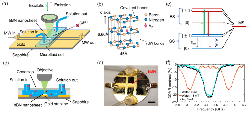
The spin defect is a color center in hBN consisting of a missing boron atom at its lattice site in a negatively charged state [7, 39, 40, 41, 42, 43] (Figure 1(b)). The symmetry axis of defects is always along the c-axis perpendicular to the hBN 2D lattice, making well suited for ensemble measurements. The has a = 1 triplet ground state with a zero field splitting of 3.47 GHz (Figure 1 (c)). The spin dependent PL emission, together with spin polarization via laser excitation, enables optically detected magnetic resonance (ODMR) experiments. Our sensor consist of an ensemble of defects created by low energy (600 eV) helium ions with a large dose density of about cm-2. The average depth of created defects in an hBN nanosheet is 6.4 nm, allowing for a small sensor-sample separation [44]. The thickness of the hBN nanosheet is typically in the range of 30-50 nm for achieving a strong plasmonic enhancement of the PL intensity on a gold microwave waveguide [12]. The lateral size of the hBN nanosheet is usually larger than 10 m.
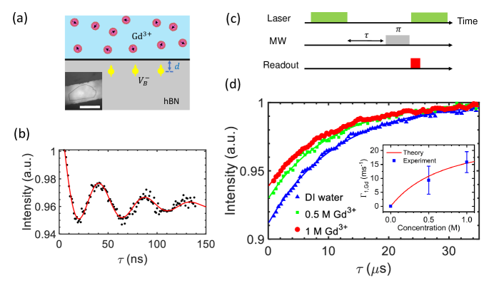
Figure 1(a)(d) illustrate our experimental setup for sensing paramagnetic ions with shallow spin defects in hBN. First, we transfer an hBN nanosheet with defects onto a gold microwave transmission line with a width of 35 m. A coverslip is then placed on top of the microwave waveguide, spaced by double-side tapes. Two pipes are connected to the two sides of the colver slip for delivering and changing solutions, and the entire device is sealed with epoxy to form a micorfluid cell. Figure 1(e) shows a picture of the prepared device. In the experiment, we use a green laser (532 nm) to excite the defects, and collect photon emission with a 750 nm long pass filter. A confocal PL map shows the nearly homogeneous distribution of defects over the hBN nanosheet. We first characterize spin defects in the transferred hBN nanosheet in air. We then slowly inject a deionized (DI) water into the microfluid cell. Continuous wave (CW) ODMR measurements confirm that defects maintain high-contrast (about 20%) ODMR signals in the liquid environment (Figure 1(f)). In the following discussion, all experiments are performed in liquids, and DI water is used to characterize defects as a reference.
We use spin relaxometry to detect surrounding paramagnetic ions by measuring the spin relaxation time () of shallow spin defects (Figure 2(a)). In a low magnetic field, free diffusing paramagnetic ions exhibit a zero average magnetic field but non-zero root-mean-square (RMS) field due to spin fluctuations. Such stochastic fields can speed up spin relaxation of nearby spin qubits. In our experiment, we first perform coherent control of the spins and extract the Rabi frequency for generating a pulse for relaxometry measurements. A 1.5-s green laser pulse is used for spin initialization and readout, and a microwave pulse is used to drive the spin to oscillate between the and states. As shown in Figure 2(b), by varying the microwave pulse duration, we see a oscillation of the PL intensity with a period of around 46.3 ns ( = 21.6 MHz). Then the longitudinal relaxation time is characterized by using the pulse sequence as depicted in Figure 2(c). In DI water, the is measured to be 11.240.17 s, which is close to in air for this hBN nanosheet. They are relatively short due to the high doping density that we used to obtain high PL count rates [45]. In the presence of Gd3+ ions (prepared by dissolving Gd(NO3)3 in water), we observe reduced relaxation times. The measured spin relaxation times are s for 1 M Gd3+ ions and s for 0.5 M Gd3+ ions (Figure 2(d)). Using DI water as the reference, the Gd3+ induced spin relaxation rates can be obtained as . They are found to be 15.83.8 ms-1 for 1 M Gd3+ ions and 9.45.0 ms-1 for 0.5 M Gd3+ ions. The measured average values are significantly larger than the uncertainties of our measurements, showing that we have detected paramagnetic ions in water with hBN spin defects for the first time.
The paramagnetic-ion-induced spin relaxation rates depends on the corresponding RMS magnetic field and its spectral density , where is the Larmor frequency of Gd3+. is the composite relaxation rate of Gd3+ spins, which is on the order of tens of gigahertz. In a low magnetic field, is negligible compares to , and hence is dominated by statistical polarization and substantial broadening effects of fluctuations [30]. When the thickness of the Gd3+ solution is far larger than the average depth of spins, we can assume that Gd3+ ions exist everywhere in the half infinite space above hBN. Then the Gd3+ induced decay rate of spins is [30]
| (1) |
where is the Gd3+ concentration in moll-1, is the Avogadro number, is the vacuum permeability, is average mean depth of the defects, and GHz/T is the gyromagnetic ratio of electrons. s-1 + (77 s-1M-1) [30]. Invoking this relaxation model, the theoretical prediction shows good agreement with the experimental results as shown in the inset of Figure 2(d). This agreement further confirms that we have observed paramagnetic spin noise in liquids with hBN spin defects.
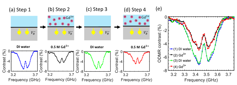
After demonstrating the capability of spin relaxometry, we develop another detection method based on the reduction of the ODMR contrast of hBN spin defects due to paramagnetic ions. In the pulsed spin relaxometry measurements, we observe a decrease in the contrast of spin relaxation signal in the presence of Gd3+ ions (Figure 2(d)). Similar phenomena can also be observed in CW ODMR experiments. In DI water, defects exhibit 7 ODMR contrast under a 50 mW microwave driving (Figure 3(a)). Once the 0.5 M Gd3+ water solution replaces the DI water, we observe a dramatic reduction of ODMR contrast to less than 5 (Figure 3(b)). The ODMR contrast will resume after pumping the DI water back into the microfluid cell (Figure 3(c)) and will decrease again in the presence of Gd3+ solution (Figure 3(d)). Thus the reduction of ODMR contrast due to paramagnetic ions is reversible and repeatable (Figure 3(e)). This method allows us to detect paramagnetic ions by monitoring the change in CW ODMR contrasts.
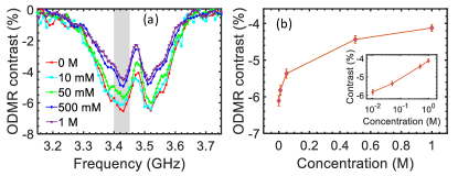
To quantify the dependence of the ODMR contrast on paramagnetic ion concentration, we perform the CW ODMR experiments in five different solutions: 0 mM Gd3+ (DI water), 10 mM Gd3+, 50 mM Gd3+, 500 mM Gd3+, and 1 M Gd3+. As the concentration of Gd3+ increases, we observe a continuous reduction in the ODMR contrast (Figure 4). To quantify this concentration dependency, we select data points within the frequency range from 3.40 GHz to 3.45 GHz and calculate the average ODMR contrasts (Figure 4(b)). The effect of Gd3+ ions at a concentration as low as 10 mM is observable. This method requires only a few ODMR data points to calculate the average integrated ODMR contrast and does not rely on pulsed laser excitation, precise microwave manipulation, fast photodetectors, or multichannel pulse generators. This simplicity allows for rapid detection of paramagnetic ions in liquids, making it ideal for real-world applications.
The reduction of the ODMR contrast can be explained by the Gd3+ induced depopulation of the ground state during the laser initialization, which has also been observed with diamond NV centers [35, 36]. While a green laser tries to initialize spin defects to the state, high-frequency magnetic noise will drive spin transitions from the state to the states. In a solution of paramagnetic ions, a higher spin concentration gives rise to more magnetic noise due to spin fluctuations. A stronger spin relaxation effect due to spin noise will result in a lower spin polarization level for a given laser power, which gives rise to the concentration-dependent ODMR contrast reduction in shallow hBN spin defects. Additionally, Gd3+ ions can affect solution conductivity and cause slight microwave absorption, which can also impact the ODMR contrast of hBN spin defects. In the future, to mitigate the potential microwave absorption effect of Gd3+ ions, we can reduce the width of the microfluid channel to expose only a small portion of the microwave waveguide to the ionic solution. Furthermore, we can employ two hBN nanosheets with spin defects at two different depths: shallow spin defects can detect the spin noise of paramagnetic ions, while deep spin defects can monitor any potential ODMR contrast reduction due to microwave absorption by the ionic solution. Overall, the concentration-sensitive ODMR contrast allows for efficient detection of paramagnetic ions in liquids using CW ODMR, which is much simpler to implement than pulsed sensing protocols.
In conclusion, we have demonstrated the first detection of spin noise from paramagnetic ions in liquids using a vdW sensor based on hBN spin defects. Our spin relaxometry measurements reveal the characteristic behavior of the Gd3+-induced spin relaxation, which increases with increasing Gd3+ concentration. By using CW ODMR technique, we are also able to detect the Gd3+ ions via the contrast reduction of the spin-dependent PL. Our work represents an initial demonstration of the potential of hBN spin defects for paramagnetic ion sensing in liquids. There are many ways for further improvement. For instance, the sensitivity can be further improved by optimizing the hBN sensor, including reducing the depth of spin defects and improving the collection efficiency of the spin-dependent fluorescence. Future studies can also explore the sensing performance of hBN spin defects in more complex chemical and biological environments.
Note added: While finalizing this paper, we became aware of a related paper on detecting paramagnetic ions with hBN spin defects [46]
Acknowledgments
T.L. thanks the Purdue Quantum Science and Engineering Institute (PQSEI) for support through the seed grant, and the DARPA ARRIVE program. Y.P.C. acknowledges support by the Quantum Science Center, a U.S. Department of Energy, Office of Science, National Quantum Information Science Research Center.
References
- Degen et al. [2017] C. L. Degen, F. Reinhard, and P. Cappellaro, Quantum sensing, Reviews of modern physics 89, 035002 (2017).
- Budker and Romalis [2007] D. Budker and M. Romalis, Optical magnetometry, Nature physics 3, 227 (2007).
- Schirhagl et al. [2014] R. Schirhagl, K. Chang, M. Loretz, and C. L. Degen, Nitrogen-vacancy centers in diamond: nanoscale sensors for physics and biology, Annu. Rev. Phys. Chem 65, 83 (2014).
- Kucsko et al. [2013] G. Kucsko, P. C. Maurer, N. Y. Yao, M. Kubo, H. J. Noh, P. K. Lo, H. Park, and M. D. Lukin, Nanometre-scale thermometry in a living cell, Nature 500, 54 (2013).
- Casola et al. [2018] F. Casola, T. Van Der Sar, and A. Yacoby, Probing condensed matter physics with magnetometry based on nitrogen-vacancy centres in diamond, Nature Reviews Materials 3, 17088 (2018).
- Shi et al. [2018] F. Shi, F. Kong, P. Zhao, X. Zhang, M. Chen, S. Chen, Q. Zhang, M. Wang, X. Ye, Z. Wang, et al., Single-dna electron spin resonance spectroscopy in aqueous solutions, Nature methods 15, 697 (2018).
- Gottscholl et al. [2020] A. Gottscholl, M. Kianinia, V. Soltamov, S. Orlinskii, G. Mamin, C. Bradac, C. Kasper, K. Krambrock, A. Sperlich, M. Toth, et al., Initialization and read-out of intrinsic spin defects in a van der Waals crystal at room temperature, Nature materials 19, 540 (2020).
- Gottscholl et al. [2021a] A. Gottscholl, M. Diez, V. Soltamov, C. Kasper, A. Sperlich, M. Kianinia, C. Bradac, I. Aharonovich, and V. Dyakonov, Room temperature coherent control of spin defects in hexagonal boron nitride, Science Advances 7, eabf3630 (2021a).
- Mendelson et al. [2021] N. Mendelson, D. Chugh, J. R. Reimers, T. S. Cheng, A. Gottscholl, H. Long, C. J. Mellor, A. Zettl, V. Dyakonov, P. H. Beton, et al., Identifying carbon as the source of visible single-photon emission from hexagonal boron nitride, Nature materials 20, 321 (2021).
- Chejanovsky et al. [2021] N. Chejanovsky, A. Mukherjee, J. Geng, Y.-C. Chen, Y. Kim, A. Denisenko, A. Finkler, T. Taniguchi, K. Watanabe, D. B. R. Dasari, et al., Single-spin resonance in a van der Waals embedded paramagnetic defect, Nature materials 20, 1079 (2021).
- Vaidya et al. [2023] S. Vaidya, X. Gao, S. Dikshit, I. Aharonovich, and T. Li, Quantum sensing and imaging with spin defects in hexagonal boron nitride, arXiv preprint arXiv:2302.11169 (2023).
- Gao et al. [2021] X. Gao, B. Jiang, A. E. Llacsahuanga Allcca, K. Shen, M. A. Sadi, A. B. Solanki, P. Ju, Z. Xu, P. Upadhyaya, Y. P. Chen, et al., High-contrast plasmonic-enhanced shallow spin defects in hexagonal boron nitride for quantum sensing, Nano Letters 21, 7708 (2021).
- Novoselov et al. [2016] K. S. Novoselov, A. Mishchenko, A. Carvalho, and A. H. Castro Neto, 2D materials and van der Waals heterostructures, Science 353, aac9439 (2016).
- Huang et al. [2022] M. Huang, J. Zhou, D. Chen, H. Lu, N. J. McLaughlin, S. Li, M. Alghamdi, D. Djugba, J. Shi, H. Wang, et al., Wide field imaging of van der Waals ferromagnet Fe3GeTe2 by spin defects in hexagonal boron nitride, Nature communications 13, 5369 (2022).
- Healey et al. [2023] A. Healey, S. Scholten, T. Yang, J. Scott, G. Abrahams, I. Robertson, X. Hou, Y. Guo, S. Rahman, Y. Lu, et al., Quantum microscopy with van der Waals heterostructures, Nature Physics 19, 87–91 (2023).
- Gottscholl et al. [2021b] A. Gottscholl, M. Diez, V. Soltamov, C. Kasper, D. Krauße, A. Sperlich, M. Kianinia, C. Bradac, I. Aharonovich, and V. Dyakonov, Spin defects in hBN as promising temperature, pressure and magnetic field quantum sensors, Nature communications 12, 4480 (2021b).
- Liu et al. [2021] W. Liu, Z.-P. Li, Y.-Z. Yang, S. Yu, Y. Meng, Z.-A. Wang, Z.-C. Li, N.-J. Guo, F.-F. Yan, Q. Li, et al., Temperature-dependent energy-level shifts of spin defects in hexagonal boron nitride, ACS Photonics 8, 1889 (2021).
- Yang et al. [2022] T. Yang, N. Mendelson, C. Li, A. Gottscholl, J. Scott, M. Kianinia, V. Dyakonov, M. Toth, and I. Aharonovich, Spin defects in hexagonal boron nitride for strain sensing on nanopillar arrays, Nanoscale 14, 5239 (2022).
- Lyu et al. [2022] X. Lyu, Q. Tan, L. Wu, C. Zhang, Z. Zhang, Z. Mu, J. Zúñiga-Pérez, H. Cai, and W. Gao, Strain quantum sensing with spin defects in hexagonal boron nitride, Nano Letters 22, 6553 (2022).
- Gao et al. [2022] X. Gao, S. Vaidya, K. Li, P. Ju, B. Jiang, Z. Xu, A. E. L. Allcca, K. Shen, T. Taniguchi, K. Watanabe, et al., Nuclear spin polarization and control in hexagonal boron nitride, Nature Materials 21, 1024 (2022).
- Thomas [2015] D. D. Thomas, Breathing new life into nitric oxide signaling: a brief overview of the interplay between oxygen and nitric oxide, Redox biology 5, 225 (2015).
- Bogdan [2015] C. Bogdan, Nitric oxide synthase in innate and adaptive immunity: an update, Trends in immunology 36, 161 (2015).
- Griendling et al. [2016] K. K. Griendling, R. M. Touyz, J. L. Zweier, S. Dikalov, W. Chilian, Y.-R. Chen, D. G. Harrison, and A. Bhatnagar, Measurement of reactive oxygen species, reactive nitrogen species, and redox-dependent signaling in the cardiovascular system: a scientific statement from the american heart association, Circulation research 119, e39 (2016).
- Weinmann et al. [1984] H.-J. Weinmann, R. C. Brasch, W.-R. Press, and G. E. Wesbey, Characteristics of gadolinium-DTPA complex: a potential NMR contrast agent, American journal of roentgenology 142, 619 (1984).
- Chan and Wong [2007] K. W.-Y. Chan and W.-T. Wong, Small molecular gadolinium (iii) complexes as mri contrast agents for diagnostic imaging, Coordination Chemistry Reviews 251, 2428 (2007).
- Tweedle [2021] M. F. Tweedle, Gadolinium retention in human brain, bone, and skin, Radiology 300, 570 (2021).
- Dos Santos et al. [2022] T. J. P. Dos Santos, A. V. Parambathu, C. C. Fraenza, C. Walsh, S. G. Greenbaum, W. G. Chapman, D. Asthagiri, and P. M. Singer, Thermal and concentration effects on 1H NMR relaxation of Gd3+-aqua using MD simulations and measurements, Physical Chemistry Chemical Physics 24, 27964 (2022).
- Ma et al. [2021] L. Ma, M. G. Azad, M. Dharmasivam, V. Richardson, R. Quinn, Y. Feng, D. Pountney, K. Tonissen, G. Mellick, I. Yanatori, et al., Parkinson’s disease: alterations in iron and redox biology as a key to unlock therapeutic strategies, Redox Biology 41, 101896 (2021).
- Samrot et al. [2021] A. V. Samrot, C. S. Sahithya, J. Selvarani, S. K. Purayil, and P. Ponnaiah, A review on synthesis, characterization and potential biological applications of superparamagnetic iron oxide nanoparticles, Current Research in Green and Sustainable Chemistry 4, 100042 (2021).
- Steinert et al. [2013] S. Steinert, F. Ziem, L. Hall, A. Zappe, M. Schweikert, N. Götz, A. Aird, G. Balasubramanian, L. Hollenberg, and J. Wrachtrup, Magnetic spin imaging under ambient conditions with sub-cellular resolution, Nature communications 4, 1607 (2013).
- Ziem et al. [2013] F. C. Ziem, N. S. Gotz, A. Zappe, S. Steinert, and J. Wrachtrup, Highly sensitive detection of physiological spins in a microfluidic device, Nano letters 13, 4093 (2013).
- Ermakova et al. [2013] A. Ermakova, G. Pramanik, J.-M. Cai, G. Algara-Siller, U. Kaiser, T. Weil, Y.-K. Tzeng, H.-C. Chang, L. McGuinness, M. B. Plenio, et al., Detection of a few metallo-protein molecules using color centers in nanodiamonds, Nano letters 13, 3305 (2013).
- Shi et al. [2015] F. Shi, Q. Zhang, P. Wang, H. Sun, J. Wang, X. Rong, M. Chen, C. Ju, F. Reinhard, H. Chen, et al., Single-protein spin resonance spectroscopy under ambient conditions, Science 347, 1135 (2015).
- Simpson et al. [2017] D. A. Simpson, R. G. Ryan, L. T. Hall, E. Panchenko, S. C. Drew, S. Petrou, P. S. Donnelly, P. Mulvaney, and L. C. Hollenberg, Electron paramagnetic resonance microscopy using spins in diamond under ambient conditions, Nature Communications 8, 458 (2017).
- Gorrini et al. [2019] F. Gorrini, R. Giri, C. Avalos, S. Tambalo, S. Mannucci, L. Basso, N. Bazzanella, C. Dorigoni, M. Cazzanelli, P. Marzola, et al., Fast and sensitive detection of paramagnetic species using coupled charge and spin dynamics in strongly fluorescent nanodiamonds, ACS applied materials & interfaces 11, 24412 (2019).
- Radu et al. [2019] V. Radu, J. C. Price, S. J. Levett, K. K. Narayanasamy, T. D. Bateman-Price, P. B. Wilson, and M. L. Mather, Dynamic quantum sensing of paramagnetic species using nitrogen-vacancy centers in diamond, ACS sensors 5 (2019).
- Liu et al. [2022] W. Liu, N.-J. Guo, S. Yu, Y. Meng, Z. Li, Y.-Z. Yang, Z.-A. Wang, X.-D. Zeng, L.-K. Xie, J.-F. Wang, et al., Spin-active defects in hexagonal boron nitride, Materials for Quantum Technology 1, 032002 (2022).
- Xu et al. [2023] X. Xu, A. B. Solanki, D. Sychev, X. Gao, S. Peana, A. S. Baburin, K. Pagadala, Z. O. Martin, S. N. Chowdhury, Y. P. Chen, et al., Greatly enhanced emission from spin defects in hexagonal boron nitride enabled by a low-loss plasmonic nanocavity, Nano Letters 23, 25–33 (2023).
- Ivády et al. [2020] V. Ivády, G. Barcza, G. Thiering, S. Li, H. Hamdi, J.-P. Chou, Ö. Legeza, and A. Gali, Ab initio theory of the negatively charged boron vacancy qubit in hexagonal boron nitride, npj Computational Materials 6, 41 (2020).
- Mathur et al. [2022] N. Mathur, A. Mukherjee, X. Gao, J. Luo, B. A. McCullian, T. Li, A. N. Vamivakas, and G. D. Fuchs, Excited-state spin-resonance spectroscopy of defect centers in hexagonal boron nitride, Nature Communications 13, 3233 (2022).
- Baber et al. [2021] S. Baber, R. N. E. Malein, P. Khatri, P. S. Keatley, S. Guo, F. Withers, A. J. Ramsay, and I. J. Luxmoore, Excited state spectroscopy of boron vacancy defects in hexagonal boron nitride using time-resolved optically detected magnetic resonance, Nano Letters 22, 461 (2021).
- Mu et al. [2022] Z. Mu, H. Cai, D. Chen, J. Kenny, Z. Jiang, S. Ru, X. Lyu, T. S. Koh, X. Liu, I. Aharonovich, et al., Excited-state optically detected magnetic resonance of spin defects in hexagonal boron nitride, Physical Review Letters 128, 216402 (2022).
- Yu et al. [2022] P. Yu, H. Sun, M. Wang, T. Zhang, X. Ye, J. Zhou, H. Liu, C.-J. Wang, F. Shi, Y. Wang, et al., Excited-state spectroscopy of spin defects in hexagonal boron nitride, Nano Letters 22, 3545 (2022).
- Ziegler et al. [2010] J. F. Ziegler, M. D. Ziegler, and J. P. Biersack, SRIM–the stopping and range of ions in matter (2010), Nuclear Instruments and Methods in Physics Research Section B: Beam Interactions with Materials and Atoms 268, 1818 (2010).
- Gong et al. [2022] R. Gong, G. He, X. Gao, P. Ju, Z. Liu, B. Ye, E. A. Henriksen, T. Li, and C. Zu, Coherent dynamics of strongly interacting electronic spin defects in hexagonal boron nitride, arXiv preprint arXiv:2210.11485 (2022).
- Robertson et al. [2023] I. O. Robertson, S. C. Scholten, P. Singh, A. J. Healey, F. Meneses, P. Reineck, H. Abe, T. Ohshima, M. Kianinia, I. Aharonovich, et al., Detection of paramagnetic spins with an ultrathin van der waals quantum sensor, arXiv preprint arXiv:2302.10560 (2023).