Transferring Models Trained on Natural Images to 3D MRI via Position Encoded Slice Models
Abstract
Transfer learning has remarkably improved computer vision. These advances also promise improvements in neuroimaging, where training set sizes are often small. However, various difficulties arise in directly applying models pretrained on natural images to radiologic images, such as MRIs. In particular, a mismatch in the input space (2D images vs. 3D MRIs) restricts the direct transfer of models, often forcing us to consider only a few MRI slices as input. To this end, we leverage the 2D-Slice-CNN architecture of Gupta et al. (2021), which embeds all the MRI slices with 2D encoders (neural networks that take 2D image input) and combines them via permutation-invariant layers. With the insight that the pretrained model can serve as the 2D encoder, we initialize the 2D encoder with ImageNet pretrained weights that outperform those initialized and trained from scratch on two neuroimaging tasks — brain age prediction on the UK Biobank dataset and Alzheimer’s disease detection on the ADNI dataset. Further, we improve the modeling capabilities of 2D-Slice models by incorporating spatial information through position embeddings, which can improve the performance in some cases.
Index Terms— MRI, deep learning, machine learning, neuroimaging, transfer learning
1 Introduction
Even though tremendous advances have been made in pretraining computer vision models that work well for natural images such as photographs, it is unclear how easily these pretrained models can be adapted to perform tasks on radiologic images such as MRIs due to domain differences. Moreover, natural images are 2-dimensional (2D), whereas brain MRIs are typically 3-dimensional (3D), making the direct finetuning of the model pretrained on 2D images challenging. This has led to an active debate in the radiology field on how to pretrain deep learning methods for MRI-based tasks, such as disease classification and staging, identifying pathology, and anatomical segmentation (e.g., [1, 2, 3]). Many workarounds have been proposed, such as deriving specialized pretraining datasets such as YouTube videos [4] or making predictions from only a few MRI slices [5, 6, 7, 8]. Using fewer slices severely limits the information available for a machine learning model to make predictions, leading to suboptimal performance.
To this end, we consider recently proposed 2D-Slice-CNN models that can consider full 3D MRI scans as the input. These models process each 2D slice via a slice encoder (usually a 2D CNN) and aggregate the resulting slice embeddings via permutation invariant operations, such as by computing the mean of the embeddings or using self-attention over the embeddings [9], max-pooling [10], or RNNs [11]. These models can access information from all the slices during training, thus exploiting a richer feature set than models that work with a single slice, and the neural networks pretrained on 2D natural images are excellent candidates to use as the slice encoder. Our first contribution is to study the effect of replacing the slice encoder with a model pretrained on ImageNet.
Our second contribution is incorporating positional encoding in the 2D-Slice model before slice embedding aggregation to preserve spatial information. The permutation-invariant operations can remove information about the ordering of the slices, thus limiting learning capabilities. Adding positional encoding, i.e., a unique vector corresponding to each position, can help, especially if predictive features are reliably located in specific parts of the images. Positional encoding allows the model to learn spatial information if needed (e.g., [12]).
We extensively evaluate the above-discussed models and a 3D-CNN with comparable architecture for brain age prediction (the common benchmarking task of predicting a person’s age from their MRI scan) and an Alzheimer’s disease diagnosis (binary classification) task. Incorporating positional encodings improved test performance in some cases. Contrary to [9], we find that 2D-Slice-CNNs perform comparably to 3D-CNN for brain age prediction1113D-CNN results improved due to expanded hyperparameter search.. Further, 2D-Slice-CNN models initialized randomly did not perform at par with 3D-CNNs for AD detection. However, the 2D-Slice-CNN models with ImageNet pretrained ResNet-18 encoder outperformed all the models for both tasks, showing that it is possible to transfer inductive biases from natural images to 3D neuroimaging tasks.
2 2D-slice CNN with Positional Encodings
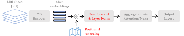
The go-to approach to train models that take raw 3D MRIs as input is to use 3D convolutional layers (e.g., [13, 14, 15]). Instead, [9] used 2D-CNNs to encode individual slices and combined the slice representations with permutation-invariant operations such as mean or attention. Compared to 3D-CNNs, these 2D-Slice-based models tend to be less accurate as they may lose spatial information due to permutation invariance. To this end, we encode positional information in the model by introducing positional encodings similar to transformers [16, 17]. In particular, these are added to the slices’ representations and trained end-to-end with other model parameters (see Fig. 1). Suppose the MRI consists of 2D slices () that are each embedded to a d-dimensional representation, with the 2D slice encoder. The model will compute , where is a trainable vector and depends only on . Fixed positional encodings can also be used. However, this work considers positional encodings as trainable.
Further, motivated by the transformer architecture, we also added a feed-forward layer and layer norm after the attention (i.e., for attention-based aggregation) as that improved the performance on brain age prediction slightly (2.86 reported in [9] vs. 2.83 MAE in our case).
3 Experiment Setup
3.1 Datasets
We consider two tasks — Brain age prediction and Alzheimer’s disease (AD) detection from brain MRIs. Our setup for brain age prediction is the same as [9]. We used MRI scans from a subset of healthy subjects from the UK Biobank dataset [18] with no psychiatric diagnosis. The training, test, and validation set sizes were 7,312; 940; and 2,194; with a mean chronological age and standard deviation of 62.6 and 7.4 years.
We used the same dataset as [19] for the AD task. In particular, we use MRI scans from three phases of the Alzheimer’s Disease Neuroimaging Initiative (ADNI), known as ADNI1, ADNI2/GO, and ADNI3. These datasets have repeated scans of the same subject. However, the subjects in train/test/validation sets do not overlap. We used 4,561 scans (2,372/873/916 in train/validation/test). The test set contains 244 scans with AD and 672 with a healthy diagnosis.
All the scans were reoriented to a standard brain MRI template, and the final dimensions of each scan in both datasets were .
3.2 Model & Training
We evaluate the 2D-Slice-CNNs under different settings — with and without positional encodings and with and without pretrained encoders. We consider two encoder architectures. We use a 5-layer 2D-CNN encoder (similar to [9, 11]) adapted from the 3D-CNN baseline of [13] to benchmark the effect of incorporating positional encodings.
We evaluate the effect of pretraining on natural images (2D) with pretrained ResNets [20]. We use ResNets pretrained on ImageNet-1K [21] from the PyTorch model hub222https://pytorch.org/hub/ as the 2D slice encoder. To get the slice embeddings from ResNets, we remove the final feed-forward layers and consider the output from the last layer (i.e., the average pooling layer). Therefore, the embedding sizes are 512 and 2048 for ResNet-18 and ResNet-50 encoders. For the 5-layer 2D-CNN encoder, the embedding size is 32. When using pretrained weights, we finetune the entire model, including the encoder. Our implementation is available at https://github.com/umgupta/2d-slice-set-networks.
For 2D-Slice models, we find that slicing MRI along the sagittal axis works best for brain age prediction, as also observed by [9]. However, this was not the case for the AD task. Therefore we report the results of slicing along all three axes for AD. We use the same hyperparameters as [9], except for the optimizer and learning rate. We search in {(ADAM, 10-4), (SGD, 10-3/ 10-4/ 10-5)} for the brain age prediction task. ADAM optimizer with a learning rate of 10-4 works best for all the models except for the 3D-CNN. We report results for all models on the AD task with a learning rate of 10-4 and ADAM optimizer333We evaluated 3D-CNN with SGD on the AD task, but ADAM worked best.. All the models are trained for 100 epochs, and the best model was chosen based on performance on the validation set at the end of every epoch.
| Method | Bal. Accuracy | F1 Score | Avg. Precision | |||
| Encoder | Pos. Enc. | Pretrained | Axis | |||
| 3D-CNN | - | ✗ | - | 88.40 0.703 | 84.44 1.028 | 92.33 0.492 |
| 2D-CNN (Mean) | ✗ | ✗ | sagittal | 87.53 1.311 | 84.40 1.707 | 94.86 0.946 |
| 2D-CNN (Mean) | ✗ | ✗ | coronal | 85.78 2.811 | 81.38 3.610 | 90.57 2.976 |
| 2D-CNN (Mean) | ✗ | ✗ | axial | 86.34 1.445 | 82.21 1.573 | 90.81 0.677 |
| \cdashline2-7 2D-CNN (Mean) | ✓ | ✗ | sagittal | 87.05 1.115 | 83.40 0.873 | 93.40 0.956 |
| 2D-CNN (Mean) | ✓ | ✗ | coronal | 85.41 2.521 | 80.62 2.521 | 89.86 0.771 |
| 2D-CNN (Mean) | ✓ | ✗ | axial | 86.61 0.942 | 81.20 1.971 | 91.44 2.123 |
| 2D-ResNet-18 (Mean) | ✗ | ✗ | sagittal | 85.71 4.834 | 80.89 6.111 | 91.12 2.880 |
| 2D-ResNet-18 (Mean) | ✗ | ✗ | coronal | 85.73 1.433 | 81.30 1.802 | 90.60 2.544 |
| 2D-ResNet-18 (Mean) | ✗ | ✗ | axial | 84.61 2.099 | 79.21 2.906 | 88.95 1.741 |
| \cdashline2-7 2D-ResNet-18 (Mean) | ✓ | ✗ | sagittal | 86.57 2.871 | 81.69 3.972 | 91.80 2.150 |
| 2D-ResNet-18 (Mean) | ✓ | ✗ | coronal | 84.34 1.706 | 79.75 2.258 | 90.29 0.645 |
| 2D-ResNet-18 (Mean) | ✓ | ✗ | axial | 86.55 1.059 | 81.39 1.752 | 90.82 0.701 |
| 2D-ResNet-18 (Mean) | ✗ | ✓ | sagittal | 87.09 1.192 | 82.07 1.167 | 90.80 1.601 |
| 2D-ResNet-18 (Mean) | ✗ | ✓ | coronal | 87.31 0.971 | 83.19 1.518 | 91.80 1.626 |
| 2D-ResNet-18 (Mean) | ✗ | ✓ | axial | 88.59 1.187 | 85.10 1.265 | 93.11 0.482 |
| \cdashline2-7 2D-ResNet-18 (Mean) | ✓ | ✓ | sagittal | 87.19 2.037 | 83.16 2.158 | 91.75 1.291 |
| 2D-ResNet-18 (Mean) | ✓ | ✓ | coronal | 86.95 2.750 | 82.33 2.811 | 90.96 1.541 |
| 2D-ResNet-18 (Mean) | ✓ | ✓ | axial | 88.60 2.058 | 84.52 2.389 | 92.56 0.563 |
| Method | Mean Absolute Err. (MAE) | Root Mean Square Err. (RMSE) | # Params | ||
| Encoder | Pos. Enc. | Pretrained | |||
| 3D-CNN | - | ✗ | 2.792 0.032 | 3.521 0.023 | 3M |
| 2D-CNN (Mean) | ✗ | ✗ | 2.826 0.021 | 3.582 0.027 | 1M |
| 2D-CNN (Mean) | ✓ | ✗ | 2.847 0.051 | 3.617 0.067 | 1M |
| 2D-CNN (Attention) | ✗ | ✗ | 2.839 0.009 | 3.590 0.014 | 1M |
| 2D-CNN (Attention) | ✓ | ✗ | 2.888 0.067 | 3.655 0.075 | 1M |
| 2D-ResNet-18 (Mean) | ✗ | ✗ | 2.911 0.039 | 3.684 0.043 | 12M |
| 2D-ResNet-18 (Mean) | ✗ | ✓ | 2.715 0.029 | 3.426 0.044 | 12M |
| 2D-ResNet-18 (Mean) | ✓ | ✓ | 2.721 0.045 | 3.439 0.053 | 12M |
| 2D-ResNet-50 (Mean) | ✗ | ✓ | 2.743 0.018 | 3.468 0.029 | 26M |
4 Results
Tables 3.2 and 2 summarize the performance of different models for AD and brain age prediction, respectively. We observed that the 2D-Slice-CNN was better at predicting brain age when using the same hyperparameters as [9]. However, our 3D-CNN results are better than 2D-Slice-CNN (Table 2), and the reported numbers in [9] (3.02 vs. 2.792 MAE) due to switching from PyTorch’s default initialization to explicitly using He initialization [22] and using SGD optimizer. Similarly, 3D-CNN outperforms the 2D-Slice-CNN for AD prediction (88.40 vs. 87.53 accuracy). We hypothesized that this might be due to 2D-Slice-CNN combining slice embeddings with permutation invariant operation and consequently losing information about the position of the slice in MRI. We, thus, incorporate positional encodings in the 2D-Slice model.
4.1 Effect of Position Encodings on the 2D-Slice-CNN.
Introducing position encodings improved 2D-Slice models for AD prediction in some cases. When using 2D-ResNet-18 encoder (not pretrained), we see improvements in AD prediction when slices along sagittal and axial directions are used. However, in other cases, the performance on the AD prediction task did not improve when incorporating position encodings. For brain age prediction, the model performance stayed identical (ResNet Encoders) or deteriorated (5-layer CNN) when using position encodings. Brain MRIs are usually aligned to a standard template, so the position of specific patterns can provide extra information. However, it may also make the model more sensitive to minor changes in the alignment template and cause overfitting. This could be a potential reason why the performance did not improve on incorporating position encoding. Here, we incorporate position encodings in the higher layers. Thus the model may not be able to exploit the spatial information best. Placement of position encodings is crucial [23] and left for future work. Overall, position encodings helped in limited cases, and the gains may be task and model dependent.
4.2 Can Encoders Pretrained on ImageNet improve 3D Deep Neuroimaging?
We answer this in the affirmative. We finetune 2D-Slice models with ResNet encoders initialized with ImageNet-1K pretrained weights. Initializing with pretrained weights consistently outperforms random initialization (i.e., training from scratch) in all cases (See Tables 3.2 and 2). These results validate our hypothesis that a) models trained on natural images (2D) can be helpful for neuroimaging tasks and b) 2D-Slice-CNNs can be used to transfer 2D models to 3D data directly.
Our main goal with these experiments is to demonstrate improvement in performance due to natural image pretraining. Nevertheless, another interesting outcome is that 2D-Slice models with pretrained encoders are the best for both tasks, outperforming the 3D-CNN as well. For the AD task, we observe that 3D-CNN has a balanced accuracy of 88.40. In contrast, models with pretrained ResNet-18 encoder model have a balanced accuracy of 88.6 when using slices along the axial direction (both with and without position encodings). Similarly, the best MAE with the pretrained model for brain age prediction is 2.715 compared to 2.792 MAE with 3D-CNN. Finally, We also evaluated if increasing the size of the pretrained encoder may lead to more gain by employing ResNet-50 as the encoder for brain age prediction. We did not see significant improvements over ResNet-18, and we leave further exploration for future work.
5 Discussion
In this paper, we extended the 2D-Slice-based architecture of [9] by incorporating positional embeddings.we demonstrated improved brain age prediction and AD detection performance by employing slice encoders pretrained on ImageNet-1K, a large dataset of natural 2D images.
Our work contributes to the growing literature on using pretraining for machine learning on radiologic images, for which training datasets are often small. Since most large image datasets are 2D natural images, it is natural to pretrain on natural images. However, there are two main challenges with this — a) Domain mismatch, i.e., radiologic images vs. natural images; b) Input dimension mismatch, i.e., 2D vs. 3D images. Most prior works using off-the-shelf vision models (i.e., pretrained with natural image datasets such as ImageNet) consider a single or a few 2D slices of the MRI scan as the input to avoid the problem of input mismatch [5, 6, 7, 8]. These may then aggregate the results from different slices during inference only. Such approaches are limiting and lead to suboptimal performance.
In contrast, our approach only substitutes encoders with pretrained counterparts. The model is trained end-to-end and considers the whole MRI as input. It addresses the input dimension mismatch problem without limiting or compromising the information available to the model to make predictions. Despite domain mismatch, pretraining outperforms the models trained from scratch.
Very few datasets exist for pretraining with raw 3D MRI images directly [24]. Only recently very large radiologic 2D image datasets become have publicly available for pretraining models [25, 26]. It would be interesting to pretrain 2D encoders with such datasets in the future to alleviate the domain mismatch problem. In this work, we used models pretrained on the supervised classification task. In future, it would be interesting to evaluate self-supervised pretraining with in- and out-domain images (e.g., [10]).
6 Compliance with Ethical Standards
This is a study of previously collected, anonymized and de-identified data available in a public repository.
7 Acknowledgements
We thank the ADNI investigators and their public and private funders for creating and publicly disseminating the ADNI dataset. This work is supported by NIH (U01AG068057, P01AG055367).
References
- [1] Laith Alzubaidi, Ye Duan, Ayad Al-Dujaili, Ibraheem Kasim Ibraheem, Ahmed H Alkenani, Jose Santamaría, and Others, “Deepening into the suitability of using pre-trained models of imagenet against a lightweight convolutional neural network in medical imaging: an experimental study,” PeerJ Computer Science, vol. 7, pp. e715, 2021.
- [2] Alexander Ke, William Ellsworth, Oishi Banerjee, Andrew Y Ng, and Pranav Rajpurkar, “CheXtransfer: performance and parameter efficiency of imagenet models for chest X-ray interpretation,” in Proceedings of the Conference on Health, Inference, and Learning, 2021, pp. 116–124.
- [3] Juan Miguel Valverde, Vandad Imani, Ali Abdollahzadeh, Riccardo De Feo, Mithilesh Prakash, Robert Ciszek, and Jussi Tohka, “Transfer learning in magnetic resonance brain imaging: a systematic review,” Journal of Imaging, vol. 7, no. 4, pp. 66, 2021.
- [4] Nahiyan Malik and Danilo Bzdok, “From YouTube to the brain: Transfer learning can improve brain-imaging predictions with deep learning,” Neural Networks, vol. 153, pp. 325–338, 2022.
- [5] M. Hon and N. M. Khan, “Towards Alzheimer’s disease classification through transfer learning,” in 2017 IEEE International Conference on Bioinformatics and Biomedicine (BIBM), 2017, pp. 1166–1169.
- [6] Aly Valliani and Ameet Soni, “Deep residual nets for improved Alzheimer’s diagnosis,” in Proceedings of the 8th ACM International Conference on Bioinformatics, Computational Biology, and Health Informatics, 2017, pp. 615–615.
- [7] Jyoti Islam and Yanqing Zhang, “Brain MRI analysis for Alzheimer’s disease diagnosis using an ensemble system of deep convolutional neural networks,” Brain Informatics, vol. 5, no. 2, pp. 2, 2018.
- [8] Vishnu M Bashyam, Guray Erus, Jimit Doshi, Mohamad Habes, Ilya M Nasrallah, Monica Truelove-Hill, and Others, “MRI signatures of brain age and disease over the lifespan based on a deep brain network and 14468 individuals worldwide,” Brain, vol. 143, no. 7, pp. 2312–2324, 06 2020.
- [9] Umang Gupta, Pradeep K Lam, Greg Ver Steeg, and Paul M Thompson, “Improved brain age estimation with slice-based set networks,” in 2021 IEEE 18th International Symposium on Biomedical Imaging (ISBI), 2021, pp. 840–844.
- [10] Nikhil J Dhinagar, Sophia I Thomopoulos, Priya Rajagopalan, Dimitris Stripelis, Jose Luis Ambite, Greg Ver Steeg, and Paul M Thompson, “Evaluation of transfer learning methods for detecting Alzheimer’s disease with brain MRI,” bioRxiv, 2022.
- [11] Pradeep K Lam, Vigneshwaran Santhalingam, Parth Suresh, Rahul Baboota, Alyssa H Zhu, Sophia I Thomopoulos, Neda Jahanshad, and Paul M Thompson, “Accurate brain age prediction using recurrent slice-based networks,” in 16th International Symposium on Medical Information Processing and Analysis, 2020, vol. 11583, pp. 11–20.
- [12] Jo Schlemper, Ozan Oktay, Michiel Schaap, Mattias Heinrich, Bernhard Kainz, Ben Glocker, and Daniel Rueckert, “Attention gated networks: Learning to leverage salient regions in medical images,” Medical Image Analysis, vol. 53, pp. 197–207, 2019.
- [13] Han Peng, Weikang Gong, Christian F. Beckmann, Andrea Vedaldi, and Stephen M. Smith, “Accurate brain age prediction with lightweight deep neural networks,” Medical Image Analysis, vol. 68, pp. 101871, 2021.
- [14] Mengjiao Hu, Kang Sim, Juan Helen Zhou, Xudong Jiang, and Cuntai Guan, “Brain MRI-based 3D convolutional neural networks for classification of schizophrenia and controls,” in 2020 42nd Annual International Conference of the IEEE Engineering in Medicine & Biology Society (EMBC), 2020, pp. 1742–1745.
- [15] Hao Guan, Li Wang, Dongren Yao, Andrea Bozoki, and Mingxia Liu, “Learning transferable 3D-CNN for MRI-based brain disorder classification from scratch: An empirical study,” in Machine Learning in Medical Imaging, 2021, pp. 10–19.
- [16] Ashish Vaswani, Noam Shazeer, Niki Parmar, Jakob Uszkoreit, Llion Jones, Aidan N Gomez, L ukasz Kaiser, and Illia Polosukhin, “Attention is all you need,” in Advances in Neural Information Processing Systems, 2017.
- [17] Alexey Dosovitskiy, Lucas Beyer, Alexander Kolesnikov, Dirk Weissenborn, Xiaohua Zhai, Thomas Unterthiner, and Others, “An image is worth 16x16 words: Transformers for image recognition at scale,” in International Conference on Learning Representations, 2021.
- [18] Karla L Miller, Fidel Alfaro-Almagro, Neal K Bangerter, David L Thomas, Essa Yacoub, Junqian Xu, and Others, “Multimodal population brain imaging in the uk biobank prospective epidemiological study,” Nature Neuroscience, vol. 19, no. 11, pp. 1523–1536, 2016.
- [19] Pradeep Lam, Alyssa H Zhu, Iyad Ba Gari, Neda Jahanshad, and Paul M Thompson, “3D grid-attention networks for interpretable age and Alzheimer’s disease prediction from structural MRI,” arXiv preprint arXiv:2011.09115, 2020.
- [20] Kaiming He, Xiangyu Zhang, Shaoqing Ren, and Jian Sun, “Deep residual learning for image recognition,” in Proceedings of the IEEE Conference on Computer Vision and Pattern Recognition, 2016, pp. 770–778.
- [21] Jia Deng, Wei Dong, Richard Socher, Li-Jia Li, Kai Li, and Li Fei-Fei, “ImageNet: A large-scale hierarchical image database,” in 2009 IEEE Conference on Computer Vision and Pattern Recognition, 2009, pp. 248–255.
- [22] Kaiming He, Xiangyu Zhang, Shaoqing Ren, and Jian Sun, “Delving deep into rectifiers: Surpassing human-level performance on imagenet classification,” in 2015 IEEE International Conference on Computer Vision (ICCV), 2015, pp. 1026–1034.
- [23] Pu-Chin Chen, Henry Tsai, Srinadh Bhojanapalli, Hyung Won Chung, Yin-Wen Chang, and Chun-Sung Ferng, “Demystifying the better performance of position encoding variants for transformer,” arXiv preprint arXiv:2104.08698, 2021.
- [24] Sihong Chen, Kai Ma, and Yefeng Zheng, “Med3D: Transfer learning for 3D medical image analysis,” arXiv preprint arXiv:1904.00625, 2019.
- [25] Xueyan Mei, Zelong Liu, Philip M. Robson, Brett Marinelli, Mingqian Huang, Amish Doshi, and Others, “RadImageNet: An open radiologic deep learning research dataset for effective transfer learning,” Radiology: Artificial Intelligence, vol. 4, no. 5, pp. e210315, 2022.
- [26] Laith Alzubaidi, J Santamaría, Mohamed Manoufali, Beadaa Mohammed, Mohammed A Fadhel, Jinglan Zhang, and Others, “MedNet: Pre-trained convolutional neural network model for the medical imaging tasks,” arXiv preprint arXiv:2110.06512, 2021.