Isotopic effects in molecular attosecond photoelectron interferometry.
Isotopic substitution in molecular systems can affect fundamental molecular properties including the energy position and spacing of electronic, vibrational and rotational levels, thus modifying the dynamics associated to their coherent superposition. In extreme ultraviolet spectroscopy, the photoelectron leaving the molecule after the absorption of a single photon can trigger an ultrafast nuclear motion in the cation, which can lead, eventually, to molecular fragmentation. This dynamics depends on the mass of the constituents of the cation, thus showing, in general, a significant isotopic dependence. In time-resolved attosecond photoelectron interferometry, the absorption of the extreme ultraviolet photon is accompanied by the exchange of an additional quantum of energy (typically in the infrared spectral range) with the photoelectron-photoion system, offering the opportunity to investigate in time the influence of isotopic substitution on the characteristics of the photoionisation dynamics. Here we show that attosecond photoelectron interferometry is sensitive to isotopic substitution by investigating the two-color photoionisation spectra measured in a mixture of methane (CH4) and deuteromethane (CD4). The isotopic dependence manifests itself in the modification of the amplitude and contrast of the oscillations of the photoelectron peaks generated in the two-color field with the two isotopologues. The observed effects are interpreted considering the differences in the time evolution of the nuclear autocorrelation functions of the two molecules.
Attosecond and strong-field spectroscopy have been widely used for the investigation of correlated electronic dynamics in atoms [1] and correlated electronic-nuclear dynamics in molecules [2]. In atoms, strong field photoionisation can create a coherent superposition of electronic states of the cation, leading to electronic charge oscillations that have been resolved in time by attosecond time-resolved transient absorption [3]. In molecules, it was shown that the process of tunnelling ionisation launches a correlated electronic-nuclear dynamics [4], offering the possibility to use the electronic wave packet recolliding with the parent cation for imaging molecular structures [5] and ultrafast nuclear dynamics [6]. The latter is expected to play a crucial role for the observation of electronic processes in complex molecules [7, 8].
An approach for studying the effect of nuclear motion on the correlated electronic-nuclear dynamics is the investigation of the response of molecules presenting the same structure and chemical composition, but with an isotopic substitution of one or more constituents. Under these conditions, the chemical properties of the systems, i.e. their electronic properties, are typically not significantly affected, giving the possibility to isolate the effect of the different timescale of the nuclear response in the isotopologues. Isotopic effects in high-order harmonic generation (HHG) have been first predicted [9] and then experimentally observed in hydrogen, deuterium and methane [10, 11]. Nuclear-motion effects in HHG appear to be universal [12], and have been demonstrated in molecules as large as toluene [13]. Recently, different decay times in the relaxation dynamics of the two isotopologues C2H4 and C2D4 have been observed in extreme ultraviolet (XUV)-infrared (IR) pump-probe experiments [14]. Finally, the relevance of isotopic effects in methane (CH4) and deuteromethane (CD4) in the reorganization of the molecular structure of the cation after sudden ionisation [15] was investigated theoretically [16].
In attosecond photoelectron spectroscopy, the reconstruction of attosecond beating by interference of two-photon transitions (RABBIT) technique [17] has been widely used first for attosecond metrology applications and, later, for the investigation of attosecond electronic dynamics in all states of matter [18]. In particular, RABBIT measurements in molecules have evidenced the role played on photoionisation delays by shape-resonances [19, 20] and by the emission direction of the outgoing photoelectron [21, 22]. Moreover, the effect of the vibrational degrees of freedom and nuclear dynamics on the photoionisation phases was observed in nitrogen [23] and hydrogen [24, 25].
Methane and deuteromethane are ideal candidates for the investigation of coupled electronic-nuclear dynamics on an ultrashort timescale, due to the ultrafast non-adiabatic dynamics triggered by the photoionisation process. It manifests itself in the coupling between degenerate electronic and vibrational degrees of freedom, resulting in a reduced symmetry in the configuration of the molecular cation [26]. Exploiting the correlated dynamics of the electron-nuclear wave packet launched by tunnelling ionisation in an intense IR field, a different efficiency of the HHG process driven by the same intense IR field in these isotopologues was observed [10].
Here, we characterise the effect of isotopic substitution in attosecond photoelectron interferometry by measuring, under the same experimental conditions, the photoelectron spectrograms generated in methane and its deuterated counterpart combining an attosecond pulse train and a synchronised IR field. The experimental data evidence an effect on the amplitude and on the contrast of oscillations of the photoelectron yield generated in the two-color photoionisation process. The experimental data can be interpreted considering the nuclear autocorrelation function of the two molecules, together with the different extension of the vibrational ground state.
XUV-only photoelectron-photoion coincidence spectroscopy of methane and deuteromethane
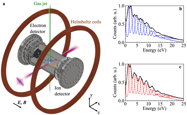
We used a photoelectron-photoion coincidence spectrometer (reaction microscope, ReMi) [27, 28, 29, 30] to disentangle the photoelectron spectra resulting from the photoionisation of an equal mixture of CH4 and CD4 molecules (see Fig. 1a). The ion spectra measured using the XUV attosecond pulse train alone are dominated by the ionic fragments CH-CH and CD-CD [31, 32]. Figure 1b and c show the total photoelectron spectra (black lines), and those measured in coincidence with the ions CH and CH (blue dashed and dotted line, respectively) (Fig. 1b), and CD and CD (red dashed and dotted line, respectively) (Fig. 1c). The channel-resolved photoelectron spectra present a clear harmonic structure. The results are consistent with the measurements obtained by photoelectron-photoion coincidence spectroscopy in combination with monochromatic XUV radiation [33]. The capability to measure in coincidence the photoelectrons and the corresponding ions is fundamental for characterising the response of the two molecules under the same conditions, thus ruling out the effect of experimental instabilities, and to disentangle the contribution of the dissociating and non dissociating ionization channels.
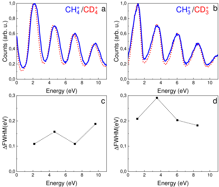
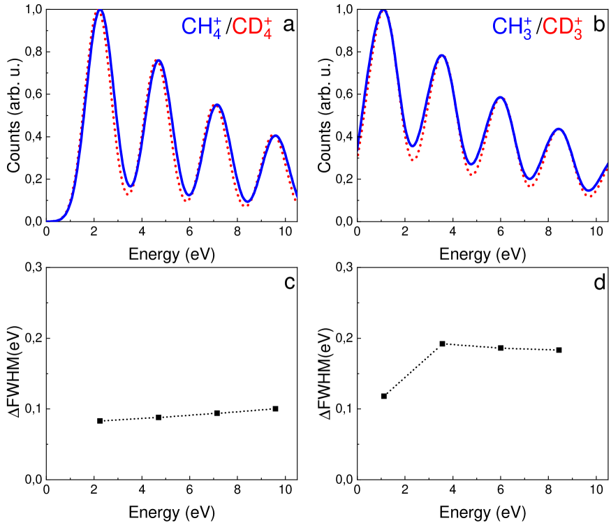
The width of a single photoelectron peak is broader than the bandwidth of the corresponding XUV harmonic, estimated in about 200 meV (full-width at half maximum; FWHM). The broadening is due to the combination of the finite resolution of the photoelectron spectrometer (see Methods section) and the spectral width of the vibronic absorption band of methane and deuteromethane [33]. We observe that the width of the single photoelectron peak for the XUV-only photoionisation process presents an isotopic dependence as shown in Extended Data Fig. 1a,b. In particular, the FWHM of the single photoelectron peak measured in coincidence with the ionic channels CH and CH (blue solid lines in Extended Data Fig. 1a and b, respectively) are larger than the corresponding quantities measured in coincidence with the ions CD and CD (red dotted lines), respectively. This is demonstrated in Extended Data Fig. 1c,d, which report the difference of the FWHM (FWHM=FWHM-FWHM) for the photoelectron peaks measured in coincidence with ions associated to the CH4 and CD4 molecules. As shown later, the broadening of the photoelectron peaks and the different widths can be explained considering the autocorrelation function of the two isotopologues.
Attosecond photoelectron interferometry in methane and
deuteromethane
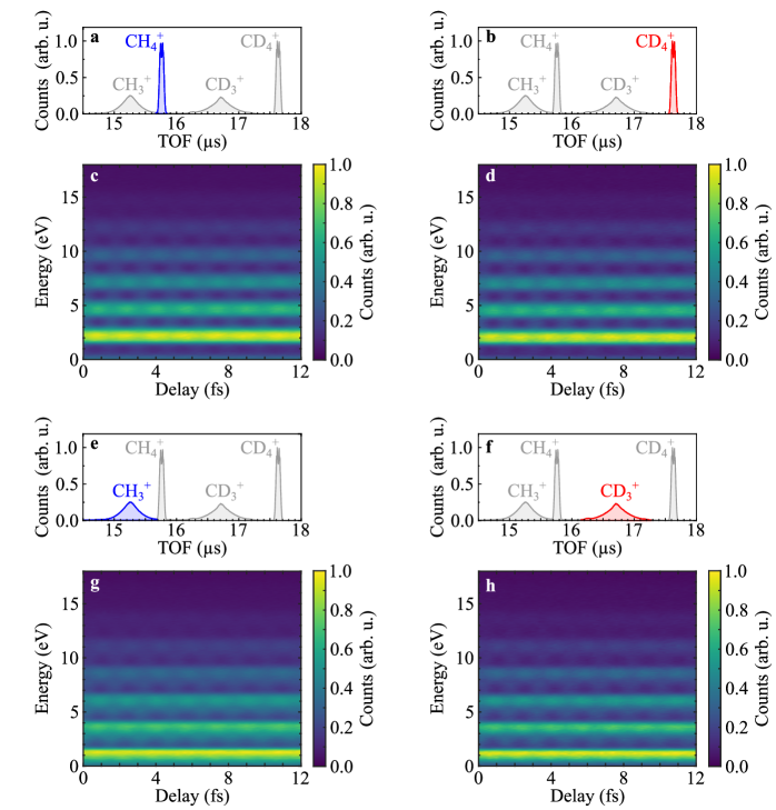
When photoionisation takes place in the presence of a synchronised IR field, additional photoelectron peaks (sidebands) appear between the main photoelectron lines, due to the interference between two photoionisation pathways leading to the same final state. These involve the absorption of an XUV photon from consecutive harmonics and the absorption or emission of an additional IR photon by the outgoing photoelectron [17]. The yield of the sidebands measured in coincidence with the ions CH, CD, CH, and CD (see ion TOF presented in Fig. 2a,b,e and f) clearly oscillate as a function of the relative delay between the attosecond pulse train and the IR field, as shown in Fig. 2c,d,g and h, respectively . The amplitude offset (), oscillation amplitude () and phase offset () of these oscillations were obtained by integrating the signal over an energy window of meV around the sideband maxima and by fitting the oscillations with the function:
| (1) |
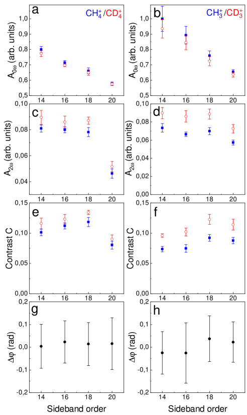
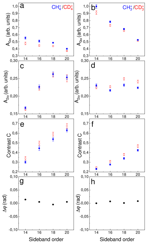
where indicates the frequency of the IR field driving the HHG process, is the kinetic energy of the photoelectron and is the sideband order. In Fig. 3, we present the comparison between the coefficients (Fig. 3a,b), (Fig. 3c,d) and the ratio (Fig. 3e,f), which corresponds to the contrast of the sideband oscillations, for the ionic channels resulting from photoionisation of CH4 (blue full squares) and CD4 (red open circles). Moreover, we also show the channel-resolved difference of the phase of the sideband oscillations for the channels CH-CD (Fig. 3g) and CH-CD (Fig. 3h). A detailed description of the analysis followed to isolate the different terms is presented in the supplementary information (SI).
The amplitude offsets are slighlty larger for the CH channel than for CD (Fig. 3a), as well as for CH with respect to CD (Fig. 3b). The amplitude of the oscillations presents an opposite behaviour, as shown in Fig. 3c and d, with a clear isotopic difference between CH and CD (Fig. 3d). As a result, the contrast of the sideband oscillations turns out to be larger in the fragments originating from the CD4 molecule (Fig. 3e,f). Interestingly, the differences of the phases for the pairs CH-CD, and CH-CD do not present a significant variation (see Fig. 3g and h). The experimental data indicate the presence of isotopic effects in the two-color photoionisation process that manifest themselves in the amplitude and contrast of the sideband oscillations, and not in the phase of the oscillations. The effect on the contrast suggests that the coherence properties of the two-color photoionisation [34] process are affected by the isotopic substitution.
Theoretical modelling and comparison with the experiment
The RABBIT traces were simulated using perturbation theory, including up to second-order effect to take into account the exchange of two photons with the XUV-IR field (see Theoretical Model in Supplementary Information) [35]. One- and two-photon electronic matrix elements were calculated using the stationary multiphoton molecular R-Matrix approach [36, 37]. The effect of nuclear dynamics in the XUV-only and in the XUV-IR photoionisation processes was modelled using the vibronic auto-correlation function [38], where indicates the time elapsed since the absorption of the XUV photon. The autocorrelation function expresses the overlap between the time dependent nuclear wave packet at the instant and the initial wavepacket , created in the cation at the time zero by the absorption of the energetic XUV photon ( indicate the nuclear coordinates). A closely related approach was already adopted to describe nuclear-motion effects in HHG [12, 16, 39, 40] and attosecond electron-hole migration [8, 41, 42]. The modulation depth of the sideband as a function of the final electron momentum () is proportional to the product of the electronic matrix element and the Fourier transform of the nuclear autocorrelation :
| (2) |
where is the vibrational energy and is averaged over the ground state (zero-point-energy) vibrational function (see Supplementary Equations (7,8)).
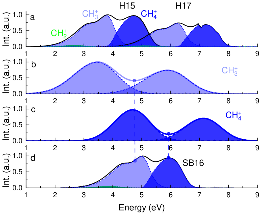
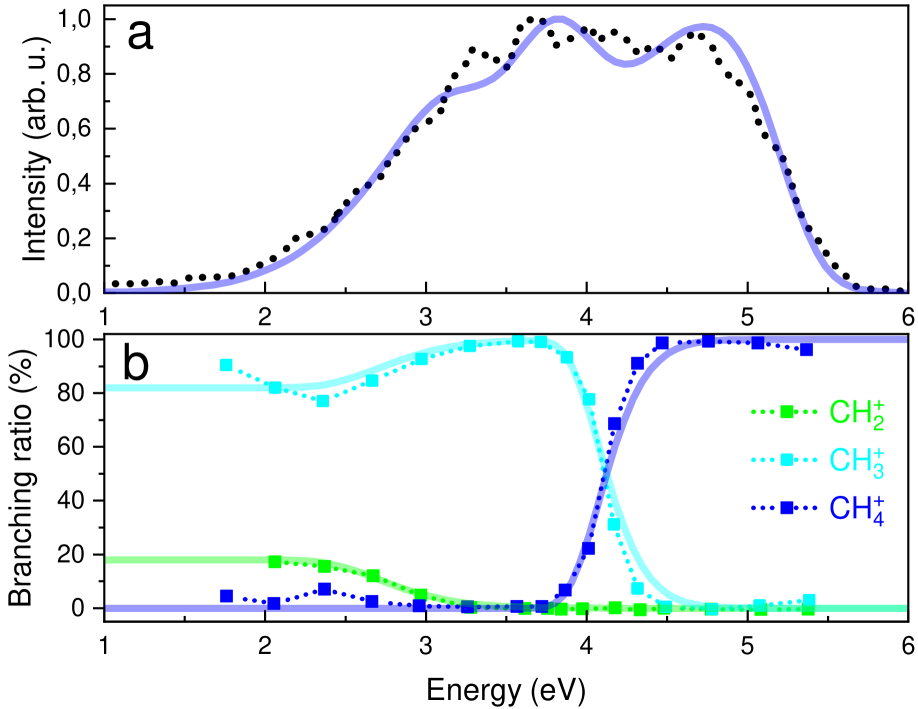
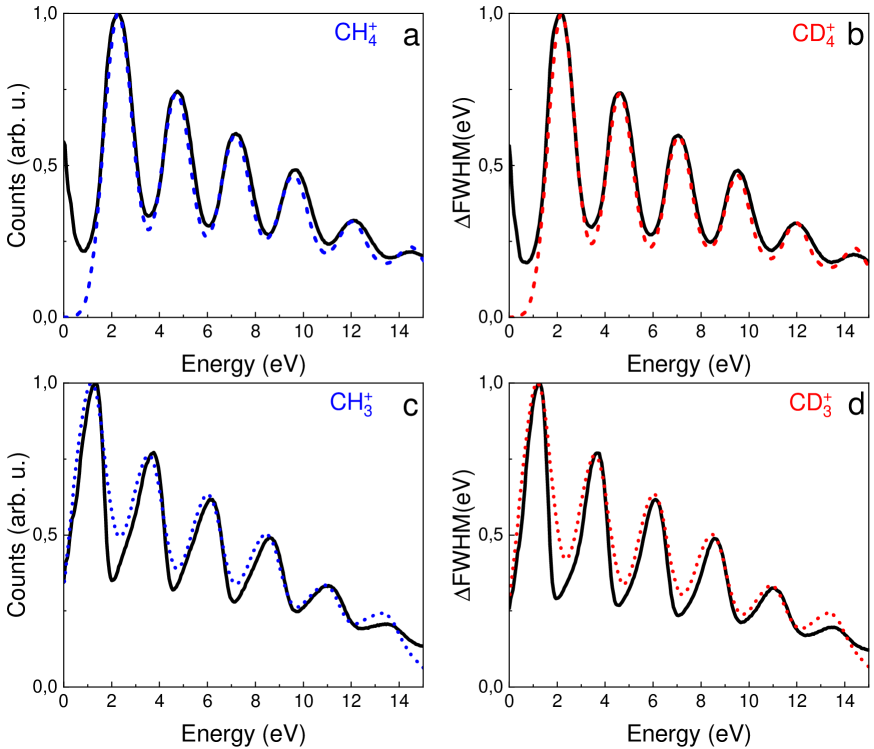
The simulated photoelectron spectra due to the absorption of a single XUV photon from the harmonic-order and are presented in Fig. 4a (black lines). The overall spectral shape is dominated by the shape of the function (with the photoelectron kinetic energy ) and matches the photoelectron spectra measured with monochromatic XUV radiation well [33] (see Extended Data Fig. 2a). The overall photoelectron spectra can be decomposed into three contributions corresponding to the dissociative (CH and CH) and non-dissociative (CH) channels [43], respectively. We used filters based on the measured branching ratios [33] to isolate these three contributions (see Extended Data Fig. 2b), thus reproducing the ion-resolved photoelectron detection of the experiment. The result is represented by the shaded areas shown in Fig. 4a for the channel CH (green area), CH (light-blue area), and CH (blue area). The XUV-only spectrum is constructed as the sum of the contributions of the different harmonics (H15 and H17 in Fig. 4), convoluted with the response function of the photoelectron spectrometer, as shown for the XUV-only spectrum associated with the ions CH (Fig. 4b) and CH (Fig. 4c), respectively. A similar procedure, considering the autocorrelation function for CD4 was used for simulating the XUV spectra associated with the ions CD and CD.
The simulated XUV-only spectra reproduce the experimental photoelectron spectra accurately (see Extended Data Fig. 3a,b). The difference of the spectral widths of the simulated peaks match qualitatively those of the experimental photoelectron lines measured in coincidence with the ions CH-CD and CH-CD, as shown by the comparison between Extended Data Fig. 1c,d and Extended Data Fig. 3c,d, respectively. In particular, the larger difference in the dissociating channels (CH-CD; Extended Data Fig. 3d) with respect to the non-dissociating one (CH-CD; Extended Data Fig. 3c) is reproduced in the simulations.
The good agreement between the channel-resolved experimental data measured in the XUV-only case and the simulated spectra allows one to extend the approach to the two-color photoionisation process for both isotopologues, including also the contribution of the sideband photoelectron peaks. Figure 4d presents the simulated spectrum for the sideband SB16, together with the filtered contributions corresponding to the ionic channels CH (green area), CH (light-blue area), and CH (blue area). As expected, the maxima of the sidebands associated to the CH (light-blue) and CH (blue) channels correspond to the minima between consecutive harmonics of the corresponding XUV spectrum, as indicated by the vertical dashed lines in Fig. 4b,c,d. We constructed the complete RABBIT traces generated by the two-color process as the incoherent sum of the high (low) energy contributions of the main photoelectron lines and of the sideband peaks for the ionic channels CH (CH). The incoherent sum is justified by the observation that the one- and two-photon pathways leading to the same final photoelectron energy, will access different vibrational states of the ion, therefore suppressing the interference between the two paths. The validity of this approach is supported by the good agreement between the delay () integrated experimental and simulated RABBIT traces (see Extended Data Fig. 4).
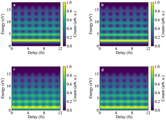
The simulated RABBIT spectrograms (reported in Extended Data Fig. 5) were analysed according to Eq. 1 to extract the coefficients , , and . The results are presented in Fig. 5 and are in close qualitative agreement with the experimental findings. The model reproduces the differences in between the ionic channels observed in the experiment (see Fig. 5a,b). In particular, the amplitude offsets of the sidebands associated with the ionic fragments CH (Fig. 5a) and CH (Fig. 5b) are larger than the corresponding quantities in CD and CD. Furthermore, the model reproduces the trends in the amplitudes , with a negligible difference for the pair CH-CD (Fig. 5c) and a significant one for the ionic fragments CH-CD (Fig. 5d). These observations are in good agreement with the experimental results shown in Fig. 4c,d. As in the experiment, the final result is an increased contrast of the oscillations for the sidebands originating from the CD4 molecule (Fig. 5e,f), which is particularly pronounced in the comparison CD-CH (Fig. 5f). In general, the amplitude of the oscillations at frequency and the contrast estimated from the numerical model are higher than those measured in the experiment. We attribute these differences to the effect of averaging over the interaction volume and small distortions in the electric and magnetic fields used in the photoelectron spectrometer, which might lead to a partial smearing out of the sideband oscillations in the experimental data.
Finally, the model also reproduces the absence of significant variation in the phase difference between homologue isotopic species (CH-CD and CH-CD) (Fig. 5g,h).
Interpretation
The isotopic differences observed in the experiment can be interpreted considering the autocorrelation function and its Fourier Transform in the spectral domain, together with the decomposition of the photoelectron spectra in the high- and low-energy components presented in Fig. 4 (hereafter, we will use the subscripts and to indicate quantities related to the isotopologues CH4 and CD4, respectively).
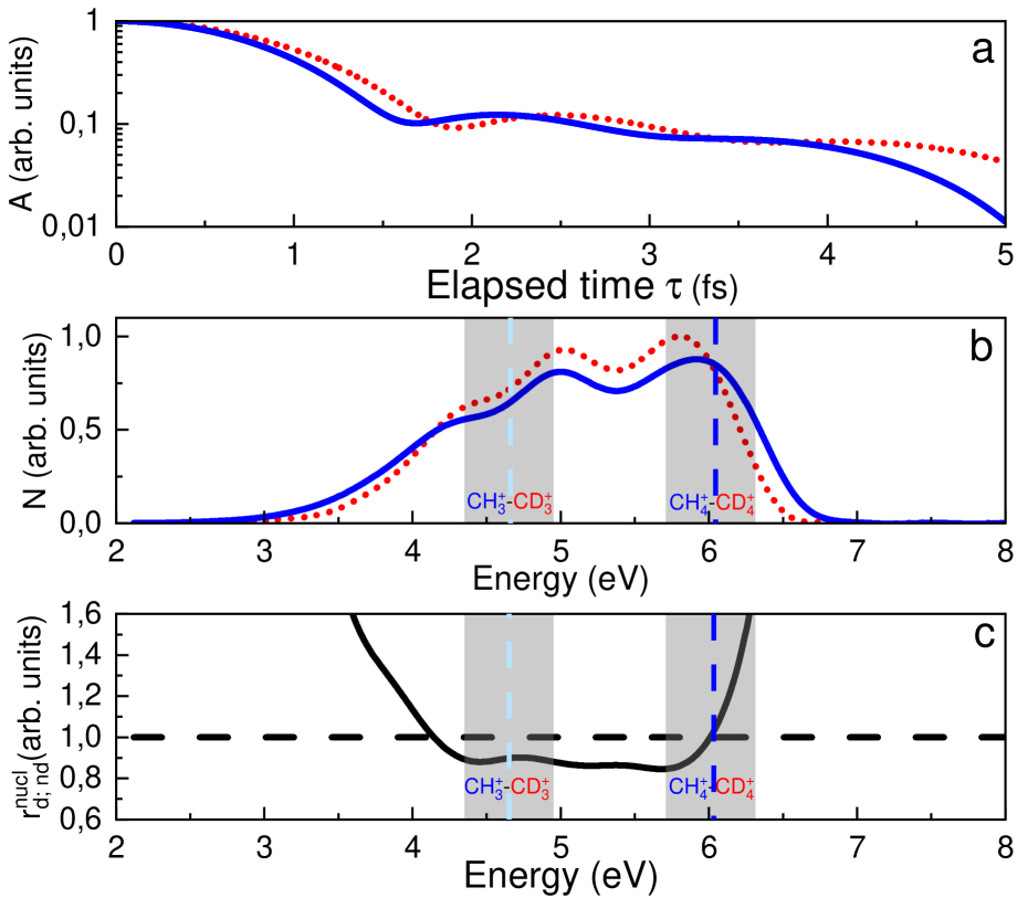
Due to its lighter mass, the dynamics triggered in the cation CH by the absorption of an XUV photon is faster than in CD, leading to a faster decay in the very first few femtoseconds of the autocorrelation function with respect to (blue solid line and red dotted line, respectively in Fig. 6a).
In the energy domain, the difference between the decay times determines a different broadening of their Fourier transforms, resulting in the function (blue solid line) being broader than (red dotted line) both at low and at high energies, as shown in Fig. 6b.
As shown in Extended Data Fig. 2, the shape of the functions reproduces well the photoelectron spectra generated by each harmonic of the XUV spectrum. Moreover, the single photoelectron peak can be decomposed in a high energy part associated to the ionic channels CH-CD and low energy contribution associated (mainly) to CH-CD (see Fig. 4). The combination of these observations indicates that the larger broadening of the photoelectron peaks measured in coincidence with the ionic channel CH with respect to CD is due to the larger width of the function at high energies (see Extended Data Fig. 1a,c). Similarly, the larger broadening of the photoelectron peaks measured in coincidence with the ionic channel CH is due to the differences of the functions and for the two isotopologues at low energies (see Extended Data Fig. 1b,d).
In the two-color photoionisation measurements, the differences observed in the amplitudes can be also explained by the different spectral width of the functions and . The larger component in CH and CH with respect to their deuterated counterparts can be ascribed to the larger width of the XUV-only spectra, which determines a larger contribution of the photoelectron released by the absorption of a single XUV photon to the energy intervals between consecutive harmonics, where the two-photon (XUV-IR) sideband signal is located (see Fig. 4d).
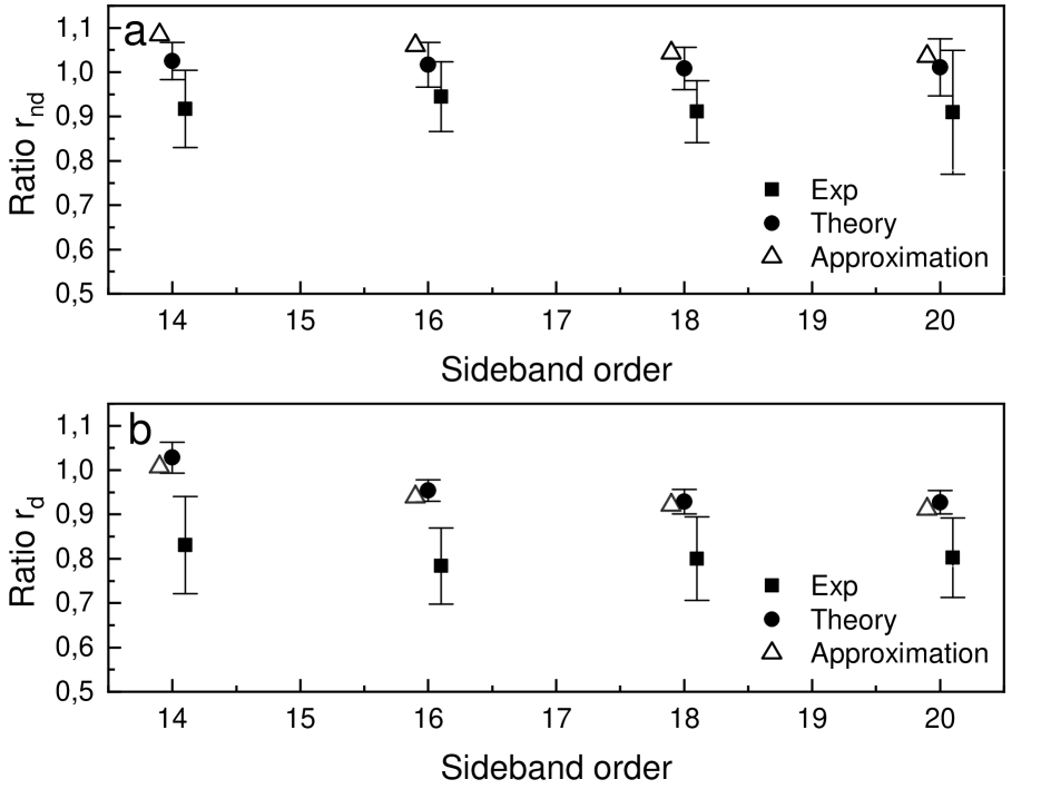
For the interpretation of the evolution in the components, we observe that the difference between the non-dissociating channels CH-CD is significantly smaller than for the dissociating channels CH-CD, both in the experiment (Fig. 3c,d) and in the simulations (Fig. 5c,d). We can quantify this difference introducing the ratio between the amplitude of the sideband oscillations for the dissociating () and non-dissociating () channels. The ratio is very close to one and larger than both in the experiment and simulations, as shown in Extended Data Tab. 1and Extended Data Fig. 6a,b.
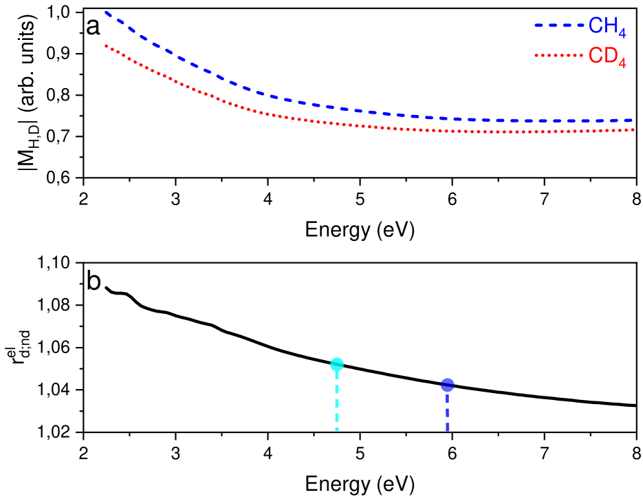
In our numerical model the amplitude of the oscillations of the sideband intensities is proportional to the modulus of the Fourier transform of the modulation depth at the frequency (see Eq. 2). This can be expressed as the product of the modulus of an electronic term () (shown in Extended Data Fig. 7a) and a nuclear contribution given by the function . (see Supplementary Information). As a result, the ratios and can be expressed as the product of a ratio for the electronic contribution () and one for the nuclear part ():
| (3) |
In the model, the ratio of the electronic terms depends only weakly on the dissociating or non-dissociating channel, through its energy dependence. For example, they assume the values and at the center of the sideband SB16 for the dissociating and non dissociating channels, respectively (see Extended Data Fig. 7b). The values for the other sidebands are reported in Table 1, indicating that the electronic part of the amplitude of the sideband oscillation in CH4 is larger than in CD4. This ratio is modified by the nuclear contribution as shown in Fig. 6b; at the center of the region corresponding to the sidebands measured in coincidence with the ion CH-CD (indicated by the light blue vertical dashed line) the function (blue solid line) is smaller than (red dotted line), leading to a ratio of the nuclear contribution lower than one, which compensates the ratio of the electronic part . For the non-dissociating channel CH-CD (vertical blue dashed line), the ratio of the two curves is close to one.
We note that both in the experiment and in the simulations the sideband photoelectrons are integrated over an energy region (grey shaded areas in Fig. 6b,c) of about meV around the sideband maxima. In the case of the dissociation channel (CH and CD) (grey shaded area on the left handside), the function remains smaller than over the entire interval, while for the non dissociating channel (CH and CD; grey shaded area on the right-hand side), the two curves cross. The nuclear contribution can be approximated as the ratio of the integrals of the curves over the energy intervals corresponding to the sidebands, returning the values and . The results are summarized in Extended Data Table 1. The factorisation of the ratio in an electronic and nuclear contribution and its approximate estimation are in good quantitative agreement with the results from the theoretical simulations and in fair agreement with the experimental results (see Extended Data Fig. 6). This analysis clearly indicates that the nuclear dynamics after the XUV photoionisation event plays an important role in determining the channel- and isotope-dependence of the amplitude of oscillations of the sidebands.
In the theoretical model, the phase difference between the phases of the sideband oscillations of different isotopic channels originates only from the energy dependence of the electronic part of the matrix dipole moment, as the nuclear contribution given by the functions presents only a real part. As a result, the differences are on the order of rad (Fig. 5g,h) and are well within the typical error bar ( rad) of our experimental data (Fig. 3g,h).
Conclusions.
We have shown that the nuclear dynamics after XUV absorption significantly affects the amplitude of the two-color photoionisation signal in attosecond photoelectron interferometry in molecules. The effects can be interpreted in the spectral domain considering the Fourier transform of the autocorrelation function. The implementation of two isotopologues allows one to highlight the effect of the nuclear dynamics exploiting the different decay times determined by the isotopic substitution. The short duration of the autocorrelation in the time domain (only a few femtoseconds) introduces a finite coherence time for the interaction of the attosecond pulse trains with the molecule. The coherence of the correlated electronic-nuclear wave packets is expected to play a major role in several molecular systems characterized by an ultrafast dynamics after the photoionisation event [8] and for the advancement of attosecond metrology [34].
Acknowledgments
This project has received funding from the European Union’s Horizon 2020 research and innovation programme under the Marie Sklodowska-Curie grant agreement no. 641789 MEDEA. D.B. acknowledges support from the Swedish Research Council grant 2020-06384. G.S. acknowledges financial support by the Deutsche Forschungsgemeinschaft project Priority Program 1840 (QUTIF), and grant 429805582 (Project SA 3470/4-1). I.M. and G.S. acknowledge financial support by the BMBF project 05K19VF1, the Deutsche Forschungsgemeinschaft project Research Training Group DynCAM (RTG 2717), and Georg H. Endress Foundation. D.E. and G.S. acknowledge support and funding by the DFG project International Research Training Group (IRTG) CoCo 2079 and INST 39/1079 (High-Repetition-Rate Attosecond Source for Coincidence Spectroscopy). Z.M. acknowledges support of the PRIMUS (20/SCI/003) project and Czech Science Foundation (20-15548Y). Computational resources were supplied by the project “e-Infrastruktura CZ” (e-INFRA LM2018140) provided within the program Projects of Large Research, Development and Innovations Infrastructures. This work was also supported by the Ministry of Education, Youth and Sports of the Czech Republic through the e-INFRA CZ (ID:90140).
Author Contributions Statement
D.E, M.S., H.A., M.M. developed the experimental setup. F.F. and L.P. collaborated in the development of the XUV spectrometer. C.D.S., T.P., R.M. and G.S. contributed to the development of the ReMi spectrometer.
D.E, D.B., H.A., M.M. and I.M. performed the experiments. D.E. and D.B analysed the data. J.B. and Z.M developed the numerical simulations for the calculation of the R-Matrix dipole element. S.P. developed the theory and the code for the inclusion of the nuclear dynamics and the influence of the vibrational ground state. G.S. conceived the idea of the experiment, supervised the work and wrote the manuscript, which was discussed and agreed by all coauthors.
Competing Interests Statement
The authors declare no competing interests.
Correspondence and requests for materials
Correspondence and requests for materials should be addressed to giuseppe.sansone@physik.uni-freiburg.de
References
- 1. Sansone, G., Pfeifer, T., Simeonidis, K. & Kuleff, A. I. Electron correlation in real time. ChemPhysChem 13, 661–680 (2012).
- 2. Nisoli, M., Decleva, P., Calegari, F., Palacios, A. & Martín, F. Attosecond Electron Dynamics in Molecules (2017).
- 3. Goulielmakis, E. et al. Real-time observation of valence electron motion. Nature 466, 739–743 (2010).
- 4. Niikura, H. et al. Probing molecular dynamics with attosecond resolution using correlated wave packet pairs. Nature 421, 826–829 (2003).
- 5. Meckel, M. et al. Laser-induced electron tunneling and diffraction. Science 320, 1478–1482 (2008).
- 6. Wolter, B. et al. Ultrafast electron diffraction imaging of bond breaking in di-ionized acetylene. Science 354, 308–312 (2016).
- 7. Lara-Astiaso, M., Palacios, A., Decleva, P., Tavernelli, I. & Martín, F. Role of electron-nuclear coupled dynamics on charge migration induced by attosecond pulses in glycine. Chemical Physics Letters 683, 357–364 (2017).
- 8. Vacher, M., Bearpark, M. J., Robb, M. A. & Malhado, J. a. P. Electron dynamics upon ionization of polyatomic molecules: Coupling to quantum nuclear motion and decoherence. Phys. Rev. Lett. 118, 083001 (2017).
- 9. Lein, M. Attosecond probing of vibrational dynamics with high-harmonic generation. Phys. Rev. Lett. 94, 053004 (2005).
- 10. Baker, S. et al. Probing proton dynamics in molecules on an attosecond time scale. Science 312, 424–427 (2006).
- 11. Haessler, S. et al. Attosecond chirp-encoded dynamics of light nuclei. Journal of Physics B: Atomic, Molecular and Optical Physics 42, 134002 (2009).
- 12. Patchkovskii, S. Nuclear dynamics in polyatomic molecules and high-order harmonic generation. Phys. Rev. Lett. 102, 253602 (2009).
- 13. Austin, D. R. et al. Extracting sub-cycle electronic and nuclear dynamics from high harmonic spectra. Sci Rep-Uk 11, 2485 (2021).
- 14. Vacher, M., Boyer, A., Loriot, V., Lépine, F. & Nandi, S. Few-femtosecond isotope effect in polyatomic molecules ionized by extreme ultraviolet attosecond pulse trains. The Journal of Physical Chemistry A 126, 5692–5701 (2022).
- 15. Gonçalves, C. E. M., Levine, R. D. & Remacle, F. Ultrafast geometrical reorganization of a methane cation upon sudden ionization: an isotope effect on electronic non-equilibrium quantum dynamics. Phys. Chem. Chem. Phys. 23, 12051–12059 (2021).
- 16. Patchkovskii, S. & Schuurman, M. S. Full-dimensional treatment of short-time vibronic dynamics in a molecular high-order-harmonic-generation process in methane. Phys. Rev. A 96, 053405 (2017).
- 17. Paul, P. M. et al. Observation of a train of attosecond pulses from high harmonic generation. Science 292, 1689–1692 (2001).
- 18. Krausz, F. & Ivanov, M. Attosecond physics. Reviews of Modern Physics 81, 163–234 (2009).
- 19. Huppert, M., Jordan, I. & Wörner, H. J. Attosecond beamline with actively stabilized and spatially separated beam paths. Rev. Sci. Instrum. 86, 123106 (2015).
- 20. Nandi, S. et al. Attosecond timing of electron emission from a molecular shape resonance. Sci. Adv. 6, 7762–7793 (2020).
- 21. Vos, J. et al. Orientation-dependent stereo Wigner time delay and electron localization in a small molecule. Science (80-. ). 360, 1326–1330 (2018).
- 22. Ahmadi, H. et al. Attosecond photoionisation time delays reveal the anisotropy of the molecular potential in the recoil frame. Nat. Commun. 13, 1242 (2022).
- 23. Haessler, S. et al. Phase-resolved attosecond near-threshold photoionization of molecular nitrogen. Phys. Rev. A 80, 011404 (2009).
- 24. Cattaneo, L. et al. Attosecond coupled electron and nuclear dynamics in dissociative ionization of H2. Nat. Phys. 14, 733–738 (2018).
- 25. Cattaneo, L. et al. Isolating Attosecond Electron Dynamics in Molecules where Nuclei Move Fast. Phys. Rev. Lett. 128 (2022).
- 26. Rabalais, J. W., Bergmark, T., Werme, L. O., Karlsson, L. & Siegbahn, K. The jahn-teller effect in the electron spectrum of methane. Phys. Scr. 3, 13–18 (1971).
- 27. Moshammer, R., Unverzagt, M., Schmitt, W., Ullrich, J. & Schmidt-Böcking, H. A 4 recoil-ion electron momentum analyzer: a high-resolution “microscope” for the investigation of the dynamics of atomic, molecular and nuclear reactions. Nucl. Instruments Methods Phys. Res. Sect. B Beam Interact. with Mater. Atoms 108, 425–445 (1996).
- 28. Ullrich, J. et al. Recoil-ion momentum spectroscopy. J. Phys. B At. Mol. Opt. Phys. 30, 2917–2974 (1997).
- 29. Dörner, R. et al. Cold Target Recoil Ion Momentum Spectroscopy: A ’momentum microscope’ to view atomic collision dynamics. Phys. Rep. 330, 95–192 (2000).
- 30. Ullrich, J. et al. Recoil-ion and electron momentum spectroscopy: reaction-microscopes. Reports Prog. Phys. 66, 1463–1545 (2003).
- 31. Chupka, W. A. Mass-spectrometric study of the photoionization of methane. J. Chem. Phys. (1968).
- 32. Latimer, C. J., Mackie, R. A., Sands, A. M., Kouchi, N. & Dunn, K. F. The dissociative photoionization of methane in the VUV. J. Phys. B At. Mol. Opt. Phys. 32, 2667–2676 (1999).
- 33. Field, T. A. & Eland, J. H. The fragmentation of CH4+ ions from photoionization between 12 and 40 eV. J. Electron Spectros. Relat. Phenomena 73, 209–216 (1995).
- 34. Bourassin-Bouchet, C. et al. Quantifying decoherence in attosecond metrology. Phys. Rev. X 10, 031048 (2020).
- 35. Patchkovskii, S., Benda, J., Ertel, D. & Busto, D. Theory of nuclear motion in rabbitt spectra (2022).
- 36. Benda, J. & Mašín, Z. Multi-photon above threshold ionization of multi-electron atoms and molecules using the r-matrix approach. Sci Rep-Uk 11, 11686 (2021).
- 37. Benda, J., Mašín, Z. & Gorfinkiel, J. D. Analysis of rabitt time delays using the stationary multiphoton molecular r-matrix approach. Phys. Rev. A 105, 053101 (2022).
- 38. Heller, E. J. The semiclassical way to molecular spectroscopy. Accounts of Chemical Research 14, 368–375 (1981).
- 39. Mondal, T. & Varandas, A. J. C. Structural evolution of the methane cation in subfemtosecond photodynamics. The Journal of Chemical Physics 143, 014304 (2015).
- 40. Mondal, T. & Varandas, A. J. C. On extracting subfemtosecond data from femtosecond quantum dynamics calculations: The methane cation. Journal of Chemical Theory and Computation 10, 3606–3616 (2014).
- 41. Arnold, C., Vendrell, O. & Santra, R. Electronic decoherence following photoionization: Full quantum-dynamical treatment of the influence of nuclear motion. Phys. Rev. A 95, 033425 (2017).
- 42. Ruberti, M., Patchkovskii, S. & Averbukh, V. Quantum coherence in molecular photoionization. Phys. Chem. Chem. Phys. 24, 19673–19686 (2022).
- 43. Mahan, B. H. & O’Keefe, A. Electron impact dissociation of CH4(CD4): Laser induced fluorescence of product CH+(CD+). Chem. Phys. 69, 35–44 (1982).
Methods
Methods
Experimental Methods
Trains of attosecond pulses with photon energies up 50 eV were generated in krypton using a 20 fs driving infrared pulses centered at nm at 50 kHz repetition rate. The intensity of the field driving the HHG process was estimated in I=1014 W/cm2 The temporal delay between the XUV radiation and the IR field was changed in a collinear geometry using a pair of drilled plates [1, 2]. The two-color field was focused in the interaction point of the Reaction Microscope using a toroidal mirror operating in one-to-one imaging at an incidence angle of 84∘. The gas target was composed by an equal mixture of CH4 and CD4 molecules. The typical count rate in the measurements was 5-6 kHz and data were acquired for 96 hours. The FWHM of the response function of the REMI photoelectron spectrometer was estimated in 1000 meV. The data discussed in the manuscript were integrated over all emission directions and all orientations of the molecules.
Theoretical Methods
One- and two-photon photoionization matrix elements were calculated using development version of UKRMol+ code [3] and aug-cc-pVTZ atomic basis set aug-cc-pVTZ for the bound states. The continuum basis consisted of a mixed set of Gaussians reaching to the distance 7.5 atomic units, followed by 10 B-splines spanning the remaining distance to the boundary of the R-matrix inner region at atomic units. The calculated energy of the ground neutral state was adjusted so that it was exactly 14.4 eV below the calculated energy of the ground ionic state. Bound molecular orbitals were calculated using the 3-state state-averaged complete active space self-consistent field method with restricted open-shell Hartree-Fock reference for CH in Psi4 v. 1.5 [4]. The lowest three states were equally weighted. Carbon 1s orbitals were frozen in the close-coupling calculations. The next 11 lowest molecular orbitals were used as active, both for the bound-state complete active space calculation as well as to build the square-integrable part of the close-coupling expansion. The continuum part of the expansion employed 100 cationic states. Equilibrium geometries and force-fields for the neutral species were calculated at the MP2(fc)/aug-cc-pVTZ level. The quadratic vibronic Hamiltonian for the cation was obtained by diabatizing MR-CIS/CASSCF(7,4) energies of the levels in the vicinity of the neutral equilibrium structure. The autocorrelation functions were evaluated for 300 atomic units of time ( fs), correponding to maximum spectral resolution of eV. Zero-point corrections to the electronic matrix were evaluated using finite-displacements along normal modes, with 7 distorted structures required for each isotopomer. Orientational averaging was performed using order-17 Lebedev grids. Further details of the numerical parameters and procedures are given in Supplementary Information.
References
- 1. Ahmadi, H., et al. Collinear setup for delay control in two-color attosecond measurements. J. Phys.-Photonics 2, 024006 (2020).
- 2. Ertel, D., et al. Ultrastable, high-repetition-rate attosecond beamline for time-resolved XUV-IR coincidence spectroscopy. https://arxiv.org/abs/2212.04742 (2022).
- 3. Mašín, Z., Benda, J., Gorfinkiel, J. D., Harvey, A. G., Tennyson, J. Ukrmol+: A suite for modelling electronic processes in molecules interacting with electrons, positrons and photons using the r-matrix method. Computer Physics Communications 249, 107092 (2020).
- 4. Smith, D. G. A. et al. PSI4 1.4: Open-source software for high-throughput quantum chemistry. J. Chem. Phys. 152, 184108 (2020).
| Ratio | ||||
|---|---|---|---|---|
| SB order | ||||
| Exp. | ||||
| Theory | ||||
| Ratio | ||||
| SB order | ||||
| Exp. | ||||
| Theory | ||||