Optimization of Permalloy properties for magnetic field sensors using He+ irradiation
Abstract
Permalloy, despite being a widely utilized soft magnetic material, still calls for optimization in terms of magnetic softness and magnetostriction for its use in magnetoresistive sensor applications. Conventional annealing methods are often insufficient to locally achieve the desired properties for a narrow parameter range. In this study, we report a significant improvement of the magnetic softness and magnetostriction in a 30 nm Permalloy film after He+ irradiation. Compared to the as-deposited state, the irradiation treatment reduces the induced anisotropy by a factor ten and the hard axis coercivity by a factor five. In addition, the effective magnetostriction of the film is significantly reduced by a factor ten - below - after irradiation. All the above mentioned effects can be attributed to the isotropic crystallite growth of the Ni-Fe alloy and to the intermixing at the magnetic layer interfaces under light ion irradiation. We support our findings with X-ray diffraction analysis of the textured Ni81Fe19 alloy. Importantly, the sizable magnetoresistance is preserved after the irradiation. Our results show that compared to traditional annealing methods, the use of He+ irradiation leads to significant improvements in the magnetic softness and reduces strain cross sensitivity in Permalloy films required for 3D positioning and compass applications. These improvements, in combination with the local nature of the irradiation process make our finding valuable for the optimization of monolithic integrated sensors, where classic annealing methods cannot be applied due to complex interplay within the components in the device.
I Introduction
Permalloy, a typical soft magnetic Ni-Fe alloy is employed as an active sense layer in several magnetoresistive (MR) sensor applicationsKhan et al. (2021). To have a small magnetostriction and low coercivity, most of these devices are designed around the alloy composition of Ni81Fe19 which also possesses significant anisotropic magnetoresistance (AMR). Optimization of Permalloy for AMR sensors has been studied for a long time Kwiatkowski and Tumanski (1986); Groenland et al. (1992); Trützschler et al. (2014) and includes different aspects: primarily, improvement of magnetic softness and low magnetostriction. To reach that, negligible crystalline anisotropy is firstly required. The single thin film elements typically feature a stripe shaped geometry to induce a strong shape anisotropy, providing the sensor with a well-defined orientation of sensitivity. Furthermore, this design of the sensitive elements ensures a fixed configuration of the magnetic domains, thus enabling a very high signal-to-noise ratio. Additional anisotropies of other sources, if not oriented in the same direction as the shape anisotropy, would hinder this sensitivity direction Jogschies et al. (2015). Moreover, to achieve low hysteresis, the coercivity in the hard axis magnetization direction must be very low and the specific AMR must be as high as possibleLenssen et al. (2000) to maximize sensitivity. Eventually, to avoid parasitic anisotropies, low magnetostriction (source of magnetoelastic anisotropy) is required. In this case, strain in the material has small or negligible impact on the magnetic properties. The low magnetoelastic anisotropy is particularly important for sensors on flexible substrates Khan et al. (2021); Cañón Bermúdez and Makarov (2021); Oliveros Mata et al. (2021); Melzer, Makarov, and Schmidt (2019); Amara et al. (2018) that have attracted great attention in recent years in wearable electronics and biomedical applications. To obtain this particular material property, growth optimizationWang et al. (2012) and annealingIwata and Hagedorn (1969) are viable options. However, none of these technique allow for a local treatment of the film.
It is well known that ion irradiation is an excellent tool to tune locally the magnetic and structural properties of thin films through orderingJiang et al. (2018); Jaafar et al. (2011); Devolder et al. (2013); Ravelosona et al. (2000) and interface intermixingMasciocchi et al. (2022); De Jong et al. (2022); Zhao et al. (2019); Diez et al. (2019). In Permalloy films, ion irradiation has been shown to change the magnetic anisotropyWoods et al. (2002); Schindler, Kernohan, and Weertman (1964); Mougin et al. (2001) and the magneto-resistive response in the presence of exchange biasTrützschler et al. (2014, 2016). However, most of these works use ion implantation Fassbender et al. (2006a); Fassbender and McCord (2006); Fassbender et al. (2006b) or heavy ionsGupta et al. (2008), which can result in significant damage to the sample. This can be avoided by using lighter ions - like He+ - with energies in the range of 10-30 keV Fassbender, Ravelosona, and Samson (2004); Masciocchi et al. (2022). In this way, collision cascades are absent and the structural modifications are confined to the vicinity of the ion path in a metal. Furthermore, the effect of irradiation on the magneto-elastic properties of single Permalloy films and a direct comparison between field free ion irradiation and annealing has not yet been reportedBaglin et al. (1997).
In this work, we propose and explore the use of He+ ion irradiation on sputtered layer of Ni81Fe19(30 nm) as material preparation for magnetic field sensors and we compare it with standard field free annealing. Using Kerr microscopy and Vibrating Sample Magnetometry (VSM) we show that 20 keV He+ ions significantly reduce the coercivity and the induced magnetic anisotropy of our magnetic material. The result is a soft magnetic film with in-plane magnetic anisotropy J/m3 and coercive field mT, which is a further improvement over the values that can be obtained by field-free annealing process by a factor 5 and 10, respectively. The anisotropy measurements are supported by a detailed comparison using the remanent domain pattern. Additionally, we show that the polycrystalline magnetostriction can be progressively reduced by a factor ten for irradiation doses of cm-2. This reduction in magnetoelastic coupling is attributed to crystallization and changes to the interface magnetostriction caused by intermixing at the magnetic layer boundaries. We support our findings with structural characterization performed using X-ray diffraction (XRD). The results show an overall improvement in the crystallization after irradiation and annealing. We attribute the reduction in magnetic anisotropy to the absence of a preferential direction of atomic ordering and to stress relaxation during irradiation. As post growth He+ ion irradiation improves magnetic softness and minimizes strain cross sensitivity of Permalloy, AMR magnetic sensors with high sensitivity and low hysteresis can be envisioned even for integrated devices.
II Experimental methods
The samples have been prepared by DC magnetron sputtering using a Singulus Rotaris system on a 1.5 m thick, thermally oxidized SiOx on top of a 625 m thick substrate. A layer of Ni81Fe19 (30 nm) is sputtered at room temperature in the presence of a rotating magnetic field of 5 mT on a NiFeCr (5 nm) seed layer and capped with 4 nm of Ta as shown in Fig. 1 (b). The following sputtering conditions were used for the magnetic layer growth: base pressure mbar, sputtering power 1200 W and Ar+ flow 90 sccm. The seed layer is used to promote a NiFe (111) texture during growth and it is known to improve magnetoresistanceFassbender et al. (2006a). After deposition, optical lithography and ion etching have been used to pattern arrays of disks (80 m of diameter and 3 m of spacing) on the samples in order to probe the local film properties. Multiple copies of the samples have been irradiated at an energy of 20 keV with different fluences of He+ ions from to cm-2. At these irradiation conditions, the majority of the ions reach the substrate (roughly 94 from Monte Carlo TRIM Ziegler, Ziegler, and Biersack (2010) simulations, not shown), resulting in homogeneous irradiation of the entire layer stack.
To compare the effect of ion irradiation to thermal annealing, the same magnetic material has been consecutively annealed for three hours at 200, 265 and 300∘C at a pressure of 10-7 mbar. In order to avoid a magnetization induced preferential direction of orderingFassbender and McCord (2006); Okay et al. (2018), external magnetic fields have been minimized during the irradiation and annealing steps. The thin film magnetic properties have been measured with Kerr microscopy and VSM. The magnetic properties of our films are summarized in Table 1. Due to the negligible implantationFassbender, Ravelosona, and Samson (2004), the value of the Young’s modulus is assumed to be unaffected by our irradiation and annealing step. Electrical measurement of anisotropic magnetoresistance (AMR) have been performed with four contacts in line in the presence of a rotating magnetic field of 10 mT.
| (T) | (J/m3) | (mT) | x | (GPa) | |
|---|---|---|---|---|---|
| as-deposited | 0.95(1) | 78(5) | 0.20(5) | -0.7(1) | 200Klokholm and Aboaf (1981) |
| Ann. 265∘C | 0.95(1) | 70(5) | 0.15(5) | +0.04(9) | 200Klokholm and Aboaf (1981) |
| He+ cm-2 | 0.91(1) | 8(7) | 0.05(5) | +0.01(9) | 200Klokholm and Aboaf (1981) |
To apply strain to our devices, the substrate was bent mechanically with a three-point bending method. As reported in our previous work Masciocchi et al. (2021) a tensile and uniaxial strain is generatedRaghunathan, Snyder, and Jiles (2009). Moreover the strain is uniform in the central area of the sample and thus in the measured region. As the thin films are in total 40 nm thick, we assume that the strain is entirely transferred from the substrate and that shear strain is negligible. Structural modifications caused by ion irradiation and annealing were probed by X-Ray Diffraction (XRD) using a Bruker D8 Discover system. Angular scans and rocking curve measurements were performed on 1 by 1 cm samples.
III Results and discussion
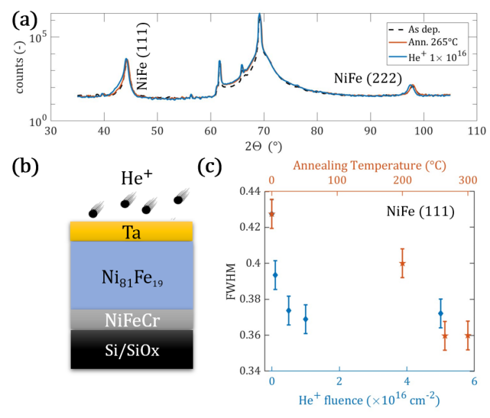
To compare the structural modifications induced by different material treatment on a Ni-Fe alloy, XRD measurements on the Ni81Fe19 (30 nm) film as-deposited and after irradiation and annealing are performed and reported in Fig. 1. Fig. 1 (a) shows angular scan of the Permalloy film. A well defined crystalline texture of NiFe (111) (and its second order peak) is present for the material in the as-deposited state and persists after irradiation and annealing in all the fluence and temperature range explored. The full width at half maximum (FWHM) of the (111) peak is reported in Fig. 1 (c) as a function of the irradiation fluence (blue diamonds) and the temperature during annealing (orange pentagrams). In both cases, the FWHM of the (111) peak decreases by about 15 with increasing ion fluence and annealing temperature with respect to the as-deposited case. The crystallite size (or the size of a coherently diffracting domain in the material) is a fundamental property that can be extracted from XRD profileAhmadipour et al. (2016). According to the Scherrer equationZak et al. (2011),
| (1) |
the size of crystallites is inversely proportional to the FWHM of a diffraction peak. Here is a dimensionless shape factor, D the crystallite size, the wavelength of the Cu-K radiation, the diffraction angle and is the line broadening at FWHM of the XRD peak in radians, after subtracting the instrumental line broadening. As our measurements show, both annealing at 265∘C and ion irradiation with a fluence of cm-2 increase the size of crystallites in our films. The estimated size of the diffracting domains using eq. 1 is 22(1) nm for the as-deposited case and 24(1) nm after the two material treatments. Additionally, rocking curve measurements of the NiFe (111) peak were preformed and more information can be found in section S1 of the supplementary material. Both for the irradiated samples and for the annealed ones a decrease in the FWHM of the rocking curve is observed indicating improvement in the film crystalline phasePark, Norton, and Selvamanickam (2005).
The major effect of room temperature irradiation has been shown to be improved material uniformityvan der Jagt et al. (2022) and interface intermixingMasciocchi et al. (2022). In the same way, thermal annealing is widely used to induce crystallizationWang et al. (2009) and promote atomic diffusionSchulz et al. (2021). Similar effects have been observed in literature for amorphous alloys, where annealing McCord et al. (2005) and He+ irradiationDevolder et al. (2013); Ravelosona et al. (2000) providing high short range atomic mobility allow a mechanism for growth of the ordered phase at the expense of its disordered or less ordered counterpart.
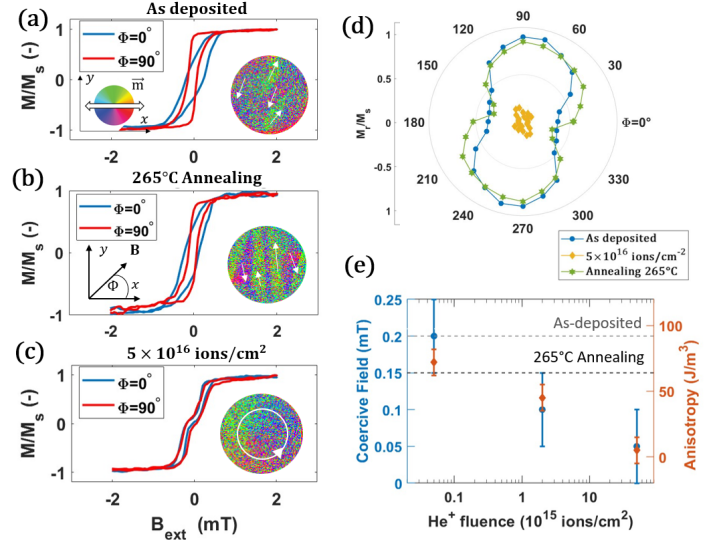
The thin film magnetic properties have been measured with Kerr microscopy and are reported in Fig. 2. Figs. 2 (a)-(c) report the hysteresis curves for the NiFeCr(5 nm)/Ni81Fe19(30 nm)/Ta(4 nm) sample for two perpendicular in-plane directions of the applied magnetic field: (a) for the as-deposited state, (b) after annealing and (c) after irradiation. The curves refer to the magnetic contrast of the structured film into 80 m disks.
The magnetic response of the Permalloy film in the as-deposited state can be seen in Fig. 2 (a). As the magnetization curves at and are different, a weak uniaxial magnetic anisotropy , is present in the as-deposited Ni81Fe19 and might be associated to internal stresses during the material growth or asymmetries in the deposition systemZou, Yu, and Bain (2002). The value of has been obtained subtracting the area between the easy and hard axis loop of the as-deposited state. The direction of the magnetic easy axis anisotropy can be seen in the orientation of the magnetic domains at the remanent state (inset of Fig. 2 (a)). The field was applied along and then reduced to zero. A vector image of the in-plane magnetization is obtained by the sum of the horizontal and vertical components of the magnetic contrast. In this case, the domains align along the easy axis direction. The measurement has been repeated for the same film after annealing and is reported in Fig. 2 (b). After the annealing, the in-plane hysteresis loops still show the presence of uniaxial magnetic anisotropy. This is confirmed by the remanent magnetic state (inset of Fig. 2 (b)) as the magnetic domains again orient in the easy axis direction . Interestingly, the magnetic response of the irradiated Permalloy reported in Fig. 2 (c) is significantly different with respect to the as-deposited and annealed case. The hysteresis loops now show a negligible angular dependence on . Both the magnetic anisotropy and the hard axis coercivity are significantly reduced. A confirmation of the extremely low magnetic anisotropy of the irradiated Permalloy can be seen in the inset of Fig. 2 (c). The remanent magnetic configuration is a vortex state, which is formed as the low induced anisotropy is negligible compared to the shape anisotropy of the patterned disks.
Fig. 2 (d) reports the angular plot of the normalized remanent magnetization for the three samples considered. The as-deposited and the annealed case (in blue and green, respectively), show a signature of uniaxial magnetic anisotropy with easy axis and sizable remanent magnetization at . The irradiated sample instead, shows reduced remanent magnetization for all the angles. The low remanent magnetization is typical for the vortex state in inset of Fig. 2 (c). To further understand the improvement to the magnetic softness of our Permalloy after irradiation, we have gradually increased the He+ fluence (ions/cm2) keeping ion energy constant. The measurements of and as a function of the fluence of He+ ions during irradiation are reported in Fig. 2 (e). The values of the film as-deposited and after annealing are given for comparison by dashed lines. For low fluences, no sizable effects are noted. At fluences larger than cm-2 the coercivity and the anisotropy are progressively reduced as the He+ fluence is increased. For the maximum fluence of cm-2, is five times lower compared to the as-deposited state while the induced anisotropy is decreased by a factor ten. We do not observe a similar substantial reduction of these magnetic parameters after the annealing.
A possible explanation for this dissimilarity is the different mechanism of ordering promoted during irradiation and field-free annealing. Improved atomic ordering in Permalloy after annealing and irradiation with different ionsGupta et al. (2008, 2005) has been reported in literature. Some of these studies on polycrystalline filmsJian, Liu, and Mayer (1989); Liu, Nastasi, and Mayer (1987) show that crystalline grain growth is more homogeneous for irradiation than for thermal annealing in the temperature range from 200 to 300∘ C. This difference originates from the distinct mechanism with which chemical ordering of the alloy is changed during the two processesLiu and Mayer (1987). As we see from these studies, radiation-enhanced mobility is more isotropic in the absence of an applied magnetic field, if compared to heat-induced mobilitySchindler, Kernohan, and Weertman (1964); Liu and Mayer (1987). Accordingly, a stronger reduction in the magnetic anisotropy for the irradiated samples can be expected.
Recentlyvan der Jagt et al. (2022), a comparison between ion irradiation and thermal annealing analyzing the microscopic pinning parameters for DW motion has been conducted. In this work, the annealed sample shows strong but widely distributed pinning sites. In contrast to this, the irradiated sample exhibits weaker defects with a higher density. A possible explanation for the observed reduction in coercivity in our irradiated samples, is therefore an overall smoother DW energy landscape after irradiation, which allows for domain formation and switching of the magnetization at lower magnetic fields. In addition to that, the release of internal stresses in the film, that has been reported during irradiationDevolder (2000); Maziewski et al. (2012), can also be responsible for improvements to the soft magnetic properties of our PermalloyZou, Yu, and Bain (2002).
To evaluate the effect of ion irradiation and annealing on the magnetoelastic coupling of a thin magnetic Ni-Fe alloy, the strain-dependent magnetic properties have been investigated. Uniaxial in-plane strain is applied to a full film of NiFeCr(5 )/Ni81Fe19(30 )/Ta(4 ) by three point bending method as previously reportedMasciocchi et al. (2021). Since the magnetization is coupled to the external strain via the expression of the anisotropy energy, the magnetic anisotropy before and after the application of strain is measured using Kerr microscopy. A strain of (tensile) is applied along the in-plane direction . The expression for the magnetoelastic anisotropy depends on the saturation magnetostriction of the material according toFinizio et al. (2014)
| (2) |
where is the Young’s modulus and is the uniaxial tensile strain. Using eq. 2 and the values of the Young’s modulus in Table 1, we calculate the effective magnetostriction of the film for different He+ fluences. The calculated values are reported in Fig. 3 (a). In the as-deposited state, as well as for He+ fluences in the range of cm-2, is negative. In this case, a tensile strain increases the anisotropy field in the direction . For larger fluences of ion during irradiation, the magnetostriction is progressively reduced and reaches values close to zero for a fluence of cm-2. In this case, the magnetoelastic anisotropy is negligible and the material is insensitive to the applied strain. For this reason, the magnetization curves before and after the application of are almost unchanged. The saturation magnetostriction of the magnetic layer after annealing has been measured and is reported in Fig. 3 (a) for comparison. After the annealing is reported.
An additional confirmation of the magnetic behavior of the stack under strain is obtained by imaging domain formation using the magneto-optical Kerr effect (MOKE). The MOKE images shown in Figs. 3(c)–(e) show how the magnetoelastic anisotropy alters the preferential direction of magnetic domains before (left) and after (right) the application of strain. Let us first consider the as-deposited state (Fig. 3(c)). Before the application of strain, the magnetization aligns to the deposition-induced anisotropy easy axis. After the application of strain, the negative magnetostriction of the as-deposited sample orients the magnetic domains along the y direction, perpendicular to the uniaxial strain . Fig. 3(d) shows instead the domain pattern for a sample annealed at C. In this case the remanent magnetic state is almost not altered by the applied strain. This is in agreement with the extremely low magnetostriction measured, that results in negligible magnetoelastic anistropy . The remanent state for the sample irradiated with He+ fluence cm-2 (Fig. 3 (e)) exhibits instead a magnetic vortex state that is not altered after the application of . The initial vortex state, unchanged under the application of strain, highlights that the contribution of induced and magnetoelastic anisotropy have been reduced to a point that only the shape anisotropy determines the remaining domain pattern.
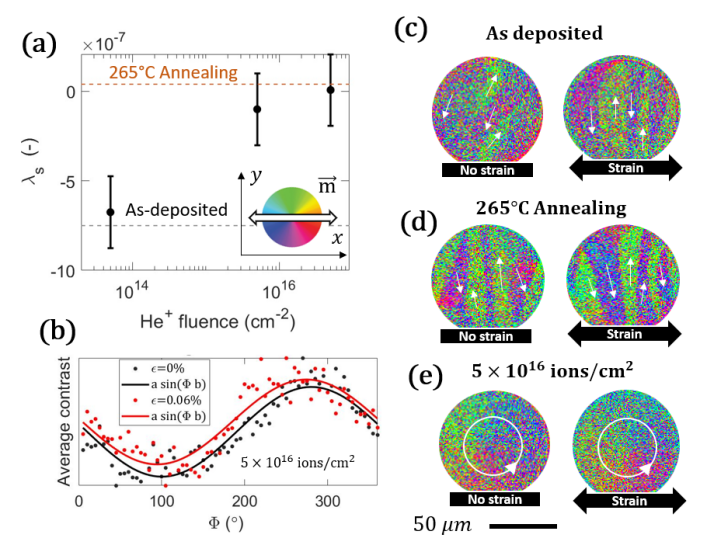
In order to compare more quantitatively the MOKE images and the vortex state of the irradiated sample, the average radial magnetization was calculated from the longitudinal component of the vector image for different in-plane directionsGilbert et al. (2016). The average contrast is calculated for a single 80 m disk for the images in Fig. 3 (e) and is reported in Fig. 3 (b). For the unstrained state seen in Fig. 3 (e) left, the disk’s magnetization is a circularly-symmetric vortex, and the average contrast varies periodically with angular position on the disk. The values well follow the expression , black line in Fig. 3 (b). After the application of strain, as a consequence of the extremely small magnetostriction, the average contrast, red line in Fig. 3 (b), still follows the periodic behavior .
A possible explanation for the reported reduction in saturation magnetostriction after ion irradiation and annealing is the growth in size of the crystallites in the NiFeCr(5 nm)/Ni81Fe19(30 nm)/Ta(4 nm) sample, already highlighted in Fig. 1 (c). The magnetostriction of isotropic oriented cubic crystallites can be written as the combination of the saturation-magnetostriction constants and in the (100) and (111) directions, respectivelyBozorth (1993)
| (3) |
In Permalloy, the two component of the magnetostriction change significantly over the relative Ni-Fe composition range altering the effective magnetostriction, . The composition used in this work, Ni81Fe19, is predicted to have close to zero Judy (1994). In our XRD measurement, a reduction to the (111) peak FWHM is observed after irradiation and annealing. This crystallization can alter the relative contribution of and in the magnetic layer. Following eq. 3, the effective magnetostriction of the film is changed. As shown in Fig. 3 (a), the magnetostriction is progressively reduced for higher fluences and annealing temperatures as the size of crystallites caused by irradiation and annealing increases. On top of that, increased intermixing at the magnetic layer boundaries, could alter the interface magnetostrictionSingleton and Duxstad (2002) (inversely proportional to the film thicknessChoe (1999); Song, Ballentine, and O’Handley (1994)) thus playing a role in the effective magnetostriction of the film.
To validate the usability of our Permalloy layer for sensing application, transport measurements have been conducted. The electrical characterization confirms that the NiFeCr(5 nm)/Ni81Fe19(30 nm)/Ta(4 nm) sample has sizable AMR . As the AMR does not change after irradiation, the proposed material treatment is suitable for improving magnetic properties of magnetic material for magnetic sensing applications. AMR measurements can be found in section S2 of the supplementary material.
IV Conclusions
In conclusion, we have investigated the effects of He+ irradiation and thermal annealing on the magnetic properties of NiFeCr(5 nm)/Ni81Fe19(30 nm)/Ta(4 nm). Our XRD analysis suggests that both irradiation and annealing promote crystalline growth of the textured Ni81Fe19 alloy. While the irradiation treatment strongly reduces the hard axis coercivity down to m and the deposition induced anisotropy by a factor ten, the field-free annealing does not significantly improve the magnetic softness. We mainly attribute this to stress relaxation in the film after irradiation and to the different mechanism for atomic ordering, that is completely isotropic in the case of irradiation only. In addition, the effective magnetostriction of the film is significantly reduced by a factor ten after irradiation and annealing as confirmed by anisotropy measurement in the presence of in-plane strain. Importantly, we have shown that the sizable magnetoresistance is preserved after the irradiation. As a result, post growth He+on irradiation is an excellent tool to improve magnetic softness and minimize strain cross sensitivity of Permalloy. In contrast to thermal annealing, ion irradiation offers the advantage of performing a local material treatmentWoods et al. (2002); McCord et al. (2005); Fassbender and McCord (2008) to adjust the anisotropy and write magnetic domain patterns directly into thin film structured devices. As a consequence, we can locally tune the properties of a magnetic material to make it suitable, for instance, for high sensitivity and low-hysteresis integrated AMR sensors that are insensitive to stain.
Supplementary Material
See supplementary material for the rocking curve and AMR measurements.
Acknowledgements.
This project has received funding from the European Union’s Horizon 2020 research and innovation program under the Marie Skłodowska-Curie grant agreement No 860060 “Magnetism and the effect of Electric Field” (MagnEFi), the Deutsche Forschungsgemeinschaft (DFG, German Research Foundation) - TRR 173 - 268565370 (project A01 and B02), the DFG funded collaborative research center (CRC)1261 / project A6 and the Austrian Research Promotion Agency (FFG). The authors acknowledge support by the chip production facilities of Sensitec GmbH (Mainz, DE), where part of this work was carried out and the Max-Planck Graduate Centre with Johannes Gutenberg University.References
- Khan et al. (2021) M. A. Khan, J. Sun, B. Li, A. Przybysz, and J. Kosel, “Magnetic sensors-a review and recent technologies,” Engineering Research Express 3, 022005 (2021).
- Kwiatkowski and Tumanski (1986) W. Kwiatkowski and S. Tumanski, “The permalloy magnetoresistive sensors-properties and applications,” Journal of Physics E: Scientific Instruments 19, 502 (1986).
- Groenland et al. (1992) J. Groenland, C. Eijkel, J. Fluitman, and R. de Ridder, “Permalloy thin-film magnetic sensors,” Sensors and Actuators A: Physical 30, 89–100 (1992).
- Trützschler et al. (2014) J. Trützschler, K. Sentosun, M. Langer, I. Mönch, R. Mattheis, J. Fassbender, and J. McCord, “Optimization of magneto-resistive response of ion-irradiated exchange biased films through zigzag arrangement of magnetization,” Journal of Applied Physics 115, 103901 (2014).
- Jogschies et al. (2015) L. Jogschies, D. Klaas, R. Kruppe, J. Rittinger, P. Taptimthong, A. Wienecke, L. Rissing, and M. C. Wurz, “Recent developments of magnetoresistive sensors for industrial applications,” Sensors 15, 28665–28689 (2015).
- Lenssen et al. (2000) K.-M. Lenssen, D. Adelerhof, H. Gassen, A. Kuiper, G. Somers, and J. Van Zon, “Robust giant magnetoresistance sensors,” Sensors and Actuators A: Physical 85, 1–8 (2000).
- Cañón Bermúdez and Makarov (2021) G. S. Cañón Bermúdez and D. Makarov, “Magnetosensitive E-Skins for interactive devices,” Advanced Functional Materials 31, 2007788 (2021).
- Oliveros Mata et al. (2021) E. S. Oliveros Mata, G. S. Cañón Bermúdez, M. Ha, T. Kosub, Y. Zabila, J. Fassbender, and D. Makarov, “Printable anisotropic magnetoresistance sensors for highly compliant electronics,” Applied Physics A 127, 1–6 (2021).
- Melzer, Makarov, and Schmidt (2019) M. Melzer, D. Makarov, and O. Schmidt, “A review on stretchable magnetic field sensorics,” Journal of Physics D: Applied Physics 53, 083002 (2019).
- Amara et al. (2018) S. Amara, G. A. T. Sevilla, M. Hawsawi, Y. Mashraei, H. Mohammed, M. E. Cruz, Y. P. Ivanov, S. Jaiswal, G. Jakob, M. Kläui, et al., “High-performance flexible magnetic tunnel junctions for smart miniaturized instruments,” Advanced Engineering Materials 20, 1800471 (2018).
- Wang et al. (2012) G. Wang, C. Dong, W. Wang, Z. Wang, G. Chai, C. Jiang, and D. Xue, “Observation of rotatable stripe domain in permalloy films with oblique sputtering,” Journal of Applied Physics 112, 093907 (2012).
- Iwata and Hagedorn (1969) T. Iwata and F. Hagedorn, “Annealing behavior of induced anisotropy and related magnetic properties in permalloy films,” Journal of Applied Physics 40, 2258–2266 (1969).
- Jiang et al. (2018) X. Jiang, X. Fan, J. Huang, C. Zhang, and D. Xue, “Improving the quality of AMR of permalloy films by N+ ion irradiation,” Nuclear Instruments and Methods in Physics Research Section B: Beam Interactions with Materials and Atoms 430, 54–58 (2018).
- Jaafar et al. (2011) M. Jaafar, R. Sanz, J. McCord, J. Jensen, R. Schäfer, M. Vázquez, and A. Asenjo, “Pattern-induced magnetic anisotropy in FePt thin films by ion irradiation,” Physical Review B 83, 094422 (2011).
- Devolder et al. (2013) T. Devolder, I. Barisic, S. Eimer, K. Garcia, J.-P. Adam, B. Ockert, and D. Ravelosona, “Irradiation-induced tailoring of the magnetism of CoFeB/MgO ultrathin films,” Journal of applied Physics 113, 203912 (2013).
- Ravelosona et al. (2000) D. Ravelosona, C. Chappert, V. Mathet, and H. Bernas, “Chemical order induced by ion irradiation in FePt (001) films,” Applied Physics Letters 76, 236–238 (2000).
- Masciocchi et al. (2022) G. Masciocchi, J. W. van der Jagt, M.-A. Syskaki, A. Lamperti, N. Wolff, A. Lotnyk, J. Langer, L. Kienle, G. Jakob, B. Borie, et al., “Control of magnetoelastic coupling in Ni/Fe multilayers using He+ ion irradiation,” Applied Physics Letters 121, 182401 (2022).
- De Jong et al. (2022) M. C. De Jong, M. J. Meijer, J. Lucassen, J. Van Liempt, H. J. Swagten, B. Koopmans, and R. Lavrijsen, “Local control of magnetic interface effects in chiral Ir| Co| Pt multilayers using Ga+ ion irradiation,” Physical Review B 105, 064429 (2022).
- Zhao et al. (2019) X. Zhao, B. Zhang, N. Vernier, X. Zhang, M. Sall, T. Xing, L. H. Diez, C. Hepburn, L. Wang, G. Durin, et al., “Enhancing domain wall velocity through interface intermixing in W-CoFeB-MgO films with perpendicular anisotropy,” Applied Physics Letters 115, 122404 (2019).
- Diez et al. (2019) L. H. Diez, M. Voto, A. Casiraghi, M. Belmeguenai, Y. Roussigné, G. Durin, A. Lamperti, R. Mantovan, V. Sluka, V. Jeudy, et al., “Enhancement of the Dzyaloshinskii-Moriya interaction and domain wall velocity through interface intermixing in Ta/CoFeB/MgO,” Physical Review B 99, 054431 (2019).
- Woods et al. (2002) S. Woods, S. Ingvarsson, J. Kirtley, H. Hamann, and R. Koch, “Local magnetic anisotropy control in NiFe thin films via ion irradiation,” Applied physics letters 81, 1267–1269 (2002).
- Schindler, Kernohan, and Weertman (1964) A. Schindler, R. Kernohan, and J. Weertman, “Effect of irradiation on magnetic properties of Fe-Ni alloys,” Journal of Applied Physics 35, 2640–2646 (1964).
- Mougin et al. (2001) A. Mougin, T. Mewes, M. Jung, D. Engel, A. Ehresmann, H. Schmoranzer, J. Fassbender, and B. Hillebrands, “Local manipulation and reversal of the exchange bias field by ion irradiation in FeNi/FeMn double layers,” Physical Review B 63, 060409 (2001).
- Trützschler et al. (2016) J. Trützschler, K. Sentosun, B. Mozooni, R. Mattheis, and J. McCord, “Magnetic domain wall gratings for magnetization reversal tuning and confined dynamic mode localization,” Scientific reports 6, 30761 (2016).
- Fassbender et al. (2006a) J. Fassbender, J. Von Borany, A. Mücklich, K. Potzger, W. Möller, J. McCord, L. Schultz, and R. Mattheis, “Structural and magnetic modifications of Cr-implanted permalloy,” Physical Review B 73, 184410 (2006a).
- Fassbender and McCord (2006) J. Fassbender and J. McCord, “Control of saturation magnetization, anisotropy, and damping due to Ni implantation in thin NiFe layers,” Applied Physics Letters 88, 252501 (2006).
- Fassbender et al. (2006b) J. Fassbender, A. Mücklich, K. Potzger, and W. Möller, “Mixing and subsequent amorphization of ultrathin NiFe/Ta bilayers by 30 keV Ni implantation,” Nuclear Instruments and Methods in Physics Research Section B: Beam Interactions with Materials and Atoms 248, 343–346 (2006b).
- Gupta et al. (2008) R. Gupta, K. Lieb, Y. Luo, G. Müller, P. Schaaf, and K. Zhang, “Argon and krypton ion-induced changes in permalloy thin films,” The European Physical Journal B 63, 501–506 (2008).
- Fassbender, Ravelosona, and Samson (2004) J. Fassbender, D. Ravelosona, and Y. Samson, “Tailoring magnetism by light-ion irradiation,” Journal of Physics D: Applied Physics 37, R179 (2004).
- Baglin et al. (1997) J. Baglin, M. Tabacniks, R. Fontana, A. Kellock, and T. Bardin, “Effects of ion irradiation on ferromagnetic thin films,” in Materials Science Forum, Vol. 248 (Trans Tech Publ, 1997) pp. 87–94.
- Ziegler, Ziegler, and Biersack (2010) J. F. Ziegler, M. D. Ziegler, and J. P. Biersack, “SRIM-the stopping and range of ions in matter (2010),” Nuclear Instruments and Methods in Physics Research Section B: Beam Interactions with Materials and Atoms 268, 1818–1823 (2010).
- Okay et al. (2018) C. Okay, P. Aksu, C. Deger, and F. Yildiz, “Tailoring the magnetic anisotropy of cobalt-gold thin films,” Turkish Journal of Physics 42, 335–341 (2018).
- Klokholm and Aboaf (1981) E. Klokholm and J. Aboaf, “The saturation magnetostriction of permalloy films,” Journal of Applied Physics 52, 2474–2476 (1981).
- Masciocchi et al. (2021) G. Masciocchi, M. Fattouhi, A. Kehlberger, L. Lopez-Diaz, M.-A. Syskaki, and M. Kläui, “Strain-controlled domain wall injection into nanowires for sensor applications,” Journal of Applied Physics 130, 183903 (2021).
- Raghunathan, Snyder, and Jiles (2009) A. Raghunathan, J. E. Snyder, and D. Jiles, “Comparison of alternative techniques for characterizing magnetostriction and inverse magnetostriction in magnetic thin films,” IEEE Transactions on Magnetics 45, 3269–3273 (2009).
- Ahmadipour et al. (2016) M. Ahmadipour, M. J. Abu, M. F. Ab Rahman, M. F. Ain, and Z. A. Ahmad, “Assessment of crystallite size and strain of prepared via conventional solid-state reaction,” Micro & Nano Letters 11, 147–150 (2016).
- Zak et al. (2011) A. K. Zak, W. A. Majid, M. E. Abrishami, and R. Yousefi, “X-ray analysis of ZnO nanoparticles by Williamson-Hall and size-strain plot methods,” Solid State Sciences 13, 251–256 (2011).
- Park, Norton, and Selvamanickam (2005) S. Park, D. Norton, and V. Selvamanickam, “Ion-beam texturing of uniaxially textured Ni films,” Applied Physics Letters 87, 031907 (2005).
- van der Jagt et al. (2022) J. W. van der Jagt, V. Jeudy, A. Thiaville, M. Sall, N. Vernier, L. H. Diez, M. Belmeguenai, Y. Roussigné, S. M. Chérif, M. Fattouhi, et al., “Revealing nanoscale disorder in W/Co-Fe-B/MgO ultrathin films using domain-wall motion,” Physical Review Applied 18, 054072 (2022).
- Wang et al. (2009) W. Wang, J. Jordan-Sweet, G. Miao, C. Ni, A. Rumaiz, L. Shah, X. Fan, P. Parsons, R. Stearrett, E. Nowak, et al., “In-situ characterization of rapid crystallization of amorphous CoFeB electrodes in CoFeB/MgO/CoFeB junctions during thermal annealing,” Applied Physics Letters 95, 242501 (2009).
- Schulz et al. (2021) F. Schulz, R. Lawitzki, H. Głowiński, F. Lisiecki, N. Träger, P. Kuświk, E. Goering, G. Schütz, and J. Gräfe, “Increase of Gilbert damping in permalloy thin films due to heat-induced structural changes,” Journal of Applied Physics 129, 153903 (2021).
- McCord et al. (2005) J. McCord, T. Gemming, L. Schultz, J. Fassbender, M. O. Liedke, M. Frommberger, and E. Quandt, “Magnetic anisotropy and domain patterning of amorphous films by He-ion irradiation,” Applied Physics Letters 86, 162502 (2005).
- Zou, Yu, and Bain (2002) P. Zou, W. Yu, and J. A. Bain, “Influence of stress and texture on soft magnetic properties of thin films,” IEEE Transactions on Magnetics 38, 3501–3520 (2002).
- Gupta et al. (2005) R. Gupta, K.-H. Han, K. Lieb, G. Müller, P. Schaaf, and K. Zhang, “Influence of ion implantation on the magnetic properties of thin FeCo films,” Journal of Applied Physics 97, 073911 (2005).
- Jian, Liu, and Mayer (1989) L. Jian, J. Liu, and J. Mayer, “Ar+ ion irradiation induced grain growth in Au and Pt thin films,” Nuclear Instruments and Methods in Physics Research Section B: Beam Interactions with Materials and Atoms 36, 306–311 (1989).
- Liu, Nastasi, and Mayer (1987) J. C. Liu, M. Nastasi, and J. Mayer, “Ion irradiation induced grain growth in Pd polycrystalline thin films,” Journal of Applied Physics 62, 423–428 (1987).
- Liu and Mayer (1987) J. C. Liu and J. Mayer, “Ion irradiation induced grain growth in Ni polycrystalline thin films,” Nuclear Instruments and Methods in Physics Research Section B: Beam Interactions with Materials and Atoms 19, 538–542 (1987).
- Devolder (2000) T. Devolder, “Light ion irradiation of Co/Pt systems: Structural origin of the decrease in magnetic anisotropy,” Physical Review B 62, 5794 (2000).
- Maziewski et al. (2012) A. Maziewski, P. Mazalski, Z. Kurant, M. Liedke, J. McCord, J. Fassbender, J. Ferré, A. Mougin, A. Wawro, L. Baczewski, et al., “Tailoring of magnetism in Pt/Co/Pt ultrathin films by ion irradiation,” Physical Review B 85, 054427 (2012).
- Finizio et al. (2014) S. Finizio, M. Foerster, M. Buzzi, B. Krüger, M. Jourdan, C. A. Vaz, J. Hockel, T. Miyawaki, A. Tkach, S. Valencia, et al., “Magnetic anisotropy engineering in thin film Ni nanostructures by magnetoelastic coupling,” Physical Review Applied 1, 021001 (2014).
- Gilbert et al. (2016) I. Gilbert, A. C. Chavez, D. T. Pierce, J. Unguris, W.-Y. Sun, C.-Y. Liang, and G. P. Carman, “Magnetic microscopy and simulation of strain-mediated control of magnetization in PMN-PT/Ni nanostructures,” Applied Physics Letters 109, 162404 (2016).
- Bozorth (1993) R. M. Bozorth, Ferromagnetism, p. 263 (1993).
- Judy (1994) J. W. Judy, “Magnetic microactuators with polysilicon flexures,” Masters Report (1994).
- Singleton and Duxstad (2002) E. Singleton and K. Duxstad, “Interfacial contributions to magnetostriction of ferromagnetic layers for magnetoresistive sensors,” MRS Online Proceedings Library 721, 1–6 (2002).
- Choe (1999) G. Choe, “Giant interface magnetostriction and temperature dependence in NiFe films encapsulated with Ta and AlOx layers,” IEEE Transactions on Magnetics 35, 3838–3840 (1999).
- Song, Ballentine, and O’Handley (1994) O. Song, C. Ballentine, and R. O’Handley, “Giant surface magnetostriction in polycrystalline Ni and NiFe films,” Applied Physics Letters 64, 2593–2595 (1994).
- Fassbender and McCord (2008) J. Fassbender and J. McCord, “Magnetic patterning by means of ion irradiation and implantation,” Journal of Magnetism and Magnetic Materials 320, 579–596 (2008).