Infrared-induced ultrafast melting of nanostructured platinum films probed by an X-ray free-electron laser
Abstract
Understanding melting in metals is a hot topic of present research. This may be accomplished by pumping the system with infrared (IR) laser radiation, and probing it with hard X-rays produced by an X-ray Free-Electron Laser (XFEL). In this work we studied nanostructured polycrystalline thin films of platinum that were illuminated by IR radiation of increasing fluences. We characterized the structural response as well as the nucleation and propagation of the liquid phase as a function of time delay between the IR pump and X-ray probe. We observed partial melting of the samples for IR fluences higher than 200 mJcm-2. To fit the contribution of the liquid phase to the scattering pattern in platinum we applied a model of liquid metal. The two-temperature model simulations were performed to understand the solid-state fraction of the sample heating process as a function of time delay and fluence.
Introduction
When infrared (IR) light from a femtosecond laser interacts with free electrons in metals, it rapidly raises their kinetic energy, which is then partially transferred to the atomic lattice via electron-phonon coupling [1]. This excess of energy perturbs the atomic arrangement, whose temporal evolution can be probed, for example, by hard X-ray pulses generated by ultrashort X-ray Free-Electron Lasers (XFELs) [2, 3, 4, 5, 6]. Due to the exceptional properties in terms of brightness, temporal pulse duration and transverse coherence [7, 8, 9], hard XFELs are in fact unique tools to characterize structure and dynamics of condensed matter [10, 11, 12, 13].
One important metal for pump-probe experiments, performed, for example, at XFELs, is platinum. Platinum is a noble metal with numerous scientific and technological applications. It is used, for example, for medical devices [14], in nanomedicine [15] and has a central role in cancer chemotherapy [16, 17]. It is essential for the catalysis of the hydrogen oxidation and oxygen reduction reactions, at the core of fuel cells and electrolysers [18, 19, 20]. Due to its excellent performance and thermal stability, platinum is also extensively employed within automotive catalytic converters [21, 22], and to catalyse several other industrially-relevant reactions, for example, in the oil industry [23]. The elevated chemical inertness, oxidation resistance, melting temperature and phase stability of platinum [24] make it perfect to produce melting equipment. For example, it is used to manufacture glass fibres, for temperature sensors or for space industry applications [25]. Because of its properties, platinum is also used as reference for several applications, ranging from electrodes and thermometers to pressure calibrants [26].
Pump-probe investigations have been performed earlier on aluminum [27, 28] and gold (for example, evidence for bond hardening [29], identification of heterogeneous to homogeneous melting transition [30] and elucidation of the role of grain boundaries on melting [31]). Gold is the most widely studied element and possesses an atomic electronic configuration similar to the one of platinum, which differs from the former by only one electron. However, theoretical calculations predict for platinum a completely different behavior in terms of electron-phonon coupling, and in turn of electron-lattice energy exchange [32].
With our experiment and its interpretation, we aim at increasing the understanding of platinum properties, and in particular of the crystalline-liquid phase transition in nanostructured thin films. Furthermore, given platinum phase stability and resistance to oxidation, we aspire at using this experiment as a model for the melting process, whose detailed understanding is of great scientific importance. Apart from increasing the comprehension of platinum and addressing thus a fundamental problem in material science, that is the nucleation and propagation of a liquid phase in films characterized by nanostructured domains, the presented results are potentially of technological relevance, given the increasing demand of ultrafast laser manufacturing [33, 34, 35, 36, 37, 38].
Results
Platinum films of 100 nm thickness were grown on silicon nitride membranes by electron beam evaporation. The resulting lateral dimension of the grains was of the order of 10 nm and they were preferentially columnar with respect to the film surface .
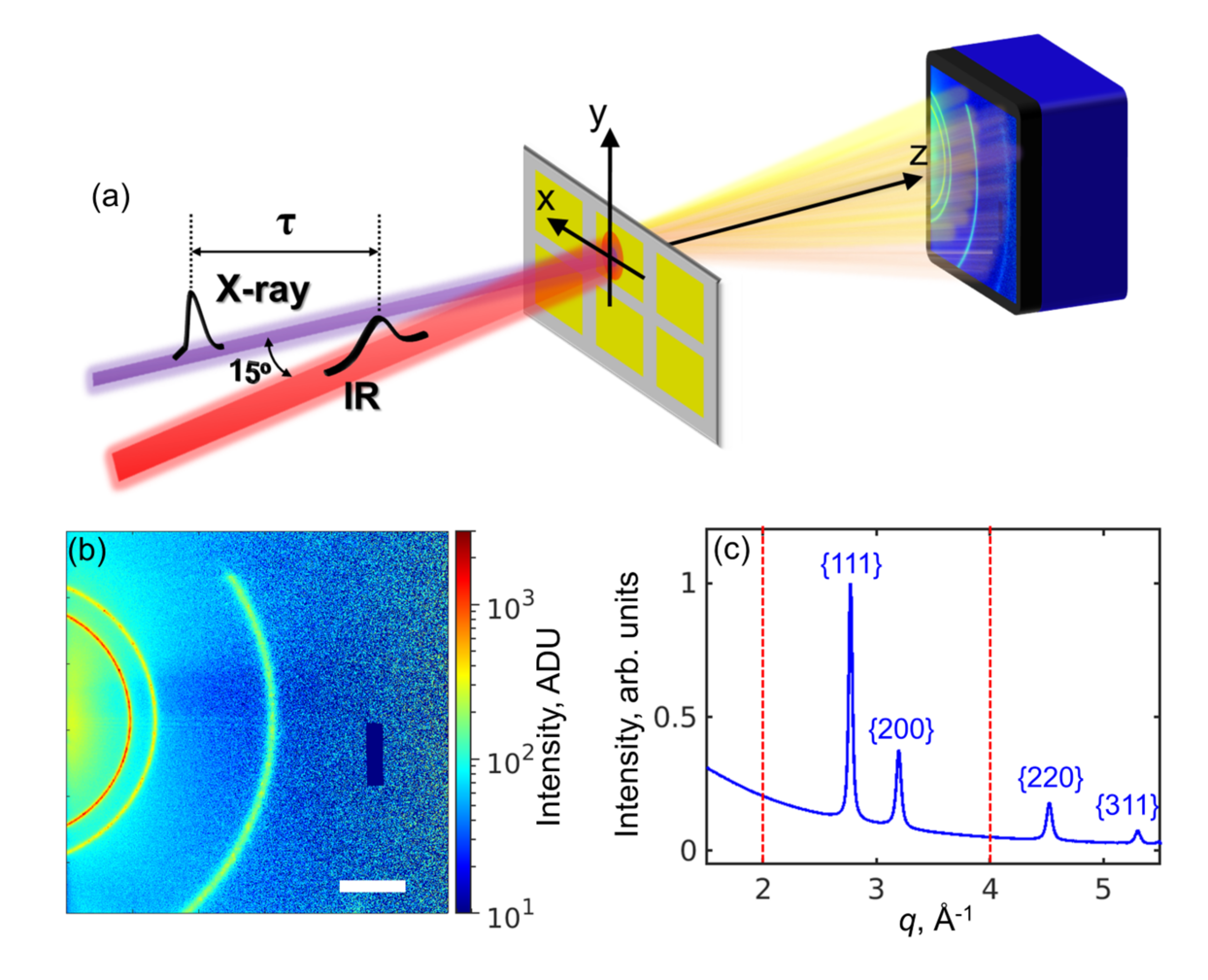
The experiment was performed at the Nano-crystallography and Coherent Imaging instrument (NCI) of the Pohang Accelerator Laboratory X-ray Free-Electron Laser (PAL-XFEL) [39, 4]. A schematic of the experiment setup is shown in Fig. 1a. The details of both the experiment and data analysis are provided in the Methods section.
| , mJcm-2 | , µJ | , kJ/kg |
| 100 | 28.4 | 139 |
| 200 | 57.0 | 279 |
| 300 | 85.0 | 416 |
| 400 | 113.0 | 553 |
| 500 | 141.0 | 691 |
| 1,000 | 283.0 | 1,386 |
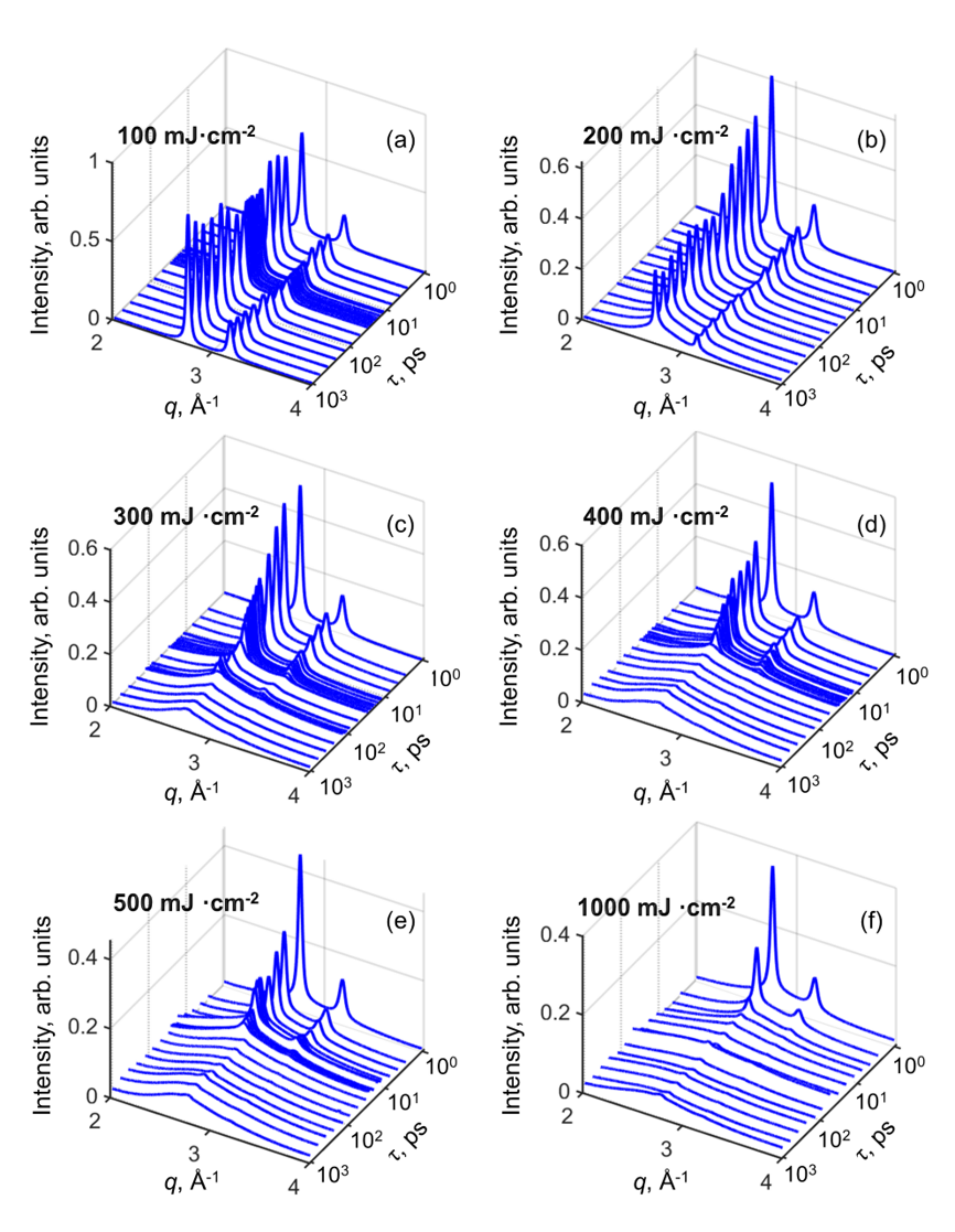
The scattering curves collected during this experiment, with the subtracted background, are shown in Fig. 2 for the different IR fluences used in this experiment. By visual inspection, at the lowest IR fluence of 100 mJcm-2 no significant differences in the scattering data are observed at different time delays. However, starting with the IR fluence of 200 mJcm-2 we observe a gradual decay of Bragg peak intensities with the increase of time delay. While at this IR fluence both the (111) and (200) Bragg peaks corresponding to face-centered-cubic (fcc) Pt structure are visible for all time delays, already at 300 mJcm-2 the higher order peak (200) starts to disappear at about 100 ps. In general, the decrease of intensities is faster the higher the IR fluence. For example, at the fluence of 1,000 mJcm-2 Bragg peaks are practically vanishing after a few picoseconds from the IR pulse. At the same time, we would like to note here that some intensity corresponding to the (111) Bragg peak is observed for every fluence and time delay, indicating that some residual solid fraction is always present in the region of the sample probed by X-rays. In fact, we interpret the smooth continuous scattering curve which appears as the intensity of Bragg peaks decreases, as the fingerprint of the liquid platinum phase. By that, we conclude that platinum films are partially melted for fluences higher than 200 mJcm-2. Below, we will present a detailed analysis of the results obtained.
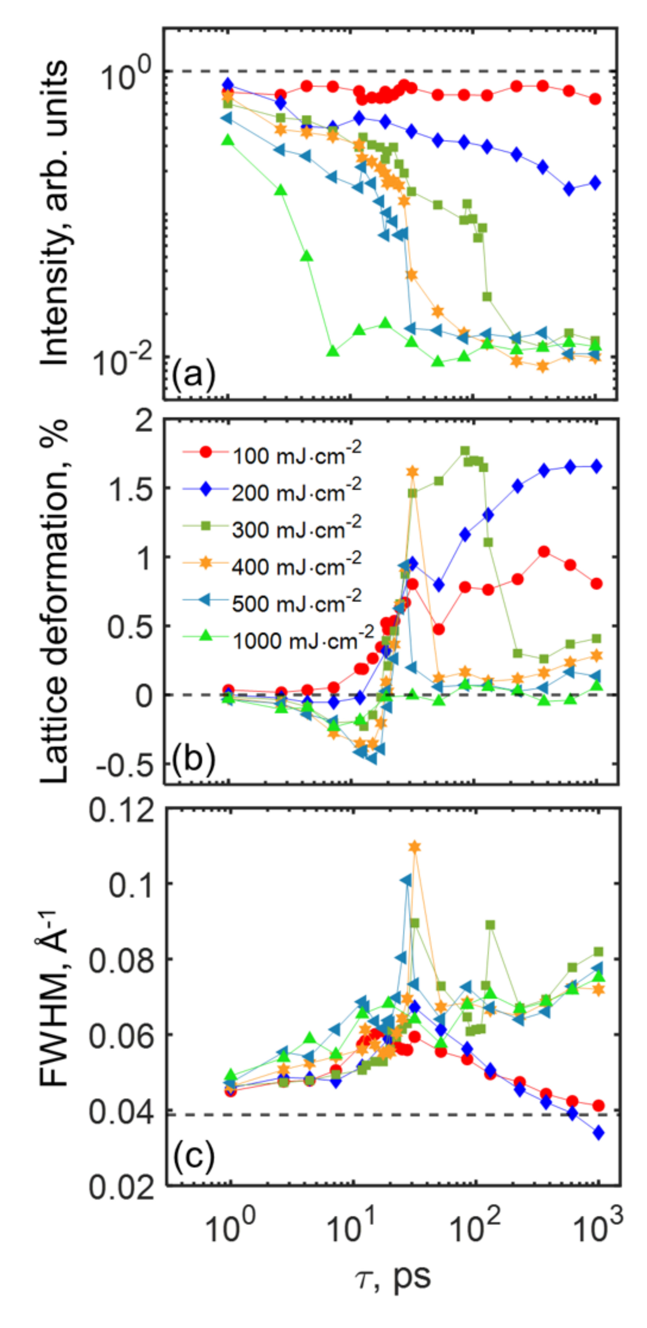
The parameters extracted from the fit of scattering curves are reported in Fig. 3 as a function of pump-probe time delays. Each point shown in this figure is the median of the corresponding dataset . Even if both the (111) and (200) Bragg peaks are present in the investigated momentum-transfer range, we restrict our analysis below to the former, since the behaviour of the two is highly correlated within the resolution of our measurements .
We begin our discussion from the integrated intensity of the (111) Bragg peak (Fig. 3a), which is proportional to the amount of crystalline fcc platinum in the probed region of the sample. As we can see from this figure, for each value of the incident IR fluence this quantity is reduced with respect to the reference value (that is measured only with XFEL radiation without illuminating the sample with IR). As discussed above, the decrease within the investigated temporal range is negligible when the IR fluence is equal to 100 mJcm-2 and modest when it is 200 mJcm-2 (these will be named as ”low fluences”). On the contrary, for higher values (”high fluences”), in our experiment, actually starting from 300 mJcm-2, the integrated intensity drops to about 1% of the reference value, and the decay is faster with higher fluence. This occurs from about one hundred picoseconds at 300 mJcm-2 to a few picoseconds for the 1,000 mJcm-2 case.
The position of a Bragg peak is related to the lattice parameter of a cubic lattice through the known relation . Variations of the strain value as a function of the time delay, defined as , are shown in Fig. 3b. For low fluences, it can be observed that the lattice is expanded for each time delay longer than approximately 10 ps. This is compatible with an increase of the sample temperature. Assuming the expansion to be purely thermal, a maximum temperature increase at one nanosecond, at the end of our observation, was estimated to be about 1,300 K and 1,800 K for IR fluences of 100 mJcm-2 and 200 mJcm-2, respectively. The thermal expansion coefficient was obtained from Ref.[40]. The general trend for each IR fluence greater than 100 mJcm-2 is that the lattice is progressively becoming more compressed in the first 10 - 20 ps, up to approximately -0.5% at the IR fluence of 500 mJcm-2. For a purely elastic deformation, with the Young’s modulus equal to[41] 168 GPa this value corresponds to the stress value of about 0.8 GPa. Compression is increasing with IR fluence, except for the 1,000 mJcm-2 case, likely due to the extremely small residual solid state fraction (see Fig. 3a). After this initial phase, for each IR fluence the lattice starts to rapidly expand up to time delays of approximately 30 ps. This is about the time necessary for a sound wave to transverse the thickness of the platinum sample, which is 100 nm in our case. Indeed, if a deformation wave is traveling through the platinum film at the (longitudinal) speed of sound [42] of 3,260 m/s, this time is 31 ps. After this point, for low fluences the strain values decrease slightly up to time delays of about 50 ps, and then they increase again. For high fluences, in general, the expansion diminishes, following the same trend already observed for the residual fcc solid state fraction.
The FWHM of the 111 reflection is shown in Fig. 3c. It is inversely proportional to the size of the coherently scattering domains and proportional to the microstrain and thermal vibrations in the sample [43]. For each value of the IR fluence, the peak width is initially slightly increased with respect to the reference value. However, at about 30 ps we observe a steep increase of the peak width followed by a fast decay. The curve has a maximum at ps for fluences lower than 500 mJcm-2.This is likely due to extremely small residual fcc fraction at this time delay (see Fig. 3a), which might also explain why the peak width for the IR fluence of 1,000 mJcm-2 does not follow a similar trend. It is interesting to note, that for low fluences, the FWHM at ns is practically returning to its initial value. In particular, for the 200 mJcm-2 case the final FWHM value in the sub-nanosecond time delay region is even lower than the reference one, possibly indicating an increase of the crystallite size due to sintering of smaller grains or relaxation of residual stresses. At the same time, for high fluences, the values of FWHM after the peak at 31.6 ps continue to increase.
The Kirchhoff’s law predicts, for a constant pressure process, the change of the temperature of the system as a function of enthalpy to be . We assume also here that all the energy absorbed by the system is increasing its temperature (see Table 1). The above equation gives us an estimate of the sample temperature, it should reach a maximum temperature of K, and therefore remain solid, when irradiated with an IR fluence of 100 mJcm-2 (we remind that the melting temperature for platinum at atmospheric pressure is =2,041.3 K [11]). It should be noted that this is the same temperature estimated from the position of the (111) Bragg peak assuming ambient pressure. For the IR fluence of 200 mJcm-2 the melting should be partial, with coexistence of the solid and liquid phases, whereas 300 mJcm-2 is sufficient to transform the solid into liquid. This picture is apparently consistent with Fig. 3a for long time delays although an extremely small residual fcc solid state fraction is always present according to our experimental findings. We speculate that this might be due to chemical stabilization of platinum at its interface with Si3N4 film, inhibiting crystal to liquid phase transition.
The scattering curve attributed to liquid, which is clearly distinguishable from the Bragg peaks, was fitted using the model developed by Ashcroft [44, 45] (see Methods section). It should be noted that, especially for low fluences and short pump-probe delays, this model is likely to fit the diffuse scattering due to various defects including thermal vibrations, and might therefore not provide correct information on the liquid phase. The area under the broad Ashcroft’s curve, which is proportional to the amount of liquid phase, was estimated by integrating the corresponding analytical model from the origin of momentum-transfer space to infinity. To compensate for fluctuations of the incoming X-ray intensities, we divided the integral by the total intensity in the background for each diffraction pattern.
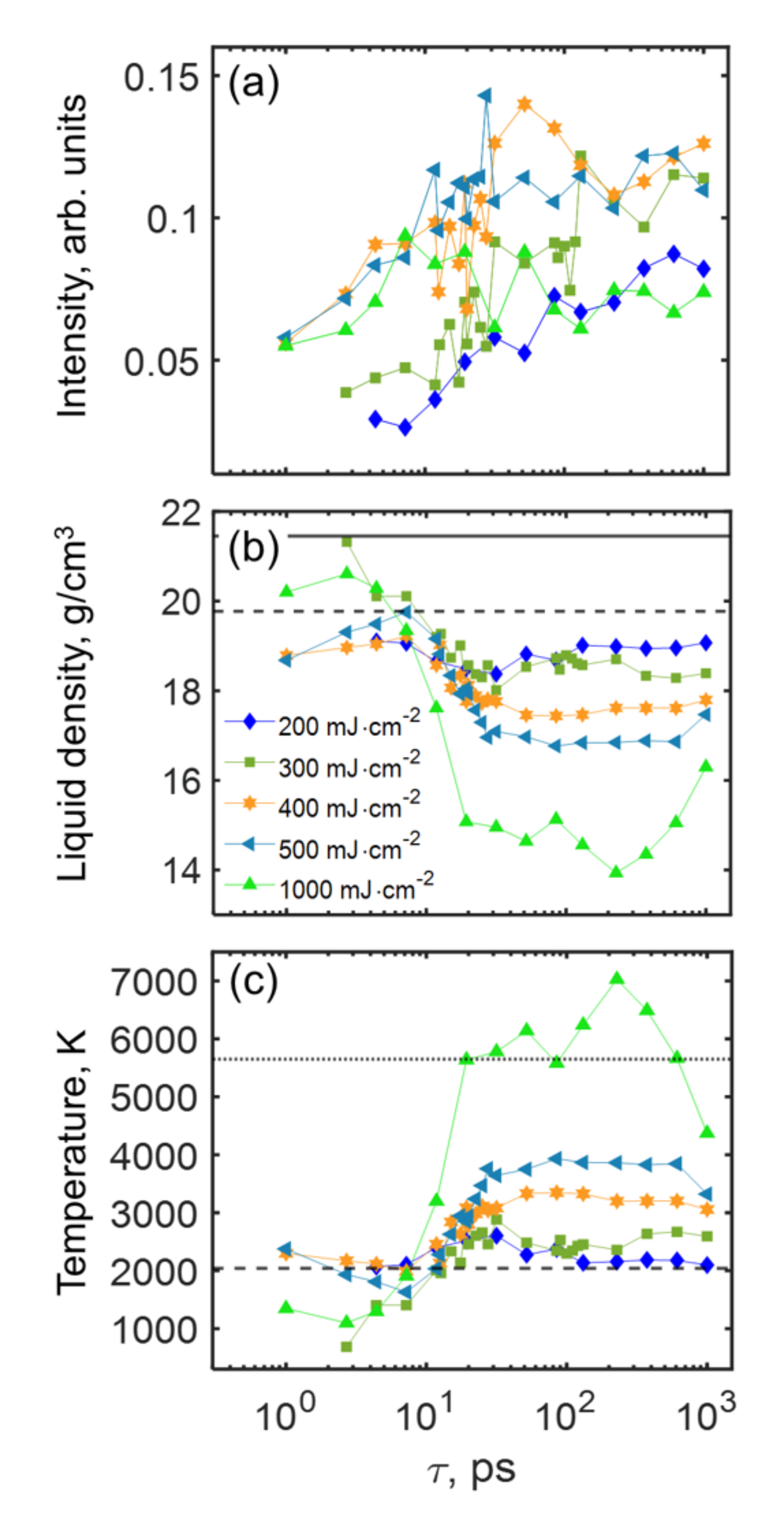
The result is shown in Fig. 4a. The general trend is that the integral of this liquid curve increases as a function of the time delay, indicating an increase of the liquid fraction. It also increases as a function of the IR fluence, although there is no significant difference between 400 mJcm-2 and 500 mJcm-2. Moreover, at time delays of hundreds of picoseconds the values of integrated intensities for the IR fluence of 1,000 mJcm-2 are similar to the ones for 200 mJcm-2. To explain this behaviour, we evaluated the conditions for ablation using the approach described in Ref.[46] . According to this, the estimated crater depth is about 1 nm and 10 nm for the IR fluences of 500 mJcm-2 and 1,000 mJcm-2, respectively .
In the model developed by Ashcroft [44, 45], the profile of the scattering curve of a liquid of hard spheres is defined by their diameter and packing density . The two parameters are in turn related to the liquid density of platinum by the relation , where g/mol is the atomic weight of platinum. The density of the liquid phase, shown in Fig. 4b, does not show an obvious trend for time delays shorter than about 10 ps. As we can see from the same figure, in the same region of time delays, the liquid density is lower the higher the IR fluence. This observation is compatible with an increase of the liquid temperature. However, if we exclude the data for 200 mJcm-2 and 300 mJcm-2, associated with a small liquid fraction (see Fig. 4a), then the higher the fluence the less dense is the liquid. We speculate that this might be due to the residual solid fraction (see Fig. 3a) mechanically constraining the liquid phase. On the contrary, for all IR fluences and time delays larger than approximately 10 ps, the liquid density is lower than the reference value at the melting point[47] of 19.77 . In the same region of time delays, it is also lower the higher is the IR fluence, compatibly with an increase of the liquid temperature.
The temperature of the liquid phase can be estimated from Ref.[48] as , where the volume is calculated from the density as obtained from the Ashcroft model, and . It should be noted that this equation is valid in the interval of temperatures from 2,042 K to 5,650 K, and it assumes a constant pressure process. Values of temperature of the liquid phase, shown in Fig. 4c, outside this range are in general the ones corresponding to time delays shorter than 10 ps, or to the highest IR fluence of 1,000 mJcm-2. The values within the range of validity of the equation, in Fig. 4c, confirm that the temperature of the liquid phase increases with the IR fluence. In particular, it is approximately the melting temperature for the IR fluence of 200 mJcm-2, the temperature at which solid and liquid coexist, and it reaches almost 4,000 K for the 500 mJcm-2 case. Interestingly, the temporal region between approximately 10 and 30 ps corresponds to both a transition between two different states of density and temperature, and of strain in the lattice (see, for example, Fig. 3b), suggesting that the solid and liquid phase might experience the same mechanical pressure.
To support the interpretation of experimental results, we performed two-temperature model (TTM) simulations[49] . The TTM is a phenomenological description of the spatio-temporal evolution of the temperature in a metal following its ultrafast excitation. In this picture, the laser pulse is absorbed by free electrons in a thin layer of about 10 nm and rapidly increases their energy. This is then partially transferred to the lattice via electron-phonon coupling. The TTM assumes a continuous media and does not include any defects, like the grain boundaries, present in the investigated samples. These grain boundaries can act as nucleation seeds for the liquid phase, lowering the amount of energy required for the phase transition (heterogeneous melting)[31].
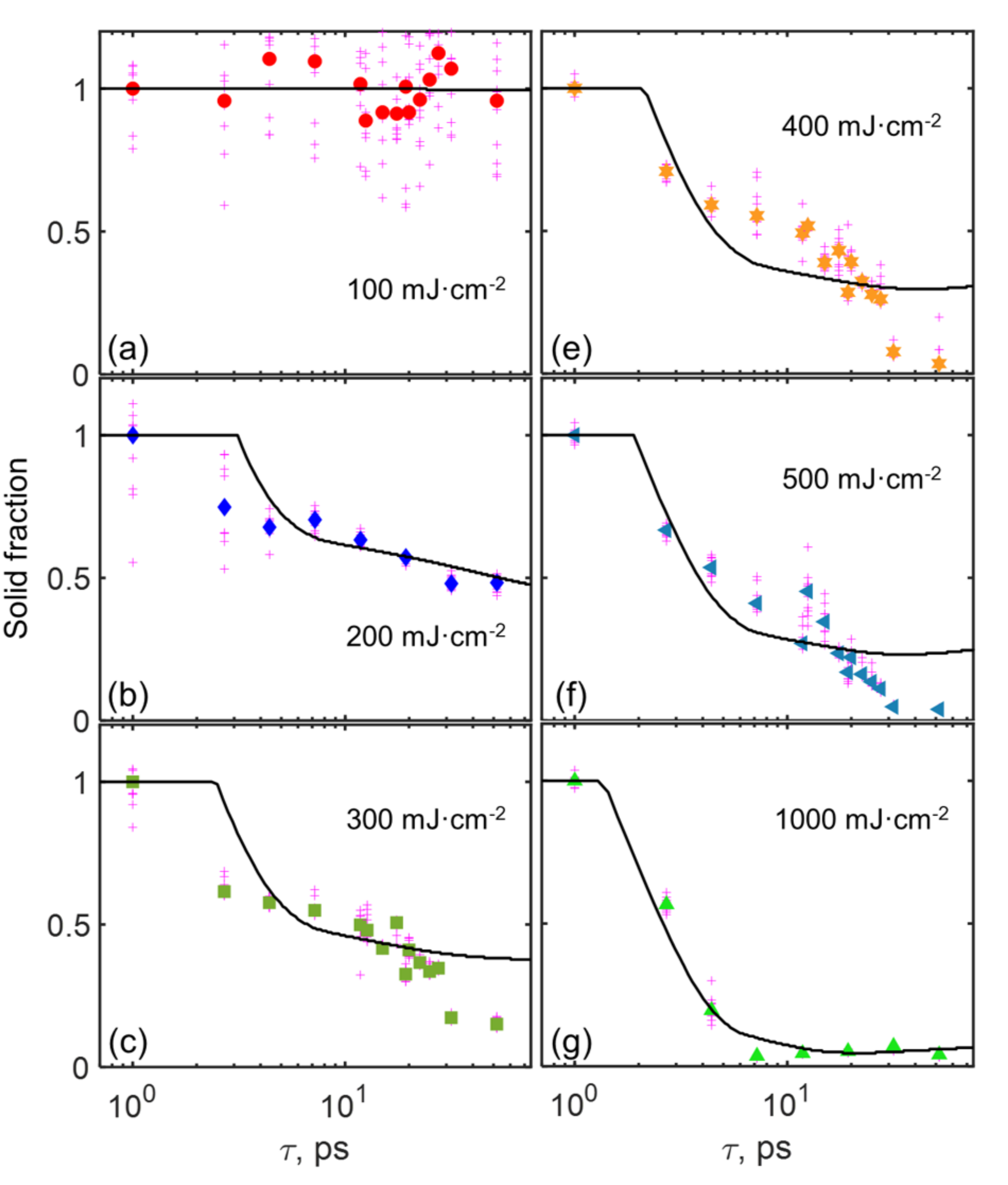
In Fig. 5 we compare the fraction of fcc solid state estimated from the experimental data and the one obtained from the TTM model. The values of experimental data are calculated assuming that the integral of the (111) Bragg peak is proportional to the amount of solid phase, and that there is no liquid platinum at the time delay of 1 ps. The value of the peak integral at this time delay is, therefore, used to normalize all the other values. For the case of the TTM simulations, the fraction of solid is simply the thickness of the solid phase at a given time from the IR pulse divided by the total thickness of the simulation domain (100 nm). It is important to note here that we did not introduce any fitting parameters in the TTM simulations. All parameters, including the electron-phonon coupling parameter , were taken from the known references . As can be observed from Fig. 5, the model qualitatively captures the trends observed experimentally. For the IR fluence of 100 mJcm-2 it does not predict any formation of the liquid phase, which is compatible to our experimental findings. From the fluence of 200 mJcm-2 the liquid phase is formed starting from about a few picoseconds. The time delay for the formation of the liquid phase is becoming shorter with increasing the IR fluence. For example, for the 1,000 mJcm-2 case the solid fraction is transformed to liquid already at time delays of about 2 ps. For all fluences, experimental results seem to indicate a faster nucleation of the liquid phase with respect to simulations. This could either be because our TTM model does not include any structural defects, which are present in the samples (homogeneous versus heterogeneous melting), or because some of the simulation parameters are not accurate. In particular, a relevant role in defining the temporal evolution of the atomic lattice is played by the electron-phonon coupling. Interestingly, our simulations show, that increasing/reducing the electron-phonon coupling parameter by twice is reducing/increasing the start of the melt process by 10 ps. For IR fluences from 300 mJcm-2 to 500 mJcm-2 we observe a second drop of the experimental solid fraction at about 20 - 30 ps, that is not explained by the TTM. This drop might be related to propagation of the sound wave through the 100 nm Pt sample layer. At the same time, we observe excellent agreement between the solid fraction calculated from experimental data and the TTM for the highest IR fluence of 1,000 mJcm-2 , although, we recall, the sample is partially ablated.
In summary, we described the evolution of platinum nanostructured films illuminated by ultrashort IR radiation at several fluences and time delays. We observed a deformation wave traveling perpendicular to the film at the speed of sound, and the temperature of the films increasing as a function of time delay. The sample remained solid when the fluence was 100 mJcm-2, and it was partially liquid for higher fluences. We observed that lattice deformations before the sound wave travelled through the entire sample were mostly compressive and after that they were causing expansion. Analysis of our results with the Ashcroft’s model suggested that the density of the liquid phase was dropping after the sound wave propagated through the sample.
To support experimental findings, we performed TTM simulations. They predicted the sample to be solid at the IR fluence of 100 mJcm-2, and partially liquid for higher fluences, with the solid fraction decreasing the more, the higher the fluence. We also observed an additional drop of the solid fraction that is not described by TTM simulations at fluences from 300 mJcm-2 to 500 mJcm-2 and time delays longer than 30 ps. This is most probably related to propagation of the sound wave through the 100 nm platinum sample layer. Our TTM simulations, based on a model of a uniform layer, are not sufficient to capture the complexity of the solid-liquid phase transition in polycrystalline films illuminated by femtosecond IR light. In particular, the ablation process or the presence of defects such as grain boundaries are not taken into account in TTM model.
Finally, we believe that a deep understanding of the results presented here will need more extensive experimental and theoretical exploration in the future. We think that research performed at XFEL facilities will be instrumental in answering questions outlined in this work.
Methods
Experiment
The X-ray beam propagating perpendicular to the sample surface was focused using a Kirkpatrick-Baez mirror system [50] on an area of approximately 5 m in diameter on the sample . The average X-ray photon energy during the experiment, measured by an on-line spectrometer, was 9.7 keV . The resolution of the on-line spectrometer was estimated to be eV Full-Width at Half-Maximum (FWHM) [51]. Scattering data were collected on a Rayonix MX225-HS detector (2,8802,880 pixels of size 7878 µm2 with a 22 binning mode). The detector was positioned downstream from the interaction region at a distance of mm and its center was shifted from the X-ray path by mm and mm along the and axes, respectively (the reference frame is shown in Fig. 1a). The sample-detector distance was determined by fitting platinum Bragg peaks of the reference sample and assuming the lattice constant to be 3.9236 Å [52]. A typical scattering pattern measured by the detector is shown in Fig. 1b. Before each shot, the sample was moved in the plane perpendicular to the X-ray beam (the -plane in Fig. 1a) so to illuminate a different window .
A Ti:sapphire laser (800 nm central wavelength) [53], generating 100 fs (FWHM) pulses and focused on an area of about 110 µm (FWHM) , was used to increase the temperature of the samples. The direction of the IR pulses was 15∘ from the sample normal on the horizontal plane (see Fig. 1a). The values of IR laser fluences, reported in Table 1, were varied from 100 mJcm-2 to 1,000 mJcm-2. The time-delay between the IR pump and X-ray probe was varied from 1 ps to 1,000 ps with logarithmic sampling. At least 10 patterns were collected for each combination of fluence and time-delay, moving to a different window after each pulse. Before each X-ray pulse, the sample position along the X-ray beam (-axis) was adjusted in order to optimize the focal plane as seen by an inline microscope. Given the uncertainty on the focal plane position and a drift of time zero across the experiment of about 0.5 ps, the error in time delay was estimated to be of the order of 1 ps.
Data analysis
The first step of the analysis of scattering data was the removal of the pedestal (dark signal) and bad pixels. Each frame was further corrected by normalizing data by solid angle, X-ray polarization, linear absorption of X-rays by the sample, and air scattering in the sample-detector path . Some regions of the detector were masked because the scattered intensity was shadowed by the sample holder (see, for example, Fig. 1b ). A few detector frames (less than 7% of the total) were considered as outliers and discarded, for example, when the X-ray beam was too close to an edge of the silicon frame. The average and standard deviation of the radial profile of each image were finally computed using the wavelength deduced from the in-line spectrometer. The momentum transfer axis () was binned with a bin size of 0.005 Å-1. A typical radial profile for the reference sample, obtained by applying the above-mentioned pipeline to the detector images in Fig. 1b, is shown in Fig. 1c. High- data were typically noisy and therefore the momentum range for further analysis was restricted to the interval from 2 Å-1 to 4 Å-1 (see Fig. 1c). In this region, face-centered cubic (fcc) platinum has two Bragg reflections, the one associated to the (value of 2.774 Å-1) and (value of 3.203 Å-1) family of planes. To account for the contribution of scattering of X-rays by air in the optical path and the silicon nitride substrate, a membrane (no sample) was measured and fitted with a 4-th order Chebyshev polynomial of the first kind. The result of the fit, , was then used to construct a general background function .
To fit the scattering curves we defined the following model as the sum of three contributions: (i) the background (2 free parameters: and ), (ii) a pseudo-Voigt function for each Bragg peak (4 free parameters: the central position , FWHM, area of the curve, and the Lorentzian fraction) and (iii) the melting model proposed by Ashcroft [44, 45] for the liquid signal (3 free parameters). Ashcroft derived the liquid structure factor of a system of hard-spheres as a function of their diameter and packing density [44]. We additionally multiplied the structure factor by a scaling parameter and the squared modulus of the platinum atomic form factor (coefficients were obtained from Ref. [54]).
Competing Interests
The authors declare that they have no competing interests.
Funding
LG, YYK, RK, and IAV acknowledge the support by the Helmholtz Associations Initiative and Networking Fund. DN was supported by the NRF of Korea (Grant No. 2021R1F1A1051444 and 2022M3H4A1A04074153). SWL and CUK acknowledge the support by the National Research Foundation of Korea (NRF) grant funded by the Korea government (NRF-2022R1A2C2091815).
Acknowledgements.
The use of the Maxwell computational resources operated at Deutsches Elektronen-Synchrotron (DESY) is acknowledged. The authors would like to thank S. Lazarev for the stimulating discussions while planning the experiment. The authors acknowledge fruitful discussions with M. Altarelli and I.K. Robinson. The authors acknowledge careful reading of the manuscript by H.-P. Liermann and G. Hinsley.References
- [1] Sergei I. Anisimov and Baerbel Rethfeld. Theory of ultrashort laser pulse interaction with a metal. In Vitali I. Konov and Mikhail N. Libenson, editors, Nonresonant Laser-Matter Interaction (NLMI-9), volume 3093, pages 192 – 203. International Society for Optics and Photonics, SPIE, 1997.
- [2] P. Emma, R. Akre, J. Arthur, R. Bionta, C. Bostedt, J. Bozek, A. Brachmann, P. Bucksbaum, R. Coffee, F.-J. Decker, et al. First lasing and operation of an ångstrom-wavelength free-electron laser. Nature Photonics, 4:641–647, 2010.
- [3] Tetsuya Ishikawa, Hideki Aoyagi, Takao Asaka, Yoshihiro Asano, Noriyoshi Azumi, Teruhiko Bizen, Hiroyasu Ego, Kenji Fukami, Toru Fukui, Yukito Furukawa, et al. A compact x-ray free-electron laser emitting in the sub-ångström region. Nature Photonics, 6:540–544, 2012.
- [4] Heung-Sik Kang, Chang-Ki Min, Hoon Heo, Changbum Kim, Haeryong Yang, Gyujin Kim, Inhyuk Nam, Soung Youl Baek, Hyo-Jin Choi, Geonyeon Mun, et al. Hard x-ray free-electron laser with femtosecond-scale timing jitter. Nature Photonics, 11:708–713, 2017.
- [5] W. Decking, S. Abeghyan, P. Abramian, A. Abramsky, A. Aguirre, C. Albrecht, P. Alou, M. Altarelli, P. Altmann, K. Amyan, et al. A mhz-repetition-rate hard x-ray free-electron laser driven by a superconducting linear accelerator. Nature Photonics, 14:391 – 397, 2020.
- [6] Eduard Prat, Rafael Abela, Masamitsu Aiba, Arturo Alarcon, Jürgen Alex, Yunieski Arbelo, Christopher Arrell, Vladimir Arsov, Camila Bacellar, Carl Beard, et al. A compact and cost-effective hard x-ray free-electron laser driven by a high-brightness and low-energy electron beam. Nature Photonics, 14:748 – 754, 2020.
- [7] I. A. Vartanyants, A. Singer, A. P. Mancuso, O. M. Yefanov, A. Sakdinawat, Y. Liu, E. Bang, G. J. Williams, G. Cadenazzi, B. Abbey, H. Sinn, D. Attwood, K. A. Nugent, E. Weckert, T. Wang, D. Zhu, B. Wu, C. Graves, A. Scherz, J. J. Turner, W. F. Schlotter, M. Messerschmidt, J. Lüning, Y. Acremann, P. Heimann, D. C. Mancini, V. Joshi, J. Krzywinski, R. Soufli, M. Fernandez-Perea, S. Hau-Riege, A. G. Peele, Y. Feng, O. Krupin, S. Moeller, and W. Wurth. Coherence properties of individual femtosecond pulses of an x-ray free-electron laser. Phys. Rev. Lett., 107:144801, Sep 2011.
- [8] Ruslan Khubbutdinov, Natalia Gerasimova, Giuseppe Mercurio, Dameli Assalauova, Jerome Carnis, Luca Gelisio, Loïc Le Guyader, Alexandr Ignatenko, Young Yong Kim, Benjamin E. Van Kuiken, Ruslan P. Kurta, Dmitry Lapkin, Martin Teichmann, Alexander Yaroslavtsev, Oleg Gorobtsov, Alexey P. Menushenkov, Matthias Scholz, Andreas Scherz, and Ivan A. Vartanyants. High spatial coherence and short pulse duration revealed by the hanbury brown and twiss interferometry at the european xfel. Structural Dynamics, 8(4):044305, 2021.
- [9] Young Yong Kim, Ruslan Khubbutdinov, Jerome Carnis, Sangsoo Kim, Daewoong Nam, Inhyuk Nam, Gyujin Kim, Chi Hyun Shim, Haeryong Yang, Myunghoon Cho, Chang-Ki Min, Changbum Kim, Heung-Sik Kang, and Ivan A. Vartanyants. Statistical analysis of hard X-ray radiation at the PAL-XFEL facility performed by Hanbury Brown and Twiss interferometry. Journal of Synchrotron Radiation, 29(6):1465–1479, Nov 2022.
- [10] Clark J.N., L. Beitra, G. Xiong, A. Higginbotham, D. M. Fritz, H. T. Lemke, D. Zhu, M. Chollet, G. J. Williams, M. Messerschmidt, B. Abbey, R. J. Harder, A. M. Korsunsky, J. S. Wark, and I. K. Robinson. Ultrafast three-dimensional imaging of lattice dynamics in individual gold nanocrystals. Science, 341(6141):56–59, 2013.
- [11] Nicolas Bergeard, Stefan Schaffert, Víctor López-Flores, Nicolas Jaouen, Jan Geilhufe, Christian M. Günther, Michael Schneider, Catherine Graves, Tianhan Wang, Benny Wu, Andreas Scherz, Cédric Baumier, Renaud Delaunay, Franck Fortuna, Marina Tortarolo, Bharati Tudu, Oleg Krupin, Michael P. Minitti, Joe Robinson, William F. Schlotter, Joshua J. Turner, Jan Lüning, Stefan Eisebitt, and Christine Boeglin. Irreversible transformation of ferromagnetic ordered stripe domains in single-shot infrared-pump/resonant-x-ray-scattering-probe experiments. Phys. Rev. B, 91:054416, Feb 2015.
- [12] G. Schiwietz, D. Kühn, A. Föhlisch, K. Holldack, T. Kachel, and N. Pontius. Laser-pump/X-ray-probe experiments with electrons ejected from a Cu(111) target: space-charge acceleration. Journal of Synchrotron Radiation, 23(5):1158–1170, Sep 2016.
- [13] Nastasia Mukharamova, Sergey Lazarev, Janne-Mieke Meijer, Oleg Yu. Gorobtsov, Andrej Singer, Matthieu Chollet, Michael Bussmann, Dmitry Dzhigaev, Yiping Feng, Marco Garten, Axel Huebl, Thomas Kluge, Ruslan P. Kurta, Vladimir Lipp, Robin Santra, Marcin Sikorski, Sanghoon Song, Garth Williams, Diling Zhu, Beata Ziaja-Motyka, Thomas E. Cowan, Andrei V. Petukhov, and Ivan A. Vartanyants. Femtosecond laser produced periodic plasma in a colloidal crystal probed by XFEL radiation. Scientific Reports, 10:10780, 2020.
- [14] Alison Cowley and Brian Woodward. A healthy future: Platinum in medical applications. Platinum Metals Review, 55(2):98–107, 2011.
- [15] Deborah Pedone, Mauro Moglianetti, Elisa De Luca, Giuseppe Bardi, and Pier Paolo Pompa. Platinum nanoparticles in nanobiomedicine. Chem. Soc. Rev., 46:4951–4975, 2017.
- [16] Dong Wang and Stephen J Lippard. Cellular processing of platinum anticancer drugs. Nat. Rev. Drug Discov., 4(4):307–320, 2005.
- [17] Lloyd Kelland. The resurgence of platinum-based cancer chemotherapy. Nat. Rev. Cancer, 7(8):573–584, Aug 2007.
- [18] Jianbo Wu and Hong Yang. Platinum-based oxygen reduction electrocatalysts. Accounts of Chemical Research, 46(8):1848–1857, 08 2013.
- [19] Wenchao Sheng, Zhongbin Zhuang, Minrui Gao, Jie Zheng, Jingguang G. Chen, and Yushan Yan. Correlating hydrogen oxidation and evolution activity on platinum at different ph with measured hydrogen binding energy. Nat. Comm., 6(1):5848, 2015.
- [20] Jiantao Fan, Ming Chen, Zhiliang Zhao, Zhen Zhang, Siyu Ye, Shaoyi Xu, Haijiang Wang, and Hui Li. Bridging the gap between highly active oxygen reduction reaction catalysts and effective catalyst layers for proton exchange membrane fuel cells. Nature Energy, 6(5):475–486, 2021.
- [21] Hom Sharma and Ashish Mhadeshwar. A detailed microkinetic model for diesel engine emissions oxidation on platinum based diesel oxidation catalysts (doc). Applied Catalysis B: Environmental, 127:190–204, 2012.
- [22] Young Yong Kim, Thomas F. Keller, Tiago J. Goncalves, Manuel Abuin, Henning Runge, Luca Gelisio, Jerome Carnis, Vedran Vonk, Philipp N. Plessow, Ivan A. Vartaniants, and Andreas Stierle. Single alloy nanoparticle x-ray imaging during a catalytic reaction. Scientific Advances, 7(40):eabh0757, 2021.
- [23] Anthony E. Hughes, Nawshad Haque, Stephen A. Northey, and Sarbjit Giddey. Platinum group metals: A review of resources, production and usage with a focus on catalysts. Resources, 10(9), 2021.
- [24] Simone Anzellini, Virginia Monteseguro, Enrico Bandiello, Agnès Dewaele, Leonid Burakovsky, and Daniel Errandonea. In situ characterization of the high pressure –high temperature melting curve of platinum. Sci. Rep., 9(1):13034, 2019.
- [25] B. Fischer. New platinum materials for high temperature applications. Advanced Engineering Materials, 3(10):811–820, 2001.
- [26] D. E. Fratanduono, M. Millot, D. G. Braun, S. J. Ali, A. Fernandez-Pañella, C. T. Seagle, J.-P. Davis, J. L. Brown, Y. Akahama, R. G. Kraus, M. C. Marshall, R. F. Smith, E. F. O’Bannon, J. M. McNaney, and J. H. Eggert. Establishing gold and platinum standards to 1 terapascal using shockless compression. Science, 372(6546):1063–1068, 2021.
- [27] S. Williamson, G. Mourou, and J. C. M. Li. Time-resolved laser-induced phase transformation in aluminum. Phys. Rev. Lett., 52:2364–2367, Jun 1984.
- [28] Bradley J. Siwick, Jason R. Dwyer, Robert E. Jordan, and R. J. Dwayne Miller. An atomic-level view of melting using femtosecond electron diffraction. Science, 302(5649):1382–1385, 2003.
- [29] Ralph Ernstorfer, Maher Harb, Christoph T. Hebeisen, Germán Sciaini, Thibault Dartigalongue, and R. J. Dwayne Miller. The formation of warm dense matter: Experimental evidence for electronic bond hardening in gold. Science, 323(5917):1033–1037, 2009.
- [30] M. Z. Mo, Z. Chen, R. K. Li, M. Dunning, B. B. L. Witte, J. K. Baldwin, L. B. Fletcher, J. B. Kim, A. Ng, R. Redmer, A. H. Reid, P. Shekhar, X. Z. Shen, M. Shen, K. Sokolowski-Tinten, Y. Y. Tsui, Y. Q. Wang, Q. Zheng, X. J. Wang, and S. H. Glenzer. Heterogeneous to homogeneous melting transition visualized with ultrafast electron diffraction. Science, 360(6396):1451–1455, 2018.
- [31] Tadesse A. Assefa, Yue Cao, Soham Banerjee, Sungwon Kim, Dongjin Kim, Heemin Lee, Sunam Kim, Jae Hyuk Lee, Sang-Youn Park, Intae Eom, Jaeku Park, Daewoong Nam, Sangsoo Kim, Sae Hwan Chun, Hyojung Hyun, Kyung sook Kim, Pavol Juhas, Emil S. Bozin, Ming Lu, Changyong Song, Hyunjung Kim, Simon J. L. Billinge, and Ian K. Robinson. Ultrafast x-ray diffraction study of melt-front dynamics in polycrystalline thin films. Sci. Adv., 6(3), 2020.
- [32] Zhibin Lin, Leonid V. Zhigilei, and Vittorio Celli. Electron-phonon coupling and electron heat capacity of metals under conditions of strong electron-phonon nonequilibrium. Phys. Rev. B, 77:075133, Feb 2008.
- [33] Mangirdas Malinauskas, Albertas Žukauskas, Satoshi Hasegawa, Yoshio Hayasaki, Vygantas Mizeikis, Ričardas Buividas, and Saulius Juodkazis. Ultrafast laser processing of materials: from science to industry. Light-Sci. Applications, 5(8):e16133–e16133, Aug 2016.
- [34] Anatoliy Y. Vorobyev and Chunlei Guo. Direct femtosecond laser surface nano/microstructuring and its applications. Laser & Photonics Reviews, 7(3):385–407, 2013.
- [35] A. Y. Vorobyev and Chunlei Guo. Metal pumps liquid uphill. Appl. Phys. Lett., 94(22):224102, 2009.
- [36] R A Green, P B Matteucci, C W D Dodds, J Palmer, W F Dueck, R T Hassarati, P J Byrnes-Preston, N H Lovell, and G J Suaning. Laser patterning of platinum electrodes for safe neurostimulation. J. Neural Eng., 11(5):056017, sep 2014.
- [37] Linze Li, Changqing Jiang, and Luming Li. Hierarchical platinum–iridium neural electrodes structured by femtosecond laser for superwicking interface and superior charge storage capacity. Bio-Design and Manufacturing, 2021.
- [38] N. G. Semaltianos. Nanoparticles by laser ablation. Critical Reviews in Solid State and Materials Sciences, 35(2):105–124, 2010.
- [39] Jaehyun Park, Sangsoo Kim, Ki-Hyun Nam, Bongsoo Kim, and In Soo Ko. Current status of the cxi beamline at the pal-xfel. Journal of the Korean Physical Society, 69(6):1089–1093, Sep 2016.
- [40] R. K. Kirby. Platinum—a thermal expansion reference material. International Journal of Thermophysics, 12(4):679–685, 1991.
- [41] François Cardarelli. Materials Handbook: A Concise Desktop Reference, 2nd edition. London: Springer–Verlag, 2008.
- [42] M. de Podesta. Understanding the Properties of Matter. CRC Press, https://doi.org/10.1201/9781315274751, 2nd edition, 2002.
- [43] B. E. Warren. X-ray diffraction. Addison-Wesley Publishing Company, 1969.
- [44] N. W. Ashcroft and J. Lekner. Structure and resistivity of liquid metals. Phys. Rev., 145:83–90, May 1966.
- [45] J. S. Pedersen. Determination of size distribution from small-angle scattering data for systems with effective hard-sphere interactions. J. Appl. Crystallogr., 27(4):595–608, Aug 1994.
- [46] E. G. Gamaly, A. V. Rode, B. Luther-Davies, and V. T. Tikhonchuk. Ablation of solids by femtosecond lasers: Ablation mechanism and ablation thresholds for metals and dielectrics. Physics of Plasmas, 9(3):949–957, 2002.
- [47] Shahid Mehmood, Ulrich E. Klotz, and Gernot Pottlacher. Thermophysical properties of platinum-copper alloys. Metallurgical and Materials Transactions A, 43(13):5029–5037, 2012.
- [48] R. S. Hixson and M. A. Winkler. Thermophysical properties of liquid platinum. Int. J. Thermophys., 14(3):409–416, May 1993.
- [49] SI Anisimov, BL Kapeliovich, TL Perelman, et al. Electron emission from metal surfaces exposed to ultrashort laser pulses. Zh. Eksp. Teor. Fiz, 66(2):375–377, 1974.
- [50] Paul Kirkpatrick and A. V. Baez. Formation of optical images by x-rays. J. Opt. Soc. Am., 38(9):766–774, Sep 1948.
- [51] Inhyuk Nam, Chang-Ki Min, Bonggi Oh, Gyujin Kim, Donghyun Na, Young Jin Suh, Haeryong Yang, Myung Hoon Cho, Changbum Kim, Min-Jae Kim, Chi Hyun Shim, Jun Ho Ko, Hoon Heo, Jaehyun Park, Jangwoo Kim, Sehan Park, Gisu Park, Seonghan Kim, Sae Hwan Chun, HyoJung Hyun, Jae Hyuk Lee, Kyung Sook Kim, Intae Eom, Seungyu Rah, Deming Shu, Kwang-Je Kim, Sergey Terentyev, Vladimir Blank, Yuri Shvyd’ko, Sang Jae Lee, and Heung-Sik Kang. High-brightness self-seeded x-ray free-electron laser covering the 3.5 kev to 14.6 kev range. Nature Photonics, 15(6):435–441, 2021.
- [52] J. W. Arblaster. Crystallographic properties of platinum. Platinum Metals Rev., 41(1):12 –, 1997.
- [53] Minseok Kim, Chang-Ki Min, and Intae Eom. Laser systems for time-resolved experiments at the Pohang Accelerator Laboratory X-ray Free-Electron Laser beamlines. J. Synchrotron Radiat., 26(3):868–873, May 2019.
- [54] P. J. Brown, A. G. Fox, E. N. Maslen, M. A. O’Keefe, and B. T. M. Willis. Intensity of diffracted intensities, pages 554–595. Springer Netherlands, Dordrecht, 2006.