Molecular Modeling of Aquaporins and Artificial Transmembrane Channels:
a mini-review and perspective for plants
Abstract
Aquaporins (AQPs) are a family of transmembrane channels that are found from archaea, eubacteria, and fungi kingdoms to plants and animals. These proteins play a major role in water and small solutes transport across biological cell membranes and maintain the osmotic balance of living cells. In this sense, many works in recent years have been devoted to understanding their behavior, including in plants, where 5 major groups of AQPs have been identified, whose physiological function details still have open questions waiting for an answer. In this direction, we observed in the literature very few Molecular Modeling studies focusing on plant AQPs. It creates a gap in the proper depiction of AQPs since Molecular Simulations allow us to get information that is usually inaccessible by experiments. Likewise, many efforts have been made to create artificial nanochannels with improved properties. It has the potential to help humanity (and plants) to face water stress – a current problem that will be worsened by Climate Change. In this short review, we will revisit and discuss important computational studies about plant aquaporins and artificial transmembrane channels. With this, we aim to show how the Molecular Modeling community can (and should) help to understand plants’ AQPs properties and function and how we can create new nanotechnology-based artificial channels.
1 A brief introduction to aquaporin transmembrane channels in plants
Ion channels are structures formed when specific proteins are incorporated into the phospholipid membrane (Hille, 2001). The channels serve to establish an electrostatic potential gradient across the cell membrane by allowing a specific flux to pass through the membrane. There are many different ion channels in living cells. They differ in composition, pore structure, and ion selectivity (Fu et al., 2000a ). Among the many channels in biological membranes, the aquaporin (AQP) family of transmembrane proteins selectively transports water and small solutes across biological cell membranes and maintains the osmotic balance of living cells. Particularly, plants contain a large number of AQPs with different selectivity. These channels generally conduct water, but some additionally conduct physiologically important molecules such as ammonia (NH3), carbon dioxide (CO2), hydrogen peroxide (H2O2), and O2 (Törnroth-Horsefield et al., 2006; Ludewig and Dynowski, 2009; Nyblom et al., 2009; Khandelia et al., 2009; Cordeiro, 2015; Chaumont et al., 2005; Dynowski et al., 2008a ).
The number of distinct channels in the AQPs is expressive since it is present in all kingdoms, including archaea, eubacteria, fungi, plants and animal (Kruse et al., 2006). For vertebrates, there are more than ten AQPs (Salman et al., 2022), while in plants there are 5 major groups of AQPs: Plasma-Membrane Intrinsic proteins (PIPs), Tonoplast Intrinsic Proteins (TIPs), Nodulin-26 like Intrinsic Proteins ( NIPs), Small Basic Intrinsic Proteins (SIPs) and X-intrinsic proteins (XIPs) (Kruse et al., 2006; Kaldenhoff and Fischer, 2006; Fox et al., 2017; Vajpai et al., 2018). PIPs and TIPs contribute to intracellular water balance and transcellular water flow (Maurel et al., 2008). AQPs located in the plasma membrane, the PIPs, are mainly located in organs characterized by large fluxes of water, such as vascular tissues, guard cells, and flowers Kapilan et al., 2018. However, they are usually subdivided into two subgroups, PIP1 and PIP2, such that the morphology and physiological functions of the organs where the proteins are present can be completely distinct regarding to water or CO2 transport (Kaldenhoff and Fischer, 2006). TIPs are also permeable to small solutes such as urea, ammonia, and hydrogen peroxide (Dynowski et al., 2008a ; Mao and Sun, 2015). NIPs were initially found in the peribacteroid membrane of legume symbiotic root nodules – nodulin proteins are expressed by plants and transferred to the membranes during the formation of the nodules (Fortin et al., 1985). They are permeable to numerous small molecules, including both beneficial and toxic metalloids, but usually have small water permeability (Lopez et al., 2012). SIPs are not well characterized, and their physiological functions are not clear (Ishikawa et al., 2005), while XIPs are permeable to various solutes but only moderately to water (Lopez et al., 2013), being evolved in osmotic regulation, H2O2 transport and metal homeostasis (Noronha et al., 2016) and have been characterized in protozoa, fungi, mosses, and dicots (Park et al., 2010).
Sure, the role and even the type of AQPs depends on the location in the plant (Ding et al., 2019), with AQPs being crucial for water and CO2 transport. Despite that a higher water transport rate is observed inside PIP channels, there is evidence of CO2 permeable AQPs in leaves in mesophyll cells and stomatal guard cells (Terashima and Ono, 2002; Groszmann et al., 2017). Also, plants counteract variations in water supply by regulating all AQPs in the cell (Törnroth-Horsefield et al., 2006). In this sense, experimental results indicate that PIPs are responsible for the water transport in Arabidopsis under hydric stress (Martre et al., 2002) and to control the hydrogen peroxide transport into plant roots (Israel et al., 2022). Also, AQPs are evolved in the roots branching patterns in heterogeneous soil water conditions. Recently, the water flux was linked with a dynamic hormone redistribution: by closing the pores the ability of the hormone auxin to initiate lateral root development in dry soil is blocked (Mehra et al., 2022). Also, PIPs regulate water transfer from the stigma to pollen in the early events of pollination in the crucifer family (Ikeda et al., 1997).
Recent work on plants in the situation of oxygen deficiency, the so-called hypoxi, indicates a compensatory mechanism that increases the ability of AQPs to conduct water to compensate for the decline in hydraulic conductivity (Kudoyarova et al., 2022). Organs responsible for the mechanical movement of the leaf and for the leaf hydraulic conductance also have AQPs in a key role (Moshelion et al., 2002; Lopez et al., 2013). Studies in the area of hydraulic resistance have a direct impact once water stress usually leads to damage to the crop, directly impacting plant life and crop yield, and recent findings indicate the importance of understanding AQPs to improve plantation productivity in the face of adverse situations (Ahmed et al., 2021). O The identification of AQPs in different tissues (Sakurai et al., 2008), the response behavior of the plant to environmental stresses (Kapilan et al., 2018; Yu et al., 2006) in addition to the crucial process of plant flowering (Li et al., 2022b ) among other processes have currently been reported. One of the other several examples to be cited is about reproduction, this process in plants is related to the movement of water between cells or tissues, which action has already been observed in relationships with AQPs (Bots et al., 2005).
Therefore, due to its importance and relevance, a great effort has been made to understand the structure and water conduction and selectivity mechanisms of AQPs in the last decades (Fu et al., 2000b ; Murata et al., 2000; Sui et al., 2001; de Groot and Grubmuller, 2001; Tajkhorshid et al., 2002; Tournaire-Roux et al., 2003; Hub and De Groot, 2008; Verdoucq et al., 2008; Fischer and Kaldenhoff, 2008; Dynowski et al., 2008b ; Aponte-Santamaría et al., 2010; Qiu et al., 2010; Nyblom et al., 2009; Neto and Cordeiro, 2016; Törnroth-Horsefield et al., 2006; Ludewig and Dynowski, 2009; Khandelia et al., 2009; Cordeiro, 2015; Chaumont et al., 2005; Dynowski et al., 2008a ; Wang et al., 2021b ; Hadidi et al., 2021; Maroli and Kolandaivel, 2020; Yadav et al., 2020; Hadidi et al., 2022; Hub and de Groot, 2006; Mangiatordi et al., 2016; Zwiazek et al., 2017). Unfortunately, due to the insensitivity of existing experimental methods such as X-ray crystallography or NMR spectroscopy to the relatively short timeframes (in the range of nanoseconds) of water conduction events, this aim remains largely unfulfilled. In addition, experiments at the molecular level cannot explain how water organize diffuses, and flux in a confined situation. In this context, simulations computational, the front-line approach for modeling the behavior of bio-macromolecules at the molecular level, has played an unprecedented role (Mangiatordi et al., 2016). Among the many possibilities that computer simulations bring, the creation of artificial nanoscience-based pores is possibly one of the most exciting.
Despite the relevance, there are few simulational works focusing on plant AQPs or in using Molecular Simulation to design new artificial nanopores to mimic, and even overcome, plants AQPs properties. In the next sections, we summarize the recent findings on Molecular Dynamics (MD) simulations of AQPs and water flow inside synthetic transmembrane channels. Next, we discuss some perspectives in this research field and how Molecular Simulations can play a significant role to increase our understanding of plant Aquaporins and lead to new technologies.
2 Molecular dynamics simulations on aquaporins
Molecular Simulations have emerged as a powerful tool to study the physical behavior of fluids by modeling the motion and interactions of atoms and molecules. Classical MD simulations lie in a computational technique that uses Newton’s laws of motion to predict the position and momentum space trajectories of a system of classical particles. Therefore, it is necessary to inform the intermolecular potential of particles, which are used to calculate potential energies and forces, and thus obtain the thermodynamic, transport, and structural properties of the system. This potential function accounts for all the atomic interactions, such as stretching, bending, torsional interactions, van der Waals forces, and long-range electrostatic Coulomb interactions.
An important advantage of MD is to accurately describe the complex structure of AQPs, and model the collective behavior of water in this complex structure. First, water is a complex substance to model, because of the competing effects of hydrogen bonding and van der Waals interactions. In addition, the structure AQPs exist as tetrameric assemblies in which monomers act as independent water channels. Each monomer is formed by six tilted transmembrane helices (H1-H6), two short helices (HB and HE), and five interconnecting loops (LA-LE). Two characteristic domains regulate water permeation through AQPs (de Groot and Grubmuller, 2001; Hashido et al., 2005; Qiu et al., 2010; Cordeiro, 2015). One domain has two highly conserved Asn-Pro-Ala (NPA) motifs, which are held together in the middle of the membrane layer at the two short helices, HB and HE. This pathway consists of a 2nm-long pore that connects the cytoplasmic and extracellular vestibules of the protein. The formation of a narrow water file is assisted by a series of backbone carbonyl groups and hydrophilic side chains placed along the pore. At the pore center, two highly conserved NPA motifs provide selectivity against the passage of H+ and other ions. Close to the extracellular exit of the channel, the so-called aromatic/arginine (ar/R) constriction region also contributes to selectivity. Some AQPs are permeated not only by water, but also by gasses, ions, glycerol, and other small molecules. There is a difference in the structure between mammalian and plant AQPs. In mammals, the formation of the tetrameric complex is driven solely by van der Waals interactions between the monomeric subunits. Thus mammalian AQPs are constitutively open channels and transport water whenever under osmotic stress. In the case of plants AQPs, monomers are held together by disulfide bonds between conserved cysteine residues at the extracellular loops LA forming are gated channels that can be regulated by various mechanisms (Törnroth-Horsefield et al., 2006; Nyblom et al., 2009), for example, regulating the phosphorylation of specific serine residues (Törnroth-Horsefield et al., 2006; Ludewig and Dynowski, 2009; Cordeiro, 2015). In the closed, unphosphorylated state, loop LD caps the cytoplasmic exit of the channel and blocks water passage. In this context, the water transport through AQPs across plant cells is regulated by gating mechanisms. Therefore, obtaining the crystal structure plant AQP is very complex. On the basis of these atomic structures, MD simulations have been widely employed to investigate water dynamics in AQPs, and have provided new insights into the mechanism of permeation and selectivity of AQPs (Fu et al., 2000b ; Murata et al., 2000; Sui et al., 2001; de Groot and Grubmuller, 2001; Tajkhorshid et al., 2002; Tournaire-Roux et al., 2003; Hashido et al., 2005; Hub and De Groot, 2008; Verdoucq et al., 2008; Fischer and Kaldenhoff, 2008; Dynowski et al., 2008b ; Aponte-Santamaría et al., 2010; Qiu et al., 2010; Törnroth-Horsefield et al., 2006; Ludewig and Dynowski, 2009; Hub et al., 2009; Khandelia et al., 2009; Cordeiro, 2015; Alishahi and Kamali, 2018, 2019; Gravelle et al., 2014; Hadidi et al., 2022; Hall et al., 2019; Hub and de Groot, 2006; AlJensen MØ, 2006; Jensen et al., 2008; Lohrasebi and Koslowski, 2019; Mangiatordi et al., 2016; Neumann et al., 2020; Ozu et al., 2013; Roux and Schulten, 2004; Wambo et al., 2017; Wang and Tajkhorshid, 2007; Zhu et al., 2004).
2.1 Molecular mechanisms of water transport
The description at atomic resolution via MD of how water and solutes are transported in AQPS has been derived from landmark structural studies on the microbial and animal prototypes, GlpF and AQP-1 (Fu et al., 2000b ; Murata et al., 2000; Sui et al., 2001; de Groot and Grubmuller, 2001; Tajkhorshid et al., 2002; Tournaire-Roux et al., 2003; Hashido et al., 2005; Hub and De Groot, 2008; Verdoucq et al., 2008; Fischer and Kaldenhoff, 2008; Dynowski et al., 2008b ; Aponte-Santamaría et al., 2010; Qiu et al., 2010; Cordeiro, 2015). The pioneering work of Groot e Grubmüller (de Groot and Grubmuller, 2001) described the mechanism of water permeation through the pore of an AQPS. The selectivity of this pore is established by a two-stage filter. The first stage of the filter is located in the central part of the channel at the NPA motifs located at the first intracellular and the third extracellular loop. Both loops are hydrophobic and dip into the membrane lining the NPA boxes directly on top of each other forming the selectivity region. The second stage is located on the extracellular face of the channel in the aromatic/arginine (ar/R) constriction region and operates as a filter that blocks the passage of protons and other cations. These hydrophobic regions near the NPA motifs are rate-limiting water barriers and reduce interactions between water molecules (hydrogen bonds). These authors showed that water permeates in a single-file arrangement and that a fine-tuned water dipole rotation during passage is essential for water selectivity (de Groot and Grubmuller, 2001). The alignment pattern was attributed to the electric fields arising from the macrodipoles of the two half helices HB and HE (de Groot and Grubmuller, 2001).
Törnroth-Horsefield et al. (Törnroth-Horsefield et al., 2006) reported seven water molecules in the spinach leaf SoPIP2;1 protein channel via MD. The structure reveals that SoPIP2;1 is not a constitutively open channel and its conformation fluctuates between closed and open states depending on the conformation of a 20-residue cytoplasmic loop, the D-loop (Törnroth-Horsefield et al., 2006). The authors observed the phosphorylation of the serine residues favors the open conformation of the channel, therefore, the realization of water transport. During the simulations, this configuration exhibited a highly correlated motion along the pore axis. In addition, a specific orientation of water molecules across the NPA motif is observed indicating the presence of a positive electrostatic potential at the NPA region to prevent proton translocation in AQPs, as reported in of Groot e Grubmüller (de Groot and Grubmuller, 2001). Later, Khandelia et al. (Khandelia et al., 2009), investigated via MD the possibility of driving the conformations equilibrium of the SoPIP2;1 protein channel toward a constitutive open state, introducing two separate mutations in the D-loop while being in the closed conformation. The simulations suggest the permeability of the open conformation of SoPIP2;1 permeability of the open conformation cm.
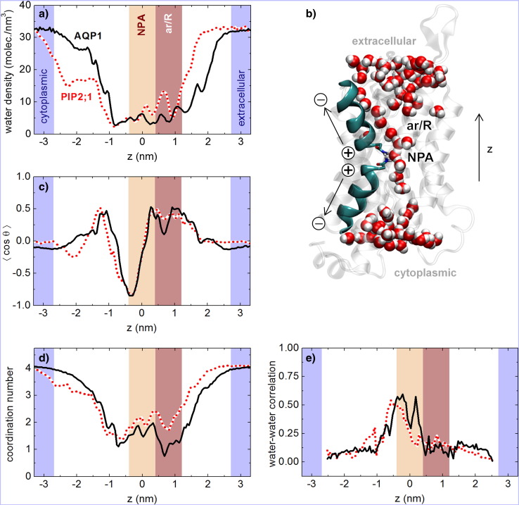
Cordeiro (Cordeiro, 2015) calculated via MD the distribution and organization of water molecules in AQP1 and plant PIP2;1 (Spinacia oleracia plasma membrane intrinsic protein) channels using interatomic interactions described with the GROMOS 54A7 force field for AQPS, and SPC water model (Cordeiro, 2015). Inside the pore, water molecules were organized as a narrow file. Their dipoles were oriented according to the electrostatic macrodipoles generated by helices HB and HE (Fig. 1a-c). Water dipoles in different halves of the channel were oppositely oriented, meaning that molecules were forced to reorient as they passed through the NPA region. The average coordination number to other water molecules had values between 1 and 2 due to occasional disruptions in the water file structure (Fig. 1d). In the case of AQP1, disruptions were more significant at the ar/R constriction region. The motion of water molecules close to the NPA region was highly correlated, indicating that molecules moved collectively along the pore (Fig. 1e)(Cordeiro, 2015).
The permeability of AQPs is based on the collective motion of water molecules along the pore. The quantitative values of water permeability, , for the single channel osmotic permeability of mammalian AQP-1 around 3.6 (Cordeiro, 2015) 7.1 (Zhu et al., 2004), 10.3 (Hashido et al., 2007) and cm (de Groot and Grubmuller, 2001). These works showed excellent agreement with the experimentally determined 5.4 x 10⁻¹⁴ cm3/s (Engel and Stahlberg, 2002). In the case of AQP-0 (the membrane protein of the lens fiber cells) transports water more slowly than other AQPs such as AQP-1. In the literature were reported of AQP-0 based on MD simulations are 0.2 (Hashido et al., 2005, 2007) and 0.28 x 10⁻¹⁴ cm )Jensen et al., 2008) , while reported experimental value is 0.25 x 10⁻¹⁴ cm (Qiu et al., 2010). In the AqpZ and Glpf was related around 16-14 cm (Hashido et al., 2007) The AQPs showed a wide range of values due that differences in the channel structures would determine the values.
2.2 Molecular mechanisms of physiologically molecules transport
The physiological function of AQPs in conducting gas molecules is not fully known (Alishahi and Kamali, 2019). MD modeling identified that there are possible routes for CO2 to cross in a membrane (Wang et al., 2007). In addition, AQPs are capable of transporting ROS across phospholipid membranes. MD simulations (Cordeiro, 2015), showed that water and ROS had similar transport features and might be transported by mammalian and plant AQPs. In addition, recent work (Alishahi and Kamali, 2019) has shown that CO2 molecules may pass through all AQP-5 monomer channels, such as the central pore. The core pore of AQP-5, like other AQPs, is known to be impervious to water molecules (Janosi and Ceccarelli, 2013). CO2 molecules, unlike water molecules, may readily pass through the AQP-5 central pore. The hydrophobic property of AQP’s central pore has no influence on the passage of nonpolar CO2 molecules across this channel (Alishahi and Kamali, 2019). A number of CO2 molecules diffuse through the palmitoyloleyl phosphatidylethanolamine (POPE) bilayer due to the free volume in the lipid structure. The greatest energy barrier for CO2 molecules in the channel for a specific CO2 diffusion with velocity 1 Å/ns is 8.8kcal/mol, but the energy barrier of the central pore does not surpass 5.1Kcal/mol for the same simulation Alishahi and Kamali, 2019. As a result, the hydrophobic central pore is the best route for CO2 molecules to pass through the AQP5 (Alishahi and Kamali, 2019).
Despite the MD simulations to quantitatively reproduce experimental parameters, even when different force field parameters and conditions were used. On the other hand, MD neglects the quantum mechanical effects of the systems. Therefore, it is impossible to consider the effects of chemical bond formation and breakage, and consequently, it was not possible to obtain proton diffusion. Furthermore, the literature lacks a greater discussion of theoretical and experimental work on water transport in open channels of AQPs plants. This is attributed to the great difficulty in obtaining the structure of these plants.
2.3 H+ exclusion by aquaporins
The plants AQPs membrane channels as their opening and closing gating is regulated by multiple effectors including H+, phosphorylation, and others. Since the determination of X-ray structures of spinach SoPIP2;1 in an open pore and a closed pore conformation, and MD of water transport in the open pore (Törnroth-Horsefield et al., 2006; Khandelia et al., 2009). However,the effect of phosphorylation on AQPS should be further explored in simulations. In addition, there is no experimental evidence of the efficient transport of H+ that any plant AQPs has, until the moment. In simulations of AQP-1 (De Groot et al., 2003; Burykin and Warshel, 2003; Chakrabarti et al., 2004; Kato et al., 2006), showed the large electrostatic barrier impeded H+ flux, together with the dehydration cost of moving a proton into the narrow hydrophobic channel. The electrostatic barrier excluded protons from permeating even in the presence of an intact proton wire (Chen et al., 2006; Kato et al., 2006; Hub et al., 2009).
3 Computational studies on water flow in synthetic ionic nanochannels and membranes
Artificial nanochannels mimicking water transport of organic membranes have been extensively investigated in the last few decades. AQP analogs are expected to help materials science and nanofluidics to establish a new era for desalination and water purification technologies (Kocsis et al., 2018). It also means that we now have access to what extent the charge distribution, atomic arrangement, and molecular architecture are determinants for the water conduction within our very cells. There is no surprise that so many condensed matter groups worldwide are now focusing efforts on investigating the interaction at solid-liquid interfaces of solid state-based nanochannels and membranes. These artificial membranes open several new potential advantages, such as improved stability, simple and scalable fabrication, and controlled functionalization (Kolahalam et al., 2019; Baig et al., 2021; Speranza, 2021; Leão et al., 2023) – and no toxicity (da Rosa et al., 2021).
The research on synthetic aquaporin-like nanopores is focused mainly on the design and synthesis of novel conducting channels with improved water transport performances and solute rejection properties. It is important to note that a possible knowledge gap between solid-state nanochannels and naturally occurring AQPs lies in the enhanced dielectric exclusion due to the cylindrical shape of most of the artificial nanochannels being studied today (Yaroshchuk, 2000). In fact, ionic rejection is very sensitive to these factors (Bordin et al., 2012).
Computational simulations based on classical MD or first-principles DFT have been vastly employed to decipher the details regarding water conduction in nanochannels composed of different atomic arrangements. Most recently, some groups started using artificial intelligence based on graph neural networks to enhance the search for new, improved materials to be used in desalination plants (Wang et al., 2021a ). Porosity and adsorption properties are also being studied with the assistance of machine and deep learning (Jian et al., 2022; Ogoke et al., 2022).
3.1 Water structure under subnanometer confinement
The confinement of water in nanochannels (e.g., nanotubes and 2D materials) is able to freeze water into crystalline ice-like solids, and it can happen at ambient pressure and temperature conditions (Köhler et al., 2017; Corti et al., 2021). Koga et al. (Koga et al., 2000) found ice structures inside carbon nanotubes (CNTs) characterized as stacked, ordered polygonal rings of water molecules. Further analysis of ice structures suggests the existence of many ice phases inside nanotubes (Takaiwa et al., 2008). Over the past decades, a vast ensemble of structures has been found inside these nanotubes, making them a prominent tool for exploring water anomalies. The number, shape, and other structural properties of the confined water are extremely dependent on the size of the nanochannels.
Highly permeable water channels based on CNTs are at the forefront of translocation and separation studies. Inside their smooth structure, water behaves differently. It performs disruptive dynamics, freezing, and even multi-phases (Kotsalis et al., 2004; Bordin et al., 2014). Recently, Farimani and Aluru (Barati Farimani and Aluru, 2016) showed the existence of multiple phases of water at CNTs under atmospheric conditions. They found vapor, high-density ice, and liquid water phases to coexist in the region within 1 nm from the surface of the nanotube. These results can explain, for example, the no-slip phenomena (Secchi et al., 2016) and the fast transport of water in bundles of CNTs (Majumder et al., 2005).
3.2 Water flux in synthetic nanochannels
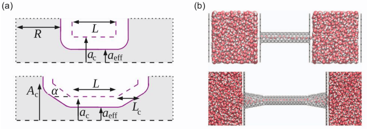
The inner walls of a CNT are known for being electrically and mechanically smooth. It allows for the formation of a depletion layer at the interface that results in high water flux (Köhler and Bordin, 2018). However, the smooth inner surface contrasts with often abrupt entrances. The problem at a usual entrance of a CNT is that much of the energy that would be converted in a highly organized stream flow is lost through friction due to collisions of water molecules and carbon atoms. Interestingly, nanotubes built from carbon atoms assembled in an AQP architecture (conical entrances with varying opening angles, see Figure 2) exhibited increased water permeability as compared to pristine CNTs, as shown in a theoretical study by Gravelle et al. (Gravelle et al., 2013). It suggests that the hourglass shape present in a significant number of AQPs may be the result of a natural selection process toward optimal permeability. The same group further combined MD simulations with finite element (FE) calculations to explore the implications of friction and energy dissipation at the nanopore/reservoir interface (Gravelle et al., 2014). All to confirm that the hourglass shape inspired in AQPs enhances the water transportation efficiency down to a single-file regime, with a maximum at shallow opening angles of around 5º. This illustrates the importance of entrance dissipation in nanofluidic systems and stresses how the fine-tuning of the nanopore geometry is necessary for optimizing water transport. More recently, inspired by the work of Gravelle’s group, Hadidi and Kamali (Hadidi and Kamali, 2020) have shown that an external pressure difference can lead to a water flux enhancement. A similar enhancement is observed when an external electric field is applied. The high flux regimes can be explained in terms of a reduction in the steric crowding effect of water molecules and an increase in the average number of hydrogen bonds, both occurring at the entrance of the nanochannel.
Aquaporins’ capacity for mediating highly selective and fast water transport has also inspired the advance and development of different supramolecular ion-excluding artificial water channels. In a recent contribution, Roy et al. (Roy et al., 2021) studied different types of foldamer-derived polymer-based synthetic water channels to find that tuning of interior groups and attachment of carboxylic acid-based lipid anchors lead them to an outstanding transport of about water molecules per second. They also found these membranes to possess a high capacity to reject both salts and protons.
3.3 Nanopore selectivity
The specificities of the nanopore architecture play a huge role in preventing or accelerating ionic passage. Many computational studies have been employed to understand this topic using artificial Bio-inspired nanopores. Bordin and co-authores have conducted simulations using a cylinder made by annular rings of Lennard-Jones spheres as the ionic channel (Bordin et al., 2012), the dielectric constant of water equals to bulk water value, K+ ions with several concentrations, so they could compare their results with experiments of gramicidin A (gA). The binding sites and the selectivity in the artificial channel were obtained by inserting negative charges in two specific points of the hydrophobic nanotube structure - similar to a functionalized carbon nanotube. They found a similar current versus voltage behavior between simulations and experiments. The resemblance was verified to different concentrations.
Among the computational alternatives available to investigate ionic transport and rejection in nanochannels, Brownian Dynamics (BD) is currently one of the most suitable tools because of its integration scheme that involves only the ionic motion. At the same time, the degrees of freedom of proteins and other bodies are kept fixed. In this method, the water is modeled as a continuum external field with a proper dielectric constant assumed (Carr et al., 2011; Bordin et al., 2016).
Many computational studies have employed CNTs to mimic organic ion channels for ion selectivity. For example, AQPs and other biological systems need selectivity between cations and anions for processes of organelle acidification or maintaining an electrochemical gradient across cells (Hille, 1978; Wang and Tajkhorshid, 2007; Warshel, 2005).
A typical ionic exclusion and selectivity prototype involves a nanochannel small enough to offer a natural mechanical barrier (Köhler et al., 2019; Abal et al., 2020). Since strongly hydrated ions prefer to stay that way (preserving their electrostatic configuration), the efficiency of the membrane will depend on the hydration level (Thomas et al., 2014). Hydrated ions have their passage facilitated in larger pores due to an energetic penalty associated with the unbinding of water molecules from de solvation shell. However, the passage of those ions through smaller nanopores is completely hindered since they have to dissociate from water. This mechanism explains why synthetic bio-inspired membranes with nanochannels can efficiently prevent the passage of ions while allowing for unimpeded water transport.
4 Conclusions and Perspectives
Despite its relevance, there is a clear lack of Molecular Simulation works devoted to unraveling the details of the AQP channel’s function in plants. The filling of this gap could greatly increase our understanding of the permeation of distinct molecules, such as water, CO2, H2O2, ammonia, and urea, in PIPs and TIPs, and even shade some light in the SIPs physiological functions. Beyond that, we face a period in history where climate changes are accelerating extreme weather events, such as long periods of drought or violent storms. Plants will have to adapt or their very existence will be threatened (Pörtner et al., 2022).
Nanotechnology and nanoscience have a central role in this urgent demand. Many studies indicate that we can insert artificial nanopores with specific functions into cellular membranes (Höfinger et al., 2011; Dutt et al., 2011; Baoukina et al., 2013; Thomas et al., 2015; Geng et al., 2014; Kanno et al., 2022; Wang et al., 2022a ). Then, if they can mimic the behavior of distinct transmembrane channels, why can we not improve their performance? Recent advances in nanotube functionalization uncovered new possibilities (Hirsch, 2002; Bordin and Barbosa, 2017; Kharlamova et al., 2022; Hosseini and Ghaffarzadeh, 2022; Pardehkhorram and Andrieu-Brunsen, 2022; Li et al., 2022a ; Xu et al., 2022; Turhan et al., 2022).
Now, we can use them to control, for instance, the water permeation into plant roots under water stress by using artificial channels with distinct functionalization. We can increase or decrease the permeation to specific molecules in roots and leaves, including releasing fertilizers and nutrients at a fixed, well-defined rate (Yanovska et al., 2021; Vakal et al., 2022). Even the pesticide delivery can be controlled (Singh et al., 2022; Wang et al., 2022b ; Bhattacharya et al., 2022), minimizing the health and environmental damages associated with the misuse of pesticides (Li and Fantke, 2022; Cara et al., 2022; Tudi et al., 2022).
Finally, we address the fact that Molecular Modeling is a powerful tool to design nanomaterials with specific functions, providing insights that are often inaccessible in experiments. This mini-review and perspective aimed to show how urgent it is to increase the collaboration between Molecular Simulations and Plant Physiology communities, filling the gaps and increasing our understanding of molecular details of the plant machinery, and proposing ways to help to overcome the current and next challenges that plants will face, mainly due the Climate Changes.
Acknowledgments
JRB is grateful to Gustavo M, Souza for the invitation to submit this minireview and pescpective, and for the stimulating discussions about Biology, Physics, Life and everything else.
Without public funding, this research would be impossible. JRB is grateful to the Brazilian National Council for Scientific and Technological Development (CNPq), PQ Grant nr. 304958/2022-0, and Research Support Foundation of the State of Rio Grande do Sul (FAPERGS), PqG Grant n2. 21/2551-0002024-5, for funding support. MHK and JRB thanks CNPq Universal grant nr. 306709/2021-0. MHK thanks FAPERGS PqG Grant nr. 21/2551-0002023-7. LAP and TG thank CNPq and AVI, PRBC, and WSO thank the Coordination for the Improvement of Higher Education Personnel (CAPES, financing Code 001) for the Scholarship.
Declarations
The authors disclose no conflicts of interest. All authors contribute equally to this paper.
References
- Abal et al., (2020) Abal, J. P. K., Bordin, J. R., and Barbosa, M. C. (2020). Salt parameterization can drastically affect the results from classical atomistic simulations of water desalination by MoS2 nanopores. Physical Chemistry Chemical Physics, 22(19):11053–11061.
- Ahmed et al., (2021) Ahmed, S., Kouser, S., Asgher, M., and Gandhi, S. G. (2021). Plant aquaporins: A frontward to make crop plants drought resistant. Physiologia Plantarum, 172(2):1089–1105.
- Alishahi and Kamali, (2018) Alishahi, M. and Kamali, R. (2018). Forced diffusion of water molecules through aquaporin-5 biomembrane; a molecular dynamics study. Biophysics and Physicobiology, 15:255–262.
- Alishahi and Kamali, (2019) Alishahi, M. and Kamali, R. (2019). A novel molecular dynamics study of co2 permeation through aquaporin-5. The European Physical Journal E, 42(11):1–8.
- AlJensen MØ, (2006) AlJensen MØ, M. O. (2006). Single-channel water permeabilities of escherichia coli aquaporins aqpz and glpf. TBiophys J, 90(7):2270–84.
- Aponte-Santamaría et al., (2010) Aponte-Santamaría, C., Hub, J. S., and de Groot, B. L. (2010). Dynamics and energetics of solute permeation through the plasmodium falciparum aquaglyceroporin. Physical Chemistry Chemical Physics, 12(35):10246–10254.
- Baig et al., (2021) Baig, N., Kammakakam, I., and Falath, W. (2021). Nanomaterials: a review of synthesis methods, properties, recent progress, and challenges. Materials Advances, 2(6):1821–1871.
- Baoukina et al., (2013) Baoukina, S., Monticelli, L., and Tieleman, D. P. (2013). Interaction of pristine and functionalized carbon nanotubes with lipid membranes. The Journal of Physical Chemistry B, 117(40):12113–12123.
- Barati Farimani and Aluru, (2016) Barati Farimani, A. and Aluru, N. R. (2016). Existence of multiple phases of water at nanotube interfaces. The Journal of Physical Chemistry C, 120(41):23763–23771.
- Bhattacharya et al., (2022) Bhattacharya, N., Cahill, D. M., Yang, W., and Kochar, M. (2022). Graphene as a nano-delivery vehicle in agriculture – current knowledge and future prospects. Critical Reviews in Biotechnology, pages 1–19.
- Bordin and Barbosa, (2017) Bordin, J. R. and Barbosa, M. C. (2017). Flow and structure of fluids in functionalized nanopores. Physica A: Statistical Mechanics and its Applications, 467:137–147.
- Bordin et al., (2012) Bordin, J. R., Diehl, A., Barbosa, M. C., and Levin, Y. (2012). Ion fluxes through nanopores and transmembrane channels. Phys. Rev. E, 85:031914.
- Bordin et al., (2014) Bordin, J. R., Krott, L. B., and Barbosa, M. C. (2014). Surface phase transition in anomalous fluid in nanoconfinement. The Journal of Physical Chemistry C, 118(18):9497–9506.
- Bordin et al., (2016) Bordin, J. R., Podgornik, R., and Holm, C. (2016). Static polarizability effects on counterion distributions near charged dielectric surfaces: A coarse-grained molecular dynamics study employing the drude model. The European Physical Journal Special Topics, 225(8-9):1693–1705.
- Bots et al., (2005) Bots, M., Vergeldt, F., Wolters-Arts, M., Weterings, K., van As, H., and Mariani, C. (2005). Aquaporins of the PIP2 Class Are Required for Efficient Anther Dehiscence in Tobacco. Plant Physiology, 137(3):1049–1056.
- Burykin and Warshel, (2003) Burykin, A. and Warshel, A. (2003). What really prevents proton transport through aquaporin? charge self-energy versus proton wire proposals. Biophysical journal, 85(6):3696–3706.
- Cara et al., (2022) Cara, I. G., \textcommabelowTopa, D., Puiu, I., and Jităreanu, G. (2022). Biochar a promising strategy for pesticide-contaminated soils. Agriculture, 12(10):1579.
- Carr et al., (2011) Carr, R., Comer, J., Ginsberg, M. D., and Aksimentiev, A. (2011). Atoms-to-microns model for small solute transport through sticky nanochannels. Lab on a Chip, 11(22):3766–3773.
- Chakrabarti et al., (2004) Chakrabarti, N., Tajkhorshid, E., Roux, B., and Pomès, R. (2004). Molecular basis of proton blockage in aquaporins. Structure, 12(1):65–74.
- Chaumont et al., (2005) Chaumont, F., Moshelion, M., and Daniels, M. J. (2005). Regulation of plant aquaporin activity. Biology of the Cell, 97(10):749–764.
- Chen et al., (2006) Chen, H., Wu, Y., and Voth, G. A. (2006). Origins of proton transport behavior from selectivity domain mutations of the aquaporin-1 channel. Biophysical journal, 90(10):L73–L75.
- Cordeiro, (2015) Cordeiro, R. M. (2015). Molecular dynamics simulations of the transport of reactive oxygen species by mammalian and plant aquaporins. Biochimica et Biophysica Acta (BBA)-General Subjects, 1850(9):1786–1794.
- Corti et al., (2021) Corti, H. R., Appignanesi, G. A., Barbosa, M. C., Bordin, J. R., Calero, C., Camisasca, G., Elola, M. D., Franzese, G., Gallo, P., Hassanali, A., Huang, K., Laria, D., Menéndez, C. A., de Oca, J. M. M., Longinotti, M. P., Rodriguez, J., Rovere, M., Scherlis, D., and Szleifer, I. (2021). Structure and dynamics of nanoconfined water and aqueous solutions. The European Physical Journal E, 44(11).
- da Rosa et al., (2021) da Rosa, P. C. C., Leão, M. B., Corte, C. L. D., and de Matos, C. F. (2021). Evaluation of the carbon nanostructures toxicity as a function of their dimensionality using model organisms: a review. Water, Air, & Soil Pollution, 232(9).
- De Groot et al., (2003) De Groot, B. L., Frigato, T., Helms, V., and Grubmüller, H. (2003). The mechanism of proton exclusion in the aquaporin-1 water channel. Journal of molecular biology, 333(2):279–293.
- de Groot and Grubmuller, (2001) de Groot, B. L. and Grubmuller, H. (2001). Water permeation across biological membranes: mechanism and dynamics of aquaporin-1 and glpf. Science, 294(5550):2353–2357.
- Ding et al., (2019) Ding, L., Uehlein, N., Kaldenhoff, R., Guo, S., Zhu, Y., and Kai, L. (2019). Aquaporin pip2;1 affects water transport and root growth in rice (oryza sativa l.). Plant Physiology and Biochemistry, 139:152–160.
- Dutt et al., (2011) Dutt, M., Nayhouse, M. J., Kuksenok, O., Little, S. R., and Balazs, A. C. (2011). Interactions of end-functionalized nanotubes with lipid vesicles: Spontaneous insertion and nanotube self-organization. Current Nanoscience, 7(5):699–715.
- (29) Dynowski, M., Mayer, M., Moran, O., and Ludewig, U. (2008a). Molecular determinants of ammonia and urea conductance in plant aquaporin homologs. FEBS letters, 582(16):2458–2462.
- (30) Dynowski, M., Schaaf, G., Loque, D., Moran, O., and Ludewig, U. (2008b). Plant plasma membrane water channels conduct the signalling molecule h2o2. Biochemical Journal, 414(1):53–61.
- Engel and Stahlberg, (2002) Engel, A. and Stahlberg, H. (2002). Aquaglyceroporins: channel proteins with a conserved core, multiple functions, and variable surfaces. International review of cytology, 215:75–104.
- Fischer and Kaldenhoff, (2008) Fischer, M. and Kaldenhoff, R. (2008). On the ph regulation of plant aquaporins. Journal of Biological Chemistry, 283(49):33889–33892.
- Fortin et al., (1985) Fortin, M. G., Zelechowska, M., and Verma, D. P. S. (1985). Specific targeting of membrane nodulins to the bacteroid-enclosing compartment in soybean nodules. The EMBO Journal, 4(12):3041–3046.
- Fox et al., (2017) Fox, A. R., Maistriaux, L. C., and Chaumont, F. (2017). Toward understanding of the high number of plant aquaporin isoforms and multiple regulation mechanisms. Plant Science, 264:179–187.
- (35) Fu, D., Libson, A., Miercke, L. J., Weitzman, C., Nollert, P., Krucinski, J., and Stroud, R. M. (2000a). Structure of a glycerol-conducting channel and the basis for its selectivity. science, 290(5491):481–486.
- (36) Fu, D., Libson, A., Miercke, L. J., Weitzman, C., Nollert, P., Krucinski, J., and Stroud, R. M. (2000b). Structure of a glycerol-conducting channel and the basis for its selectivity. science, 290(5491):481–486.
- Geng et al., (2014) Geng, J., Kim, K., Zhang, J., Escalada, A., Tunuguntla, R., Comolli, L. R., Allen, F. I., Shnyrova, A. V., Cho, K. R., Munoz, D., Wang, Y. M., Grigoropoulos, C. P., Ajo-Franklin, C. M., Frolov, V. A., and Noy, A. (2014). Stochastic transport through carbon nanotubes in lipid bilayers and live cell membranes. Nature, 514(7524):612–615.
- Gravelle et al., (2013) Gravelle, S., Joly, L., Detcheverry, F., Ybert, C., Cottin-Bizonne, C., and Bocquet, L. (2013). Optimizing water permeability through the hourglass shape of aquaporins. Proceedings of the National Academy of Sciences, 110(41):16367–16372.
- Gravelle et al., (2014) Gravelle, S., Joly, L., Ybert, C., and Bocquet, L. (2014). Large permeabilities of hourglass nanopores: From hydrodynamics to single file transport. The Journal of chemical physics, 141(18):18C526.
- Groszmann et al., (2017) Groszmann, M., Osborn, H. L., and Evans, J. R. (2017). Carbon dioxide and water transport through plant aquaporins. Plant, Cell & Environment, 40(6):938–961.
- Hadidi and Kamali, (2020) Hadidi, H. and Kamali, R. (2020). Non-equilibrium molecular dynamics simulations of water transport through plate-and hourglass-shaped cnts in the presence of pressure difference and electric field. Computational Materials Science, 185:109978.
- Hadidi et al., (2021) Hadidi, H., Kamali, R., and Binesh, A. (2021). Investigation of the aquaporin-2 gating mechanism with molecular dynamics simulations. Proteins: Structure, Function, and Bioinformatics, 89(7):819–831.
- Hadidi et al., (2022) Hadidi, H., Kamali, R., and Binesh, A. (2022). Dynamics and energetics of water transport through aquaporin mutants causing nephrogenic diabetes insipidus (ndi): A molecular dynamics study. Journal of Biomolecular Structure and Dynamics, 40(3):1273–1284.
- Hall et al., (2019) Hall, J. E., Freites, J. A., and Tobias, D. J. (2019). Experimental and simulation studies of aquaporin 0 water permeability and regulation. Chemical reviews, 119(9):6015–6039.
- Hashido et al., (2005) Hashido, M., Ikeguchi, M., and Kidera, A. (2005). Comparative simulations of aquaporin family: Aqp1, aqpz, aqp0 and glpf. FEBS letters, 579(25):5549–5552.
- Hashido et al., (2007) Hashido, M., Kidera, A., and Ikeguchi, M. (2007). Water transport in aquaporins: osmotic permeability matrix analysis of molecular dynamics simulations. Biophysical journal, 93(2):373–385.
- Hille, (1978) Hille, B. (1978). Ionic channels in excitable membranes. current problems and biophysical approaches. Biophysical journal, 22(2):283–294.
- Hille, (2001) Hille, B. (2001). Ion Channels of Excitable Membranes, 3rd ed. Sinauer Associates, Sunderland, MA.
- Hirsch, (2002) Hirsch, A. (2002). Functionalization of single-walled carbon nanotubes. Angewandte Chemie International Edition, 41(11):1853–1859.
- Hosseini and Ghaffarzadeh, (2022) Hosseini, H. and Ghaffarzadeh, M. (2022). Surface functionalization of carbon nanotubes via plasma discharge: A review. Inorganic Chemistry Communications, 138:109276.
- Hub and de Groot, (2006) Hub, J. S. and de Groot, B. L. (2006). Does co2 permeate through aquaporin-1? Biophysical Journal, 91(3):842–848.
- Hub and De Groot, (2008) Hub, J. S. and De Groot, B. L. (2008). Mechanism of selectivity in aquaporins and aquaglyceroporins. Proceedings of the National Academy of Sciences, 105(4):1198–1203.
- Hub et al., (2009) Hub, J. S., Grubmüller, H., and Groot, B. L. d. (2009). Dynamics and energetics of permeation through aquaporins. what do we learn from molecular dynamics simulations? Aquaporins, pages 57–76.
- Höfinger et al., (2011) Höfinger, S., Melle-Franco, M., Gallo, T., Cantelli, A., Calvaresi, M., Gomes, J. A., and Zerbetto, F. (2011). A computational analysis of the insertion of carbon nanotubes into cellular membranes. Biomaterials, 32(29):7079–7085.
- Ikeda et al., (1997) Ikeda, S., Nasrallah, J. B., Dixit, R., Preiss, S., and Nasrallah, M. E. (1997). An aquaporin-like gene required for the brassica self-incompatibility response. Science, 276(5318):1564–1566.
- Ishikawa et al., (2005) Ishikawa, F., Suga, S., Uemura, T., Sato, M. H., and Maeshima, M. (2005). Novel type aquaporin sips are mainly localized to the er membrane and show cell-specific expression in arabidopsis thaliana. FEBS Letters, 579(25):5814–5820.
- Israel et al., (2022) Israel, D., Lee, S. H., Robson, T. M., and Zwiazek, J. J. (2022). Plasma membrane aquaporins of the pip1 and pip2 subfamilies facilitate hydrogen peroxide diffusion into plant roots. BMC Plant Biology, 22(1):1–15.
- Janosi and Ceccarelli, (2013) Janosi, L. and Ceccarelli, M. (2013). The gating mechanism of the human aquaporin 5 revealed by molecular dynamics simulations. PLoS One, 8(4):e59897.
- Jensen et al., (2008) Jensen, M. Ø., Dror, R. O., Xu, H., Borhani, D. W., Arkin, I. T., Eastwood, M. P., and Shaw, D. E. (2008). Dynamic control of slow water transport by aquaporin 0: implications for hydration and junction stability in the eye lens. Proceedings of the National Academy of Sciences, 105(38):14430–14435.
- Jian et al., (2022) Jian, Y., Wang, Y., and Barati Farimani, A. (2022). Predicting co2 absorption in ionic liquids with molecular descriptors and explainable graph neural networks. ACS Sustainable Chemistry & Engineering, 10(50):16681–16691.
- Kaldenhoff and Fischer, (2006) Kaldenhoff, R. and Fischer, M. (2006). Functional aquaporin diversity in plants. Biochimica et Biophysica Acta (BBA) - Biomembranes, 1758(8):1134–1141. Aquaporins.
- Kanno et al., (2022) Kanno, S., Peng, Z., Shimba, K., Miyamoto, Y., and Yagi, T. (2022). Transmembrane carbon nanotubes for intracellular stimulation. In 2022 14th Biomedical Engineering International Conference (BMEiCON), pages 1–4.
- Kapilan et al., (2018) Kapilan, R., Vaziri, M., and Zwiazek, J. J. (2018). Regulation of aquaporins in plants under stress. Biological research, 51(1):1–11.
- Kato et al., (2006) Kato, M., Pisliakov, A. V., and Warshel, A. (2006). The barrier for proton transport in aquaporins as a challenge for electrostatic models: The role of protein relaxation in mutational calculations. Proteins: Structure, Function, and Bioinformatics, 64(4):829–844.
- Khandelia et al., (2009) Khandelia, H., Jensen, M. Ø., and Mouritsen, O. G. (2009). To gate or not to gate: using molecular dynamics simulations to morph gated plant aquaporins into constitutively open conformations. The Journal of Physical Chemistry B, 113(15):5239–5244.
- Kharlamova et al., (2022) Kharlamova, M. V., Paukov, M., and Burdanova, M. G. (2022). Nanotube functionalization: Investigation, methods and demonstrated applications. Materials, 15(15):5386.
- Kocsis et al., (2018) Kocsis, I., Sun, Z., Legrand, Y. M., and Barboiu, M. (2018). Artificial water channels—deconvolution of natural aquaporins through synthetic design. npj Clean Water, 1(1):13.
- Koga et al., (2000) Koga, K., Parra, R. D., Tanaka, H., and Zeng, X. C. (2000). Ice nanotube: What does the unit cell look like? The Journal of Chemical Physics, 113(12):5037–5040.
- Köhler and Bordin, (2018) Köhler, M. H. and Bordin, J. R. (2018). Surface, density, and temperature effects on the water diffusion and structure inside narrow nanotubes. The Journal of Physical Chemistry C, 122(12):6684–6690.
- Köhler et al., (2017) Köhler, M. H., Bordin, J. R., da Silva, L. B., and Barbosa, M. C. (2017). Breakdown of the stokes-einstein water transport through narrow hydrophobic nanotubes. Phys. Chem. Chem. Phys., 19:12921–12927.
- Köhler et al., (2019) Köhler, M. H., Bordin, J. R., de Matos, C. F., and Barbosa, M. C. (2019). Water in nanotubes: The surface effect. Chemical Engineering Science, 203:54–67.
- Kolahalam et al., (2019) Kolahalam, L. A., Viswanath, I. K., Diwakar, B. S., Govindh, B., Reddy, V., and Murthy, Y. (2019). Review on nanomaterials: Synthesis and applications. Materials Today: Proceedings, 18:2182–2190.
- Kotsalis et al., (2004) Kotsalis, E., Walther, J. H., and Koumoutsakos, P. (2004). Multiphase water flow inside carbon nanotubes. International Journal of Multiphase Flow, 30(7-8):995–1010.
- Kruse et al., (2006) Kruse, E., Uehlein, N., and Kaldenhoff, R. (2006). The aquaporins. Genome Biol, 7(206).
- Kudoyarova et al., (2022) Kudoyarova, G., Veselov, D., Yemelyanov, V., and Shishova, M. (2022). The role of aquaporins in plant growth under conditions of oxygen deficiency. International Journal of Molecular Sciences, 23(17).
- Leão et al., (2023) Leão, M. B., Bordin, J. R., and de Matos, C. F. (2023). Specific surface area versus adsorptive capacity: an application view of 3d graphene-based materials for the removal of emerging water pollutants. Water, Air, & Soil Pollution, 234(2).
- (77) Li, C., Guan, X., Sun, Y., and Li, J. (2022a). Recent progress in the construction of artificial nanochannels for biosensing. Analysis & Sensing, 2(6).
- (78) Li, Q., Tong, T., Jiang, W., Cheng, J., Deng, F., Wu, X., Chen, Z.-H., Ouyang, Y., and Zeng, F. (2022b). Highly conserved evolution of aquaporin pips and tips confers their crucial contribution to flowering process in plants. Frontiers in plant science, 12:2945.
- Li and Fantke, (2022) Li, Z. and Fantke, P. (2022). Toward harmonizing global pesticide regulations for surface freshwaters in support of protecting human health. Journal of Environmental Management, 301:113909.
- Lohrasebi and Koslowski, (2019) Lohrasebi, A. and Koslowski, T. (2019). Modeling water purification by an aquaporin-inspired graphene-based nano-channel. Journal of Molecular Modeling, 25(9):1–9.
- Lopez et al., (2012) Lopez, D., Bronner, G., Brunel, N., Auguin, D., Bourgerie, S., Brignolas, F., Carpin, S., Tournaire-Roux, C., Maurel, C., Fumanal, B., Martin, F., Sakr, S., Label, P., Julien, J., Gousset-Dupont, A., and Venisse, J. (2012). Insights into Populus XIP aquaporins: evolutionary expansion, protein functionality, and environmental regulation. Journal of Experimental Botany, 63(5):2217–2230.
- Lopez et al., (2013) Lopez, D., Venisse, J.-S., Fumanal, B., Chaumont, F., Guillot, E., Daniels, M. J., Cochard, H., Julien, J.-L., and Gousset-Dupont, A. (2013). Aquaporins and Leaf Hydraulics: Poplar Sheds New Light. Plant and Cell Physiology, 54(12):1963–1975.
- Ludewig and Dynowski, (2009) Ludewig, U. and Dynowski, M. (2009). Plant aquaporin selectivity: where transport assays, computer simulations and physiology meet. Cellular and Molecular Life Sciences, 66(19):3161–3175.
- Majumder et al., (2005) Majumder, M., Chopra, N., Andrews, R., and Hinds, B. J. (2005). Enhanced flow in carbon nanotubes. Nature, 438(7064):44–44.
- Mangiatordi et al., (2016) Mangiatordi, G. F., Alberga, D., Trisciuzzi, D., Lattanzi, G., and Nicolotti, O. (2016). Human aquaporin-4 and molecular modeling: historical perspective and view to the future. International Journal of Molecular Sciences, 17(7):1119.
- Mao and Sun, (2015) Mao, Z. and Sun, W. (2015). Arabidopsis seed-specific vacuolar aquaporins are involved in maintaining seed longevity under the control of ABSCISIC ACID INSENSITIVE 3. Journal of Experimental Botany, 66(15):4781–4794.
- Maroli and Kolandaivel, (2020) Maroli, N. and Kolandaivel, P. (2020). Comparative study of stability and transport of molecules through cyclic peptide nanotube and aquaporin: a molecular dynamics simulation approach. Journal of Biomolecular Structure and Dynamics, 38(1):186–199. PMID: 30678549.
- Martre et al., (2002) Martre, P., Morillon, R., Barrieu, F., North, G. B., Nobel, P. S., and Chrispeels, M. J. (2002). Plasma Membrane Aquaporins Play a Significant Role during Recovery from Water Deficit. Plant Physiology, 130(4):2101–2110.
- Maurel et al., (2008) Maurel, C., Verdoucq, L., Luu, D.-T., and Santoni, V. (2008). Plant aquaporins: Membrane channels with multiple integrated functions. Annual Review of Plant Biology, 59(1):595–624. PMID: 18444909.
- Mehra et al., (2022) Mehra, P., Pandey, B. K., Melebari, D., Banda, J., Leftley, N., Couvreur, V., Rowe, J., Anfang, M., Gernier, H. D., Morris, E., Sturrock, C. J., Mooney, S. J., Swarup, R., Faulkner, C., Beeckman, T., Bhalerao, R. P., Shani, E., Jones, A. M., Dodd, I. C., Sharp, R. E., Sadanandom, A., Draye, X., and Bennett, M. J. (2022). Hydraulic flux responsive hormone redistribution determines root branching. Science, 378(6621):762–768.
- Moshelion et al., (2002) Moshelion, M., Becker, D., Biela, A., Uehlein, N., Hedrich, R., Otto, B., Levi, H., Moran, N., and Kaldenhoff, R. (2002). Plasma membrane aquaporins in the motor cells of samanea saman: diurnal and circadian regulation. The Plant cell, 14(1):727–739.
- Murata et al., (2000) Murata, K., Mitsuoka, K., Hirai, T., Walz, T., Agre, P., Heymann, J. B., Engel, A., and Fujiyoshi, Y. (2000). Structural determinants of water permeation through aquaporin-1. Nature, 407(6804):599–605.
- Neto and Cordeiro, (2016) Neto, A. J. and Cordeiro, R. M. (2016). Molecular simulations of the effects of phospholipid and cholesterol peroxidation on lipid membrane properties. Biochimica et Biophysica Acta (BBA)-Biomembranes, 1858(9):2191–2198.
- Neumann et al., (2020) Neumann, L. S., Dias, A. H., and Skaf, M. S. (2020). Molecular modeling of aquaporins from leishmania major. The Journal of Physical Chemistry B, 124(28):5825–5836.
- Noronha et al., (2016) Noronha, H., Araújo, D., Conde, C., Martins, A. P., Soveral, G., Chaumont, F., Delrot, S., and Gerós, H. (2016). The grapevine uncharacterized intrinsic protein 1 (vvxip1) is regulated by drought stress and transports glycerol, hydrogen peroxide, heavy metals but not water. PLOS ONE, 11(8):1–18.
- Nyblom et al., (2009) Nyblom, M., Frick, A., Wang, Y., Ekvall, M., Hallgren, K., Hedfalk, K., Neutze, R., Tajkhorshid, E., and Törnroth-Horsefield, S. (2009). Structural and functional analysis of sopip2; 1 mutants adds insight into plant aquaporin gating. Journal of molecular biology, 387(3):653–668.
- Ogoke et al., (2022) Ogoke, O. F., Johnson, K., Glinsky, M., Laursen, C., Kramer, S., and Farimani, A. B. (2022). Deep-learned generators of porosity distributions produced during metal additive manufacturing. Additive Manufacturing, 60:103250.
- Ozu et al., (2013) Ozu, M., Alvarez, H. A., McCarthy, A. N., Grigera, J. R., and Chara, O. (2013). Molecular dynamics of water in the neighborhood of aquaporins. European Biophysics Journal, 42(4):223–239.
- Pardehkhorram and Andrieu-Brunsen, (2022) Pardehkhorram, R. and Andrieu-Brunsen, A. (2022). Pushing the limits of nanopore transport performance by polymer functionalization. Chemical Communications, 58(34):5188–5204.
- Park et al., (2010) Park, W., Scheffler, B., Bauer, P., and Campbell, B. T. (2010). Identification of the family of aquaporin genes and their expression in upland cotton (gossypium hirsutuml.). BMC Plant Biol, 10:142.
- Pörtner et al., (2022) Pörtner, H.-O., Roberts, D., Tignor, M., Poloczanska, E., Mintenbeck, K., Alegría, A., Craig, M., Langsdorf, S., Löschke, S., Möller, V., Okem, A., and (eds.), B. R. (2022). IPCC, 2022: Climate Change 2022: Impacts, Adaptation, and Vulnerability. Contribution of Working Group II to the Sixth Assessment Report of the Intergovernmental Panel on Climate Change. Cambridge University Press, Cambridge, UK and New York, NY, USA.
- Qiu et al., (2010) Qiu, H., Ma, S., Shen, R., and Guo, W. (2010). Dynamic and energetic mechanisms for the distinct permeation rate in aqp1 and aqp0. Biochimica et Biophysica Acta (BBA)-Biomembranes, 1798(3):318–326.
- Roux and Schulten, (2004) Roux, B. and Schulten, K. (2004). Computational studies of membrane channels. Structure, 12(8):1343–1351.
- Roy et al., (2021) Roy, A., Shen, J., Joshi, H., Song, W., Tu, Y.-M., Chowdhury, R., Ye, R., Li, N., Ren, C., Kumar, M., et al. (2021). Foldamer-based ultrapermeable and highly selective artificial water channels that exclude protons. Nature Nanotechnology, 16(8):911–917.
- Sakurai et al., (2008) Sakurai, J., Ahamed, A., Murai, M., Maeshima, M., and Uemura, M. (2008). Tissue and Cell-Specific Localization of Rice Aquaporins and Their Water Transport Activities. Plant and Cell Physiology, 49(1):30–39.
- Salman et al., (2022) Salman, M. M., Kitchen, P., Yool, A. J., and Bill, R. M. (2022). Recent breakthroughs and future directions in drugging aquaporins. Trends in Pharmacological Sciences, 43(1):30–42.
- Secchi et al., (2016) Secchi, E., Marbach, S., Niguès, A., Stein, D., Siria, A., and Bocquet, L. (2016). Massive radius-dependent flow slippage in carbon nanotubes. Nature, 537(7619):210–213.
- Singh et al., (2022) Singh, G., Ramadass, K., Sooriyakumar, P., Hettithanthri, O., Vithange, M., Bolan, N., Tavakkoli, E., Zwieten, L. V., and Vinu, A. (2022). Nanoporous materials for pesticide formulation and delivery in the agricultural sector. Journal of Controlled Release, 343:187–206.
- Speranza, (2021) Speranza, G. (2021). Carbon nanomaterials: Synthesis, functionalization and sensing applications. Nanomaterials, 11(4):967.
- Sui et al., (2001) Sui, H., Han, B.-G., Lee, J. K., Walian, P., and Jap, B. K. (2001). Structural basis of water-specific transport through the aqp1 water channel. Nature, 414(6866):872–878.
- Tajkhorshid et al., (2002) Tajkhorshid, E., Nollert, P., Jensen, M. Ø., Miercke, L. J., O’Connell, J., Stroud, R. M., and Schulten, K. (2002). Control of the selectivity of the aquaporin water channel family by global orientational tuning. Science, 296(5567):525–530.
- Takaiwa et al., (2008) Takaiwa, D., Hatano, I., Koga, K., and Tanaka, H. (2008). Phase diagram of water in carbon nanotubes. Proceedings of the National Academy of Sciences, 105(1):39–43.
- Terashima and Ono, (2002) Terashima, I. and Ono, K. (2002). Effects of HgCl2 on CO2 Dependence of Leaf Photosynthesis: Evidence Indicating Involvement of Aquaporins in CO2 Diffusion across the Plasma Membrane. Plant and Cell Physiology, 43(1):70–78.
- Thomas et al., (2014) Thomas, M., Corry, B., and Hilder, T. A. (2014). What have we learnt about the mechanisms of rapid water transport, ion rejection and selectivity in nanopores from molecular simulation? Small, 10(8):1453–1465.
- Thomas et al., (2015) Thomas, M., Enciso, M., and Hilder, T. A. (2015). Insertion mechanism and stability of boron nitride nanotubes in lipid bilayers. The Journal of Physical Chemistry B, 119(15):4929–4936.
- Törnroth-Horsefield et al., (2006) Törnroth-Horsefield, S., Wang, Y., Hedfalk, K., Johanson, U., Karlsson, M., Tajkhorshid, E., Neutze, R., and Kjellbom, P. (2006). Structural mechanism of plant aquaporin gating. Nature, 439(7077):688–694.
- Tournaire-Roux et al., (2003) Tournaire-Roux, C., Sutka, M., Javot, H., Gout, E., Gerbeau, P., Luu, D.-T., Bligny, R., and Maurel, C. (2003). Cytosolic ph regulates root water transport during anoxic stress through gating of aquaporins. Nature, 425(6956):393–397.
- Tudi et al., (2022) Tudi, M., Li, H., Li, H., Wang, L., Lyu, J., Yang, L., Tong, S., Yu, Q. J., Ruan, H. D., Atabila, A., Phung, D. T., Sadler, R., and Connell, D. (2022). Exposure routes and health risks associated with pesticide application. Toxics, 10(6):335.
- Turhan et al., (2022) Turhan, E. A., Pazarçeviren, A. E., Evis, Z., and Tezcaner, A. (2022). Properties and applications of boron nitride nanotubes. Nanotechnology, 33(24):242001.
- Vajpai et al., (2018) Vajpai, M., Mukherjee, M., and Sankararamakrishnan, R. (2018). Cooperativity in plant plasma membrane intrinsic proteins (pips): Mechanism of increased water transport in maize pip1 channels in hetero-tetramers.
- Vakal et al., (2022) Vakal, S., Vakal, V., Artyukhov, A., Shkola, V., and Yanovska, A. (2022). New method for obtaining “green” encapsulated fertilizers with nanoporous structure within the concept of sustainable development. Clean Technologies and Environmental Policy.
- Verdoucq et al., (2008) Verdoucq, L., Grondin, A., and Maurel, C. (2008). Structure–function analysis of plant aquaporin at pip2; 1 gating by divalent cations and protons. Biochemical Journal, 415(3):409–416.
- Wambo et al., (2017) Wambo, T. O., Rodriguez, R. A., and Chen, L. Y. (2017). Computing osmotic permeabilities of aquaporins aqp4, aqp5, and glpf from near-equilibrium simulations. Biochimica et Biophysica Acta (BBA)-Biomembranes, 1859(8):1310–1316.
- (124) Wang, Q., Sun, J., Xue, W., Zhao, G., Ding, W., Zhang, K., and Wang, S. (2022a). An ideal artificial water channel—carbon nanotube porins (cntps) for preparing highly permeable reverse osmosis membrane with excellent antifouling capacity. SSRN Electronic Journal.
- (125) Wang, Y., Cao, Z., and Barati Farimani, A. (2021a). Efficient water desalination with graphene nanopores obtained using artificial intelligence. npj 2D Materials and Applications, 5(1):66.
- Wang et al., (2007) Wang, Y., Cohen, J., Boron, W. F., Schulten, K., and Tajkhorshid, E. (2007). Exploring gas permeability of cellular membranes and membrane channels with molecular dynamics. Journal of structural biology, 157(3):534–544.
- Wang and Tajkhorshid, (2007) Wang, Y. and Tajkhorshid, E. (2007). Molecular mechanisms of conduction and selectivity in aquaporin water channels. The Journal of nutrition, 137(6):1509S–1515S.
- (128) Wang, Y., Tian, J., Wang, Z., Li, C., and Li, X. (2022b). Crop-safe pyraclostrobin-loaded multiwalled carbon nanotube delivery systems: Higher fungicidal activity and lower acute toxicity. ACS Agricultural Science & Technology, 2(3):534–545.
- (129) Wang, Z., Zhao, T., Hu, Y., Zou, L., Wang, X., and Zhang, Y. (2021b). Molecular dynamics simulations of the permeation and distribution of plasma ros in aquaporin-1. Physics of Plasmas, 28(8):083509.
- Warshel, (2005) Warshel, A. (2005). Inverting the selectivity of aquaporin 6: Gating versus direct electrostatic interaction. Proceedings of the National Academy of Sciences, 102(6):1813–1814.
- Xu et al., (2022) Xu, T., Zhang, K., Cai, Q., Wang, N., Wu, L., He, Q., Wang, H., Zhang, Y., Xie, Y., Yao, Y., and Chen, Y. (2022). Advances in synthesis and applications of boron nitride nanotubes: A review. Chemical Engineering Journal, 431:134118.
- Yadav et al., (2020) Yadav, D. K., Kumar, S., Choi, E.-H., Chaudhary, S., and Kim, M.-H. (2020). Computational modeling on aquaporin-3 as skin cancer target: A virtual screening study. Frontiers in Chemistry, 8.
- Yanovska et al., (2021) Yanovska, A., Artyukhov, A., Vakal, S., Vakal, V., and Shkola, V. (2021). Encapsulated organic–mineral fertilizers with nanoporous structure. Applied Nanoscience, 12(4):1275–1283.
- Yaroshchuk, (2000) Yaroshchuk, A. E. (2000). Dielectric exclusion of ions from membranes. Advances in colloid and interface science, 85(2-3):193–230.
- Yu et al., (2006) Yu, X., Peng, Y. H., Zhang, M. H., Shao, Y. J., Su, W. A., and Tang, Z. C. (2006). Water relations and an expression analysis of plasma membrane intrinsic proteins in sensitive and tolerant rice during chilling and recovery. Cell research, 16(6):599–608.
- Zhu et al., (2004) Zhu, F., Tajkhorshid, E., and Schulten, K. (2004). Theory and simulation of water permeation in aquaporin-1. Biophysical Journal, 86(1):50–57.
- Zwiazek et al., (2017) Zwiazek, J. J., Xu, H., Tan, X., Navarro-Ródenas, A., and Morte, A. (2017). Significance of oxygen transport through aquaporins. Scientific reports, 7(1):1–11.