Deep Learning and Medical Imaging for COVID-19 Diagnosis: A Comprehensive Survey
Abstract.
COVID-19 (Coronavirus disease 2019) has been quickly spreading since its outbreak, impacting financial markets and healthcare systems globally. Countries all around the world have adopted a number of extraordinary steps to restrict the spreading virus, where early COVID-19 diagnosis is essential. Medical images such as X-ray images and Computed Tomography scans are becoming one of the main diagnostic tools to combat COVID-19 with the aid of deep learning-based systems. In this survey, we investigate the main contributions of deep learning applications using medical images in fighting against COVID-19 from the aspects of image classification, lesion localization, and severity quantification, and review different deep learning architectures and some image preprocessing techniques for achieving a preciser diagnosis. We also provide a summary of the X-ray and CT image datasets used in various studies for COVID-19 detection. The key difficulties and potential applications of deep learning in fighting against COVID-19 are finally discussed. This work summarizes the latest methods of deep learning using medical images to diagnose COVID-19, highlighting the challenges and inspiring more studies to keep utilizing the advantages of deep learning to combat COVID-19.
1. Introduction
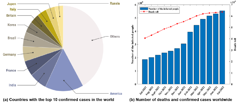
COVID-19 (Coronavirus Disease 2019), an acute respiratory infection caused by a novel coronavirus, was first reported in Wuhan, China, in December 2019 (Qin et al., 2020; Kannan et al., 2020). It is highly contagious and the World Health Organization (WHO) declared that the outbreak was a Public Health Emergency of International Concern (PHEIC) on January 30, 2020 and one and a half month later, a pandemic. The number of confirmed and death cases has increased rapidly since its outbreak as shown in Figure 1 (WHO, 2022). As of July 31, 2022, more than 574 million confirmed cases and more than 6.3 million fatalities had been documented, severely impacting people’s daily lives. The healthcare systems of many countries are perilously close to collapse, and the world financial market is suffered a negative impact (Torales et al., 2020; Pak et al., 2020; Nicola et al., 2020; Shorten et al., 2021). To break the viral cycle of transmission, early screening and severity evaluation for COVID-19 patients are essential (Tayarani, 2020; Das et al., 2022; Farhat et al., 2020).
RT-PCR (Reverse Transcription-Polymerase Chain Reaction) is a technology that combines reverse transcription (RT) of RNA and polymerase chain reaction (PCR) of cDNA (Mak et al., 2020; He et al., 2020). Due to its high sensitivity, wide dynamic range, and accurate detection, the RT-PCR is widely used in identifying COVID-19 subjects (Singanayagam et al., 2020; Wu et al., 2020; Huggett et al., 2005). It, however, has a lot of limitations. First, the quality of sample collection has a significant impact on RT-PCR results, and the false negative rate is extremely high (Shah et al., 2021a; Wang et al., 2020; Zhou et al., 2021c; Mukherjee et al., 2021). Second, the reaction time for RT-PCR is generally lengthy, and the standards for testing certification are rather high (Rahman et al., 2021b). Third, the suspected patients are vulnerable to cross-infection during the RT-PCR test, which will exacerbate the spreading virus (Alafif et al., 2021).
To solve the above problem, medical imaging is an available diagnostic method, which can be regarded as an important complement to RT-PCR in COVID-19 detection. It can signal lesion manifestations of different infected levels, aiding doctors to make an appropriate diagnosis(Mei et al., 2020; Pascual et al., 2021). Meanwhile, many studies attempt to use deep learning technologies and medical imaging for COVID-19 early screening, which can greatly prevent the further spread of the epidemic. And the DL-based diagnostic methods have shown great potential in image classification, lesion localization and severity evaluation (Srivastava, 2022; Polat et al., 2021; Rahman et al., 2021b; Alyasseri et al., 2022; Agrawal et al., 2022).
There are some surveys about using deep learning and medical imaging to fight against COVID-19 (Bhattacharya et al., 2021; Hryniewska et al., 2021; Liu et al., 2022; Soomro et al., 2021). Bhattacharya et al. (Bhattacharya et al., 2021) summarize recent studies related to the applications of deep learning based on medical image processing and discuss their potential to combat COVID-19. Hryniewska et al. (Hryniewska et al., 2021) summarize the deep neural network methods using medical images for COVID-19 diagnosis and show how to use interpretable AI techniques in these models. Soomro et al. (Soomro et al., 2021) perform the image analysis based on CT and X-rays images from both traditional image system and AI-system directions, and summarize the various applications of AI in COVID-19 diagnosis. Liu et al. (Liu et al., 2022) review existing deep learning models and medical image analysis methods for COVID-19 diagnosis, relating them to hot issues in deep learning research, including interpretable deep learning, and fair deep learning. While most of these surveys focus on the various applications of deep learning using medical images in fighting against COVID-19, there is a lack of discussion on how to utilize deep learning technologies to achieve a preciser diagnosis. Therefore, it is meaningful to summarize recent studies using deep learning and medical imaging to diagnose COVID-19, and explore how to utilize existing deep learning models to achieve more accurate diagnosis.
The contributions of this paper are summarized as follows:
-
First, we gather open source medical image datasets used in various studies, which mainly contain CT and X-ray images, to assist new studies in finding relevant and reliable medical images quickly.
-
Second, we systematically summarize various applications of deep learning using medical images for COVID-19 diagnosis. Different deep learning architectures and the methods used to improve the performance of deep models are reviewed.
-
Third, we discuss the limitations and future work of using deep learning techniques to diagnose COVID-19. And we hope that deep learning can play a crucial role in fighting against COVID-19 in the future.
2. COVID-19 Dataset and Medical Image Manifestations
Deep learning (DL) has demonstrated extraordinary capabilities in medical image processing. The performance of deep learning models, however, is significantly influenced by the quality of annotated data (Ker et al., 2017; Chen et al., 2021; Naudé, 2020). A large amount of well-annotated data can improve network performance and avoid overfitting (Bhattacharya et al., 2021; Zhou et al., 2021b). Due to the sudden outbreak of the epidemic, the available datasets are limited and still in the development stage (Kim et al., 2021). Hence, it is a difficult but significant work to collect open source medical image datasets. In this section, we summarize various medical image datasets used for COVID-19 diagnosis in different papers, mainly containing CT and X-ray images. Additionally, we describe the manifestations of CT and X-ray images as well as the lesion information provided by these images.
2.1. CT manifestations of COVID-19 and resource description
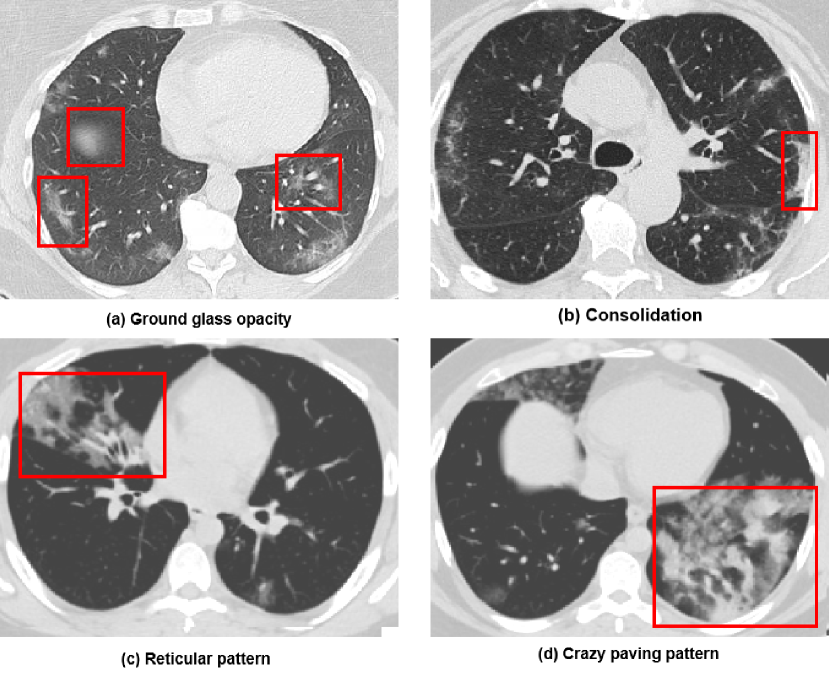
Computed tomography (CT) is one of the landmark advances in medical technology, providing radiologists with a useful imaging tool. Some pathological manifestations, such as ground glass opacity (GGO) and consolidation (C), are typically presenting in COVID-19 patients (Ye et al., 2020; Kwee and Kwee, 2020). Additionally, chest CT images may show diverse radiological characteristics or patterns as the state of COVID-19 patients evolves. In the early stage of the disease, the lesions mainly are presented as ground glass opacity (GGO) and thickened small blood vessels. Consolidation may have occurred in some cases (Bao et al., 2020). The patients with severe disease start to develop into a reticular pattern with interlobular septal thickening, and consolidation shadows will appear in peripheral and central regions of their lungs (Li et al., 2021b). When the patient’s condition deteriorates further, the white lung is visible in CT images and the lesions display multiple diffuse GGO and consolidation. Typical CT manifestations of COVID-19 patients are shown in Figure 2.
Many studies attempt to diagnose COVID-19 using CT images because of the rich pathological manifestations they provide, and deep learning models based on CT images have demonstrated outstanding performance on COVID-19 diagnosis (Shi et al., 2021b; Kiruthika et al., 2021; Shi et al., 2021a). To explore these CT images, Nivetha et al. (Nivetha and Inbarani, 2022) use a public COVID-19 CT scan dataset, which contains 349 positive COVID-19 cases and 397 negative chest CT images. Their proposed NRNN model achieves the accuracy of 0.98, 0.92, 1.00, 1.00 respectively in the experiment. Choudhary et al. (Choudhary et al., 2022) use a publicly available SARS-CoV2 CT-scan dataset, which consists of 1,252 positive COVID-19 cases and 1,230 negative chest CT images. Their pruned ResNet-34 model reaches 0.9547 accuracy, 0.9216 sensitivity, 0.9567 F-score, and 0.9942 specificity. Ahrabi et al. (Sarv Ahrabi et al., 2022) establish a training dataset composed of 4,000 CT images of COVID-19 cases collected from more than 500 patients. Their proposed approach reaches 0.9712 accuracy, 0.9741 precision, 0.9659 recall and 0.9696 F-score.
Additionally, some of the most popular datasets come from authoritative hospitals. Song et al. (Song et al., 2021) collect their dataset from three hospitals, i.e., Renmin Hospital of Wuhan University, and two affiliated hospitals of the Sun Yat-sen University in Guangzhou. Shi et al. (Shi et al., 2021b) build their dataset from three hospitals, i.e., Tongji Hospital of Huazhong University of Science and Technology, Shanghai Public Health Clinical Center of Fudan University, and China–Japan Union Hospital of Jilin University. In order to make it easier for new studies to obtain available and authoritative COVID-19 CT datasets, various datasets used in different papers are gathered and listed in Table 1.
2.2. X-ray manifestations of COVID-19 and resource description
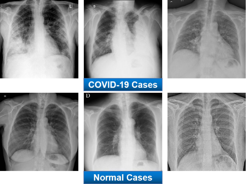
Even though CT scans provide radiologists with more information about lesions, they are more expensive and expose the patients to more radiation. The normal and COVID-19 X-ray images are shown in Figure 3. The peripheral lung opacity (PLO) and ground glass opacity (GGO) are two of these abnormalities (Kim et al., 2020; Jacobi et al., 2020). The chest X-ray images of COVID-19 patients may seem normal in the early or mild stage. About 10 to 12 days later, symptoms become more obvious. The lesions are generally distributed among the lower, peripheral, and bilateral regions (Weinstock et al., 2020; Yoon et al., 2020). In reference (Nagarajan et al., 2021), they retrospectively evaluate X-ray images of 593 patients admitted to hospital with COVID-19. The experiments demonstrate that abnormalities are mostly found in the middle and lower regions (88% of cases), and ground glass opacity (GGO) is the most common finding in abnormal X-rays (75% of cases). The next most common findings are peripheral lung opacity (PLO) and confluent consolidation, which generally indicate more severe symptoms.
The X-ray image datasets are more available than CT image datasets because capturing X-ray images is more convenient and less expensive than CT images. Hence, many studies attempt to use X-ray images rather than CT scans to diagnose COVID-19 (Shi et al., 2021a; Tang et al., 2021a). Regarding these X-ray images, Dev et al. (Dev et al., 2021) use the COVIDx dataset, which consists of 55,328 normal cases, 8,066 bacterial pneumonia cases and 358 COVID-19 cases. Their HCN-FM and HCN-DML reach 95.33% accuracy and 96.67% accuracy, respectively. Annavarapu et al. (Annavarapu et al., 2021) use a publicly accessible COVID-19 chest X-ray dataset, which consists of 2,905 images. Their model obtains 95% accuracy and 97% specificity. Turkoglu (Turkoglu, 2021) collects 6,092 X-ray images from a combination of public datasets. Their proposed model reaches 99.18% accuracy, surpassing most previous studies. Miao et al. (Miao et al., 2021) use three public COVID-19 X-ray datasets and other pneumonia datasets to construct two experimental datasets. Jain et al. (Jain et al., 2021) collect 6,432 chest X-ray images from the Kaggle repository. Various COVID-19 X-ray datasets used in different papers are listed in Table 1 to facilitate new studies to quickly find available resources.
3. Image Preprocessing Technologies
Most COVID-19 datasets contain some blurry, repetitive, and unrelated images, which will degrade the performance of deep model (Shah et al., 2021a). Image preprocessing is generally the first step in diagnosing COVID-19 using deep learning technologies. It eliminates irrelevant information in the image and recovers useful and real information, thereby improving the reliability and performance of diagnostic algorithms (Loew, 2022; Heidari et al., 2020). Nivetha et al. (Nivetha and Inbarani, 2022) use the median filter for the CT images of COVID-19 patients from a publicly accessible dataset. In this manner, the quality of COVID images is improved, and the noise is reduced without losing important information. Joshi et al. (Joshi et al., 2022) resize all images to 224×224 to maintain data consistency. And four types of transformation are employed to preprocess images. Ahrabi et al. (Sarv Ahrabi et al., 2022) use 4,000 CT images of COVID-19 cases from two publicly accessible datasets. All the images are cropped and resized to remove irrelevant regions. In this section, we will discuss extensively utilized image preprocessing technologies and their characteristics.
-
Resizing
Resizing is an essential step for image preprocessing phase in the applications of deep learning. It uniforms the size of images from different datasets. Tao et al. (Zhou et al., 2021c) resize all CT images to a uniform size (64×64) to build the COVID-19 dataset for model training. Shah et al. (Shah et al., 2021b) resize CT images from COVID-19 cases to 128×128×3 or 224×224×3 to suit the input size of different deep models. Garain et al. (Garain et al., 2021) resize each image to a dimension of 32×32 and the images are converted to greyscale.
-
Flipping and Rotating
Flipping and rotating are the commonly used data augmentation methods, which can guarantee that the deep learning model is trained with images at any angle and thus can make better prediction (Annavarapu et al., 2021). And the sufficient images can also help deep model avoid overfitting. Sedik et al. (Sedik et al., 2022) use a dataset consisting of 288 COVID-19 cases and 288 normal cases. The dataset is augmented by several rotations and scaling operations. Keles et al. (Keles et al., 2021) employ three augmentation methods, including horizontal/vertical flipping, rotation and shifting. Ahuja et al. (Ahuja et al., 2021) randomly rotate the training data between range [-90°, 90°] to resolve the overfitting problem.
-
Cropping
Cropping refers to cropping the given image to remove irrelevant regions. It greatly reduces the interference of irrelevant information and largely increases the performance and robustness of deep learning models. Balaha et al. (Balaha et al., 2022) preprocess the image data to a suitable format for CNN to further learn the latent features. The images are cropped using a bounding rectangle to reserve the region of interest. Madaan et al. (Madaan et al., 2021) use two chest X-ray image datasets in their model. Cropping is employed to reduce the noise and all images are resized. Zhang et al. (Zhang et al., 2021b) crop the rulers on the right side of the image and texts on the bottom of the image, which are irrelevant information and can impair the model performance.
-
Contrast adjusting
Medical images may have poor contrast during imaging process and data acquisition. It is essential to enhance image contrast to help deep models extract meaningful information from the region of interest. Dhaka et al. (Dhaka et al., 2021) propose that changing the contrast of X-ray images is capable of improving the performance of deep model. Tahir et al. (Tahir et al., 2022) use contrast limited adaptive histogram equalization (CLAHE) to enhance the contrast of the original X-ray image. The produced images are more natural compared with other contrast enhancement methods. Habib et al. (Habib et al., 2022) use histogram equalization (HE) and CLAHE to enhance the contrast.
-
Denoising
Image noise means the unnecessary or unwanted interference information that exists in an image. Noise can seriously affect the quality of images. Hence, it is necessary to do denoising before the downstream image processing task. Mahendran et al. (Mahendran and Kavitha, 2022) use DnCNN Algorithm to denoise CT Images before image classification. Balaha et al. (Balaha et al., 2022) convert all images to grayscale images and apply Gaussian blurring to eliminate unnecessary noise. Mishra et al. (Mishra, 2021) use a Gaussian filter to smooth the image and the background noise can be eliminated via a two-dimensional median filter.
Applying appropriate preprocessing methods needs to consider multiple factors such as the size of the dataset, image quality, input of the model, and the application scenarios. We investigate various papers using deep learning and medical imaging to diagnose COVID over the past three years. We summarize the representative preprocessing technologies in Table 2.
| Preprocessing Method | Papers | Count |
| Resizing | (Nivetha and Inbarani, 2022; Khurana and Soni, 2022; Sarv Ahrabi et al., 2022; Kumari and Jagadesh, 2022; Balaha et al., 2022; Madaan et al., 2021; Zhou et al., 2021c; Shah et al., 2021b; Garain et al., 2021; Mishra, 2021; Ravi et al., 2022; Annavarapu et al., 2021; Yasar and Ceylan, 2021a; Singh et al., 2021b; Zhang et al., 2021b; Abraham and Nair, 2022; Tan et al., 2021; Elpeltagy and Sallam, 2021; Dhaka et al., 2021; Ahuja et al., 2021; Zhang et al., 2021c; Shiri et al., 2022; Hasan et al., 2021; Akbarimajd et al., 2022; Kogilavani et al., 2022; Baghdadi et al., 2022; Zhang et al., 2022b; Zhao et al., 2021b) | 28 |
| Flipping and Rotating | (Joshi et al., 2022; Madaan et al., 2021; Sedik et al., 2022; Mishra, 2021; Annavarapu et al., 2021; Heidarian et al., 2021; Zhang et al., 2021b; Keles et al., 2021; Jangam et al., 2022; Huang et al., 2021a; Ahuja et al., 2021; Aslan et al., 2022; Kogilavani et al., 2022; Baghdadi et al., 2022; Zhao et al., 2021b) | 15 |
| Cropping | (Sarv Ahrabi et al., 2022; Balaha et al., 2022; Madaan et al., 2021; Mishra, 2021; Zhang et al., 2021b; Jangam et al., 2022; Shiri et al., 2022; Aslan et al., 2022; Kogilavani et al., 2022; Zhang et al., 2022b; Lian et al., 2022) | 11 |
| Contrast adjusting | (Tahir et al., 2022; Habib et al., 2022; Mishra, 2021; Dhaka et al., 2021; Baghdadi et al., 2022) | 5 |
| Denoising | (Nivetha and Inbarani, 2022; Mahendran and Kavitha, 2022; Balaha et al., 2022; Mishra, 2021; Zhang et al., 2022b) | 5 |
4. Image Segmentation
Accurate segmentation of lung and infection in medical images of COVID-19 patients plays a crucial role in the diagnosis of COVID-19 patients (Ma et al., 2021; Shiri et al., 2022). In specific, image segmentation identifies the region of interest (RoI) from given medical images and divides the medical image into several parts (Liu et al., 2021; Zhao et al., 2019). The segmented region can aid doctors and deep learning models more comfortably to understand lung disease type and severity (Ter-Sarkisov, 2022; Khan et al., 2022; Shi et al., 2020). Punn et al. (Punn and Agarwal, 2022) propose a deep learning based semantic hierarchical segmenter (CHS-Net) to identify the COVID-19 infected regions from chest CT images. They apply a residual inception U-Net model with spectral spatial and depth attention network (SSD) to effectively encode and decode the semantic and varying image information, and finally segment the COVID-19 infected regions in the lungs. Wang et al. (Wang et al., 2021b) propose a lesion edge detection model (COVID Edge-Net), which consists of one edge detection backbone and two novel modules, i.e., the multi-scale residual dual attention (MSRDA) module and the Canny operator module. The MSRDA module can capture rich contextual associations to generate enhanced feature representations, which are then merged with Canny features learned from the Canny operator module to extract more accurate, clearer, and sharper edges.
Chen et al. (Chen et al., 2022) propose a segmentation network based on unsupervised domain adaptation (UDA) to improve the segmentation performance of the infected regions in CT images. The generalization ability of the segmentation network is significantly enhanced by a novel domain adaption module that aligns the two domains. Their proposed method achieves high performance in lung segmentation, and it is proved that their proposed model is also suitable for large-area organs or tissues. Lashchenova et al. (Lashchenova et al., 2021) argue that an important factor affecting the real performance of the segmentation model is that the model segments the puncta (connected components) of lung and the lung lesions outside the real lung. To detect this, connected components are computed for every class and for each component to found whether it has interception with ground-truth lungs.
4.1. U-net Architecture
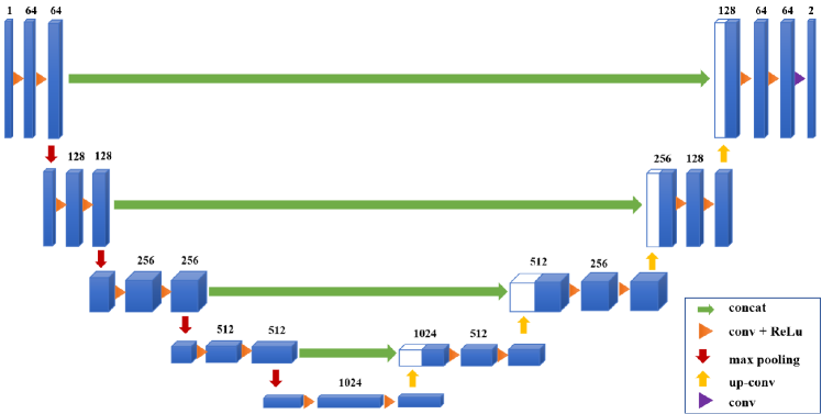
U-net is a popular image segmentation network which is developed primarily for medical image segmentation. It is built based on Convolutional Neural Network (CNN) and is upgraded to achieve better segmentation performance (Shirato et al., 2020; Minaee et al., 2022; Siddique et al., 2021). As shown in Figure 4, the U-Net (Ronneberger et al., 2015) network is U-shaped, mainly including the downsampling layer on the left and the upsampling process on the right. The structure is symmetrical from left to right. Additionally, the encoder-decoder structure and skip connection used by U-Net are core designs to achieve a more effective combination of contextual information to make precise pixel-level judgments.
Alirr (Alirr, 2022) designs a lung image segmentation method to extract the region of interest, which modifies U-net with upgraded residual block including concatenation skip connection. Additionally, the segmented region employs edge enhancing diffusion filtering (EED) to improve the infection area’s contrast. Their proposed method achieves Dice overlapping score of 96.1% and 78.0% for lung and infection areas segmentation. Diniz et al. (Diniz et al., 2021) seek to automatically identify infections caused by COVID-19 and evaluate quantitative score of the infected region. They propose a model by modifying the traditional U-net architecture via batch normalization, leaky ReLU, dropout, and residual block techniques. Their model yields an average Dice value of 77.1% and an average specificity of 99.8%.
Zhao et al. (Zhao et al., 2021b) propose a novel dilated dual attention U-Net (D2A U-Net) with the dual attention strategy and hybrid dilated convolutions to segment COVID-19 lesion region. The former dual attention strategy, which contains two attention modules, is used to enhance feature maps and reduce the semantic gap among diverse levels of feature maps. The latter hybrid dilated convolutions are used for achieving larger receptive fields. Their proposed method achieves 0.73 Dice score and 0.71 recall score. Lian et al. (Lian et al., 2022) propose an end-to-end DRD U-Net (dilated residual and deeply supervised U-Net) network to segment the lung lesions. They add residual modules to each layer of channel to accelerate convergence and avoid gradient disappearance. To extract richer feature information, the extended convolution unit is utilized to increase the receptive field without increasing the number of parameters. In the experiments, their proposed model shows impressive segmentation performance and the error can be significantly reduced compared with the original U-Net.
U-net has shown great potential in the field of image segmentation. Some variants that follow the core idea of U-net have been proposed, including attention U-net (Schlemper et al., 2019), and Residual U-net (Das et al., 2019), to complete more complex segmentation tasks. In addition to U-Net architecture, other segmentation methods such as V-Net, ResU-Net, Dense-Net, and DeepLab, are also used for the segmentation of COVID-19 infected region in different papers. Table 3 displays the various segmentation techniques employed by different papers.
| Segmentation methods | Papers | Count |
| U-Net | (Ma et al., 2021; Lashchenova et al., 2021; Diniz et al., 2021; Xiao et al., 2022; Voulodimos et al., 2021a; Jadhav et al., 2021; Avetisian et al., 2021; Punn and Agarwal, 2022; Alirr, 2022; Zhou et al., 2021a; Elharrouss et al., 2022; Kuchana et al., 2021; Trivizakis et al., 2020; Shan et al., 2021; Tahir et al., 2022; Saood, 2021; Lian et al., 2022; Voulodimos et al., 2021b) | 18 |
| V-Net | (Voulodimos et al., 2021a; Zhao et al., 2021a; Milletari et al., 2016) | 3 |
| ResU-Net | (Diniz et al., 2021; Jadhav et al., 2021; Avetisian et al., 2021) | 3 |
| Dense-Net | (Al-Antari et al., 2021; Elharrouss et al., 2022) | 2 |
| UNet++ | (Zhou et al., 2021a; Elharrouss et al., 2022) | 2 |
| Deeplab | (Diniz et al., 2021; Amin et al., 2022) | 2 |
| Attention-UNet | (Zhao et al., 2021b; Elharrouss et al., 2022) | 2 |
| SAUNet++ | (Xiao et al., 2022) | 1 |
| COVSeg-NET | (Zhang et al., 2021c) | 1 |
4.2. Loss Function
The loss function is another important component of the segmentation model. It can guide the segmentation model to learn towards a specific direction and segment the wanted infected area. Xiao et al. (Xiao et al., 2022) use focal Tversky loss (FTL) and the generalized Dice loss (GDL). The GDL can reduce the correlation between lesion size and Dice loss, and can effectively guide the model in segmenting small regions. Elharrouss et al. (Elharrouss et al., 2022) use binary cross entropy as loss function and their model reaches 78.6% for Dice, 71.1% for sensitivity metric, 99.3% for specificity, and 85.6% for precision. Alirr (Alirr, 2022) uses Dice loss as loss function and averages the DSC of each class to generate the final score. Diniz et al. (Diniz et al., 2021) use Dice loss as loss function and their proposed model reaches 77.1% for Dice, and 99.76% for average specificity. This section will introduce several representative loss functions commonly used in the medical image segmentation task.
A. Binary Cross Entropy
The pixel level cross entropy loss is commonly used in image segmentation task. For a given random variable, this loss function will examine each pixel individually and compare the difference between two probability distributions. Binary cross entropy is defined as (Yi-de et al., 2004):
| (1) |
where and represent the ground truth value and predicted value respectively.
B. Weighted Binary Cross Entropy
Weighted binary cross entropy (WCE) (Pihur et al., 2007) is a variant of binary cross entropy, which aims to solve the problem of uneven distribution of lesion information in medical image. WCE is defined as:
| (2) |
where is used to tune false negative and false positive. If we want to decrease the number of false negative, set ¿1. In contrast, setting ¡1 can decrease the number of false positive.
C.Balanced Cross Entropy
Balanced cross entropy (BCE) (Xie and Tu, 2015) is similar with WCE, which appropriately weights positive examples and negative examples. Balanced cross entropy is defined as:
| (3) |
D. Focal Loss
Focal loss (Lin et al., 2017) is often used to deal with unbalanced sample classification. It focuses on weighting loss to the sample according to the difficulty of sample discrimination. For medical images presenting small lesion information, focal loss assigns a larger weight to positive pixels and can achieve excellent performance. It is defined as:
| (4) |
where is a regulatory parameter and ¿0. generally ranges from [0,1], which is used to deal with class imbalance. Focal loss defines as:
| (5) |
where represents the predicted value of pixel classification and denotes the ground truth value.
E. Dice Loss
Dice loss (Sudre et al., 2017) is designed to calculate the similarity between two images and is utilized to guide the training process of image segmentation methods. Dice loss is defined as:
| (6) |
Here, 1 is added in numerator and denominator to ensure that the function is not undefined in edge case scenarios where .
F. Generalized Dice Loss
Dice loss is unsuitable to predict small targets. When some pixels of small targets are predicted incorrectly, Dice coefficient may greatly fluctuate. To solve these problems, GDL uses weights inversely proportional to lesion region, in order to better segment small regions (Xiao et al., 2022). When the number of classes is 2, Generalized Dice loss is defined as:
| (7) |
where and denote the true voxel values of -th position and the related probability predicted as class . represents the weight of class and is defined as:
| (8) |
where is the total number of pixels, and is often set as to prevent the loss function from dividing by 0.
G. Tversky Loss
Tversky loss (Salehi et al., 2017) is considered as a generalization of Dice loss, where the coefficient is a hyperparameter that controls the balance between false negative (FN) and false positive (FP). Tversky loss is defined as:
| (9) |
where and denote the ground truth value and predicted value respectively. When = 0.5, it can be solved into regular Dice coefficient.
H. Focal Tversky Loss
Similar to focal loss, focal Tversky loss (Abraham and Khan, 2019) is proposed to deal with class imbalance. It focuses on learning hard samples with the help of coefficient . Focal Tversky loss is often used to segment small regions and is defined as:
| (10) |
where the hyperparameter can range from [1,3], and (Tversky index) is defined as:
| (11) |
where and are probabilities that pixel belongs to the lesion class and non-lesion class , respectively. and represent probabilities for another class. is the total number of pixels.
5. Overall Analysis of DL-based Methods for COVID‑19 Diagnosis
Manual detection of COVID-19 (RT-PCR), which has extremely high accuracy and specificity, is always regarded as the gold standard for COVID-19 detection (Pu et al., 2022; Rabaan et al., 2021). However, RT-PCR is time-consuming, and the spread risk and susceptibility of the disease are greatly increased during detection (Rahman et al., 2021b; Peng et al., 2022). Hence, many studies are focusing on diagnosing COVID-19 using medical images, attempting to find a diagnostic method that can guarantee high accuracy and significantly minimize human contact during detection (Krishnamoorthy et al., 2021; Xu et al., 2022; Ibrahim et al., 2021; Shorfuzzaman, 2021). Among the many inspection methods, the deep learning models using medical images have shown excellent performance. And they are thus extensively utilized in image classification, lesion segmentation, and severity quantification to fight against COVID-19, such as Convolutional Neural Network (CNN) (Singh et al., 2021a; Pouyanfar et al., 2018; Rahman et al., 2021a), Generative Adversarial Network (GAN) (Bargshady et al., 2022; Kim et al., 2021), and Long Short-Term Memory (LSTM) (Demir, 2021; Naeem and Bin-Salem, 2021; Sheykhivand et al., 2021). In this section, we will introduce the different applications of deep learning models in diagnosing COVID-19.
5.1. CNN models for COVID-19 diagnosis
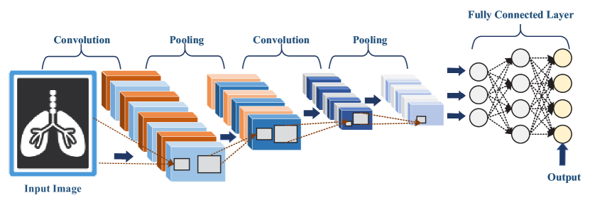
Convolutional Neural Network (CNN) is a multi-layer supervised neural network, in which the hidden convolutional and pooling layers are the core modules to realize the feature extraction function (Anwar et al., 2018; Alzubaidi et al., 2021). CNN uses the gradient descent method to minimize the loss function and error back propagation (BP) is employed to reversely adjust the weight parameters in the network layer by layer (Mishra and Kane, 2022). CNN models have demonstrated great potential in medical image processing and become a popular approach for identifying COVID-19 and localizing lesion. As Figure 5 depicts, a representative CNN model for COVID-19 diagnosis generally includes input, convolutional layers, pooling layers, fully connected layer, and output (Bhattacharya et al., 2021). Additionally, we try to record some deep learning methods and model evaluation metrics used in high quality studies and more detailed information is shown in Table 4.
Wang et al. (Wang et al., 2020) design a deep learning-based method for automatic COVID-19 diagnosis. Their model uses a pre-trained U-Net to segment the lung region. The segmented 3D lung region is input into the 3D deep neural network to predict the possibility of infection. Finally, the activation regions in the classification network and the unsupervised connected components are together used to localize the COVID-19 lesions. Their proposed method obtains an accuracy of 0.90, a positive predictive value of 0.84, and a high negative predictive value of 0.98. Turkoglu (Turkoglu, 2021) proposes an expert-designed system named COVIDetectioNet, which is consisted of three basic components. First, deep features are obtained from the pre-trained AlexNet architecture. Second, the relief selection algorithm is employed to select the most efficient features from the pre-learned deep features. Third, the support vector machine (SVM) method is used for final classification. Their proposed model is validated by classifying 6,092 X-ray images as normal (healthy), COVID-19, and pneumonia cases, respectively. In the experimental results, their proposed model achieves 99.18% accuracy and 97.1% sensitivity.
Singh et al. (Singh et al., 2021a) propose an automated COVID-19 screening model. It employs some pre-trained CNN models such as DenseNet201, ResNet152V2, and VGG16, for early detection of COVID-19 patients. The predicted outputs are fed into the ensemble DCCNs, which are designed for diagnosing the suspected objects into four classes, including COVID-19 cases, tuberculosis, pneumonia, and healthy subjects. Their proposed model achieves 98.94% accuracy, 98.94% sensitivity and 98.93% specificity, which is superior to the competitive models. Moreover, Aslan et al. (Aslan et al., 2022) firstly use ANN-based automated segmentation method to extract region of interest in the X-ray images. Second, various CNNs are used for feature extraction after data augmentation stage. Third, the features learned from each CNN model are input to four different machine learning (ML) algorithms for classification. The hyperparameters of each ML algorithm are determined by Bayesian optimization. Finally, the highest performance is obtained by the DenseNet201 model with SVM algorithm.
Kogilavani et al. (Kogilavani et al., 2022) explore the performance of a variety of convolutional neural network (CNN) models in detecting COVID-19, attempting to find the suitable deep models, including VGG16, DeseNet121, MobileNet, NASNet, Xception, and EfficientNet. Accuracy obtained for VGG16 is 97.68%, DenseNet121 is 97.53%, MobileNet is 96.38%, NASNet is 89.51%, Xception is 92.47%, and EfficientNet is 80.19%, respectively. Based on the performance analyses, it is demonstrated that the VGG16 architecture reaches the best accuracy compared to other architectures. Akbarimajd et al. (Akbarimajd et al., 2022) propose a novel CNN approach applying adaptive convolution, which intends to enhance COVID-19 identification in noisy X-ray images without reducing noise. Specifically, the impulse noise-map layer and the adaptive resizing layer are added on the traditional CNN framework. A learning-to-augment strategy which uses noisy X-ray images is designed to improve the generalization of deep model. Some pre-trained networks such as SqueezeNet, ResNet18, and ResNet50 are modified to increase their robustness for impulse noise.
| Reference | Data | Deep Learning Methods | Classification | Acc | Sn | Sp |
| Tang et al. (Tang et al., 2021a) | X-ray | Ensemble learning, COVID-Net | Multiclass | 95.0% | 96.0% | — |
| Yamaç et al. (Yamaç et al., 2021) | X-ray | CheXNet, CSEN | Multiclass | 95.9% | 98.5% | 95.7% |
| Pathak et al. (Pathak et al., 2020) | CT | CNN, Transfer learning | Binary | 93.0% | 91.5% | 94.9% |
| Rathinasamy et al. (R et al., 2022) | X-ray | CNN, Ensemble learning | Binary | 99.0% | — | — |
| Khan et al. (Khan et al., 2021) | X-ray | COVID-RENet, SVM | Binary | 98.5% | 99.0% | — |
| Wang et al. (Wang et al., 2020) | CT | DeCoVNet, U-net | Binary | 90.1% | 90.7% | 91.1% |
| Zhou et al. (Zhou et al., 2021c) | CT | AlexNet, GoogleNet, ResNet, Ensemble learning | Multiclass | 99.1% | 99.1% | 99.6% |
| Zheng et al. (Zheng et al., 2022) | CT | ResNet50, MAB, FAB | Binary | 98.2% | 98.8% | 97.3% |
| Choudhary et al. (Choudhary et al., 2022) | CT | VGG16, ResNet34 | Binary | 95.5% | 92.2% | 99.4% |
| Singh et al. (Singh et al., 2021a) | CT | CNN, Ensemble learning, Transfer learning | Multiclass | 98.8% | 98.8% | 98.8% |
| Balaha et al. (Balaha et al., 2022) | CT | CNN, Transfer learning, GAN | Binary | 98.7% | — | — |
| Turkoglu (Turkoglu, 2021) | X-ray | AlexNet, Transfer learning, SVM | Multiclass | 99.2% | 97.1% | — |
| Aslan et al. (Aslan et al., 2022) | X-ray | CNN, Transfer learning, Bayesian Optimization | Multiclass | 96.3% | 96.4% | 98.1% |
| Zhang et al. (Zhang et al., 2022b) | CT | CNN, Transfer learning, Bayesian Optimization | Binary | 92.1% | — | 91.2% |
Some existing convolutional neural network architectures have been proven to be vital in medical image feature extraction, such as ResNet (He et al., 2016), AlexNet (Krizhevsky et al., 2017), SqueezeNet (Iandola et al., 2016), Inception (Szegedy et al., 2015), DenseNet (Huang et al., 2017), VGG (Simonyan and Zisserman, 2014), and EfficientNet (Tan and Le, 2019). Various CNN models used in different papers for COVID-19 diagnosis are listed in Table 5. The ResNet is the most popular network model to detect COVID-19 and 34 papers use ResNet as backbone network. Besides, the DenseNet and the VGG are also used by many studies to detect COVID-19. In conclusion, it is significant for us to select an appropriate CNN model as backbone network to diagnose COVID-19 by considering data type, actual scale, application scenarios and other factors.
5.2. Transfer learning for COVID-19 diagnosis
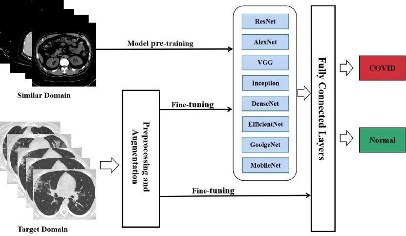
Transfer learning is a machine learning technique that can transfer existing knowledge from one domain (source domain) to another (target domain) (Zhuang et al., 2020). The source domain generally has sufficient annotated data and many existing models have learned excellent feature extraction capabilities. The target domain lacks large-scale annotated samples and the cost of obtaining annotated samples is too high (Guan and Liu, 2021; Morid et al., 2021). Transfer learning aims to use knowledge learned from the source domain to help the target learner achieve better performance. The closer the relationship between the target domain and the source domain, the better transfer learning can be achieved (Das et al., 2022). Otherwise, it may be more difficult and even has negative transfer to bring harmful effects. As shown in Figure 6, the pre-trained model has obtained excellent generalization performance in the source domain and then it is fine-tuned by using small-scale data from the target domain (Chen et al., 2021). Due to the expensive cost of capturing CT or X-ray images of COVID-19 patients, various pre-trained deep learning models have been employed for COVID-19 diagnosis.
Kabe et al. (Kashala Kabe et al., 2021) propose a novel domain transfer learning model for classifying COVID-19 cases, named feature fusion, decompose and transfer (FFDT). Their proposed FFDT gains feature enhancement by combining far-off features taken from far-off domains into a single feature space where the distribution mismatch is reduced. Additionally, they adopt modified convolutional neural network (MCNN) to extract features and the class reconstruction is used to unravel the local structure of the data distribution. Their model achieves the classification accuracy of 94.5%. Lu et al. (Lu et al., 2021) suggest that transfer learning can be used to extract features from chest CT images because it is of high complexity to train a CNN model from scratch. The pre-trained ResNet-18 and ResNet-50 models are chosen as the backbone to extract features from the CT images. To create refined image features, the retrieved features are combined using discriminant correlation analysis. Finally, in order to get a more reliable classification performance, three randomized neural networks are trained using the improved features and their predictions are combined.
Vogado et al. (Vogado et al., 2021) evaluate transfer learning techniques in five pre-trained CNN architectures (VGG-16, VGG-19, ResNet-50, Xception and DenseNet-121). In specific, they train CNNs using 1,436 images associated with COVID-19 cases, 1,932 healthy images, and 3,651 images of other pathologies. The pre-trained ResNet50 obtains the best performance for extracting deep features, and the MLP classifier has shown the best results when using the features derived by ResNet50. Makris et al. (Makris et al., 2020) use a combination of publicly available X-ray images from patients with confirmed COVID-19 cases, common bacterial pneumonia and healthy cases. Transfer learning is used for mitigating the issue of insufficient samples. In order to classify images, VGG16, VGG19, MobileNet V2, Inception V3, Xception, InceptionResNet V2, DenseNet201, and ResNet152 V2 are employed and compared to explore the best model for COVID-19 diagnosis. Specifically, VGG16 and VGG19 perform the best and achieve an overall accuracy of 95%.
Pathak et al. (Pathak et al., 2020) employ a deep transfer learning technique to classify COVID-19 infected patients. The ResNet-50 is chosen as backbone network and transfer learning is employed to adjust the initial parameters of the deep layers. The pre-trained model, which is fine-tuned by a small number of training samples, can extract correct deep features from chest CT images to detect COVID-19 infected patients. Their proposed model achieves training and testing accuracy up to 96.23% and 93.02% respectively, which is superior to the compared deep models. To detect COVID-19 by medical images, Shamsi et al. (Shamsi et al., 2021) propose a novel deep uncertainty-aware transfer learning framework. The pre-trained CNNs are used to extract features from chest X-ray and CT images. The extracted features are subsequently used to identify COVID-19 cases using various machine learning and statistical modeling techniques. It has been found that CT images can produce superior diagnosis because they contain more information than X-ray images.
| CNN model | Papers | Count |
| ResNet | (Zheng et al., 2022; Khurana and Soni, 2022; Tahir et al., 2022; Canayaz et al., 2022; Habib et al., 2022; Balaha et al., 2022; Dev et al., 2021; Zhou et al., 2021c; Madhavan et al., 2021; Shorfuzzaman, 2021; Shah et al., 2021b; Singh et al., 2021a; Annavarapu et al., 2021; Ilhan et al., 2022; Huang et al., 2021b; Keles et al., 2021; Elpeltagy and Sallam, 2021; Jangam et al., 2022; Ahuja et al., 2021; Jain et al., 2021; Turkoglu, 2021; Huang, 2020; Hira et al., 2021; Wang et al., 2020; Zhang et al., 2021a; Pathak et al., 2020; Sen et al., 2021; Chakraborty et al., 2021; Zebin and Rezvy, 2021; Song et al., 2021; Kundu et al., 2022; Paul et al., 2022; Akbarimajd et al., 2022; Aslan et al., 2022) | 34 |
| DenseNet | (Tahir et al., 2022; Habib et al., 2022; Dev et al., 2021; Shorfuzzaman, 2021; Shah et al., 2021b; Singh et al., 2021a; Mishra, 2021; Zhang et al., 2021b; Jangam et al., 2022; Hira et al., 2021; Sen et al., 2021; Kundu et al., 2022; Paul et al., 2022; Hasan et al., 2021; Aslan et al., 2022; Kogilavani et al., 2022) | 16 |
| VGG | (Khurana and Soni, 2022; Balaha et al., 2022; Shah et al., 2021b; Singh et al., 2021a; Ilhan et al., 2022; Tan et al., 2021; Jangam et al., 2022; Vidyun et al., 2021; Turkoglu, 2021; Huang, 2020; Sen et al., 2021; Chakraborty et al., 2021; Zebin and Rezvy, 2021; Paul et al., 2022; Karacı, 2022; Kogilavani et al., 2022) | 16 |
| MobileNet | (Canayaz et al., 2022; Mahendran and Kavitha, 2022; Balaha et al., 2022; Mishra, 2021; Ilhan et al., 2022; Abraham and Nair, 2022; Yasar and Ceylan, 2021b; Akbarimajd et al., 2022; Aslan et al., 2022; Kogilavani et al., 2022; Baghdadi et al., 2022; Zhang et al., 2022b) | 12 |
| Inception | (Tahir et al., 2022; Balaha et al., 2022; Shah et al., 2021b; Ilhan et al., 2022; Jain et al., 2021; Turkoglu, 2021; Hira et al., 2021; Sen et al., 2021; Kundu et al., 2022; Paul et al., 2022; Aslan et al., 2022) | 11 |
| AlexNet | (Zhang et al., 2022a; Zhou et al., 2021c; Ibrahim et al., 2021; Hira et al., 2021; Wang et al., 2020; Yasar and Ceylan, 2021b; Chakraborty et al., 2021; Aslan et al., 2022) | 8 |
| GoolgeNet | (Dev et al., 2021; Zhou et al., 2021c; Hira et al., 2021; Zhang et al., 2021a; Chakraborty et al., 2021; Akbarimajd et al., 2022; Aslan et al., 2022) | 7 |
| Xception | (Balaha et al., 2022; Shorfuzzaman, 2021; Ilhan et al., 2022; Abraham and Nair, 2022; Jain et al., 2021; Kogilavani et al., 2022) | 6 |
| EfficientNet | (Khurana and Soni, 2022; Balaha et al., 2022; Ravi et al., 2022; Abraham and Nair, 2022; Zebin and Rezvy, 2021; Kogilavani et al., 2022) | 6 |
| SqueezeNet | (Tahir et al., 2022; Dev et al., 2021; Akbarimajd et al., 2022) | 3 |
| Darknet | (Dev et al., 2021; Abraham and Nair, 2022) | 2 |
5.3. Ensemble learning for COVID-19 diagnosis
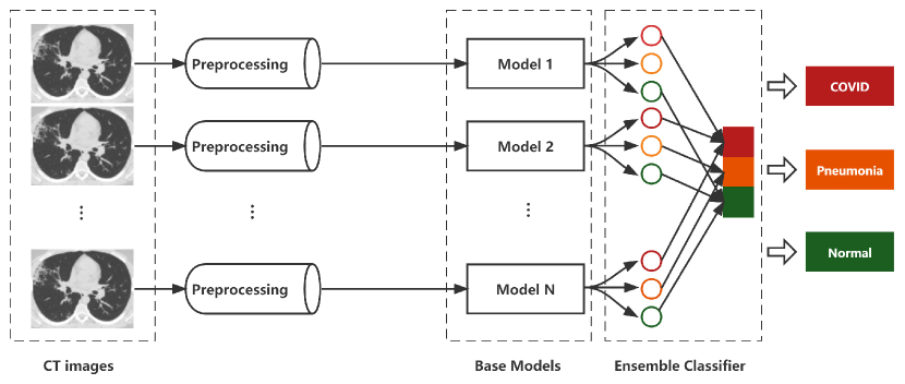
Ensemble learning is a machine learning method that combines multiple learners to complete complex learning tasks (Dong et al., 2020; Mienye and Sun, 2022). It trains multiple base learners and combines them to obtain improved generalization ability than the individual base learners (Mienye et al., 2020). Currently, the common methods to generate base learners can be divided into two categories: one is to apply different types of learning models in the same size but different samples, which are generated from the same dataset (Zhang et al., 2018). The base learners which are generated by this method are called heterogeneous learners. The other is to apply the same learning model on different training sets. The base learners which are generated by this method are called homogeneous learners. Besides, the combined strategy of base learners mainly includes simple average method, weighted average method, majority voting, plurality voting, and weighted voting. Figure 7 illustrates a representative application of ensemble learning for COVID-19 diagnosis. Multiple base models learn to diagnose COVID-19 from the medical images and output classification results. The ensemble classifier receives input from base models to make the final decision.
Chaudhary et al. (Chaudhary and Qiang, 2021) train three base models (two Efficient-Net with different initial pre-trained weights and SE-ResNext), which can classify X-ray images into COVID-19, pneumonia, and normal cases. The final results are calculated by averaging the classification outcomes produced by the three different models individually. Their proposed method achieves excellent performance with an accuracy of 0.9592, a sensitivity of 0.9592, and a specificity of 0.9597. It is proven that the ensemble model’s accuracy is greater than that of three separately trained models. Tang et al. (Tang et al., 2021a) propose the EDL-COVID model using deep learning and ensemble learning. It trains several base models to overcome the shortcomings of single model by combining their predicted outputs. A deep learning network generates several model snapshots in one training run by snapshotting. Additionally, several model snapshots are integrated to produce a preciser model and the final classification is made using the weighted average method. Their EDL-COVID model achieves 95% accuracy.
Abraham et al. (Abraham and Nair, 2022) use five pre-trained CNNs (MobilenetV2, Shufflenet, Xception, Darknet53, and EfficientnetB0) to extract features. Then the features are combined, and the ensemble classifier kernel support vector machine is used to diagnose COVID-19 cases. Their proposed model achieves 0.916 accuracy, 0.8305 kappa score, 0.91 F-score, 0.917 sensitivity, and positive predictive value of 0.904. Zhou et al. (Zhou et al., 2021c) employ three deep pre-trained models (AlexNet, GoogleNet, and ResNet) as base learners. Then the predicted outputs from three pre-trained models are input into the ensemble classifier EDL-COVID, and relative majority voting is used to determine the final outcome. Finally, by comparing the ensemble classifier with three component classifiers in some specific performance metrics, it is demonstrated that the ensemble model obtains more effective performance than three deep pre-trained models.
5.4. GAN model for COVID-19 diagnosis
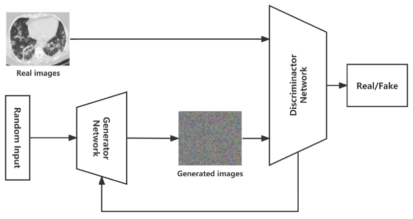
One of the main causes for poor performance and underfitting is the insufficient amount of annotated COVID-19 images (Rahman et al., 2021b; Chen et al., 2021). And the cost of collecting COVID-19 infected images is too expensive. To solve these problems, Generative Adversarial Network (GAN) is employed by many studies to generate fake COVID-19 infected images to tackle data insufficiency (Saxena and Cao, 2021; Acar et al., 2021). GAN mainly consists of two systems: a generator and a discriminator, as shown in Figure 8.
-
Generator network: Generator takes noise samples from a particular distribution (uniform distribution and Gaussian distribution) and generates results similar to the real training data. It tries to generate fake images to successfully trick the discriminator after training.
-
Discriminator network: Real data and generated data are mixed and input into the discriminator. The discriminator distinguishes whether a sample belongs to real data or generated data. If it comes from real data, then the high probability will appear to be output, otherwise the probability is low.
Jiang et al. (Jiang et al., 2021) construct a public COVID-19 CT dataset, including 1,186 CT images synthesized from a large-scale lung cancer CT dataset using CycleGAN. Their proposed model can learn the GGO style of COVID-19 so that the synthetic images are closely resembled to the real distribution. Goel et al. (Goel et al., 2021) employ generative adversarial network (GAN) to generate synthetic chest CT images during data augmentation phase. The Whale Optimization Algorithm (WOA) is used to optimize the hyperparameters of GAN. Their proposed model using GAN reaches 99.22% accuracy.
Bargshady et al. (Bargshady et al., 2022) apply generative adversarial network (GAN) and semi-supervised CycleGAN (SSA-CycleGAN) to augment the training dataset of X-ray images. Their proposed Inception-CycleGAN model achieves 94.2% accuracy, and 92.2% area under cure. Serte et al. (Serte et al., 2022) augment the number of available CT images by using generative adversarial network (GAN). By comparing the traditional deep learning methods with their proposed method using data-efficient method (GAN), it is demonstrated that their proposed data-efficient model outperforms all other traditional deep learning models. The ResNet-18 and MobileNetV2 obtain the best performance.
In addition to its popularity in data augmentation, GAN is also employed in other fields based on its adversarial training characteristics. Bhattacharyya et al. (Bhattacharyya et al., 2022) use C-GAN to segment the COVID-19 chest X-ray images. The X-ray images as input are fed into the generator and the generator network tries to produce the mask images. The discriminator tries to distinguish fake image pair (Input X-ray images and generated mask images) from the real one. Besides, Doraiswami et al. (Doraiswami et al., 2022) propose an effective prediction mechanism, where local ternary pattern (LTP) is used for feature extraction, and prediction of COVID-19 patients is performed by their proposed Jaya-TSA based GAN after acquiring features. Their proposed method reaches the accuracy of 0.87, the sensitivity of 0.85 and the specificity of 0.89.
Some of the applications of deep learning based on GAN model and their result are demonstrated in Table 6. In most of the studies, GAN is generally employed for data augmentation before model training to greatly increase the training sample space, thereby significantly improve the performance of the model (Menon et al., 2020; Li et al., 2021a; Serte et al., 2022). In reference (Menon et al., 2020), the quantitative analysis shows that their proposed MTT-GAN greatly improves the accuracy of binary classifier and multiclass classifier. In reference (Li et al., 2021a), the original CNN models with Generative Adversarial Network (GAN) increase the accuracy by 2% to 3%, the recall by 2% to 4%, and the precision by 1% to 3%.
| Reference | Data | Methods | Function | Acc | Sn | Sp |
| Zhang et al. (Zhang et al., 2021d) | CT | GAN, U-net | Segmentation | 93.2% | 69.8% | — |
| Bargshady et al. (Bargshady et al., 2022) | X-ray | CycleGAN, Inception V3 | Classification | 94.2% | 95.5% | 91.4% |
| Acar et al. (Acar et al., 2021) | CT | GAN,CNN | Classification | 95.0% | 94.2% | 95.3% |
| Goel et al. (Goel et al., 2021) | CT | GAN, WOA, ResNet-50 | Classification | 99.2% | 99.8% | 97.8% |
| Menon et al. (Menon et al., 2020) | X-ray | GAN, CNN, transfer learning | Classification | 96.3% | 100.0% | 93.2% |
| Li et al. (Li et al., 2021a) | CT | GAN, DenseNet | Classification | 93.0% | 96.0% | — |
| Serte et al. (Serte et al., 2022) | CT | GAN, CNN, Transfer learning | Classification | 74.0% | 88.0% | 68.0% |
| Bhattacharyya et al. (Bhattacharyya et al., 2022) | X-ray | C-GAN, CNN, ML | Segmentation | 96.6% | 95.0% | 97.4% |
5.5. LSTM model for COVID-19 diagnosis
Long Short-Term Memory (LSTM) is a powerful recurrent neural network, which is proposed to address the substantial limitation of neural networks when dealing with sequential data (Hasan et al., 2020). For the issue that traditional RNN structure has poor performance on long sequences, the LSTM model is capable of greatly alleviating the gradient disappearance problem, thereby supporting long-term dependence in dealing with sequential data (Sherstinsky, 2020; Meraihi et al., 2022). And it has been extensively utilized to diagnose COVID-19 and predict the prognosis of COVID-19 patients. In this section, we will introduce the application of LSTM model in detecting COVID-19, and some high quality studies using the LSTM model for COVID-19 diagnosis are listed in Table 7.
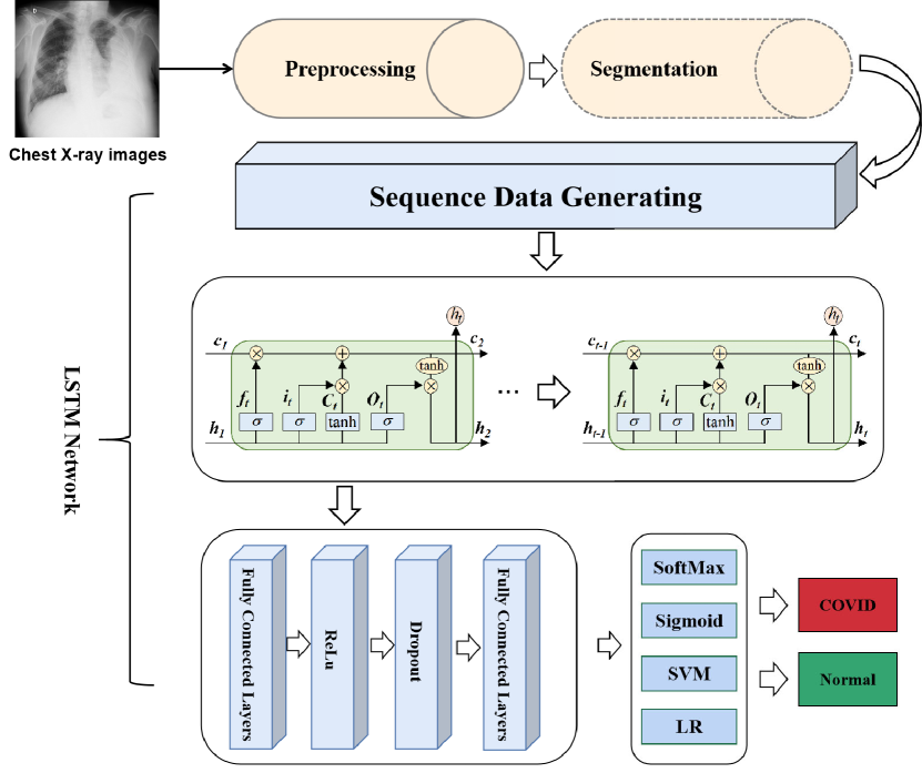
Patients with COVID-19 tend to have a dynamic condition. The information obtained from one CT image or X-ray is generally limited. Conversely, CT or X-ray sequences are capable of providing more medical information, assisting models or doctors to make a preciser diagnosis for COVID-19 patients (Xu et al., 2022). Consequently, many studies apply the LSTM model and image sequences to diagnose COVID-19. The typical architecture of the LSTM model for COVID-19 diagnosis is shown in Figure 9 (Demir, 2021). Er (Er, 2022) proposes a hybrid approach for COVID-19 classification that combines long short-term memory (LSTM) with some pre-trained deep networks. The contrast enhancing method is firstly applied to X-ray images in preprocessing phase. Then the pre-trained CNNs and LSTM model are used to learn features from the contrast enhanced chest X-rays. Finally, COVID-19, normal (healthy), and pneumonia cases are classified by softmax. Their proposed model reaches 98.97% accuracy, 98.80% precision, and 98.70% sensitivity.
Hasan et al. (Hasan et al., 2020) preprocess the CT images to reduce the effect of intensity variations. Then, histogram thresholding is used to segment the CT lung region. Each CT image is extracted feature using Q-Deformed entropy (QDE) and convolutional neural network (CNN). Then the obtained features are fused and fed into a long short-term memory (LSTM) classifier to identify COVID-19 cases. The highest accuracy for classifying the collected dataset is 99.68%. Sheykhivand et al. (Sheykhivand et al., 2021) propose an efficient deep neural network for COVID-19 automatic detection. First, the GAN model is used to generate sufficient train samples for augmenting data. Later, the pre-trained model (Inception) obtains a feature vector from the X-ray images. Then the feature vector is split into shorter vector sequences for the input of LSTM model to identify COVID-19. Their proposed model achieves more than 90% accuracy for most scenarios and the accuracy of 99% for separating COVID-19 from healthy group.
Xu et al. (Xu et al., 2022) develop a three dimensional algorithm that combines multi-instance learning with the LSTM architecture (3DMTM) to identify COVID-19 from community acquired pneumonia (CAP). The 3DMTM model employs a lesion instance generator based on a pneumonia segmentation model to generate many lesion instances, which are then combined with clinical information and input into LSTM for final classification. Their proposed model achieves 0.956 AUC, 0.862 and 0.98 specificity under the condition of relatively large data. Demir (Demir, 2021) proposes a novel LSTM-based model to automatically identify COVID-19 cases using X-ray images. Their proposed model is trained from scratch. In particular, the sobel gradient and marker-controlled watershed segmentation operations are firstly applied to raw images. The processed images are then converted to sequence data, which is fed into LSTM layer to generate the feature vector. The feature vector is finally input into softmax layer to identify COVID-19 cases. In the experiment, regarding the accuracy, sensitivity, specificity, and F-score, the proposed method achieves excellent performance.
6. Quantify the Severity of COVID-19 Patients
Early identifying COVID-19 from the patient’s CT images or X-ray images is the first and crucial step in COVID-19 diagnosis. After that, a large number of confirmed and suspected cases need to be managed properly and given appropriate treatment, which is an enormous challenge to medical resource distribution (Tang et al., 2021b; Sheela and Arun, 2022; Vasilev et al., 2022). To solve these problems, quantifying the severity of COVID-19 patients is another crucial component for COVID-19 diagnosis, which can help doctors have a better grasp of the patient’s infected condition and make the most reasonable treatment, thereby maximizing the rational distribution of medical resources.
Rana et al. (Rana et al., 2022) design a severity estimation SSD network, which uses the images collected from the detection experiment as the training set. Based on the COVID-19 positive images, their proposed model predicts different feature classes and bounding box coordinates. Then the evaluation module uses the top 36 predicted classes, ignoring the background classes, to derive severity estimates. In the end, three classifications are created based on the severity of COVID-19 patients, mainly including initial type, intermediate type, and severe type.
Shan et al. (Shan et al., 2021) develop a DL-based segmentation method using VB-Net to segment COVID-19 infection regions in CT images. Each training case’s automatic annotation is improved using the suggested HIMI (human involved model iterations). The chest CT scans are first fed into the proposed segmentation model. Then, quantitative metrics, e.g., infection volumes and POIs in the entire lung, lung lobes, and bronchopulmonary segments, are estimated to characterize the infection locations in the CT image.
Chamberlin et al. (Chamberlin et al., 2022) try to evaluate a previously trained interpretable deep learning algorithm for the diagnosis and prognosis of COVID-19. Three radiologists with cardiothoracic fellowship training systematically evaluate each chest radiograph and generated a severity score based on the region of airspace disease. And the evaluation results are compared with the identical score produced by artificial intelligence. It shows that the anticipated severity scores are compatible with professional evaluation and AI model correctly forecasts crucial clinical consequences.
Zhou et al. (Zhou et al., 2022) propose a multi-modality feature learning and fusion model for COVID-19 patient severity prediction. The CT images and electronic medical record (EMR) are used for multi-modality feature extraction. The High-order Factorization Network (HoFN) is proposed to learn the impact of a set of clinical features from an electronic medical record (EMR). Finally, the features are concatenated as the input of fully connected layer to evaluate a patient’s severity. In general, according to clinical symptoms and medical image data, their proposed model classifies patients’ severity as mild, moderate, severe, or fatal.
| Reference | Data | Methods | Acc | Sn | Sp |
| Naeem et al. (Naeem and Bin-Salem, 2021) | CT and X-ray | CNN, LSTM | 98.9% | 99.0% | — |
| Aslan et al. (Aslan et al., 2020) | X-ray | AlexNet, LSTM | 98.7% | 98.8% | 99.3% |
| Hamza et al. (Hamza et al., 2022) | X-ray | Efficient Net, LSTM | 93.4% | 93.3% | — |
| Demir (Demir, 2021) | X-ray | LSTM | 97.6% | 100.0% | 96.0% |
| Sheykhivand et al. (Sheykhivand et al., 2021) | X-ray | CNNs, GAN, LSTM | 99.5% | 100.0% | 99.0% |
| Hasan et al. (Hasan et al., 2020) | CT | LSTM, Q-Deformed Entropy | 99.7% | — | — |
| Xu et al. (Xu et al., 2022) | X-ray | LSTM, SVM | 95.3% | 86.2% | 98.0% |
| Er (Er, 2022) | X-ray | CNN,LSTM | 99.0% | 98.7% | — |
7. Challenges and Future Work
Deep learning technologies have been proven to have great potential in fighting against COVID-19 and are widely used for diagnosis and quantification. However, the applications of deep learning for COVID-19 diagnosis are still in their infancy with many shortcomings. In this section, we will detail the challenges and future work when applying deep learning technologies to diagnose COVID-19.
7.1. Challenges
At present, the applications of deep learning based on medical images for diagnosing COVID-19 mainly face eight challenges:
-
Accuracy in multiclass classification. Although the deep learning model based on medical images can identify COVID-19 from normal cases, it is still challenging to distinguish COVID-19 through different types of pneumonia, which is far less accurate than RT-PCR.
-
Unavailability of large-scale annotated data. The performance of most deep learning methods depends on large-scale annotated data. Although some studies propose their own datasets, the available data is still insufficient. Additionally, annotating data is time-consuming and requires many professional medical people.
-
Data imbalance. Due to the rapid outbreak of the epidemic, the positive COVID-19 samples are far smaller than the normal samples. This data imbalance will affect the performance of deep learning models in COVID-19 diagnosis.
-
Image quality. Artifacts and noise often appear in datasets or imaging methods or in the devices used to capture images, which may interfere with the learning direction of the deep models to make incorrect judgments. Therefore, more effective noise reduction methods and dataset cleaning technologies need to be studied.
-
Lack of multi-modality based system. Most studies employ only one of medical images (CT or X-ray images) to diagnose COVID-19, which is insufficient in complex infection situations. A multi-modality based system can take the advantages of all modalities and meet more complex requirement of diagnosis.
-
Lack of research on 3D images. With the development of medical imaging, 3D images have been used with richer medical information. However, most advanced deep learning models are trained on 2D images, which may ignore many important features.
-
The intersection of computer science and medicine fields. The applications of deep learning in the fight against COVID-19 require deep collaboration in computer science, medical imaging, bioinformatics, virology and many other related fields.
-
Data privacy. Facing the outbreak of COVID-19, most studies require a series of data such as personal information, image, and clinical record of patients. A question worth thinking is how to effectively protect the privacy and human rights of patients in the fight against COVID-19 based on deep learning.
7.2. Future Work
In order to make better use of medical images and deep learning to fight against COVID-19, future studies can study the COVID-19 diagnosis from the following different perspectives.
-
Multiclass classification. To better fight against COVID-19 and meet the complex infection situation, the future diagnostic model should consider the direction of multiclass classification. Only if the deep learning technology has a very high accuracy in multiclass classification, can it possibly outperform the existing RT-PCR.
-
Non-contact detection. During CXR and CT image inspection, the non-contact image acquisition can significantly decrease the infection risk between radiologists and patients in the COVID-19 pandemic.
-
Data fusion. Numerous annotated samples are required by deep learning models and different hospitals and institutions, which have different data collection protocols. The types and formats of collected data can be significantly different from each other. Hence, a very meaningful direction is to use data fusion methods to build a large-scale dataset.
-
Transfer learning. Most studies employ a small-scale dataset, which is likely to lead to model underfitting and poor performance. Transfer learning is worth studying to speed up the training of models and improve the performance by transferring knowledge from similar domains.
-
Incremental learning. In almost all areas of the fight against COVID-19, the dataset and new studies are growing steadily. One question worth noticing is how to improve the ability to optimize old knowledge while absorbing new knowledge. Thus we suggest all the models should be implemented in an incremental way.
-
Multi-modality. A single-modality based prediction systems generally have limited predictive performance. In the future, we can consider integrating multiple image information, and even combine with clinical information, which will greatly improve the performance of model and interpretability.
-
Optimization. Many problems in deep learning are optimization problems, and existing methods such as gradient descent tend to fall into local optimum. It is an interesting future direction to use global search algorithms to train the deep learning models for COVID-19 diagnosis.
-
Interpretability. Deep learning has achieved excellent performance in diagnosing COVID-19. However, it is a black box, and it is difficult to understand the cause of certain predictions. Hence, explainable deep learning models are worth studying and are the key to making AI techniques truly effective.
8. Conclusion
Coronavirus disease 2019 (COVID-19) has significantly influenced healthcare systems and finical markets around the world since its outbreak. To interrupt the spreading virus, medical imaging has proven to be an important tool in early COVID-19 detection and severity evaluation. In this survey, we investigate the main scope and contribution of deep learning applications based on medical imaging in diagnosing COVID-19. Although deep learning model based on medical images cannot replace existing RT-PCR test at current stage, it has shown great potential in diagnosing COVID-19 and has become an important complement to RT-PCR. Meanwhile, we gather available datasets for diagnosing COVID-19 and point out that appropriate image preprocessing techniques can improve the generalization performance of the model. Later, we introduce the existing deep learning applications for diagnosing COVID-19, including lesion segmentation, image classification and severity quantification. Meanwhile, some methods used to improve the performance of deep model are discussed. Finally, we discuss some challenges and future directions for using deep learning technologies and medical image processing to diagnose COVID-19. We believe that with the help of deep learning and image processing technologies, and many other disciplines, the outbreak of COVID-19 will be better managed. We sincerely hope that this paper will be a good reference and will drive more new studies on deep learning and medical imaging to fight against COVID-19 epidemic and future outbreak on respiratory disease.
References
- (1)
- Abraham and Nair (2022) Bejoy Abraham and Madhu S Nair. 2022. Computer-aided detection of COVID-19 from CT scans using an ensemble of CNNs and KSVM classifier. Signal, Image and Video Processing 16, 3 (2022), 587–594.
- Abraham and Khan (2019) Nabila Abraham and Naimul Mefraz Khan. 2019. A novel focal tversky loss function with improved attention u-net for lesion segmentation. In 2019 IEEE 16th international symposium on biomedical imaging (ISBI 2019). IEEE, 683–687.
- Acar et al. (2021) Erdi Acar, Engin Şahin, and İhsan Yılmaz. 2021. Improving effectiveness of different deep learning-based models for detecting COVID-19 from computed tomography (CT) images. Neural Computing and Applications 33, 24 (2021), 17589–17609.
- Agrawal et al. (2022) Shrishtee Agrawal, Abhishek Singh, Abhishek Tiwari, Anushri Mishra, and Abhinandan Tripathi. 2022. A Systematic Survey on COVID 19 Detection and Diagnosis by Utilizing Deep Learning Techniques and Modalities of Radiology. In Proceedings of the 2022 Fourteenth International Conference on Contemporary Computing. 446–452.
- Ahuja et al. (2021) Sakshi Ahuja, Bijaya Ketan Panigrahi, Nilanjan Dey, Venkatesan Rajinikanth, and Tapan Kumar Gandhi. 2021. Deep transfer learning-based automated detection of COVID-19 from lung CT scan slices. Applied Intelligence 51, 1 (2021), 571–585.
- Akbarimajd et al. (2022) Adel Akbarimajd, Nicolas Hoertel, Mohammad Arafat Hussain, Ali Asghar Neshat, Mahmoud Marhamati, Mahdi Bakhtoor, and Mohammad Momeny. 2022. Learning-to-augment incorporated noise-robust deep CNN for detection of COVID-19 in noisy X-ray images. Journal of Computational Science 63 (2022), 101763 – 101763.
- Al-Antari et al. (2021) Mugahed A Al-Antari, Cam-Hao Hua, Jaehun Bang, and Sungyoung Lee. 2021. Fast deep learning computer-aided diagnosis of COVID-19 based on digital chest x-ray images. Applied Intelligence 51, 5 (2021), 2890–2907.
- Alafif et al. (2021) Tarik Alafif, Abdul Muneeim Tehame, Saleh Bajaba, Ahmed Barnawi, and Saad Zia. 2021. Machine and deep learning towards COVID-19 diagnosis and treatment: survey, challenges, and future directions. International journal of environmental research and public health 18, 3 (2021), 1117.
- Alirr (2022) Omar Ibrahim Alirr. 2022. Automatic deep learning system for COVID-19 infection quantification in chest CT. Multimedia Tools and Applications 81, 1 (2022), 527–541.
- Alyasseri et al. (2022) Zaid Abdi Alkareem Alyasseri, Mohammed Azmi Al-Betar, Iyad Abu Doush, Mohammed A Awadallah, Ammar Kamal Abasi, Sharif Naser Makhadmeh, Osama Ahmad Alomari, Karrar Hameed Abdulkareem, Afzan Adam, Robertas Damasevicius, et al. 2022. Review on COVID-19 diagnosis models based on machine learning and deep learning approaches. Expert systems 39, 3 (2022), e12759.
- Alzubaidi et al. (2021) Laith Alzubaidi, Jinglan Zhang, Amjad J Humaidi, Ayad Al-Dujaili, Ye Duan, Omran Al-Shamma, José Santamaría, Mohammed A Fadhel, Muthana Al-Amidie, and Laith Farhan. 2021. Review of deep learning: Concepts, CNN architectures, challenges, applications, future directions. Journal of big Data 8, 1 (2021), 1–74.
- Amin et al. (2022) Javeria Amin, Muhammad Almas Anjum, Muhammad Sharif, Amjad Rehman, Tanzila Saba, and Rida Zahra. 2022. Microscopic segmentation and classification of COVID-19 infection with ensemble convolutional neural network. Microscopy research and technique 85, 1 (2022), 385–397.
- Annavarapu et al. (2021) Chandra Sekhara Rao Annavarapu et al. 2021. Deep learning-based improved snapshot ensemble technique for COVID-19 chest X-ray classification. Applied Intelligence 51, 5 (2021), 3104–3120.
- Anwar et al. (2018) Syed Muhammad Anwar, Muhammad Majid, Adnan Qayyum, Muhammad Awais, Majdi Alnowami, and Muhammad Khurram Khan. 2018. Medical image analysis using convolutional neural networks: a review. Journal of medical systems 42, 11 (2018), 1–13.
- Apostolopoulos et al. (2020) Ioannis D. Apostolopoulos, Sokratis Aznaouridis, and Mpesiana Tzani. 2020. Extracting Possibly Representative COVID-19 Biomarkers from X-ray Images with Deep Learning Approach and Image Data Related to Pulmonary Diseases. Journal of Medical and Biological Engineering 40 (2020), 462 – 469.
- Apostolopoulos and Bessiana (2020) Ioannis D. Apostolopoulos and Tzani Bessiana. 2020. Covid-19: automatic detection from X-ray images utilizing transfer learning with convolutional neural networks. Physical and Engineering Sciences in Medicine 43 (2020), 635 – 640.
- Aslan et al. (2022) Muhammet Fatih Aslan, Kadir Sabanci, Akif Durdu, and Muhammed Fahri Unlersen. 2022. COVID-19 diagnosis using state-of-the-art CNN architecture features and Bayesian Optimization. Computers in Biology and Medicine 142 (2022), 105244 – 105244.
- Aslan et al. (2020) Muhammet Fatih Aslan, Muhammed Fahri Unlersen, Kadir Sabanci, and Akif Durdu. 2020. CNN-based transfer learning–BiLSTM network: A novel approach for COVID-19 infection detection. Applied Soft Computing 98 (2020), 106912 – 106912.
- Avetisian et al. (2021) Manvel Avetisian, Ilya Burenko, Konstantin Egorov, Vladimir Kokh, Aleksandr Nesterov, Aleksandr Nikolaev, Alexander Ponomarchuk, Elena Sokolova, Alex Tuzhilin, and Dmitry Umerenkov. 2021. CoRSAI: A system for robust interpretation of CT scans of COVID-19 patients using deep learning. ACM Transactions on Management Information Systems (TMIS) 12, 4 (2021), 1–16.
- Baghdadi et al. (2022) Nadiah A Baghdadi, Amer Malki, Sally F. Abdelaliem, Hossam Magdy Balaha, Mahmoud Mohammed Badawy, and Mostafa A. Elhosseini. 2022. An automated diagnosis and classification of COVID-19 from chest CT images using a transfer learning-based convolutional neural network. Computers in Biology and Medicine 144 (2022), 105383 – 105383.
- Balaha et al. (2022) Hossam Magdy Balaha, Eman M El-Gendy, and Mahmoud M Saafan. 2022. A complete framework for accurate recognition and prognosis of COVID-19 patients based on deep transfer learning and feature classification approach. Artificial Intelligence Review (2022), 1–46.
- Bao et al. (2020) Cuiping Bao, Xuehuan Liu, Han Zhang, Yiming Li, and Jun Liu. 2020. Coronavirus disease 2019 (COVID-19) CT findings: a systematic review and meta-analysis. Journal of the American college of radiology 17, 6 (2020), 701–709.
- Bargshady et al. (2022) Ghazal Bargshady, Xujuan Zhou, Prabal Datta Barua, Raj Gururajan, Yuefeng Li, and U Rajendra Acharya. 2022. Application of CycleGAN and transfer learning techniques for automated detection of COVID-19 using X-ray images. Pattern Recognition Letters 153 (2022), 67–74.
- Basu et al. (2020) Sanhita Basu, Sushmita Mitra, and Nilanjan Saha. 2020. Deep Learning for Screening COVID-19 using Chest X-Ray Images. 2020 IEEE Symposium Series on Computational Intelligence (SSCI) (2020), 2521–2527.
- Bhattacharya et al. (2021) Sweta Bhattacharya, Praveen Kumar Reddy Maddikunta, Quoc-Viet Pham, Thippa Reddy Gadekallu, Chiranji Lal Chowdhary, Mamoun Alazab, Md Jalil Piran, et al. 2021. Deep learning and medical image processing for coronavirus (COVID-19) pandemic: A survey. Sustainable cities and society 65 (2021), 102589.
- Bhattacharyya et al. (2022) Abhijit Bhattacharyya, Divyanshu Bhaik, Sunil Kumar, Prayas Thakur, Rahul Sharma, and Ram Bilas Pachori. 2022. A deep learning based approach for automatic detection of COVID-19 cases using chest X-ray images. Biomedical Signal Processing and Control 71 (2022), 103182.
- Brunese et al. (2020) Luca Brunese, Francesco Mercaldo, Alfonso Reginelli, and Antonella Santone. 2020. Explainable Deep Learning for Pulmonary Disease and Coronavirus COVID-19 Detection from X-rays. Computer Methods and Programs in Biomedicine 196 (2020), 105608 – 105608.
- Canayaz et al. (2022) Murat Canayaz, Sanem Şehribanoğlu, Recep Özdağ, and Murat Demir. 2022. COVID-19 diagnosis on CT images with Bayes optimization-based deep neural networks and machine learning algorithms. Neural Computing and Applications 34, 7 (2022), 5349–5365.
- Chakraborty et al. (2021) Mainak Chakraborty, Sunita Vikrant Dhavale, and Jitendra Ingole. 2021. Corona-Nidaan: lightweight deep convolutional neural network for chest X-Ray based COVID-19 infection detection. Applied Intelligence 51, 5 (2021), 3026–3043.
- Chamberlin et al. (2022) Jordan H Chamberlin, Gilberto Aquino, Sophia Nance, Andrew Wortham, Nathan Leaphart, Namrata Paladugu, Sean Brady, Henry Baird, Matthew Fiegel, Logan Fitzpatrick, et al. 2022. Automated diagnosis and prognosis of COVID-19 pneumonia from initial ER chest X-rays using deep learning. BMC Infectious Diseases 22, 1 (2022), 1–13.
- Chaudhary and Qiang (2021) Suman Chaudhary and Yan Qiang. 2021. Ensemble deep learning method for Covid-19 detection via chest X-rays. In 2021 Ethics and Explainability for Responsible Data Science (EE-RDS). IEEE, 1–3.
- Chen et al. (2022) Han Chen, Yifan Jiang, Murray Loew, and Hanseok Ko. 2022. Unsupervised domain adaptation based COVID-19 CT infection segmentation network. Applied Intelligence (Dordrecht, Netherlands) 52 (2022), 6340 – 6353.
- Chen et al. (2021) Jianguo Chen, Kenli Li, Zhaolei Zhang, Keqin Li, and Philip S Yu. 2021. A survey on applications of artificial intelligence in fighting against COVID-19. ACM Computing Surveys (CSUR) 54, 8 (2021), 1–32.
- Choudhary et al. (2022) Tejalal Choudhary, Shubham Gujar, Anurag Goswami, Vipul Mishra, and Tapas Badal. 2022. Deep learning-based important weights-only transfer learning approach for COVID-19 CT-scan classification. Applied Intelligence (2022), 1–15.
- Cohen et al. (2020) Joseph Paul Cohen, Paul Morrison, and Lan Dao. 2020. COVID-19 Image Data Collection. ArXiv abs/2003.11597 (2020).
- Das et al. (2022) Dolly Das, Saroj Kumar Biswas, and Sivaji Bandyopadhyay. 2022. Perspective of AI system for COVID-19 detection using chest images: a review. Multimedia Tools and Applications (2022), 1–31.
- Das et al. (2019) Sushmita Das, Ankur Deka, Yuji Iwahori, Manas Kamal Bhuyan, Takashi Iwamoto, and Jun Ueda. 2019. Contour-Aware Residual W-Net for Nuclei Segmentation. In International Conference on Knowledge-Based Intelligent Information & Engineering Systems.
- Demir (2021) Fatih Demir. 2021. DeepCoroNet: A deep LSTM approach for automated detection of COVID-19 cases from chest X-ray images. Applied Soft Computing 103 (2021), 107160.
- Dev et al. (2021) Kapal Dev, Sunder Ali Khowaja, Ankur Singh Bist, Vaibhav Saini, and Surbhi Bhatia. 2021. Triage of potential COVID-19 patients from chest X-ray images using hierarchical convolutional networks. Neural Computing and Applications (2021), 1–16.
- Dhaka et al. (2021) Vijaypal Singh Dhaka, Geeta Rani, Meet Ganpatlal Oza, Tarushi Sharma, and Ankit Misra. 2021. A deep learning model for mass screening of COVID-19. International journal of imaging systems and technology 31, 2 (2021), 483–498.
- Diniz et al. (2021) João OB Diniz, Darlan BP Quintanilha, Antonino C Santos Neto, Giovanni LF da Silva, Jonnison L Ferreira, Stelmo Netto, José DL Araújo, Luana B Da Cruz, Thamila FB Silva, Caio M da S Martins, et al. 2021. Segmentation and quantification of COVID-19 infections in CT using pulmonary vessels extraction and deep learning. Multimedia Tools and Applications 80, 19 (2021), 29367–29399.
- Dong et al. (2020) Xibin Dong, Zhiwen Yu, Wenming Cao, Yifan Shi, and Qianli Ma. 2020. A survey on ensemble learning. Frontiers of Computer Science 14, 2 (2020), 241–258.
- Doraiswami et al. (2022) Palanivel Rajan Doraiswami, Velliangiri Sarveshwaran, Iwin Thanakumar Joseph Swamidason, and Sona Chandra Devadass Sorna. 2022. Jaya-tunicate swarm algorithm based generative adversarial network for COVID-19 prediction with chest computed tomography images. Concurrency and Computation: Practice and Experience (2022), e7211.
- Elharrouss et al. (2022) Omar Elharrouss, Nandhini Subramanian, and Somaya Al-Maadeed. 2022. An encoder–decoder-based method for segmentation of COVID-19 lung infection in CT images. SN Computer Science 3, 1 (2022), 1–12.
- Elpeltagy and Sallam (2021) Marwa Elpeltagy and Hany Sallam. 2021. Automatic prediction of COVID- 19 from chest images using modified ResNet50. Multimedia tools and applications 80, 17 (2021), 26451–26463.
- Er (2022) Mehmet Bilal Er. 2022. COVID‐19 detection based on pre‐trained deep networks and LSTM model using X‐ray images enhanced contrast with artificial bee colony algorithm. Expert Systems (2022).
- Farhat et al. (2020) Hanan Farhat, George E Sakr, and Rima Kilany. 2020. Deep learning applications in pulmonary medical imaging: recent updates and insights on COVID-19. Machine vision and applications 31, 6 (2020), 1–42.
- Farooq and Hafeez (2020) Muhammad Shoaib Farooq and Abdul Hafeez. 2020. COVID-ResNet: A Deep Learning Framework for Screening of COVID19 from Radiographs. ArXiv abs/2003.14395 (2020).
- Garain et al. (2021) Avishek Garain, Arpan Basu, Fabio Giampaolo, Juan D Velasquez, and Ram Sarkar. 2021. Detection of COVID-19 from CT scan images: A spiking neural network-based approach. Neural Computing and Applications 33, 19 (2021), 12591–12604.
- Goel et al. (2021) Tripti Goel, R Murugan, Seyedali Mirjalili, and Deba Kumar Chakrabartty. 2021. Automatic screening of covid-19 using an optimized generative adversarial network. Cognitive computation (2021), 1–16.
- Guan and Liu (2021) Hao Guan and Mingxia Liu. 2021. Domain adaptation for medical image analysis: a survey. IEEE Transactions on Biomedical Engineering 69, 3 (2021), 1173–1185.
- Gunraj et al. (2020) Hayden Gunraj, Linda Wang, and Alexander Wong. 2020. COVIDNet-CT: A Tailored Deep Convolutional Neural Network Design for Detection of COVID-19 Cases From Chest CT Images. Frontiers in Medicine 7 (2020).
- Habib et al. (2022) Mohammed Habib, Muhammad Ramzan, and Sajid Ali Khan. 2022. A Deep Learning and Handcrafted Based Computationally Intelligent Technique for Effective COVID-19 Detection from X-ray/CT-scan Imaging. Journal of Grid Computing 20, 3 (2022), 1–20.
- Hall et al. (2020) Lawrence O. Hall, Rahul Paul, Dmitry B. Goldgof, and Gregory M. Goldgof. 2020. Finding Covid-19 from Chest X-rays using Deep Learning on a Small Dataset. ArXiv abs/2004.02060 (2020).
- Hamza et al. (2022) Ameer Hamza, Muhammad Attique Khan, Shuihua Wang, Abdullah Alqahtani, Shtwai Alsubai, Adel Binbusayyis, Hany S. Hussein, Thomas Martinetz, and Hammam A. Alshazly. 2022. COVID-19 classification using chest X-ray images: A framework of CNN-LSTM and improved max value moth flame optimization. Frontiers in Public Health 10 (2022).
- Hasan et al. (2020) Ali M Hasan, Mohammed M Al-Jawad, Hamid A Jalab, Hadil Shaiba, Rabha W Ibrahim, and Ala’a R AL-Shamasneh. 2020. Classification of Covid-19 coronavirus, pneumonia and healthy lungs in CT scans using Q-deformed entropy and deep learning features. Entropy 22, 5 (2020), 517.
- Hasan et al. (2021) Najmul Hasan, Yukun Bao, Ashadullah Shawon, and Yanmei Huang. 2021. DenseNet convolutional neural networks application for predicting COVID-19 using CT image. SN computer science 2, 5 (2021), 1–11.
- He et al. (2020) Jian-Long He, Lin Luo, Zhen-Dong Luo, Jian-Xun Lyu, Ming-Yen Ng, Xin-Ping Shen, and Zhibo Wen. 2020. Diagnostic performance between CT and initial real-time RT-PCR for clinically suspected 2019 coronavirus disease (COVID-19) patients outside Wuhan, China. Respiratory medicine 168 (2020), 105980.
- He et al. (2016) Kaiming He, Xiangyu Zhang, Shaoqing Ren, and Jian Sun. 2016. Deep residual learning for image recognition. In Proceedings of the IEEE conference on computer vision and pattern recognition. 770–778.
- Heidari et al. (2020) Morteza Heidari, Seyedehnafiseh Mirniaharikandehei, Abolfazl Zargari Khuzani, Gopichandh Danala, Yuchen Qiu, Bin Zheng School of Electrical, Computer Engineering, University of Oklahoma, Norman Usa, Department of Electrical, University of California at Santa Cruz, Santa Cruz, and Usa. 2020. Improving the performance of CNN to predict the likelihood of COVID-19 using chest X-ray images with preprocessing algorithms. International Journal of Medical Informatics 144 (2020), 104284 – 104284.
- Heidarian et al. (2021) Shahin Heidarian, Parnian Afshar, Nastaran Enshaei, Farnoosh Naderkhani, Moezedin Javad Rafiee, Faranak Babaki Fard, Kaveh Samimi, S Farokh Atashzar, Anastasia Oikonomou, Konstantinos N Plataniotis, et al. 2021. Covid-fact: A fully-automated capsule network-based framework for identification of covid-19 cases from chest ct scans. Frontiers in Artificial Intelligence 4 (2021), 598932.
- Hira et al. (2021) Swati Hira, Anita Bai, and Sanchit Hira. 2021. An automatic approach based on CNN architecture to detect Covid-19 disease from chest X-ray images. Applied Intelligence 51, 5 (2021), 2864–2889.
- Hryniewska et al. (2021) Weronika Hryniewska, Przemyslaw Bombinski, Patryk Szatkowski, Paulina Tomaszewska, Artur Przelaskowski, and Przemysław Biecek. 2021. Checklist for responsible deep learning modeling of medical images based on COVID-19 detection studies. Pattern Recognition 118 (2021), 108035 – 108035.
- Huang et al. (2017) Gao Huang, Zhuang Liu, Laurens Van Der Maaten, and Kilian Q Weinberger. 2017. Densely connected convolutional networks. In Proceedings of the IEEE conference on computer vision and pattern recognition. 4700–4708.
- Huang (2020) Huishi Huang. 2020. COVID-19 CT Image Recognition Based on Multi-stage Transfer Learning. In Proceedings of the 2020 International Conference on Aviation Safety and Information Technology. 682–688.
- Huang et al. (2021b) Ling Huang, Su Ruan, and Thierry Denoeux. 2021b. Covid-19 classification with deep neural network and belief functions. In The Fifth International Conference on Biological Information and Biomedical Engineering. 1–4.
- Huang et al. (2021a) Zhenxing Huang, Xinfeng Liu, Rongpin Wang, Mudan Zhang, Xianchun Zeng, Jun Liu, Yongfeng Yang, Xin Liu, Hairong Zheng, Dong Liang, et al. 2021a. FaNet: fast assessment network for the novel coronavirus (COVID-19) pneumonia based on 3D CT imaging and clinical symptoms. Applied Intelligence 51, 5 (2021), 2838–2849.
- Huggett et al. (2005) J Huggett, K Dheda, S Bustin, and A Zumla. 2005. Real-time RT-PCR normalisation; strategies and considerations. Genes & Immunity 6, 4 (2005), 279–284.
- Iandola et al. (2016) Forrest N Iandola, Song Han, Matthew W Moskewicz, Khalid Ashraf, William J Dally, and Kurt Keutzer. 2016. SqueezeNet: AlexNet-level accuracy with 50x fewer parameters and¡ 0.5 MB model size. arXiv preprint arXiv:1602.07360 (2016).
- Ibrahim et al. (2021) Abdullahi Umar Ibrahim, Mehmet Ozsoz, Sertan Serte, Fadi Al-Turjman, and Polycarp Shizawaliyi Yakoi. 2021. Pneumonia classification using deep learning from chest X-ray images during COVID-19. Cognitive Computation (2021), 1–13.
- Ilhan et al. (2022) Hamza Osman Ilhan, Gorkem Serbes, and Nizamettin Aydin. 2022. Decision and feature level fusion of deep features extracted from public COVID-19 data-sets. Applied Intelligence 52, 8 (2022), 8551–8571.
- Jacobi et al. (2020) Adam Jacobi, Michael Chung, Adam Bernheim, and Corey Eber. 2020. Portable chest X-ray in coronavirus disease-19 (COVID-19): A pictorial review. Clinical imaging 64 (2020), 35–42.
- Jadhav et al. (2021) Shreeraj Jadhav, Gaofeng Deng, Marlene Zawin, and Arie E Kaufman. 2021. COVID-view: Diagnosis of COVID-19 using Chest CT. IEEE transactions on visualization and computer graphics 28, 1 (2021), 227–237.
- Jain et al. (2021) Rachna Jain, Meenu Gupta, Soham Taneja, and D Jude Hemanth. 2021. Deep learning based detection and analysis of COVID-19 on chest X-ray images. Applied Intelligence 51, 3 (2021), 1690–1700.
- Jangam et al. (2022) Ebenezer Jangam, Aaron Antonio Dias Barreto, and Chandra Sekhara Rao Annavarapu. 2022. Automatic detection of COVID-19 from chest CT scan and chest X-Rays images using deep learning, transfer learning and stacking. Applied Intelligence 52, 2 (2022), 2243–2259.
- Jiang et al. (2021) Hao Jiang, Shiming Tang, Weihuang Liu, and Yang Zhang. 2021. Deep learning for COVID-19 chest CT (computed tomography) image analysis: A lesson from lung cancer. Computational and Structural Biotechnology Journal 19 (2021), 1391–1399.
- Joshi et al. (2022) Amogh Manoj Joshi, Deepak Ranjan Nayak, Dibyasundar Das, and Yu-Dong Zhang. 2022. LiMS-Net: A Lightweight Multi-Scale CNN for COVID-19 Detection from Chest CT Scans. ACM Transactions on Management Information Systems (TMIS) (2022).
- Kannan et al. (2020) SPAS Kannan, P Shaik Syed Ali, A Sheeza, and K Hemalatha. 2020. COVID-19 (Novel Coronavirus 2019)-recent trends. Eur Rev Med Pharmacol Sci 24, 4 (2020), 2006–2011.
- Karacı (2022) Abdulkadir Karacı. 2022. VGGCOV19-NET: automatic detection of COVID-19 cases from X-ray images using modified VGG19 CNN architecture and YOLO algorithm. Neural Computing and Applications 34, 10 (2022), 8253–8274.
- Kashala Kabe et al. (2021) Gedeon Kashala Kabe, Yuqing Song, and Zhe Liu. 2021. Novel Distant Domain Transfer Learning Method for COVID-19 Classification from X-rays Images. In 2021 The 5th International Conference on Algorithms, Computing and Systems. 127–134.
- Keles et al. (2021) Ayturk Keles, Mustafa Berk Keles, and Ali Keles. 2021. COV19-CNNet and COV19-ResNet: diagnostic inference Engines for early detection of COVID-19. Cognitive Computation (2021), 1–11.
- Ker et al. (2017) Justin Ker, Lipo Wang, Jai Rao, and Tchoyoson Lim. 2017. Deep learning applications in medical image analysis. Ieee Access 6 (2017), 9375–9389.
- Khan et al. (2022) Azrin Khan, Rachael Garner, Marianna La Rocca, Sana Salehi, and Dominique Duncan. 2022. A Novel Threshold-Based Segmentation Method for Quantification of COVID-19 Lung Abnormalities. Signal, Image and Video Processing (2022), 1–8.
- Khan et al. (2021) Saddam Hussain Khan, Anabia Sohail, Asifullah Khan, Mehdi Hassan, Yeon Soo Lee, Jamshed Alam, Abdul Basit, and Saima Zubair. 2021. COVID-19 detection in chest X-ray images using deep boosted hybrid learning. Computers in Biology and Medicine 137 (2021), 104816. https://doi.org/10.1016/j.compbiomed.2021.104816
- Khurana and Soni (2022) Yashika Khurana and Umang Soni. 2022. Leveraging deep learning for COVID-19 diagnosis through chest imaging. Neural Computing and Applications (2022), 1–10.
- Kim et al. (2021) Ga Young Kim, Jae Yong Kim, Chae Hyeon Kim, and Sung Min Kim. 2021. Evaluation of deep learning for COVID-19 diagnosis: impact of image dataset organization. Journal of Applied Clinical Medical Physics 22, 7 (2021), 297–305.
- Kim et al. (2020) Hyunjoong W Kim, KM Capaccione, Gen Li, Lyndon Luk, Reginald S Widemon, Ozair Rahman, Volkan Beylergil, Ryan Mitchell, Belinda M D’Souza, Jay S Leb, et al. 2020. The role of initial chest X-ray in triaging patients with suspected COVID-19 during the pandemic. Emergency radiology 27, 6 (2020), 617–621.
- Kiruthika et al. (2021) S Kiruthika, V Masilamani, and Pratik Joshi. 2021. Fusion of image quality assessment and transfer learning for COVID19 detection using CT scan image. In 12th Indian Conference on Computer Vision, Graphics and Image Processing, ICVGIP 2021.
- Kogilavani et al. (2022) Shanmuga Vadivel Kogilavani, J. Prabhu, Raman Sandhiya, M. Sandeep Kumar, Umashankar Subramaniam, Alagar Karthick, Muhammad Muhibbullah, and Sharmila Banu Sheik Imam. 2022. COVID-19 Detection Based on Lung Ct Scan Using Deep Learning Techniques. Computational and Mathematical Methods in Medicine 2022 (2022).
- Krishnamoorthy et al. (2021) Sabitha Krishnamoorthy, Sudhakar Ramakrishnan, Lanson Brijesh Colaco, Akshay Dias, Indu K Gopi, Gautham AG Gowda, KC Aishwarya, Veena Ramanan, and Manju Chandran. 2021. Comparing a deep learning model’s diagnostic performance to that of radiologists to detect Covid-19 features on chest radiographs. Indian Journal of Radiology and Imaging 31, S 01 (2021), S53–S60.
- Krizhevsky et al. (2017) Alex Krizhevsky, Ilya Sutskever, and Geoffrey E Hinton. 2017. Imagenet classification with deep convolutional neural networks. Commun. ACM 60, 6 (2017), 84–90.
- Kuchana et al. (2021) Maheshwar Kuchana, Amritesh Srivastava, Ronald Das, Justin Mathew, Atul Mishra, and Kiran Khatter. 2021. AI aiding in diagnosing, tracking recovery of COVID-19 using deep learning on Chest CT scans. Multimedia tools and applications 80, 6 (2021), 9161–9175.
- Kumari and Jagadesh (2022) L Kanya Kumari and B Naga Jagadesh. 2022. A Deep Convolutional Neural Network for COVID-19 Chest CT-Scan Image Classification. In High Performance Computing and Networking. Springer, 603–612.
- Kundu et al. (2022) Rohit Kundu, Pawan Kumar Singh, Massimiliano Ferrara, Ali Ahmadian, and Ram Sarkar. 2022. ET-NET: an ensemble of transfer learning models for prediction of COVID-19 infection through chest CT-scan images. Multimedia Tools and Applications 81, 1 (2022), 31–50.
- Kwee and Kwee (2020) Thomas C Kwee and Robert M Kwee. 2020. Chest CT in COVID-19: what the radiologist needs to know. Radiographics 40, 7 (2020), 1848.
- Lashchenova et al. (2021) D Lashchenova, A Gromov, Anton Konushin, and A Mesheryakova. 2021. The Improvement of Segmentation of Lung Pathologies and Pleural Effusion on CT-scans of Patients with Covid-19. Programming and Computer Software 47, 4 (2021), 327–333.
- Li et al. (2021b) Xue-Ying Li, Yang Zhou, Zi-Xuan Kong, Mei-Dan Hou, Ning Luo, Wei-Hang Sun, Nan Huang, Chao Yang, Ao-Dan Zhang, and Yu-Shi Li. 2021b. Exploration of CT manifestations of different clinical types of novel coronavirus pneumonia. Ann Palliat Med 10 (2021), 37–44.
- Li et al. (2021a) Zonggui Li, Junhua Zhang, Bo Li, Xiaoying Gu, and Xudong Luo. 2021a. COVID‐19 diagnosis on CT scan images using a generative adversarial network and concatenated feature pyramid network with an attention mechanism. Medical Physics 48 (2021), 4334 – 4349.
- Lian et al. (2022) Luoyu Lian, Xin Luo, Canyu Pan, Jinlong Huang, Wen juan Hong, and Zhendong Xu. 2022. Lung image segmentation based on DRD U-Net and combined WGAN with Deep Neural Network. Computer Methods and Programs in Biomedicine 226 (2022), 107097 – 107097.
- Lin et al. (2017) Tsung-Yi Lin, Priya Goyal, Ross Girshick, Kaiming He, and Piotr Dollár. 2017. Focal loss for dense object detection. In Proceedings of the IEEE international conference on computer vision. 2980–2988.
- Liu et al. (2022) Tianming Liu, Eliot Siegel, and Dinggang Shen. 2022. Deep Learning and Medical Image Analysis for COVID-19 Diagnosis and Prediction. Annual review of biomedical engineering (2022).
- Liu et al. (2021) Xiangbin Liu, Liping Song, Shuai Liu, and Yudong Zhang. 2021. A Review of Deep-Learning-Based Medical Image Segmentation Methods. Sustainability (2021).
- Loew (2022) Murray H Loew. 2022. Brief history of Image Processing at SPIE Medical Imaging. Journal of Medical Imaging 9, S1 (2022), S12209.
- Lu et al. (2021) Siyuan Lu, Di Wu, Zheng Zhang, and Shui-Hua Wang. 2021. An explainable framework for diagnosis of COVID-19 pneumonia via transfer learning and discriminant correlation analysis. ACM Transactions on Multimedia Computing, Communications, and Applications (TOMM) 17, 3s (2021), 1–16.
- Ma et al. (2021) Jun Ma, Yixin Wang, Xingle An, Cheng Ge, Ziqi Yu, Jianan Chen, Qiongjie Zhu, Guoqiang Dong, Jian He, Zhiqiang He, et al. 2021. Toward data-efficient learning: A benchmark for COVID-19 CT lung and infection segmentation. Medical physics 48, 3 (2021), 1197–1210.
- Madaan et al. (2021) Vishu Madaan, Aditya Roy, Charu Gupta, Prateek Agrawal, Anand Sharma, Cristian Bologa, and Radu Prodan. 2021. XCOVNet: chest X-ray image classification for COVID-19 early detection using convolutional neural networks. New Generation Computing 39, 3 (2021), 583–597.
- Madhavan et al. (2021) Mangena Venu Madhavan, Aditya Khamparia, Deepak Gupta, Sagar Pande, Prayag Tiwari, and M Shamim Hossain. 2021. Res-CovNet: An internet of medical health things driven COVID-19 framework using transfer learning. Neural Computing and Applications (2021), 1–14.
- Mahendran and Kavitha (2022) N Mahendran and S Kavitha. 2022. A MobileNet-V2 COVID-19: Multi-class Classification of the COVID-19 by Using CT/CXR Images. In Advances in Electrical and Computer Technologies. Springer, 727–738.
- Mak et al. (2020) Gannon CK Mak, Peter KC Cheng, Stephen SY Lau, Kitty KY Wong, CS Lau, Edman TK Lam, Rickjason CW Chan, and Dominic NC Tsang. 2020. Evaluation of rapid antigen test for detection of SARS-CoV-2 virus. Journal of Clinical Virology 129 (2020), 104500.
- Makris et al. (2020) Antonios Makris, Ioannis Kontopoulos, and Konstantinos Tserpes. 2020. COVID-19 detection from chest X-Ray images using Deep Learning and Convolutional Neural Networks. In 11th hellenic conference on artificial intelligence. 60–66.
- Mei et al. (2020) Xueyan Mei, Hao-Chih Lee, Kai-yue Diao, Mingqian Huang, Bin Lin, Chenyu Liu, Zongyu Xie, Yixuan Ma, Philip M Robson, Michael Chung, et al. 2020. Artificial intelligence–enabled rapid diagnosis of patients with COVID-19. Nature medicine 26, 8 (2020), 1224–1228.
- Menon et al. (2020) Sumeet Menon, Joshua Galita, David Chapman, Aryya Gangopadhyay, Jayalakshmi Mangalagiri, Phuong Nguyen, Yaacov Yesha, Yelena Yesha, Babak Saboury, and Michael Morris. 2020. Generating Realistic COVID19 X-rays with a Mean Teacher + Transfer Learning GAN. ArXiv abs/2009.12478 (2020).
- Meraihi et al. (2022) Yassine Meraihi, Asma Benmessaoud Gabis, Seyedali Mirjalili, Amar Ramdane-Cherif, and Fawaz E Alsaadi. 2022. Machine Learning-Based Research for COVID-19 Detection, Diagnosis, and Prediction: A Survey. SN Computer Science 3, 4 (2022), 1–35.
- Miao et al. (2021) Rui Miao, Xin Dong, Sheng-Li Xie, Yong Liang, and Sio-Long Lo. 2021. UMLF-COVID: an unsupervised meta-learning model specifically designed to identify X-ray images of COVID-19 patients. BMC medical imaging 21, 1 (2021), 1–16.
- Mienye and Sun (2022) Ibomoiye Domor Mienye and Yanxia Sun. 2022. A Survey of Ensemble Learning: Concepts, Algorithms, Applications, and Prospects. IEEE Access 10 (2022), 99129–99149.
- Mienye et al. (2020) Ibomoiye Domor Mienye, Yanxia Sun, and Zenghui Wang. 2020. Improved predictive sparse decomposition method with densenet for prediction of lung cancer. Int. J. Comput 1 (2020), 533–541.
- Milletari et al. (2016) Fausto Milletari, Nassir Navab, and Seyed-Ahmad Ahmadi. 2016. V-net: Fully convolutional neural networks for volumetric medical image segmentation. In 2016 fourth international conference on 3D vision (3DV). IEEE, 565–571.
- Minaee et al. (2022) Shervin Minaee, Yuri Boykov, Fatih Murat Porikli, Antonio J. Plaza, Nasser Kehtarnavaz, and Demetri Terzopoulos. 2022. Image Segmentation Using Deep Learning: A Survey. IEEE Transactions on Pattern Analysis and Machine Intelligence 44 (2022), 3523–3542.
- Mishra (2021) Shreyas Mishra. 2021. Deep Transfer Learning-Based Framework for COVID-19 Diagnosis Using Chest CT Scans and Clinical Information. SN Computer Science 2, 5 (2021), 1–11.
- Mishra and Kane (2022) Vidyanand Mishra and Lalit Kane. 2022. A survey of designing convolutional neural network using evolutionary algorithms. Artificial Intelligence Review (2022), 1–38.
- Morid et al. (2021) Mohammad Amin Morid, Alireza Borjali, and Guilherme Del Fiol. 2021. A scoping review of transfer learning research on medical image analysis using ImageNet. Computers in biology and medicine 128 (2021), 104115.
- Mukherjee et al. (2021) Himadri Mukherjee, Subhankar Ghosh, Ankita Dhar, Sk Md Obaidullah, KC Santosh, and Kaushik Roy. 2021. Deep neural network to detect COVID-19: one architecture for both CT Scans and Chest X-rays. Applied Intelligence 51, 5 (2021), 2777–2789.
- Naeem and Bin-Salem (2021) Hamad Naeem and Ali Abdulqader Bin-Salem. 2021. A CNN-LSTM network with multi-level feature extraction-based approach for automated detection of coronavirus from CT scan and X-ray images. Applied Soft Computing 113 (2021), 107918.
- Nagarajan et al. (2021) Bhagyam Nagarajan, Gayatri Autkar, Aarav Monga, and Nikhil Toshniwal. 2021. Lung Manifestations of COVID-19 on Chest Radiographs—Indian Experience in a High-Volume Dedicated COVID center. SN Comprehensive Clinical Medicine 3, 1 (2021), 16–21.
- Naudé (2020) Wim Naudé. 2020. Artificial intelligence vs COVID-19: limitations, constraints and pitfalls. AI & society 35, 3 (2020), 761–765.
- Nicola et al. (2020) Maria Nicola, Zaid Alsafi, Catrin Sohrabi, Ahmed Kerwan, Ahmed Al-Jabir, Christos Iosifidis, Maliha Agha, and Riaz Agha. 2020. The socio-economic implications of the coronavirus pandemic (COVID-19): A review. International journal of surgery 78 (2020), 185–193.
- Nivetha and Inbarani (2022) S Nivetha and H Hannah Inbarani. 2022. Neighborhood Rough Neural Network Approach for COVID-19 Image Classification. Neural processing letters (2022), 1–23.
- Pak et al. (2020) Anton Pak, Oyelola A Adegboye, Adeshina I Adekunle, Kazi M Rahman, Emma S McBryde, and Damon P Eisen. 2020. Economic consequences of the COVID-19 outbreak: the need for epidemic preparedness. Frontiers in public health 8 (2020), 241.
- Pascual et al. (2021) Elisa Aguirre Pascual, David Coca Robinot, C. Gallego Herrero, María Navallas Irujo, Miguel Rasero Ponferrada, and M. Pont Vilalta. 2021. Pediatric chest X-rays during the COVID-19 pandemic. Radiologia (2021).
- Pathak et al. (2020) Yadunath Pathak, Prashant Kumar Shukla, Akhilesh Tiwari, Shalini Stalin, and Saurabh Singh. 2020. Deep transfer learning based classification model for COVID-19 disease. Irbm (2020).
- Paul et al. (2022) Ashis Paul, Arpan Basu, Mufti Mahmud, M Shamim Kaiser, and Ram Sarkar. 2022. Inverted bell-curve-based ensemble of deep learning models for detection of COVID-19 from chest X-rays. Neural Computing and Applications (2022), 1–15.
- Peng et al. (2022) Yong Peng, Enbin Liu, Shanbi Peng, Qikun Chen, Dangjian Li, and Dianpeng Lian. 2022. Using artificial intelligence technology to fight COVID-19: a review. Artificial Intelligence Review (2022), 1–37.
- Pihur et al. (2007) Vasyl Pihur, Susmita Datta, and Somnath Datta. 2007. Weighted rank aggregation of cluster validation measures: a monte carlo cross-entropy approach. Bioinformatics 23, 13 (2007), 1607–1615.
- Polat et al. (2021) Hasan Polat, Mehmet Siraç Özerdem, Faysal Ekici, and Veysi Akpolat. 2021. Automatic detection and localization of COVID-19 pneumonia using axial computed tomography images and deep convolutional neural networks. International Journal of Imaging Systems and Technology 31, 2 (2021), 509–524.
- Pouyanfar et al. (2018) Samira Pouyanfar, Saad Sadiq, Yilin Yan, Haiman Tian, Yudong Tao, Maria Presa Reyes, Mei-Ling Shyu, Shu-Ching Chen, and S. S. Iyengar. 2018. A Survey on Deep Learning: Algorithms, Techniques, and Applications. ACM Comput. Surv. 51, 5, Article 92 (sep 2018), 36 pages. https://doi.org/10.1145/3234150
- Pu et al. (2022) Ruiyang Pu, Sha Liu, Xiaoyu Ren, Dian Shi, Yupei Ba, Yanbei Huo, Wenling Zhang, Lingling Ma, Yanyan Liu, Yan Yang, et al. 2022. The screening value of RT-LAMP and RT-PCR in the diagnosis of COVID-19: Systematic review and meta-analysis. Journal of virological methods 300 (2022), 114392.
- Punn and Agarwal (2022) Narinder Singh Punn and Sonali Agarwal. 2022. Chs-net: A deep learning approach for hierarchical segmentation of covid-19 via ct images. Neural Processing Letters (2022), 1–22.
- Qin et al. (2020) Chuan Qin, MD PhD Luoqi Zhou MD Ziwei, Sheng Yang MD Yu Tao, PhD Cuihong Xie MD PhD Ke, and Ma MD PhD Ke Shang. 2020. Dysregulation of immune response in patients with COVID-19 in Wuhan, China; Clinical Infectious Diseases; Oxford Academic. Clinical Infectious Diseases (2020).
- R et al. (2022) Sakthivel R, I. Sumaiya Thaseen, Vanitha M, Deepa M, Angulakshmi M, Mangayarkarasi R, Anand Mahendran, Waleed Alnumay, and Puspita Chatterjee. 2022. An efficient hardware architecture based on an ensemble of deep learning models for COVID -19 prediction. Sustainable Cities and Society 80 (2022), 103713. https://doi.org/10.1016/j.scs.2022.103713
- Rabaan et al. (2021) Ali A Rabaan, Raghavendra Tirupathi, Anupam A Sule, Jehad Aldali, Abbas Al Mutair, Saad Alhumaid, Nitin Gupta, Thoyaja Koritala, Ramesh Adhikari, Muhammad Bilal, et al. 2021. Viral dynamics and real-time RT-PCR Ct values correlation with disease severity in COVID-19. Diagnostics 11, 6 (2021), 1091.
- Rahman et al. (2021a) Md Rahman, Ahashan Habib Niloy, Shammi Akhter Shiba, SM Fahim, Faizun Nahar Faria, Emtiaz Hussain, Mohammad Zavid Parvez, et al. 2021a. CoroPy: A Deep Learning Based Comparison Between X-Ray and CT Scan Images in Covid-19 Detection and Classification. In International Conference on Bioengineering and Biomedical Signal and Image Processing. Springer, 392–404.
- Rahman et al. (2021b) Sejuti Rahman, Sujan Sarker, Md Abdullah Al Miraj, Ragib Amin Nihal, AKM Nadimul Haque, and Abdullah Al Noman. 2021b. Deep learning–driven automated detection of Covid-19 from radiography images: A comparative analysis. Cognitive Computation (2021), 1–30.
- Rana et al. (2022) Ashish Rana, Harpreet Singh, Ravimohan Mavuduru, Smita Pattanaik, and Prashant Singh Rana. 2022. Quantifying prognosis severity of COVID-19 patients from deep learning based analysis of CT chest images. Multimedia Tools and Applications 81, 13 (2022), 18129–18153.
- Ravi et al. (2022) Vinayakumar Ravi, Harini Narasimhan, Chinmay Chakraborty, and Tuan D Pham. 2022. Deep learning-based meta-classifier approach for COVID-19 classification using CT scan and chest X-ray images. Multimedia systems 28, 4 (2022), 1401–1415.
- Ronneberger et al. (2015) Olaf Ronneberger, Philipp Fischer, and Thomas Brox. 2015. U-Net: Convolutional Networks for Biomedical Image Segmentation. ArXiv abs/1505.04597 (2015).
- Salehi et al. (2017) Seyed Sadegh Mohseni Salehi, Deniz Erdogmus, and Ali Gholipour. 2017. Tversky loss function for image segmentation using 3D fully convolutional deep networks. In International workshop on machine learning in medical imaging. Springer, 379–387.
- Saood (2021) Adnan Saood. 2021. COVID-19 lung CT image segmentation using deep learning methods: U-Net versus SegNet. BMC Medical Imaging 21 (2021).
- Sarv Ahrabi et al. (2022) Sima Sarv Ahrabi, Lorenzo Piazzo, Alireza Momenzadeh, Michele Scarpiniti, and Enzo Baccarelli. 2022. Exploiting probability density function of deep convolutional autoencoders’ latent space for reliable COVID-19 detection on CT scans. The Journal of Supercomputing 78, 9 (2022), 12024–12045.
- Saxena and Cao (2021) Divya Saxena and Jiannong Cao. 2021. Generative adversarial networks (GANs) challenges, solutions, and future directions. ACM Computing Surveys (CSUR) 54, 3 (2021), 1–42.
- Schlemper et al. (2019) Jo Schlemper, Ozan Oktay, Michiel Schaap, Mattias P. Heinrich, Bernhard Kainz, Ben Glocker, and Daniel Rueckert. 2019. Attention Gated Networks: Learning to Leverage Salient Regions in Medical Images. Medical image analysis 53 (2019), 197 – 207.
- Sedik et al. (2022) Ahmed Sedik, Mohamed Hammad, Abd El-Samie, E Fathi, Brij B Gupta, Abd El-Latif, and A Ahmed. 2022. Efficient deep learning approach for augmented detection of Coronavirus disease. Neural Computing and Applications 34, 14 (2022), 11423–11440.
- Sen et al. (2021) Shibaprasad Sen, Soumyajit Saha, Somnath Chatterjee, Seyedali Mirjalili, and Ram Sarkar. 2021. A bi-stage feature selection approach for COVID-19 prediction using chest CT images. Applied Intelligence 51, 12 (2021), 8985–9000.
- Serte et al. (2022) Sertan Serte, Mehmet Alp Dirik, and Fadi Al-Turjman. 2022. Deep Learning Models for COVID-19 Detection. Sustainability 14, 10 (2022). https://doi.org/10.3390/su14105820
- Shah et al. (2021a) Faisal Muhammad Shah, Sajib Kumar Saha Joy, Farzad Ahmed, Tonmoy Hossain, Mayeesha Humaira, Amit Saha Ami, Shimul Paul, Md Abidur Rahman Khan Jim, and Sifat Ahmed. 2021a. A comprehensive survey of COVID-19 detection using medical images. SN Computer Science 2, 6 (2021), 1–22.
- Shah et al. (2021b) Vruddhi Shah, Rinkal Keniya, Akanksha Shridharani, Manav Punjabi, Jainam Shah, and Ninad Mehendale. 2021b. Diagnosis of COVID-19 using CT scan images and deep learning techniques. Emergency radiology 28, 3 (2021), 497–505.
- Shamsi et al. (2021) Afshar Shamsi, Hamzeh Asgharnezhad, Shirin Shamsi Jokandan, Abbas Khosravi, Parham Mohsenzadeh Kebria, Darius Nahavandi, Saeid Nahavandi, and Dipti Srinivasan. 2021. An Uncertainty-Aware Transfer Learning-Based Framework for COVID-19 Diagnosis. IEEE Transactions on Neural Networks and Learning Systems 32 (2021), 1408–1417.
- Shan et al. (2021) Fei Shan, Yaozong Gao, Jun Wang, Weiya Shi, Nannan Shi, Miaofei Han, Zhong Xue, Dinggang Shen, and Yuxin Shi. 2021. Abnormal lung quantification in chest CT images of COVID-19 patients with deep learning and its application to severity prediction. Medical physics 48, 4 (2021), 1633–1645.
- Sheela and Arun (2022) M. Sahaya Sheela and Chintamani Atish Arun. 2022. Hybrid PSO–SVM algorithm for Covid-19 screening and quantification. International Journal of Information Technology 14 (2022), 2049 – 2056.
- Sherstinsky (2020) Alex Sherstinsky. 2020. Fundamentals of recurrent neural network (RNN) and long short-term memory (LSTM) network. Physica D: Nonlinear Phenomena 404 (2020), 132306.
- Sheykhivand et al. (2021) Sobhan Sheykhivand, Zohreh Mousavi, Sina Mojtahedi, Tohid Yousefi Rezaii, Ali Farzamnia, Saeed Meshgini, and Ismail Saad. 2021. Developing an efficient deep neural network for automatic detection of COVID-19 using chest X-ray images. Alexandria Engineering Journal 60, 3 (2021), 2885–2903.
- Shi et al. (2020) Feng Shi, Jun Wang, Jun Shi, Ziyan Wu, Qian Wang, Zhenyu Tang, Kelei He, Yinghuan Shi, and Dinggang Shen. 2020. Review of artificial intelligence techniques in imaging data acquisition, segmentation, and diagnosis for COVID-19. IEEE reviews in biomedical engineering 14 (2020), 4–15.
- Shi et al. (2021b) Feng Shi, Liming Xia, Fei Shan, Bin Song, Dijia Wu, Ying Wei, Huan Yuan, Huiting Jiang, Yichu He, Yaozong Gao, et al. 2021b. Large-scale screening to distinguish between COVID-19 and community-acquired pneumonia using infection size-aware classification. Physics in medicine & Biology 66, 6 (2021), 065031.
- Shi et al. (2021a) Wenqi Shi, Mitali S Gupte, and May D Wang. 2021a. Learning from Heterogeneous Data via Contrastive Learning: An Application in Multi-Source COVID-19 Radiography. In 2021 IEEE EMBS International Conference on Biomedical and Health Informatics (BHI). IEEE, 1–4.
- Shirato et al. (2020) Kazuya Shirato, Naganori Nao, Harutaka Katano, Ikuyo Takayama, Shinji Saito, Fumihiro Kato, Hiroshi Katoh, Masafumi Sakata, Yuichiro Nakatsu, Yoshio Mori, et al. 2020. Development of genetic diagnostic methods for novel coronavirus 2019 (nCoV-2019) in Japan. Japanese journal of infectious diseases (2020), JJID–2020.
- Shiri et al. (2022) Isaac Shiri, Hossein Arabi, Yazdan Salimi, Amirhossein Sanaat, Azadeh Akhavanallaf, Ghasem Hajianfar, Dariush Askari, Shakiba Moradi, Zahra Mansouri, Masoumeh Pakbin, et al. 2022. COLI-Net: Deep learning-assisted fully automated COVID-19 lung and infection pneumonia lesion detection and segmentation from chest computed tomography images. International journal of imaging systems and technology 32, 1 (2022), 12–25.
- Shorfuzzaman (2021) Mohammad Shorfuzzaman. 2021. IoT-enabled stacked ensemble of deep neural networks for the diagnosis of COVID-19 using chest CT scans. Computing (2021), 1–22.
- Shorten et al. (2021) Connor Shorten, Taghi M Khoshgoftaar, and Borko Furht. 2021. Deep Learning applications for COVID-19. Journal of big Data 8, 1 (2021), 1–54.
- Shuja et al. (2021) Junaid Shuja, Eisa A. Alanazi, Waleed S. Alasmary, and Abdulaziz S. Alashaikh. 2021. COVID-19 open source data sets: a comprehensive survey. Applied Intelligence (Dordrecht, Netherlands) 51 (2021), 1296 – 1325.
- Siddique et al. (2021) Nahian Alam Siddique, Sidike Paheding, Colin P. Elkin, and Vijay Kumar Devabhaktuni. 2021. U-Net and Its Variants for Medical Image Segmentation: A Review of Theory and Applications. IEEE Access 9 (2021), 82031–82057.
- Simonyan and Zisserman (2014) Karen Simonyan and Andrew Zisserman. 2014. Very deep convolutional networks for large-scale image recognition. arXiv preprint arXiv:1409.1556 (2014).
- Singanayagam et al. (2020) Anika Singanayagam, Monika Patel, André Charlett, Jamie Lopez Bernal, Vanessa Saliba, Joanna Ellis, Shamez N. Ladhani, Maria Zambon, and Robin Gopal. 2020. Duration of infectiousness and correlation with RT-PCR cycle threshold values in cases of COVID-19, England, January to May 2020. Eurosurveillance 25 (2020).
- Singh et al. (2021b) Asu Kumar Singh, Anupam Kumar, Mufti Mahmud, M Shamim Kaiser, and Akshat Kishore. 2021b. COVID-19 infection detection from chest X-ray images using hybrid social group optimization and support vector classifier. Cognitive Computation (2021), 1–13.
- Singh et al. (2021a) Dilbag Singh, Vijay Kumar, and Manjit Kaur. 2021a. Densely connected convolutional networks-based COVID-19 screening model. Applied Intelligence 51, 5 (2021), 3044–3051.
- Soares et al. (2020) Eduardo A. Soares, Plamen P. Angelov, Sarah Biaso, Michele Higa Froes, and Daniel Kanda Abe. 2020. SARS-CoV-2 CT-scan dataset:A large dataset of real patients CT scans for SARS-CoV-2 identification. medRxiv (2020).
- Song et al. (2021) Ying Song, Shuangjia Zheng, Liang Li, Xiang Zhang, Xiaodong Zhang, Ziwang Huang, Jianwen Chen, Ruixuan Wang, Huiying Zhao, Yutian Chong, et al. 2021. Deep learning enables accurate diagnosis of novel coronavirus (COVID-19) with CT images. IEEE/ACM transactions on computational biology and bioinformatics 18, 6 (2021), 2775–2780.
- Soomro et al. (2021) Toufique A Soomro, Lihong Zheng, Ahmed J Afifi, Ahmed Ali, Ming Yin, and Junbin Gao. 2021. Artificial intelligence (AI) for medical imaging to combat coronavirus disease (COVID-19): a detailed review with direction for future research. Artificial Intelligence Review (2021), 1–31.
- Srivastava (2022) Varad Srivastava. 2022. Diagnosing Covid-19 using AI based Medical Image Analysis. In 5th Joint International Conference on Data Science & Management of Data (9th ACM IKDD CODS and 27th COMAD). 204–212.
- Sudre et al. (2017) Carole H Sudre, Wenqi Li, Tom Vercauteren, Sebastien Ourselin, and M Jorge Cardoso. 2017. Generalised dice overlap as a deep learning loss function for highly unbalanced segmentations. In Deep learning in medical image analysis and multimodal learning for clinical decision support. Springer, 240–248.
- Szegedy et al. (2015) Christian Szegedy, Wei Liu, Yangqing Jia, Pierre Sermanet, Scott Reed, Dragomir Anguelov, Dumitru Erhan, Vincent Vanhoucke, and Andrew Rabinovich. 2015. Going deeper with convolutions. In Proceedings of the IEEE conference on computer vision and pattern recognition. 1–9.
- Tahir et al. (2022) Anas M Tahir, Yazan Qiblawey, Amith Khandakar, Tawsifur Rahman, Uzair Khurshid, Farayi Musharavati, MT Islam, Serkan Kiranyaz, Somaya Al-Maadeed, and Muhammad EH Chowdhury. 2022. Deep learning for reliable classification of COVID-19, MERS, and SARS from chest X-ray images. Cognitive Computation (2022), 1–21.
- Tan and Le (2019) Mingxing Tan and Quoc Le. 2019. Efficientnet: Rethinking model scaling for convolutional neural networks. In International conference on machine learning. PMLR, 6105–6114.
- Tan et al. (2021) Wenjun Tan, Pan Liu, Xiaoshuo Li, Yao Liu, Qinghua Zhou, Chao Chen, Zhaoxuan Gong, Xiaoxia Yin, and Yanchun Zhang. 2021. Classification of COVID-19 pneumonia from chest CT images based on reconstructed super-resolution images and VGG neural network. Health Information Science and Systems 9, 1 (2021), 1–12.
- Tang et al. (2021a) Shanjiang Tang, Chunjiang Wang, Jiangtian Nie, Neeraj Kumar, Yang Zhang, Zehui Xiong, and Ahmed Barnawi. 2021a. EDL-COVID: Ensemble deep learning for COVID-19 case detection from chest X-ray images. IEEE Transactions on Industrial Informatics 17, 9 (2021), 6539–6549.
- Tang et al. (2021b) Zhenyu Tang, Wei Zhao, Xingzhi Xie, Zheng Zhong, Feng Shi, Tianmin Ma, Jun Liu, and Dinggang Shen. 2021b. Severity assessment of COVID-19 using CT image features and laboratory indices. Physics in Medicine & Biology 66, 3 (2021), 035015.
- Tayarani (2020) Mohammad Tayarani. 2020. Applications of artificial intelligence in battling against covid-19: A literature review. Chaos, Solitons & Fractals (2020).
- Ter-Sarkisov (2022) Aram Ter-Sarkisov. 2022. Covid-ct-mask-net: Prediction of covid-19 from ct scans using regional features. Applied Intelligence (2022), 1–12.
- Torales et al. (2020) Julio Torales, Marcelo O’Higgins, João Mauricio Castaldelli-Maia, and Antonio Ventriglio. 2020. The outbreak of COVID-19 coronavirus and its impact on global mental health. International journal of social psychiatry 66, 4 (2020), 317–320.
- Trivizakis et al. (2020) Eleftherios Trivizakis, Nikos Tsiknakis, Evangelia E Vassalou, Georgios Z Papadakis, Demetrios A Spandidos, Dimosthenis Sarigiannis, Aristidis Tsatsakis, Nikolaos Papanikolaou, Apostolos H Karantanas, and Kostas Marias. 2020. Advancing COVID-19 differentiation with a robust preprocessing and integration of multi-institutional open-repository computer tomography datasets for deep learning analysis. Experimental and therapeutic medicine 20, 5 (2020), 1–1.
- Turkoglu (2021) Muammer Turkoglu. 2021. COVIDetectioNet: COVID-19 diagnosis system based on X-ray images using features selected from pre-learned deep features ensemble. Applied Intelligence 51, 3 (2021), 1213–1226.
- Vasilev et al. (2022) IA Vasilev, MI Petrovskiy, IV Mashechkin, and LL Pankratyeva. 2022. Predicting COVID-19-Induced Lung Damage Based on Machine Learning Methods. Programming and Computer Software 48, 4 (2022), 243–255.
- Vidyun et al. (2021) AS Vidyun, B Srinivasa Rao, and J Harikiran. 2021. Automated Detection and Classification of COVID-19 Based on CT Images Using Deep Learning Model. In Applications of Artificial Intelligence and Machine Learning. Springer, 419–426.
- Vogado et al. (2021) Luis Vogado, Pablo Vieira, Pedro Santos Neto, Lucas Lopes, Gleison Silva, Flávio Araújo, and Rodrigo Veras. 2021. Detection of COVID-19 in chest X-ray images using transfer learning with deep convolutional neural network. In Proceedings of the 36th Annual ACM Symposium on Applied Computing. 629–636.
- Voulodimos et al. (2021a) Athanasios Voulodimos, Eftychios Protopapadakis, Iason Katsamenis, Anastasios Doulamis, and Nikolaos Doulamis. 2021a. Deep learning models for COVID-19 infected area segmentation in CT images. In the 14th PErvasive technologies related to assistive environments conference. 404–411.
- Voulodimos et al. (2021b) Athanasios Voulodimos, Eftychios E. Protopapadakis, Iason Katsamenis, Anastasios D. Doulamis, and Nikolaos D. Doulamis. 2021b. A Few-Shot U-Net Deep Learning Model for COVID-19 Infected Area Segmentation in CT Images. Sensors (Basel, Switzerland) 21 (2021).
- Wang et al. (2021b) Kang Wang, Yang Zhao, Yong Dou, Dong Wen, and Zikai Gao. 2021b. COVID Edge-Net: Automated COVID-19 Lung Lesion Edge Detection in Chest CT Images. In ECML/PKDD.
- Wang et al. (2021a) Shuai Wang, Bo Kang, Jinlu Ma, Xianjun Zeng, Mingming Xiao, Jia Guo, Meng Cai, Jingyi Yang, Yaodong Li, Xiangfei Meng, and Bo Xu. 2021a. A deep learning algorithm using CT images to screen for Corona virus disease (COVID-19). European Radiology 31 (2021), 6096 – 6104.
- Wang et al. (2020) Xinggang Wang, Xianbo Deng, Qing Fu, Qiang Zhou, Jiapei Feng, Hui Ma, Wenyu Liu, and Chuansheng Zheng. 2020. A weakly-supervised framework for COVID-19 classification and lesion localization from chest CT. IEEE transactions on medical imaging 39, 8 (2020), 2615–2625.
- Weinstock et al. (2020) Michael B Weinstock, ANA Echenique, JW Russell, ARI Leib, J Miller, D Cohen, Stephen Waite, A Frye, and F Illuzzi. 2020. Chest x-ray findings in 636 ambulatory patients with COVID-19 presenting to an urgent care center: a normal chest x-ray is no guarantee. J Urgent Care Med 14, 7 (2020), 13–18.
- WHO (2022) WHO. 2022. Coronavirus disease 2019 (covid-19). https://www.worldometers.info/coronavirus/. Last accessed on 21.10.22.
- Wu et al. (2020) Jianguo Wu, Jiasheng Liu, Shijun Li, Zhiyang Peng, Zhe man Xiao, Xufeng Wang, Ruicheng Yan, and Jianfei Luo. 2020. Detection and analysis of nucleic acid in various biological samples of COVID-19 patients. Travel Medicine and Infectious Disease 37 (2020), 101673 – 101673.
- Xiao et al. (2022) Hanguang Xiao, Zhiqiang Ran, Shingo Mabu, Yuewei Li, and Li Li. 2022. SAUNet++: an automatic segmentation model of COVID-19 lesion from CT slices. The Visual Computer (2022), 1–14.
- Xie and Tu (2015) Saining Xie and Zhuowen Tu. 2015. Holistically-nested edge detection. In Proceedings of the IEEE international conference on computer vision. 1395–1403.
- Xu et al. (2022) Fangyi Xu, Kaihua Lou, Chao Chen, Qingqing Chen, Dawei Wang, Jiangfen Wu, Wenchao Zhu, Weixiong Tan, Yong Zhou, Yongjiu Liu, et al. 2022. An original deep learning model using limited data for COVID-19 discrimination: A multicenter study. Medical Physics (2022).
- Yamaç et al. (2021) Mehmet Yamaç, Mete Ahishali, Aysen Degerli, Serkan Kiranyaz, Muhammad E. H. Chowdhury, and Moncef Gabbouj. 2021. Convolutional Sparse Support Estimator-Based COVID-19 Recognition From X-Ray Images. IEEE Transactions on Neural Networks and Learning Systems 32, 5 (2021), 1810–1820. https://doi.org/10.1109/TNNLS.2021.3070467
- Yang et al. (2020) Xingyi Yang, Jinyu Zhao, Yichen Zhang, Xuehai He, and Pengtao Xie. 2020. COVID-CT-Dataset: A CT Scan Dataset about COVID-19. ArXiv abs/2003.13865 (2020).
- Yasar and Ceylan (2021a) Huseyin Yasar and Murat Ceylan. 2021a. Deep Learning–Based Approaches to Improve Classification Parameters for Diagnosing COVID-19 from CT Images. Cognitive Computation (2021), 1–28.
- Yasar and Ceylan (2021b) Huseyin Yasar and Murat Ceylan. 2021b. A novel comparative study for detection of Covid-19 on CT lung images using texture analysis, machine learning, and deep learning methods. Multimedia Tools and Applications 80, 4 (2021), 5423–5447.
- Ye et al. (2020) Zheng Ye, Yun Zhang, Yi Wang, Zixiang Huang, and Bin Song. 2020. Chest CT manifestations of new coronavirus disease 2019 (COVID-19): a pictorial review. European radiology 30, 8 (2020), 4381–4389.
- Yi-de et al. (2004) Ma Yi-de, Liu Qing, and Qian Zhi-Bai. 2004. Automated image segmentation using improved PCNN model based on cross-entropy. In Proceedings of 2004 International Symposium on Intelligent Multimedia, Video and Speech Processing, 2004. IEEE, 743–746.
- Yoon et al. (2020) Soon Ho Yoon, Kyung Hee Lee, Jin Yong Kim, Young Kyung Lee, Hongseok Ko, Ki Hwan Kim, Chang Min Park, and Yun-Hyeon Kim. 2020. Chest radiographic and CT findings of the 2019 novel coronavirus disease (COVID-19): analysis of nine patients treated in Korea. Korean journal of radiology 21, 4 (2020), 494–500.
- Zebin and Rezvy (2021) Tahmina Zebin and Shahadate Rezvy. 2021. COVID-19 detection and disease progression visualization: Deep learning on chest X-rays for classification and coarse localization. Applied Intelligence 51, 2 (2021), 1010–1021.
- Zhang et al. (2021d) Ju Zhang, Lunduan Yu, Decheng Chen, Weidong Pan, Chao Shi, Yan Niu, Xinwei Yao, Xiaobin Xu, and Yun Cheng. 2021d. Dense GAN and multi-layer attention based lesion segmentation method for COVID-19 CT images. Biomedical Signal Processing and Control 69 (2021), 102901. https://doi.org/10.1016/j.bspc.2021.102901
- Zhang et al. (2022a) Xin Zhang, Siyuan Lu, Shui-Hua Wang, Xiang Yu, Su-Jing Wang, Lun Yao, Yi Pan, and Yu-Dong Zhang. 2022a. Diagnosis of COVID-19 pneumonia via a novel deep learning architecture. Journal of computer science and technology 37, 2 (2022), 330–343.
- Zhang et al. (2021c) XiaoQing Zhang, GuangYu Wang, and Shu-Guang Zhao. 2021c. COVSeg-NET: A deep convolution neural network for COVID-19 lung CT image segmentation. International Journal of Imaging Systems and Technology 31, 3 (2021), 1071–1086.
- Zhang et al. (2022b) Xiaorui Zhang, Jie Zhou, Wei Sun, and Sunil Kumar Jha. 2022b. A Lightweight CNN Based on Transfer Learning for COVID-19 Diagnosis. Computers, Materials & Continua (2022).
- Zhang et al. (2021a) Yu-Dong Zhang, Suresh Chandra Satapathy, Shuaiqi Liu, and Guang-Run Li. 2021a. A five-layer deep convolutional neural network with stochastic pooling for chest CT-based COVID-19 diagnosis. Machine vision and applications 32, 1 (2021), 1–13.
- Zhang et al. (2021b) Yu-Dong Zhang, Suresh Chandra Satapathy, Xin Zhang, and Shui-Hua Wang. 2021b. Covid-19 diagnosis via DenseNet and optimization of transfer learning setting. Cognitive computation (2021), 1–17.
- Zhang et al. (2018) Zhen Zhang, Yibing Li, Chao Wang, Meiyu Wang, Ya Tu, and Jin Wang. 2018. An ensemble learning method for wireless multimedia device identification. Security and communication networks 2018 (2018).
- Zhao et al. (2021a) Chen Zhao, Yan Xu, Zhuo He, Jinshan Tang, Yijun Zhang, Jungang Han, Yuxin Shi, and Weihua Zhou. 2021a. Lung segmentation and automatic detection of COVID-19 using radiomic features from chest CT images. Pattern Recognition 119 (2021), 108071 – 108071.
- Zhao et al. (2021b) Xiangyu Zhao, Peng Zhang, Fan Song, Guangda Fan, Yangyang Sun, Yujia Wang, Zheyuan Tian, Luqi Zhang, and Guanglei Zhang. 2021b. D2A U-Net: Automatic segmentation of COVID-19 CT slices based on dual attention and hybrid dilated convolution. Computers in Biology and Medicine 135 (2021), 104526 – 104526.
- Zhao et al. (2019) Zhong-Qiu Zhao, Peng Zheng, Shou-tao Xu, and Xindong Wu. 2019. Object detection with deep learning: A review. IEEE transactions on neural networks and learning systems 30, 11 (2019), 3212–3232.
- Zheng et al. (2022) BingBing Zheng, Yu Zhu, Qin Shi, Dawei Yang, Yanmei Shao, and Tao Xu. 2022. MA-Net: Mutex attention network for COVID-19 diagnosis on CT images. Applied Intelligence (2022), 1–16.
- Zhou et al. (2022) Jinzhao Zhou, Xingming Zhang, Ziwei Zhu, Xiangyuan Lan, Lunkai Fu, Haoxiang Wang, and Hanchun Wen. 2022. Cohesive Multi-Modality Feature Learning and Fusion for COVID-19 Patient Severity Prediction. IEEE Transactions on Circuits and Systems for Video Technology 32 (2022), 2535–2549.
- Zhou et al. (2021b) S Kevin Zhou, Hayit Greenspan, Christos Davatzikos, James S Duncan, Bram Van Ginneken, Anant Madabhushi, Jerry L Prince, Daniel Rueckert, and Ronald M Summers. 2021b. A review of deep learning in medical imaging: Imaging traits, technology trends, case studies with progress highlights, and future promises. Proc. IEEE 109, 5 (2021), 820–838.
- Zhou et al. (2020) Tongxue Zhou, Stéphane Canu, and Su Ruan. 2020. An automatic COVID-19 CT segmentation based on U-Net with attention mechanism. arXiv: Image and Video Processing (2020).
- Zhou et al. (2021a) Tongxue Zhou, Stéphane Canu, and Su Ruan. 2021a. Automatic COVID-19 CT segmentation using U-Net integrated spatial and channel attention mechanism. International Journal of Imaging Systems and Technology 31, 1 (2021), 16–27.
- Zhou et al. (2021c) Tao Zhou, Huiling Lu, Zaoli Yang, Shi Qiu, Bingqiang Huo, and Yali Dong. 2021c. The ensemble deep learning model for novel COVID-19 on CT images. Applied soft computing 98 (2021), 106885.
- Zhuang et al. (2020) Fuzhen Zhuang, Zhiyuan Qi, Keyu Duan, Dongbo Xi, Yongchun Zhu, Hengshu Zhu, Hui Xiong, and Qing He. 2020. A comprehensive survey on transfer learning. Proc. IEEE 109, 1 (2020), 43–76.