Anatomical Invariance Modeling and Semantic Alignment for Self-supervised Learning in 3D Medical Image Analysis
Abstract
Self-supervised learning (SSL) has recently achieved promising performance for 3D medical image analysis tasks. Most current methods follow existing SSL paradigm originally designed for photographic or natural images, which cannot explicitly and thoroughly exploit the intrinsic similar anatomical structures across varying medical images. This may in fact degrade the quality of learned deep representations by maximizing the similarity among features containing spatial misalignment information and different anatomical semantics. In this work, we propose a new self-supervised learning framework, namely Alice, that explicitly fulfills Anatomical invariance modeling and semantic alignment via elaborately combining discriminative and generative objectives. Alice introduces a new contrastive learning strategy which encourages the similarity between views that are diversely mined but with consistent high-level semantics, in order to learn invariant anatomical features. Moreover, we design a conditional anatomical feature alignment module to complement corrupted embeddings with globally matched semantics and inter-patch topology information, conditioned by the distribution of local image content, which permits to create better contrastive pairs. Our extensive quantitative experiments on three 3D medical image analysis tasks demonstrate and validate the performance superiority of Alice, surpassing the previous best SSL counterpart methods and showing promising ability for united representation learning. Codes are available at https://github.com/alibaba-damo-academy/alice.
1 Introduction
Since the advent of deep learning, the lack of high-quality annotated data has long been a thorny challenge in medical image analysis, especially for 3D tasks. Recent research efforts based on self-supervised learning (SSL) shed light on acquiring strong visual representations in an unsupervised manner [28, 22, 8, 47, 7, 17].
Nowadays, contrastive learning (CL) [3, 11, 57, 40] and masked image modeling (MIM) [2, 53, 21], together with Vision Transformers (ViTs) [16, 32], have revolutionized the field of SSL in computer vision and medical imaging, which achieve the state-of-the-art (SOTA) performance for a variety of tasks [3, 63, 21, 52, 45]. There is also a growing trend to combine CL and MIM in a self-distillation way to design more powerful SSL frameworks [63, 45, 46, 25]. Despite popularity and success, these methods still follow the self-supervised paradigm designed for specific computer vision scenarios, e.g., ImageNet (ILSVRC-2012) [41], which can be less suitable or irrational when applied to medical images. Now we analyze the drawbacks and irrationalities of existing hybrid SSL approaches, which combine CL with MIM, from the following aspects:
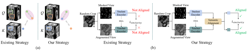
(i) Neglecting the intrinsic similar anatomical structure across varying medical image volumes. Commonly-used computed tomography (CT) and magnetic resonance (MR) images render human anatomies with intrinsic structures. As shown in Fig. 1a, the definition of positive and negative pairs in existing siamese SSL frameworks [3, 63, 46, 25, 45] ignore the semantically consistent anatomical features across different volumes and force an incorrect constraint of instance invariance. Intuitively, utilizing the intrinsic anatomical structure across different image volumes to model the class-specific invariance can help the learned representations more robust to the size, shape, and texture variances of body parts.
(ii) Lack of anatomical semantic alignment for views extracted or sampled from the same image volume. As shown in Fig. 1b, the widely used siamese architecture in hybrid SSL approaches [63, 46, 25, 12] maximize the similarity between the representations of masked view and intact view. However, a random crop of a body part may contain different organs and human tissues (some of which are small in scale). A large masking ratio would already erase these contents and make the masked view quite distinct from the intact one. Thus, maximizing the similarity between views which incorporate totally different semantics can be harmful to the learned representations. To learn good representations for downstream tasks, SSL methods for medical images should align the anatomical features of the views which form a positive pair.
Driven by the aforementioned limitations, we present a simple, effective, and dedicated self-supervised learning framework for 3D medical segmentation tasks, Alice, by explicitly fulfilling Anatomical invariance modeling and semantic alignment through elaborately combined contrastive learning and MIM. From Fig. 1, Alice leverages the structural similarity across different volumetric images to explicitly learn universal consistent features from intrinsic body structures and model the anatomical invariance which is robust to the size, shape, intensity, and texture diversity of body parts caused by inter-subject variation, organ deformation, and pathological changes.
Moreover, we design a conditional anatomical feature alignment module which complements masked views with the globally matched anatomical semantics and inter-patch topology to craft better contrastive pairs, enforcing that the positive pairs encoded to semantically consistent feature representations. This process explicitly realizes anatomical semantic alignment to further strengthen the representation with spatial sensitivity and semantic discriminability.
To adequately validate the effectiveness of Alice, we employ 3D medical image segmentation and classification as downstream tasks. We fine-tune these widely used ViT-based medical image segmentation frameworks following [60, 20, 45] with our pre-trained weights on two publicly available benchmarks: Fast and Low-resource semi-supervised Abdominal oRgan sEgmentation in CT (FLARE 2022)***https://flare22.grand-challenge.org/, and Beyond the Cranial Vault (BTCV) [29]. Our method achieves the current SOTA results, with Dice on FLARE 2022 and Dice on BTCV, surpassing previous best results by and respectively. We also evaluate transfer learning on a public COVID-19 classification benchmark [36]. Alice outperforms state-of-the-art counterpart methods by in AUC.
Our main contributions can be summarized as:
-
•
We investigate the irrationalities of commonly used siamese SSL frameworks applied to medical images. We propose Alice that is customized to leverage the anatomical similarity across volumetric medical images to model class-specific invariance.
-
•
In Alice, a conditional anatomical semantic alignment module is proposed to match the most related high-level semantics between the crafted contrastive views.
-
•
Alice consistently outperforms popular SSL methods on three public downstream benchmarks, showing its effectiveness and generality.
2 Related Work
Self-supervised Learning. Contrastive learning is the most popular method in self-supervised learning, which achieves remarkable performance on downstream tasks in computer vision [22, 8, 17, 7, 9, 11, 3]. The essence of contrastive learning aims to learn view-invariant representations by maximizing the similarity between the features extracted from different crops of the same image. More recently, some works explore how to learn representations by combining contrastive learning and MIM [46, 63, 25]. As pointed out in [39], these methods adopt random sampling to make different crops of the same image, which overlooks the semantic information and may generate views truly contain different image contents. A few newer investigations [42, 39, 59] attempt to leverage semantic guided information to crop semantically consistent views. However, they ignore the potential positive pairs in other images, restraining the diversity of learned representations. A key difference between these methods and Alice is that we mine diversified contrastive views with semantic similar contexts across varying images to encode the intrinsic structure of consistent anatomy information and facilitate the class-specific invariance.
Masked Image Modeling (MIM) accepts input image corrupted by masking and predicts the target of the masked content, which has been actively studied recently in self-supervised learning. Existing work mainly differ in their regression objectives [48, 2, 14, 50, 21, 53] or masking strategies [27, 30, 43]. In this work, Alice takes one step further by exploiting cooperations between contrastive learning and MIM to learn effective representations with both strong instance discriminability and local detail sensitive perceptibility, from varying or different image views.
Self-supervised Learning in Medical Imaging. Many works apply tailored contrastive SSL methods to medical image problems [28, 4, 44, 52] with reasonable success. Similar to MIM, image restoration is also commonly used as pre-text task to memorize spatial context from medical images. Typical attempts include inpainting tasks [5, 1, 66], Rubik’s cube problem [67] and diverse context reconstruction [62]. Most recently, DiRA [18] employs discriminative [7], restorative [6], and adversarial learning [13] objectives simultaneously in a unified SSL framework for medical image analysis. Swin UNETR [45] trains a transformer-based encoder with combination of different pre-text tasks for 3D medical image segmentation. These efforts constitute important steps toward better SSL methods for medical image analysis. Nevertheless, Alice distinguishes itself by having two key new developments: (1) explicitly leveraging the inherent anatomical consistency between different image volumes to encode the class-specific invariance; and (2) fulfilling anatomical semantic alignment to craft better contrastive pairs.
3 Method
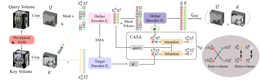
The overall framework of our method is illustrated in Fig. 2. Our Alice model consists of two branches performing masked image modeling and contrastive learning, respectively. We first mine diverse yet semantically consistent crops from varying image volumes. A query volume and a key volume are randomly picked. Then we adopt a pre-trained SAM [54] model, which performs self-supervised universal landmark detection to locate the same body part in different volumetric medical images and produce two crops, denoted as and , that depict two sub-volumes from the same body part. After that, we utilize two different data augmentations, and , to generate two views of . We denote them as and . Likewise, we also utilize two different data augmentations, and , to generate two views of , denoted as and . Here, and are random masking, which is similar to the ‘‘random sampling’’ adopted in MAE [21]. and are two different data augmentation operations. In the following, we elaborate on each component of Alice in details and describe how we optimize the relationships between and among these four views.
3.1 Network Architecture Components
Online Encoder. The online encoder takes masked view pairs ( and ) as inputs. Following MAE [21], we only feed the visible, unmasked patches to the online encoder . embeds visible tokens added with the positional embeddings and produces the output features ( and ) through a sequence of transformer blocks. In this paper, we mainly consider two vision Transformer architectures (ViT [16] and Swin Transformer [32]) as the online encoder options. We adopt the strategy in [24] to allow the Swin Transformer to discard masked patches and operate only on the visible ones. After pre-training, only the online encoder is used for extracting image representations in downstream tasks.
Online Decoder. In addition to the features of visible patches and , the online decoder receives mask tokens as inputs. We add positional embeddings to all tokens. Then the online decoder learns to reconstruct the pixel of the masked patches. Following MAE [21], we use the normalized pixel values as targets in the reconstruction task. Our loss function computes the mean squared error (MSE) on masked patches between the decoder predictions (, ) and the original input volume crops :
| (1) |
where is an indicator to select the prediction corresponding to masked tokens, and are the number of masked patches in and , respectively. This MIM objective helps the learned representations encode local context and patient-specific information of input volumes. The online decoder is set to be stacked transformer blocks.
Target Encoder. Following existing siamese frameworks [63, 46, 25], we introduce a target encoder to generate contrastive supervision for the online encoder to further strengthen the representation learned by MIM with semantic discriminability. The target encoder shares the same architecture as the online encoder. We update parameters of the target encoder using an exponential moving average (EMA) of the online encoder weights. The target encoder takes two unmasked augmented views and as inputs and embeds them into high dimensional feature representations which reserve the semantic integrity.
3.2 Anatomical Invariance Modeling
A prominent difference between previous methods and Alice is that we mine diversified views from different volumes to learn anatomical features that are universal across similar body parts. While existing methods [63, 45, 25] only operate on the same volume, we propose to explicitly model the anatomical invariance by maximizing the similarity between embeddings of views from and .
As shown in Fig. 2, we append a projection head and another projection head to the online decoder and target encoder respectively to produce positive feature pairs and . and both consist of a 3-layer MLPs with -normalized bottleneck following DINO [3]. We then adopt global average pooling to these features and obtain their global visual semantics, denoted as and . We encourage their high-level class-specific representations move closer in the corresponding feature space. This yields the loss:
| (2) |
where denotes a general cosine similarity loss [17, 10]. Through optimizing such inter-volume relationship, Alice explicitly enforces anatomical invariance.
3.3 Anatomical Semantic Alignment
To further borrow the capability of semantics abstraction acquired from self-distillation, we now consider optimizing the intra-volume relationship by maximizing the similarity between views from the same volume. Different from recent works [63, 27] that use embeddings of intact views ( and ) as teachers to supervise the masked views’ representations ( and ), we argue that it is unreasonable to directly encourage the masked views to have similar representations to the global views since their anatomical structure and semantic information may be significantly distinct.This dilemma motivates us to perform anatomical semantic alignment†††Please note that anatomical semantic alignment does not involve a registration task. Our primary objective is to extract aligned features that demonstrate the highest correlation from masked views and augmented views. We do not aim to match masked images to full images in this process. between output features from the masked view and the intact view.
One straightforward way is to contrast between the output features of the online decoder and target encoder as proposed in SIM [46]. However, we empirically observe that this choice brings little improvement for downstream tasks. Directly using the features from the online decoder for self-distillation means that the online decoder has to simultaneously optimize multiple different targets. Such multi-task learning process trained a strong online decoder and thus creates an oversimple optimizing task for the online encoder since the decoder takes the most charge of recovering details and anatomical semantics. To ensure that the online encoder effectively contributes to downstream tasks, it is crucial to develop an improved training strategy that enables the online encoder to learn rich and generalized feature representations. Taking this into account, we propose a conditional anatomical semantic alignment (CASA) module with learning capacity of reasoning about the encoded masked view’s most semantically similar anatomical features in the embeddings of the original volume. This leads to crafting more specific and aligned high-level features for self-distillation.
We take the process of aligning features from (masked view from volume crop ) and (augmented intact view from volume crop ) as an example to elaborate. Fig. 2 shows the high-level idea of the anatomical semantic alignment. We use from the online encoder as a criterion query, and generate a contrastive pair from outputs of online decoder and target encoder with aligned anatomical semantics constrained by the criterion query. Concretely, we generate teacher embeddings and student embeddings , which are aligned and capture the most semantically similar anatomical information in the original volume as expressed in the masked view’s embeddings . Here, the anatomical features from serve as the guidance for semantic alignment.
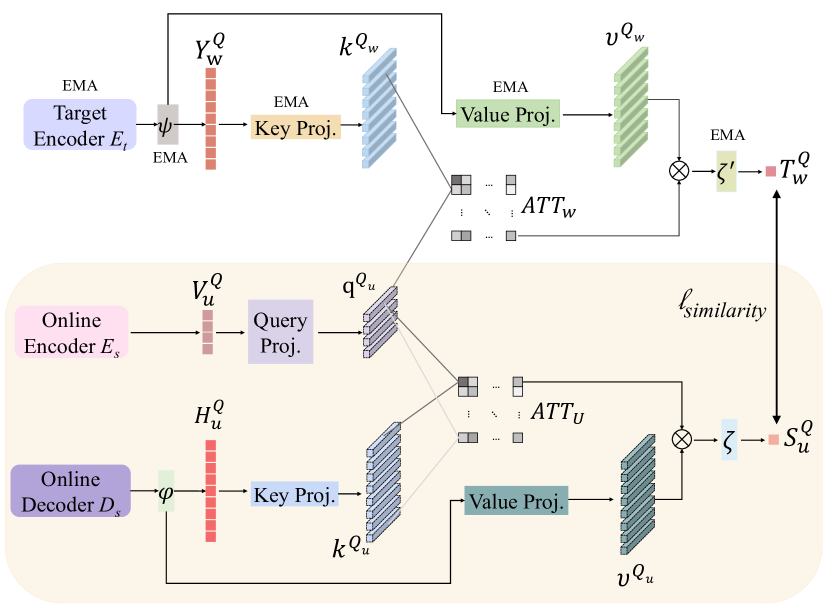
Fig. 3 shows the process of generating the student embeddings (in yellow part) and teacher embeddings . Consider generating first, we project into the query matric: . Then we project , which encodes the reconstructed features, into key and value matrices: , .
Here is the projection dimension, is a LayerNorm layer and , and are projection matrices. The query matrices are used to seek from the key matrices to attend to semantics with the highest relevance. The value matrices represent the anatomical features from which we aggregate only certain high-level global information depending on . Since is extracted from masked views, we aim to adaptively highlights the relevant features from the reconstructed views to selectively obtain anatomical semantics based on their relevance to .
In order to learn flexible conditioning between the online encoder outputs and the reconstructed features , we compute scaled dot-product attention [49] from the query-projected matrices to the key-projected matrices . The dot product attention gives relevancy weights from local fragmentary patch information to each high-level global anatomical semantics:
| (3) |
We then leverage to aggregate the value-projected matrices and get the student embeddings through a projection layer as follows:
| (4) |
The process of generating the teacher embeddings is similar and the mere difference is that we use the target encoder’s outputs to produce key and value matrices. As and are guided by the same query matrices, they learn to encode globally matched anatomical semantics and inter-patch topology information from different views conditioned by the distribution of masked view’s local image content. We then maximize the similarity between semantic-aligned teacher embeddings and student embeddings to produce more suitably learned representations.
The process of aligning features from (masked view from volume crop ) and (augmented intact view from volume crop ) is exactly the same as the above. We also obtain teacher embeddings and student embeddings from the volume . Now we have positive feature pairs and . The semantically aligned features in each pair encompass the same high-level anatomical information, we aim to reach a consensus among their representations by maximizing the similarity between them. We therefore define the intra-volume loss as:
| (5) |
By involving the inter-volume contrast (in Sec. 3.2) and intra-volume contrast (in Sec. 3.3), we explicitly model the anatomical invariance. Combining such advantage with the MIM objective, the online encoder learns to capture both high-level discriminative anatomical features and fine-grained localization-sensitive context details.
Note that, in Sec. 3.2, we do not adopt semantic alignment between views from different volumes, as they should share the same high-level class information but be distinct in local texture and shape. There is no direct relevance between the masked view from volume crop and the intact view from volume crop .
4 Experiments
4.1 Datasets & Evaluation Metrics
Pre-training Datasets. We use a total of 2,000 unlabeled CT scans from the Fast and Low-resource semi-supervised Abdominal oRgan sEgmentation in CT (FLARE 2022) challenge dataset‡‡‡https://flare22.grand-challenge.org/ to train Alice by self-supervised learning. Any (available) forms of annotations or labels of these 2,000 CTs are not employed during the pre-training stage.
Downstream Datasets & Evaluation Metrics. Three public datasets are used for downstream evaluation. For segmentation task: (1) In addition to 2,000 unlabeled CT scans, FLARE 2022 also provides a downstream training set including 50 labeled CT scans with pancreatic diseases. The offline test set incorporates 20 CT scans of patients with liver, kidney, spleen, or pancreas diseases. The segmentation targets are 13 organs: liver, spleen, pancreas, right kidney, left kidney, stomach, gallbladder, esophagus, aorta, inferior vena cava, right adrenal gland, left adrenal gland, and duodenum. (2) The Beyond the Cranial Vault (BTCV) abdomen challenge dataset [29] contains 30 subjects of abdominal CT scans where 13 organs (not the same as FLARE 2022) are annotated by interpreters under the supervision of radiologists at Vanderbilt University Medical Center. Following [52], we employ two settings of test set configurations: offline test set and online test set, for BTCV dataset. For classification task: (3) We conduct experiments on a public benchmark MosMedData: Chest CT Scans with COVID-19 Related Findings [36]. This dataset contains a total of 1,110 lung CT scans with COVID-19 related findings, as well as without such findings. We randomly split of the dataset for training, for validation and the rest for testing. Note that all downstream datasets do not have any intersection with the dataset used for pre-training. For quantitative evaluation, we adopt two segmentation metrics of Dice Similarity Coefficient (DSC) and Normalized Surface Dice (NSD), and one classification metric of the area under the receiver operator curve (AUC).
4.2 Implementation Details
Pre-training Setup. We use ViT-B [16] and Swin-B [32] as the default backbones for online encoder. Other than adopting two different random crops for the online encoder and target encoder as common practice, we utilize SAM [54] to first locate/align the same body part, then we use a default input volume crop size of to generate respective views of consistent anatomies. This avoids the large disparity between the inputs of online/target encoders when randomly cropped regions are far apart spatially, or scarcely semantically relevant. For MIM tasks, we apply augmentations and masking strategy following MAE [21] to the input of the online encoder. For the input of the target encoder, strong data augmentations, e.g. random resized rotation, flipping, intensity scaling and shifting, are adopted to avoid a trivial solution. We keep the same projection head structure as used in [11, 17]. The training loss is a summation of , , and . During pre-training, we adopt a lightweight decoder to reduce computing overhead and only use the online encoder for downstream segmentation tasks. We employ AdamW optimizer [33] with the momentum set to , and cosine learning rate schedule with a warmup of 100 epochs. The pre-training process uses a batch size of 8 per GPU and an initial learning rate of for 100K iterations. We implement Alice model in PyTorch [38]. All pre-training experiments are conducted on 8 NVIDIA A100 GPUs.
Downstream Training Setup. For segmentation tasks, we apply our pre-trained encoder weights to various ViT-based segmentation networks designed for medical tasks of UNETR [20], nnFormer [60], and Swin UNETR [45], by following their settings. We compare different SSL methods within [20, 60, 45]. Five-fold cross validation is used to train and evaluate models for all FLARE 2022 and BTCV experiments. For classification task, we utilizes unlabeled CT scans from FLARE 2022 for pre-training to evaluate the adaptability of Alice for different scenes (COVID-19 classification). Ten-fold cross validation is used for COVID-19 dataset. Detailed training hyperparameters for these downstream tasks can be found in the supplemental material.
| UNETR | Swin UNETR | nnFormer | ||||
| DSC | NSD | DSC | NSD | DSC | NSD | |
| Rand. init. | 80.953.48 | 85.233.74 | 81.043.29 | 85.603.55 | 81.333.05 | 86.053.31 |
| MoCo v3 [11] | 82.013.27 | 86.493.52 | 82.633.18 | 86.923.44 | 82.882.82 | 86.893.10 |
| DINO [3] | 82.073.15 | 86.313.40 | 82.692.99 | 87.083.17 | 82.952.74 | 87.452.92 |
| IBOT [63] | 83.153.21 | 87.613.48 | 83.773.14 | 88.343.30 | 84.042.81 | 88.463.11 |
| SIM [46] | 83.042.97 | 87.373.36 | 83.592.74 | 88.572.90 | 83.962.64 | 88.612.83 |
| MAE [21] | 83.092.92 | 87.423.43 | 83.622.66 | 88.552.92 | 84.012.59 | 88.632.75 |
| SemMAE [30] | 83.132.87 | 87.903.31 | - | - | - | - |
| CMAE [25] | 83.592.76 | 88.252.98 | 84.252.59 | 89.172.88 | 84.472.42 | 89.442.65 |
| medical MAE [64] | 83.112.91 | 87.453.39 | 83.662.64 | 88.572.86 | 84.032.57 | 88.672.71 |
| Tang et al. [45] | 82.973.22 | 87.513.50 | 83.143.01 | 88.743.37 | 83.592.89 | 89.513.23 |
| Alice | 85.812.05 | 90.032.28 | 86.751.89 | 91.222.12 | 86.871.84 | 91.282.09 |
4.3 Results
FLARE 2022 Abdominal Organ Segmentation. We conduct extensive comparison between Alice and existing SOTA methods. We first fix three ViT-based medical segmentation framework, and compare Alice with the random initialization (Rand. init.) and other advanced SSL methods designed for computer vision and medical tasks. The evaluation results on the offline test set are shown in Table 1. Compared with strong Transformer-based contrastive learning methods MoCo v3 and DINO, Alice outperforms them by at least absolute in DSC. Compared with MIM methods MAE and SemMAE, the DSC score of Alice surpasses that of MAE and SemMAE by a large margin. Furthermore, Alice also achieves at least improvement of DSC relative to hybrid SSL methods IBOT and CMAE, which combine contrastive learning and MIM. Notably, Alice is also superior to SOTA ViT based SSL methods, medical MAE [64] and pre-training framework proposed in [45], which are tailored for medical image analysis.
| Method | Pre-trained Parts | DSC | NSD |
| MoCo v2 [9] | 3D ResNet E. | 81.963.29 | 86.583.45 |
| BYOL [17] | 3D ResNet E. | 81.943.32 | 86.603.50 |
| ContrastiveCrop [39] | 3D ResNet E. | 82.553.05 | 87.263.15 |
| LoGo [59] | 3D ResNet E. | 82.533.08 | 87.083.17 |
| PCRL [62] | 3D UNet E.&D. | 83.412.81 | 87.843.00 |
| PCRLv2 [61] | 3D nsUNet E. | 84.452.49 | 89.112.68 |
| PGL [51] | 3D ResNet E. | 83.162.85 | 87.932.99 |
| Chaitanya et al. [4] | UNet E.&D. | 82.292.90 | 86.723.14 |
| nnU-Net [26] | - | 83.622.78 | 88.512.85 |
| TransVW [19] | 3D UNet E.&D. | 82.703.02 | 87.263.18 |
| SAM [54] | 3D UNet E.&D. | 82.483.06 | 87.063.21 |
| Models Gen. [66] | V-Net E.&D. | 83.352.82 | 88.122.95 |
| DiRA [18] | 3D UNet E.&D. | 82.572.88 | 87.233.01 |
| Alice | nnFormer E. | 86.871.84 | 91.282.09 |
| Method | Ensemble | Offline Test Set | Online Test Set |
| MoCo v2 [9] | 1 | 82.052.82 | - |
| MoCo v3 [11] | 1 | 82.022.77 | - |
| DINO [3] | 1 | 82.611.79 | - |
| MAE [21] | 1 | 83.162.14 | - |
| IBOT [63] | 1 | 83.282.26 | - |
| CMAE [25] | 1 | 83.471.33 | - |
| PCRL [62] | 1 | 82.732.42 | - |
| PCRLv2 [61] | 1 | 83.551.49 | - |
| PGL [51] | 1 | 82.572.60 | - |
| Chaitanya et al. [4] | 1 | 82.742.12 | - |
| medical MAE [64] | 1 | 83.232.05 | - |
| SAM [54] | 1 | 82.003.01 | 84.07 |
| DoDnet [58] | 5 | - | 86.44 |
| UNETR [20] | 5 | - | 85.55 |
| PaNN [65] | 5 | - | 85.00 |
| nnU-Net [26] | 10 | - | 87.62 |
| nnFormer [60] | 5 | 82.882.59 | - |
| DiRA [18] | 1 | 83.142.04 | - |
| Tang et al. [45] | 1 | 82.582.20 | 84.72 |
| UniMiSS [52] | 1 | 84.991.57 | 87.05 |
| Alice | 1 | 86.760.98 | 88.58 |
| CNN-based Method | AUC on COVID-19 | ViT-based Method | AUC on COVID-19 |
| BYOL [17] | 85.745.04 | DINO [3] | 86.874.35 |
| Peng et al. [39] | 87.023.11 | IBOT [63] | 87.553.63 |
| LoGo [59] | 86.953.59 | SIM [46] | 87.672.95 |
| PCRL [62] | 87.312.88 | MAE [21] | 86.623.27 |
| PCRLv2 [61] | 88.362.51 | SemMAE [30] | 86.943.44 |
| PGL [51] | 86.084.72 | CMAE [25] | 87.733.02 |
| DiRA [18] | 87.433.55 | Alice | 90.881.29 |
We also compare Alice with strong CNN-based SSL methods and baselines. The results are shown in Table 2. Alice significantly outperforms the other CNN-based SOTA methods. Alice achieves better performance compared with PCRL, TransVW, SAM, DiRA, and Models genesis, which additionally pre-train a decoder. Notably, Alice outperforms the method of Chaitanya et al. [4] which also utilizes inter-volume slices. Chaitanya et al. [4] assume that all volume images have been aligned and use simple grouping of image slices in 3D volumes to craft contrastive patches, which will lead to unaligned pairs. Differently, we adopt a learning-based module SAM [54] to get aligned sample pairs from different volumes online and we further align feature level semantics by CASA module. Our Alice is more suitable to handle real-world complex problems like changes in the range of CT scans and temporal changes.
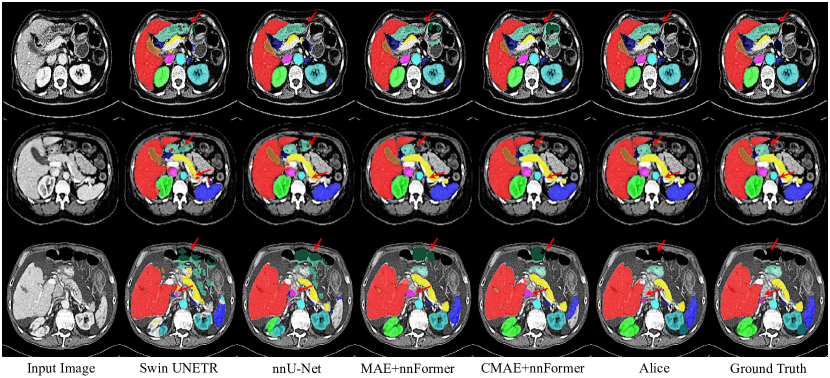
Fig. 4 shows the qualitative results and demonstrate the merits of Alice. Most competing methods suffer from segmentation target incompleteness related failures and misclassification of background regions as organs (false positives). Alice produces sharper boundaries and generates results that are more consistent with the ground truth in comparison with all other models. This success is attributed to the advantage of leveraging the intrinsic anatomical invariance across varying volumes and semantic alignment between contrastive views, which help the learned representation robust to organ deformation and pathological changes.
BTCV Multi-organ Segmentation. We also compare Alice with other SOTA SSL methods on the BTCV offline and online test set. As shown in Table 3, Alice, without using any ensemble strategy, still achieves the competitive performance with the best DSC on both offline and online test set. Note that compared to SOTA methods designed for medical images, namely UniMiSS and the self-supervised Swin UNETR (Tang et al.), using over 3D CT scans for pre-training, our Alice outperforms them using only of the data. This effectiveness can be explained that our approach is capable of learning representations that is robust to the size, shape, intensity, and texture diversity of body parts by modeling the anatomical invariance. During pre-training, we map the high-level embeddings of the same organs from varying volumes to the same point, which helps the model in downstream tasks reduce misclassification failures and noticeably improves the performance.
COVID-19 Classification. We compare Alice with the state-of-the-arts including representative CNN-based SSL methods and ViT-based SSL methods. The results are shown in Table 4. Compared with CNN-based SSL methods BYOL, PCRL, PCRLv2, PGL and DiRA, Alice outperforms them at least absolute in AUC. Notably, Alice achieves much better results than LoGo [59] and Peng et al. [39], which also design specific strategies to generate semantic-aligned contrastive view pairs. However, these two methods only operate within each image independently and ignore the inter-volume consistency. Compared against strong ViT-based SSL methods, Alice significantly outperforms them at least absolute in AUC. It proves the effectiveness of modeling anatomical invariance and performing semantic alignment to assist the SSL process. Moreover, as an inter-scene evaluation, Table 4 reveals Alice has the ability to generalize to other scenarios. More details are in our Supplementary Material.
4.4 Ablation Study
Significance in Modeling Inter-volume Anatomical Invariance. We investigate the effect of leveraging the intrinsic anatomical structures of different volumes to model the anatomical invariance. We feed the same body part of query volume and key volume to another SSL method IBOT and add the inter-volume relationship contrastive loss for it. Table 5 shows that involving feature contrasting between mined consistent views from varying aligned CT volumes can substantially improve the 3D segmentation accuracy in the downstream task for both IBOT and Alice. The performance gain on DSC is and , respectively. This shows that modeling the anatomical invariance among CT volumes to learn intrinsic high-level semantics during pre-training benefits the learned representations for downstream tasks.
| Method | Inter-Volume Relationship | FLARE 2022 | |
| DSC | NSD | ||
| IBOT [63] | 84.042.81 | 88.463.11 | |
| ✓ | 85.412.42 | 89.972.70 | |
| Alice | 85.632.11 | 90.012.32 | |
| ✓ | 86.871.84 | 91.282.09 | |
Effectiveness of Anatomical Semantic Alignment. Table 6 shows the ablation study on the effectiveness of the CASA module. Our approach can be applied to most general siamese architecture based SSL methods. All methods achieve more than absolute DSC gain on the FLARE 2022 dataset by adopting CASA. This shows applying our anatomical semantic alignment to harvest better contrastive pairs from masked and unmasked views yields significant improvements on various siamese SSL methods.
| Method | FLARE 2022 | |
| DSC | NSD | |
| IBOT (w/. vs. w/o.) | 85.342.63 vs. 84.042.81 | 89.882.89 vs. 88.463.11 |
| SIM (w/. vs. w/o.) | 85.212.36 vs. 83.962.64 | 89.892.67 vs. 88.612.83 |
| Alice (w/. vs. w/o.) | 86.871.84 vs. 85.662.18 | 91.282.09 vs. 90.022.32 |
The impact of whether to use negative samples. In Eq. 2, we utilize BYOL-style cosine loss [17] as our default choice for contrastive learning. This loss only maximizes the similarity between positive views and eliminates the use of negative pairs. Another widely used similarity loss function is the InfoNCE loss [7, 22, 37], which aims to simultaneously pull close positive views and push away negative samples. The key distinction between these two widely used types of similarity loss functions lies in the utilization of negative samples. Alice is compatible with a wide range of SSL techniques and is independent of the specific training losses used in those techniques. Thus we conduct experiments to investigate the influence of whether negative pairs are used. We discuss two widely used similarity loss functions: InfoNCE loss (exploits both positive and negative samples) and BYOL-style cosine loss (does not exploit negative samples). When using InfoNCE loss for contrastive learning, we utilize views from crops of different body parts in the same batch to compose negative pairs. Table 7 shows the ablation study on whether to use negative samples. We observed that using cosine loss in Alice achieves slightly higher performance than InfoNCE loss on FLARE 2022 test set. Thus, we do not utilize negative samples and use cosine loss as the default for the contrastive learning branch in Alice. The discussion of why cosine loss does not need negative pairs can be found in BYOL’s literature [17].
| Method | loss function | FLARE 2022 | |
| DSC | NSD | ||
| Alice | InfoNCE loss [7, 22, 37] | 86.831.88 | 91.202.12 |
| BYOL-style cosine loss [17] | 86.871.84 | 91.282.09 | |
5 Conclusions
In this work, we propose a novel self-supervised learning method (Alice) for improving the learned image representation of contrastive learning and MIM by modeling the class-specific invariance of intrinsic anatomical semantics in 3D medical images. We also introduce a conditional anatomical semantic alignment module that generates better contrastive pairs with more consistent high-level information. Extensive quantitative experiments reporting superior results validate the effectiveness of our method.
References
- [1] Shekoofeh Azizi, Basil Mustafa, Fiona Ryan, Zachary Beaver, Jan Freyberg, Jonathan Deaton, Aaron Loh, Alan Karthikesalingam, Simon Kornblith, Ting Chen, et al. Big self-supervised models advance medical image classification. In ICCV, pages 3478--3488, 2021.
- [2] Hangbo Bao, Li Dong, and Furu Wei. Beit: Bert pre-training of image transformers. arXiv preprint arXiv:2106.08254, 2021.
- [3] Mathilde Caron, Hugo Touvron, Ishan Misra, Hervé Jégou, Julien Mairal, Piotr Bojanowski, and Armand Joulin. Emerging properties in self-supervised vision transformers. In ICCV, pages 9650--9660, 2021.
- [4] Krishna Chaitanya, Ertunc Erdil, Neerav Karani, and Ender Konukoglu. Contrastive learning of global and local features for medical image segmentation with limited annotations. NeurIPS, 33:12546--12558, 2020.
- [5] Liang Chen, Paul Bentley, Kensaku Mori, Kazunari Misawa, Michitaka Fujiwara, and Daniel Rueckert. Self-supervised learning for medical image analysis using image context restoration. Medical Image Analysis, 58:101539, 2019.
- [6] Mark Chen, Alec Radford, Rewon Child, Jeffrey Wu, Heewoo Jun, David Luan, and Ilya Sutskever. Generative pretraining from pixels. In ICML, pages 1691--1703. PMLR, 2020.
- [7] Ting Chen, Simon Kornblith, Mohammad Norouzi, and Geoffrey Hinton. A simple framework for contrastive learning of visual representations. In ICML, pages 1597--1607. PMLR, 2020.
- [8] Ting Chen, Simon Kornblith, Kevin Swersky, Mohammad Norouzi, and Geoffrey E Hinton. Big self-supervised models are strong semi-supervised learners. NeurIPS, 33:22243--22255, 2020.
- [9] Xinlei Chen, Haoqi Fan, Ross Girshick, and Kaiming He. Improved baselines with momentum contrastive learning. arXiv preprint arXiv:2003.04297, 2020.
- [10] Xinlei Chen and Kaiming He. Exploring simple siamese representation learning. In CVPR, pages 15750--15758, 2021.
- [11] Xinlei Chen, Saining Xie, and Kaiming He. An empirical study of training self-supervised vision transformers. In ICCV, pages 9640--9649, 2021.
- [12] Yabo Chen, Yuchen Liu, Dongsheng Jiang, Xiaopeng Zhang, Wenrui Dai, Hongkai Xiong, and Qi Tian. Sdae: Self-distillated masked autoencoder. In ECCV, pages 108--124. Springer, 2022.
- [13] Jeff Donahue and Karen Simonyan. Large scale adversarial representation learning. NeurIPS, 32, 2019.
- [14] Xiaoyi Dong, Jianmin Bao, Ting Zhang, Dongdong Chen, Weiming Zhang, Lu Yuan, Dong Chen, Fang Wen, and Nenghai Yu. Peco: Perceptual codebook for bert pre-training of vision transformers. arXiv preprint arXiv:2111.12710, 2021.
- [15] Xiaoyi Dong, Jianmin Bao, Ting Zhang, Dongdong Chen, Weiming Zhang, Lu Yuan, Dong Chen, Fang Wen, and Nenghai Yu. Bootstrapped masked autoencoders for vision bert pretraining. arXiv preprint arXiv:2207.07116, 2022.
- [16] Alexey Dosovitskiy, Lucas Beyer, Alexander Kolesnikov, Dirk Weissenborn, Xiaohua Zhai, Thomas Unterthiner, Mostafa Dehghani, Matthias Minderer, Georg Heigold, Sylvain Gelly, et al. An image is worth 16x16 words: Transformers for image recognition at scale. In ICLR, 2020.
- [17] Jean-Bastien Grill, Florian Strub, Florent Altché, Corentin Tallec, Pierre Richemond, Elena Buchatskaya, Carl Doersch, Bernardo Avila Pires, Zhaohan Guo, Mohammad Gheshlaghi Azar, et al. Bootstrap your own latent-a new approach to self-supervised learning. NeurIPS, 33:21271--21284, 2020.
- [18] Fatemeh Haghighi, Mohammad Reza Hosseinzadeh Taher, Michael B Gotway, and Jianming Liang. Dira: Discriminative, restorative, and adversarial learning for self-supervised medical image analysis. In CVPR, pages 20824--20834, 2022.
- [19] Fatemeh Haghighi, Mohammad Reza Hosseinzadeh Taher, Zongwei Zhou, Michael B Gotway, and Jianming Liang. Transferable visual words: Exploiting the semantics of anatomical patterns for self-supervised learning. IEEE Trans. Medical Imaging, 40(10):2857--2868, 2021.
- [20] Ali Hatamizadeh, Yucheng Tang, Vishwesh Nath, Dong Yang, Andriy Myronenko, Bennett Landman, Holger R Roth, and Daguang Xu. Unetr: Transformers for 3d medical image segmentation. In WACV, pages 574--584, 2022.
- [21] Kaiming He, Xinlei Chen, Saining Xie, Yanghao Li, Piotr Dollár, and Ross Girshick. Masked autoencoders are scalable vision learners. In CVPR, pages 16000--16009, 2022.
- [22] Kaiming He, Haoqi Fan, Yuxin Wu, Saining Xie, and Ross Girshick. Momentum contrast for unsupervised visual representation learning. In CVPR, pages 9729--9738, 2020.
- [23] Nicholas Heller, Sean McSweeney, Matthew Thomas Peterson, Sarah Peterson, Jack Rickman, Bethany Stai, Resha Tejpaul, Makinna Oestreich, Paul Blake, Joel Rosenberg, et al. An international challenge to use artificial intelligence to define the state-of-the-art in kidney and kidney tumor segmentation in ct imaging., 2020.
- [24] Lang Huang, Shan You, Mingkai Zheng, Fei Wang, Chen Qian, and Toshihiko Yamasaki. Green hierarchical vision transformer for masked image modeling. arXiv preprint arXiv:2205.13515, 2022.
- [25] Zhicheng Huang, Xiaojie Jin, Chengze Lu, Qibin Hou, Ming-Ming Cheng, Dongmei Fu, Xiaohui Shen, and Jiashi Feng. Contrastive masked autoencoders are stronger vision learners. arXiv preprint arXiv:2207.13532, 2022.
- [26] Fabian Isensee, Paul F Jaeger, Simon AA Kohl, Jens Petersen, and Klaus H Maier-Hein. nnu-net: a self-configuring method for deep learning-based biomedical image segmentation. Nature methods, 18(2):203--211, 2021.
- [27] Ioannis Kakogeorgiou, Spyros Gidaris, Bill Psomas, Yannis Avrithis, Andrei Bursuc, Konstantinos Karantzalos, and Nikos Komodakis. What to hide from your students: Attention-guided masked image modeling. arXiv preprint arXiv:2203.12719, 2022.
- [28] Nikos Komodakis and Spyros Gidaris. Unsupervised representation learning by predicting image rotations. In ICLR, 2018.
- [29] Bennett Landman, Zhoubing Xu, J Igelsias, Martin Styner, T Langerak, and Arno Klein. Miccai multi-atlas labeling beyond the cranial vault--workshop and challenge. In Proc. MICCAI Multi-Atlas Labeling Beyond Cranial Vault—Workshop Challenge, volume 5, page 12, 2015.
- [30] Gang Li, Heliang Zheng, Daqing Liu, Chaoyue Wang, Bing Su, and Changwen Zheng. Semmae: Semantic-guided masking for learning masked autoencoders. arXiv preprint arXiv:2206.10207, 2022.
- [31] Zhaowen Li, Zhiyang Chen, Fan Yang, Wei Li, Yousong Zhu, Chaoyang Zhao, Rui Deng, Liwei Wu, Rui Zhao, Ming Tang, et al. Mst: Masked self-supervised transformer for visual representation. NeurIPS, 34:13165--13176, 2021.
- [32] Ze Liu, Yutong Lin, Yue Cao, Han Hu, Yixuan Wei, Zheng Zhang, Stephen Lin, and Baining Guo. Swin transformer: Hierarchical vision transformer using shifted windows. In ICCV, pages 10012--10022, 2021.
- [33] Ilya Loshchilov and Frank Hutter. Decoupled weight decay regularization. arXiv preprint arXiv:1711.05101, 2017.
- [34] Jun Ma, Yao Zhang, Song Gu, Xingle An, Zhihe Wang, Cheng Ge, Congcong Wang, Fan Zhang, Yu Wang, Yinan Xu, et al. Fast and low-gpu-memory abdomen ct organ segmentation: The flare challenge. Medical Image Analysis, 82:102616, 2022.
- [35] Jun Ma, Yao Zhang, Song Gu, Cheng Zhu, Cheng Ge, Yichi Zhang, Xingle An, Congcong Wang, Qiyuan Wang, Xin Liu, et al. Abdomenct-1k: Is abdominal organ segmentation a solved problem. IEEE TPAMI, 2021.
- [36] Sergey P Morozov, AE Andreychenko, NA Pavlov, AV Vladzymyrskyy, NV Ledikhova, VA Gombolevskiy, Ivan A Blokhin, PB Gelezhe, AV Gonchar, and V Yu Chernina. Mosmeddata: Chest ct scans with covid-19 related findings dataset. arXiv preprint arXiv:2005.06465, 2020.
- [37] Aaron van den Oord, Yazhe Li, and Oriol Vinyals. Representation learning with contrastive predictive coding. arXiv preprint arXiv:1807.03748, 2018.
- [38] Adam Paszke, Sam Gross, Francisco Massa, Adam Lerer, James Bradbury, Gregory Chanan, Trevor Killeen, Zeming Lin, Natalia Gimelshein, Luca Antiga, et al. Pytorch: An imperative style, high-performance deep learning library. NeurIPS, 32, 2019.
- [39] Xiangyu Peng, Kai Wang, Zheng Zhu, Mang Wang, and Yang You. Crafting better contrastive views for siamese representation learning. In CVPR, pages 16031--16040, 2022.
- [40] Kanchana Ranasinghe, Muzammal Naseer, Salman Khan, Fahad Shahbaz Khan, and Michael S Ryoo. Self-supervised video transformer. In CVPR, pages 2874--2884, 2022.
- [41] Olga Russakovsky, Jia Deng, Hao Su, Jonathan Krause, Sanjeev Satheesh, Sean Ma, Zhiheng Huang, Andrej Karpathy, Aditya Khosla, Michael Bernstein, et al. Imagenet large scale visual recognition challenge. IJCV, 115(3):211--252, 2015.
- [42] Ramprasaath R Selvaraju, Karan Desai, Justin Johnson, and Nikhil Naik. Casting your model: Learning to localize improves self-supervised representations. In CVPR, pages 11058--11067, 2021.
- [43] Yuge Shi, N Siddharth, Philip Torr, and Adam R Kosiorek. Adversarial masking for self-supervised learning. In ICML, pages 20026--20040. PMLR, 2022.
- [44] Aiham Taleb, Winfried Loetzsch, Noel Danz, Julius Severin, Thomas Gaertner, Benjamin Bergner, and Christoph Lippert. 3d self-supervised methods for medical imaging. NeurIPS, 33:18158--18172, 2020.
- [45] Yucheng Tang, Dong Yang, Wenqi Li, Holger R Roth, Bennett Landman, Daguang Xu, Vishwesh Nath, and Ali Hatamizadeh. Self-supervised pre-training of swin transformers for 3d medical image analysis. In CVPR, pages 20730--20740, 2022.
- [46] Chenxin Tao, Xizhou Zhu, Gao Huang, Yu Qiao, Xiaogang Wang, and Jifeng Dai. Siamese image modeling for self-supervised vision representation learning. arXiv preprint arXiv:2206.01204, 2022.
- [47] Yonglong Tian, Chen Sun, Ben Poole, Dilip Krishnan, Cordelia Schmid, and Phillip Isola. What makes for good views for contrastive learning? NeurIPS, 33:6827--6839, 2020.
- [48] Aaron Van Den Oord, Oriol Vinyals, et al. Neural discrete representation learning. NeurIPS, 30, 2017.
- [49] Ashish Vaswani, Noam Shazeer, Niki Parmar, Jakob Uszkoreit, Llion Jones, Aidan N Gomez, Łukasz Kaiser, and Illia Polosukhin. Attention is all you need. NeurIPS, 30, 2017.
- [50] Chen Wei, Haoqi Fan, Saining Xie, Chao-Yuan Wu, Alan Yuille, and Christoph Feichtenhofer. Masked feature prediction for self-supervised visual pre-training. In CVPR, pages 14668--14678, 2022.
- [51] Yutong Xie, Jianpeng Zhang, Zehui Liao, Yong Xia, and Chunhua Shen. Pgl: prior-guided local self-supervised learning for 3d medical image segmentation. arXiv preprint arXiv:2011.12640, 2020.
- [52] Yutong Xie, Jianpeng Zhang, Yong Xia, and Qi Wu. Unimiss: Universal medical self-supervised learning via breaking dimensionality barrier. In ECCV, pages 558--575. Springer, 2022.
- [53] Zhenda Xie, Zheng Zhang, Yue Cao, Yutong Lin, Jianmin Bao, Zhuliang Yao, Qi Dai, and Han Hu. Simmim: A simple framework for masked image modeling. In CVPR, pages 9653--9663, 2022.
- [54] Ke Yan, Jinzheng Cai, Dakai Jin, Shun Miao, Dazhou Guo, Adam P Harrison, Youbao Tang, Jing Xiao, Jingjing Lu, and Le Lu. Sam: Self-supervised learning of pixel-wise anatomical embeddings in radiological images. IEEE Trans. Medical Imaging, 2022.
- [55] Chenyu You, Ruihan Zhao, Fenglin Liu, Sandeep Chinchali, Ufuk Topcu, Lawrence Staib, and James S Duncan. Class-aware generative adversarial transformers for medical image segmentation. arXiv preprint arXiv:2201.10737, 2022.
- [56] Xin Yu, Qi Yang, Yinchi Zhou, Leon Y Cai, Riqiang Gao, Ho Hin Lee, Thomas Li, Shunxing Bao, Zhoubing Xu, Thomas A Lasko, et al. Unest: Local spatial representation learning with hierarchical transformer for efficient medical segmentation. arXiv preprint arXiv:2209.14378, 2022.
- [57] Sukmin Yun, Hankook Lee, Jaehyung Kim, and Jinwoo Shin. Patch-level representation learning for self-supervised vision transformers. In CVPR, pages 8354--8363, 2022.
- [58] Jianpeng Zhang, Yutong Xie, Yong Xia, and Chunhua Shen. Dodnet: Learning to segment multi-organ and tumors from multiple partially labeled datasets. In CVPR, pages 1195--1204, 2021.
- [59] Tong Zhang, Congpei Qiu, Wei Ke, Sabine Süsstrunk, and Mathieu Salzmann. Leverage your local and global representations: A new self-supervised learning strategy. In CVPR, pages 16580--16589, 2022.
- [60] Hong-Yu Zhou, Jiansen Guo, Yinghao Zhang, Lequan Yu, Liansheng Wang, and Yizhou Yu. nnformer: Interleaved transformer for volumetric segmentation. arXiv preprint arXiv:2109.03201, 2021.
- [61] Hong-Yu Zhou, Chixiang Lu, Chaoqi Chen, Sibei Yang, and Yizhou Yu. A unified visual information preservation framework for self-supervised pre-training in medical image analysis. IEEE Transactions on Pattern Analysis and Machine Intelligence, 2023.
- [62] Hong-Yu Zhou, Chixiang Lu, Sibei Yang, Xiaoguang Han, and Yizhou Yu. Preservational learning improves self-supervised medical image models by reconstructing diverse contexts. In ICCV, pages 3499--3509, 2021.
- [63] Jinghao Zhou, Chen Wei, Huiyu Wang, Wei Shen, Cihang Xie, Alan Yuille, and Tao Kong. ibot: Image bert pre-training with online tokenizer. arXiv preprint arXiv:2111.07832, 2021.
- [64] Lei Zhou, Huidong Liu, Joseph Bae, Junjun He, Dimitris Samaras, and Prateek Prasanna. Self pre-training with masked autoencoders for medical image analysis. arXiv preprint arXiv:2203.05573, 2022.
- [65] Yuyin Zhou, Zhe Li, Song Bai, Chong Wang, Xinlei Chen, Mei Han, Elliot Fishman, and Alan L Yuille. Prior-aware neural network for partially-supervised multi-organ segmentation. In ICCV, pages 10672--10681, 2019.
- [66] Zongwei Zhou, Vatsal Sodha, Jiaxuan Pang, Michael B Gotway, and Jianming Liang. Models genesis. Medical Image Analysis, 67:101840, 2021.
- [67] Jiuwen Zhu, Yuexiang Li, Yifan Hu, Kai Ma, S Kevin Zhou, and Yefeng Zheng. Rubik’s cube+: A self-supervised feature learning framework for 3d medical image analysis. Medical Image Analysis, 64:101746, 2020.