Multi-organ segmentation: a progressive exploration of learning paradigms under scarce annotation
Abstract
Precise delineation of multiple organs or abnormal regions in the human body from medical images plays an essential role in computer-aided diagnosis, surgical simulation, image-guided interventions, and especially in radiotherapy treatment planning. Thus, it is of great significance to explore automatic segmentation approaches, among which deep learning-based approaches have evolved rapidly and witnessed remarkable progress in multi-organ segmentation. However, obtaining an appropriately sized and fine-grained annotated dataset of multiple organs is extremely hard and expensive. Such scarce annotation limits the development of high-performance multi-organ segmentation models but promotes many annotation-efficient learning paradigms. Among these, studies on transfer learning leveraging external datasets, semi-supervised learning using unannotated datasets and partially-supervised learning integrating partially-labeled datasets have led the dominant way to break such dilemma in multi-organ segmentation. We first review the traditional fully supervised method, then present a comprehensive and systematic elaboration of the 3 abovementioned learning paradigms in the context of multi-organ segmentation from both technical and methodological perspectives, and finally summarize their challenges and future trends.
keywords:
Multi-organ segmentation , annotation-efficient , transfer learning , semi-supervised learning , partially-supervised learning1 Introduction
Medical image segmentation is an important task in medical image analysis and is widely applicable to images such as microscopy, X-ray, ultrasound, computed tomography (CT), magnetic resonance imaging (MRI), and positron emission tomography (PET). The tissue segmentation in the medical images can help clinicians evaluate the lesion size and morphology, and visualize different anatomical regions, which plays essential roles in computer-aided diagnosis, surgical simulation, treatment planning, and image-guided interventions [1, 2]. For instance, the success of radiotherapy treatment relies on accurate multi-organ segmentation. The accurate delineations of the organ at risk (OAR) and the target tumor contribute to precision radiotherapy treatment planning (RTP) and improvement of radiotherapy outcomes [3, 4]. In radiotherapy, to deliver a proper therapeutic dose to the target tumor while protecting OARs, multiple OARs and target tumor have to be delicately delineated by experienced experts, which is a highly time-consuming and labor-intensive task [5, 6], which may cause delays in treatment and fall far short of clinical needs. In addition, the delineation results vary among radiologists [7], and such inter-observer variation causes contour differences accompanying a certain amount of dosimetry discrepancies [8], which may cause complications and affect the prognosis [9].To this end, computer-assisted rapid, precise, and automated segmentation methods for multi-organ segmentation with short waiting time, high reproducibility, and consistency [10, 11] have proven clinical feasibility with substantial academic and clinical value [12, 13].
In recent years, thanks to the fast development of artificial intelligence in medical image processing, deep learning methods represented by convolutional neural networks (CNN) have dominated medical image segmentation [14, 15, 16, 17]. Likewise, in multi-organ segmentation, deep learning methods have achieved promising performance via supervised learning [18, 19, 20, 21, 22]. In these works, it is essential to provide pixel-level fine-grained annotations for each organ.
Despite the achievements in multi-organ segmentation of medical images using fully supervised deep learning methods, they all face the same dilemma: annotation scarcity under deep learning techniques, which often leads to model overfitting and insufficient generalization performance for unseen clinical data.
In reality, obtaining densely annotated multi-organ data is extremely expensive and challenging [23], for such reasons: 1) Experienced professionals are required for annotation, and organs in different part of the body calls for different experts with specific knowledge. 2) Due to the privacy concerns unique to medical images, extensive clinical data are kept in-house and away from public academic research. 3) Due to complex anatomical structures, with varying shapes, sizes, and contrasts in medical images, spatial occlusion, and blurring boundaries between adjacent organs make the accurate annotation of multi-organ an extremely hard and tedious task [24]. Arguably, annotation scarcity serves as a bottleneck limiting the performance of deep learning-based multi-organ segmentation models.
To alleviate the problem of annotation scarcity in multi-organ segmentation, several promising learning paradigms have been proposed and drawn attention. These new learning paradigms can be roughly divided into three categories: 1) Learning using external labeled datasets, such as transfer learning [25]. In transfer learning, fine-grained multi-organ annotation for a target task is not necessary and a pre-trained model on the external dataset is reused for a similar multi-organ segmentation task. 2) Learning using unlabeled datasets, such as semi-supervised learningg [26]. Semi-supervised learning only uses a small portion of dense annotation and a large amount of unlabeled data for model training. 3) Learning by fusing multiple partially annotated datasets, such as partially-supervised learning [27]. Partial supervision can use multiple single-organ or partial-organ datasets as valid annotations to segment multiple organs of inputs simultaneously. The above learning paradigms are ushering in new research directions for deep learning-based segmentation of multi-organ images.
Existing reviews on multi-organ segmentation have mainly focused on the area of fully supervised learning [13, 28, 29, 12], while reviews on annotation scarcity are still absent. Recently, the exploration of annotation-efficient methods has received increasing attention in medical image segmentation. Cheplygina et al. [30], Zhang et al. [31] and Tajbakhsh et al. [32] investigated non-fully supervised medical image segmentation methods including weakly supervised, transfer learning, active learning, and semi-supervised learning paradigms, etc. Ma et al. [33] established new segmentation benchmarks for semi-supervised, weakly supervised, and continuous learning to advance the unsolved problem that the existing satisfactory performance may not generalize on more diverse datasets. It can be observed that as new technologies continue to emerge, new learning paradigms gain wider popularity. Hence, a systematic review of studies on annotation-efficient multi-organ segmentation is urgent and valuable.
In this review, we comprehensively summarize different frameworks of the typical fully supervised learning and challenges in fine-grained segmentation for multi-organ segmentation tasks, and survey relevant works on transfer learning, semi-supervised learning, and partially supervised learning from the perspective of annotation scarcity problem. To our best knowledge, we are the first to conduct a review of partially supervised learning paradigms. We search by google scholar with the keywords such as semi-supervised learning, transfer learning, domain adaptation, partially supervised, and multi-organ segmentation, and got over 200 initial articles. After filtered by investigating abstract, we obtained 126 highly relevant works and our review contained 201 references.
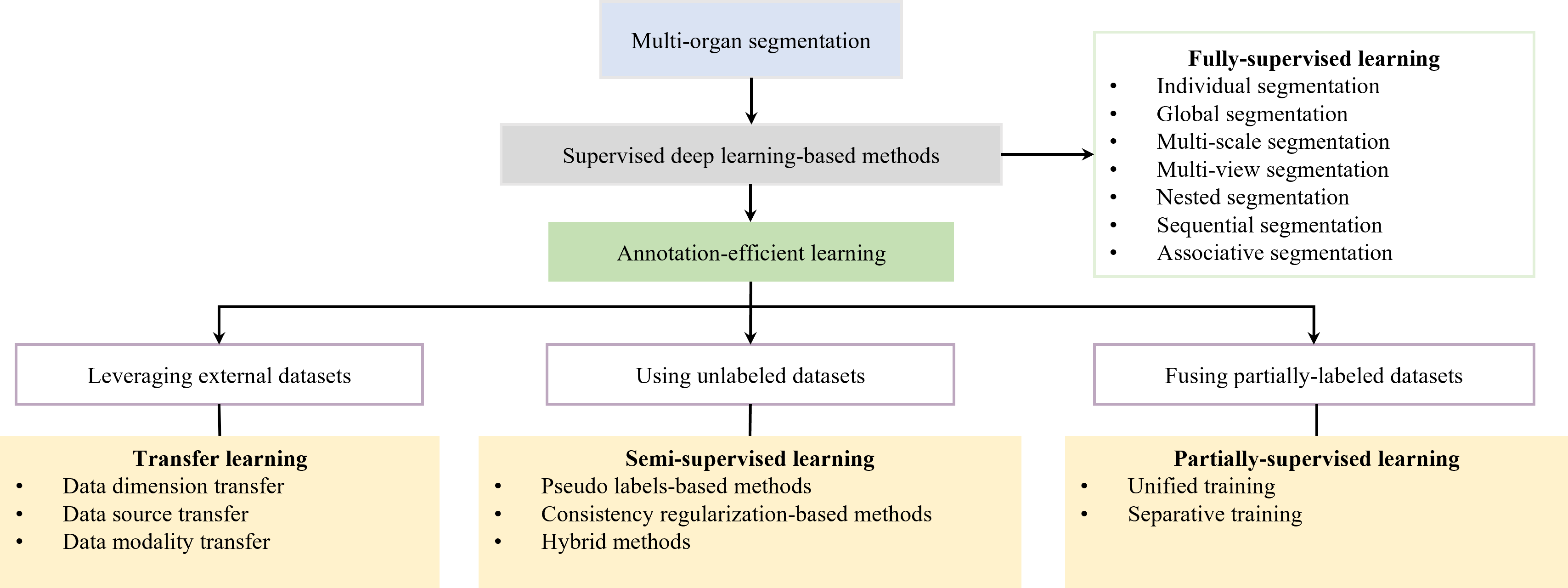
It should be noted that, on the definition of multi-organ, except for the OARs of radiotherapy, we also encompass organs & tumors or multiple tissues, in a word, we adopted segment multiple anatomical structures simultaneously as the task of multi-organ segmentation in this paper. For the sake of grouping the overarching ideas, we skip the illustration of particular challenges for different regions, such as head and neck [34, 35] or abdomen [36, 37], but explore the general problem of multi-organ segmentation under deep learning from the perspective of solving annotation scarcity. The rest of the paper is structured as follows. Section 2 introduces the general modes and challenges of multi-organ segmentation, and Section 3 describes a detailed review of transfer learning (Section 3.1), semi-supervised learning (Section 3.2), and partially supervised learning (Section 3.3). In Section 4, we discuss future trends of the three learning paradigms, and we conclude the whole paper in Section 5. And the organization of the whole review is shown in Figure 1.
2 Overview of multi-organ segmentation & problem analysis
The segmentation of multiple organs is of great clinical significance, providing quantitative information for the diagnosis and classification of diseases [38, 39, 40], and is an indispensable part of radiotherapy. Currently, deep learning-based image segmentation methods are commonly classified into convolutional neural networks (CNN) [17], fully convolutional networks (FCN) [41], and generative networks (GAN) [42] according to the structure of the network [23]. And recently, Transformer [43], which has been widely used in natural images, has also been introduced into medical organ segmentation [44, 45]. All these methods have achieved good results in the field of multi-organ segmentation. Among them, U-Net [46] and its variant structures [14, 15, 16, 17, 47], the classic fully convolutional networks, have become the prevailing method for the multi-organ segmentation of medical images owing to their powerful capabilities. Alternatively, if classified from the training procedure, apart from the basic end-to-end segmentation process, the two-stage approaches based on localization-segmentation [48, 49, 50, 51] and the cascade optimization [52, 53, 54, 55] approaches are also common in multi-organ segmentation for the enhancement of delineation accuracy.
Some approaches simplify multi-organ segmentation to multiple independent single-organ segmentation tasks [22, 48, 56, 57], which has been experimentally verified to be effective for localizing and segmenting the multi-organ [58] and some challenging organs, such as the pancreas [33]. Yet, such approaches ignore the inter-organ spatial correlation, therefore, how to make full use of such contextual information, i.e., multi-organ anatomical prior information, is an inescapable problem in multi-organ segmentation [59]. Following the definition of the categories of segmentation methods in the review by Cerrolaza et al. [12], we classify the common deep learning-based multi-organ segmentation methods using context information into six different categories similarly. And it is worth mentioning that the specific scope of our survey differs from the review of Cerrolaza et al. [12] that focuses on traditional machine learning methods.
Global segmentation can be achieved by directly inputting medical images that contain multiple targets into the model for simultaneous segmentation [60, 61, 62]. The whole-volume input preserves complete intra- and inter-organ anatomical information, which can assist in localization and segmentation [63]. However, for the full-volume input, it is difficult to extract the full inter-organ contextual information due to the limited receptive field of the convolutional kernel. Some sub-modules are proposed to exploit the structural contextual information, such as dilated convolution, pyramidal architecture, etc. [64, 65, 66, 67] and other ideas are to make use of spatial attention mechanisms [37, 53, 68, 69, 70]. The advantages of global segmentation are the simplicity of the training process and the fact that the global model can impose strong anatomical constraints for the segmentation of organs suffering from data corruption (missing, artifacts, etc.). Nevertheless, limited by the computational capability, researchers usually crop the image in preprocessing stage based on statistical and anatomical priors [71, 72, 73, 74] or coarse segmentation localization so as to further improve the foreground proportion. It is crucial for global segmentation to balance the computational consumption and the resolution-constraint segmentation accuracy.
Multi-scale segmentation improves segmentation performance by incorporating information or predictions under different resolutions [75, 76, 77, 78]. Multi-scale exploration is similar to the perception of human vision, utilizing diverse detailed information of different resolutions. At low resolution, we can easily acquire the overall inter-organ relationships, while the specific details of each organ are better examined at high resolution. Existing multi-scale studies have been conducted by fusing multi-resolution inputs [79] or multi-level feature information [80] to better segment organs of highly varying sizes simultaneously. Moreover, a deeply-supervised approach [81] can also optimize the segmentation by the combination of multi-scale outputs [82].
Multi-view segmentation enables more accurate spatial localization by using multi-view data to impose anatomical information constraints, which can effectively alleviate the problems of occlusion and confusion [83, 84, 85]. Multi-view segmentation can model spatial information with less computational consumption by constructing multi-view attention modules with inputs from transverse, coronal, and sagittal planes or more [86, 87]. Moreover, different views enable spatial-anisotropy modeling to mitigate the effects of various resolutions of different directions and collaboratively utilize high-resolution information within slices and inter-slice contextual information [84]. However, this method draws the disadvantage of training multiple separate models of different views.
Nested segmentation is a hierarchical segmentation approach with models training for both global and local organs respectively [88, 89, 21, 90]. Global organ segmentation network can impose topological constraints on anatomical context information for multi-organ segmentation, and the networks for sub-organs can further refine segmentation results. The nested segmentation allows coarse-to-fine segmentation and interaction of global and local information, where the construction of hierarchical objects can be built on spatial or physiological connections. Besides the spatial relationship, joint physiological associations can be helpful to enhance the segmentation accuracy of lesions and affected organs [62]. However, in nested segmentation, the training complexity and time increase significantly with the number of hierarchical layers.
Sequential segmentation is a way of segmenting organs from easy to hard, which can help boost the prediction of hard-to-segment organs and maximize overall segmentation accuracy[91]. In clinical practice, radiologists often use easily distinguishable structures (e.g., bones) as a reference to assist in outlining difficult targets (e.g., soft tissues of the brain). Similarly, the order of organ processing in sequential segmentation is also often determined based on difficulty, and such processing benefits from the fixed spatial prior [91, 21]. For example, combining large organ prediction in the sequential segmentation can effectively guide the small adjacent organ delineation, such as feeding with the segmentation of kidneys can help contour the adrenal gland with the help of cascade training [91]. In this case, sequential segmentation concludes locations for the subsequential task which also does good for organ-specific tumor identification, but the effect heavily depends on the accuracy of the preceding sequence with long training and inference time.
Associative segmentation delineates different organs individually but not independently by constructing bridges between models to provide anatomical interaction for simultaneous segmentation of multi-organ [92, 20]. For example, plenty of symmetric structures in the human body (kidney, optic nerve, etc. [88, 93]) can be optimally segmented by shape similarity and spatial symmetry constraint, and the lesion region can also be identified by disparity. Furthermore, an association of asymmetric organs is equally effective for better segmentation performance, which was experimental confirmed in the segmentation of highly variable and non-directly connected organs such as the pancreas and spleen [94]. Associative segmentation can add flexible inter-organ constraints to the models of each organ, allowing for higher segmentation accuracy. However, multiple coupled segmentation networks pose challenges to models’ convergence and training.
Although the above common multi-organ segmentation methods effectively leverage contextual information, there are still two issues that deserve improvement. (1) Low segmentation accuracy of tiny or low-contrast organs. Segmentation of such organs is very challenging, caused by the large variation in organ size and intensity distribution and class imbalance [20, 95]. Apart from the two-stage approach mentioned above [96, 97, 98], the additional loss function is also a tool to reduce the class imbalance, but the gain is low when dealing with too many organs [19, 99, 100]. Alternatively, organ-specific preprocessing is beneficial for problem solving [101, 51], such as intensity regularization [102] and bias correction [103], but this requires additional prior information and is not generalizable across organs. (2) Large computational resource occupation. Some existing methods sacrifice training efficiency and network simplicity for higher accuracy, making the hardware requirements (e.g., memory, computational speed) unsuitable for realistic clinical practice [104, 105, 106].
In general, extended from earlier single-organ segmentation methods, multi-organ segmentation methods have gradually combined the robustness of global multi-organ modeling with the flexibility and versatility of individual segmentation strategies to balance accuracy and computational efficiency. By incorporating context information, models are able to cope with the effects of overlap, gaps, or topological deformation, thus achieving a more precise representation of complex human anatomy. In recent years, deeper networks have been proposed and have shown stronger feature extraction capabilities. However, due to the limited quality, size, and consistency of the publicly available datasets, network overfitting, and over-parameterization have intensified resulting in insufficient generalization more so than before [107, 108, 109]. This makes annotation-efficient algorithms a key to breaking the data scarcity bottleneck and thus enabling performance improvement in the future.
3 Learning paradigms under annotation scarcity
3.1 Leveraging external annotated datasets - Transfer learning
Rarely do we access enough data and labels for the target domain with respect to multi-organ segmentation, in which case, we can segment target datasets via transfer learning. Typical transfer learning involves pre-training a model with a large external annotated dataset and then fine-tuning the model on the target dataset. The basic concept of this method comes from the fact that the same organs from various datasets may have similar anatomical morphology and surrounding structures in medical image segmentation. Such learnable knowledge can be transferred to the segmentation of the target domain data, or serve as auxiliary information contributing to performance enhancement. Inevitably, medical images from different datasets involve distribution gaps across various modalities, scanners, protocols, and clinical sites, etc., degrading the performance. How to mitigate the impact of such domain shift is an inherent problem of transfer learning. This section elaborates on the existing studies in terms of different domain adaption modes, including dimensions, sources, and modalities. We review and comparatively present the representative work on multi-organ segmentation using transfer learning in Table 1.
| Reference | Segmentation mode | Site | Domain adaption mode | Transfer technique |
|---|---|---|---|---|
| [56] | Individual | Abdomen | 2D 3D | Axis transform; pseudo-label generation |
| [110] | Multi-view | Liver and tumor | 2D 3D | Anisotropic hybrid network |
| [111] | Multi-view | Heart | 2D 3D | Pseudo-label generation |
| [112] | Global | Chest | Public clinical | Adversarial training of shared segmentor with discriminator on predicted masks |
| [113] | Multi-view | Heart | Multi-devices | Slices generation by neural style transfer |
| [114] | Global | Whole body organs | Public clinical | Filtered back projection-driven domain adaptation |
| [94] | Global | Abdomen | Clinical public | Shared knowledge transfer of fixed spatial location by solving puzzle task; resolution normalization by super-resolution network |
| [115] | Global | Abdomen | Public clinical | Human-in-the-loop |
| [116] | Global | Chest | CT X-ray | Task-driven generative network |
| [117] | Global | Abdomen | CT MR | Translation by CycleGAN |
| [118] | Global | Heart | BSSFP LGE | Translation by CycleGAN |
| [119] | Global | Abdomen | CT MR | Style-compensation generation; statics-guided feature adaption |
| [120] | Global | Heart, Abdomen | CT MR | Appearance transformation and representation transfer by GAN |
| [121] | Global | Abdomen | CT T1w T2w | Generation by VAE separating style and content |
| [122] | Global | Heart, Abdomen, Brain | CT MR | Learning domain-invariant and domain-specific components |
| [123] | Global | Abdomen | CT MR | Learning shared knowledge by vary structures in Figure 2 |
| [124] | Global | Heart | CT MR | A shared decoder to produce consistent high-level semantic embeddings |
| [125] | Global | Heart, Abdomen | CT MR | A single shared network with attention module to learn structured semantic consistency |
3.1.1 Dimension transfer
Since the annotation of 2D data is easier than that of 3D data, using 2D annotated data for the segmentation of 3D data is an effective way to reduce the annotation workload. Yu et al. [56] proposed a recurrent saliency transformation network, which transfers the third dimension of the image into a temporal axis to generate 3D pseudo-labels from 2D prediction, and converts the predicted probabilities into spatial weights with the aim of improving the results of medium and small organs via multi-stage cascade segmentation. However, this method can hardly capture 3D context information. Meanwhile, in dimension transfer, the intra-slice resolution is much higher than the inter-slice resolution, in which the direct employment of 3D convolutions may lead to distorted extraction of image features. To tackle such issues, Liu et al. [110] proposed an anisotropic hybrid network to migrate convolutional features learned from 2D images to anisotropic 3D images segmentation. The network achieved advanced results with its powerful extraction of intra-slice information and effective modeling of inter-slice information. Additionally, Zheng et al. [111] proposed to select the most representative slices for annotation and performed a 3D segmentation with these few annotation slices. This selection strategy used 20% of labeled slices to obtain results comparable to those obtained with 100% of labeled slices. In this work, they used a multi-view model to integrate contextual information and the pseudo label to implement dimension transfer.
3.1.2 Source transfer
Data from different sources often differ in data distribution due to imaging parameters, imaging devices, etc. With a view to applying the existing research achievements based on public datasets to clinical datasets, we could pre-train the model on public datasets, then transfer it to the segmentation task on new clinical private data. Dong et al. [112] argue that the prediction masks have domain-invariant anatomical similarity, and therefore implemented adversarial training on the prediction masks by discriminator to segment the chest organs for cardio-thoracic ratio estimation. Apart from the adversarial-based approach, Ma et al. [113] proposed a neural style transfer approach that reduces the data inconsistency between sources to minimize the variation in image attributes (including brightness, contrast, texture, etc.) and integrates global information using atrous convolution and pyramidal pooling. Huang et al. [114] enforced FBP (Filtered Back Projection)-driven domain adaptation techniques to minimize the gap across different CT image datasets, and fused the contextual information through global organs’ registration. Fu et al. [94] employed the relatively fixed spatial location between organs as potentially transferable shared knowledge for domain adaptation of multi-center data. It learns spatial relationships by solving a puzzle task and normalizes the spatial resolution of different sources through a super-resolution network, yielding an average of 19.6% improvement in dice similarity coefficient (DSC) on five publicly target dataset via transfer learning from a clinical labeled dataset.
Besides, human-in-the-loop is an important idea to facilitate the clinical usage of computer-aided technology, where rapid transfer of AI models from one dataset to another can be supplemented with human intervention. Ma et al. [115] proposed a novel and general human-in-the-loop scheme to transfer a model trained with small-scale labeled data to a multi-organ segmentation task of large-scale unannotated CT images, which not only improves the segmentation accuracy but also reduces the manual annotation cost from 13.87 minutes to 1.51 minutes per sample, demonstrating its potential for clinical application.

3.1.3 Modality transfer
Medical images from diverse modalities have different characteristics and complementary information. The multimodal medical images and their cross-modality information can not only support radiologists in making more accurate diagnoses but have also proven effective in automatic AI segmentation [126, 127, 128]. Cross-modality knowledge transfer is the major emphasis of transfer learning for medical images, and it’s essential to implement domain adaption by learning latent shared representations or translating images from one modality to another. The presence or absence of target domain annotation often calls for different research ideas.
Without target domain annotations: When the target domain is unlabeled, unsupervised learning is performed on the target modality. In this case, segmentation of the target domain often relies on segmentation networks trained using source domain annotations [129]. Generative adversarial network (GAN) [130] and cycle-consistent generative adversarial network (CycleGAN) [131], trained in an adversarial mode, are the commonly used methods to address domain shifts across modalities with generators and discriminators. By encouraging the generators to produce synthetic images that are sufficient to confuse the discriminators, the generators can translate images from one domain to another. Zhang et al. [116] used CT images to synthesize X-Ray like digitally reconstructed radiographs through a task-driven generative network, and then trained an X-Ray depth decomposition model based on these images [132], which was transferred to real X-ray images for multi-organ segmentation. Jiang et al. [117] proposed cross-modality educed distillation (CMEDL) based on a teacher-student network, where CycleGAN was used to achieve MRI-to-CT translation, and conducted knowledge distillation on CT and translated MRI images. Similarly, Chen et al. [118] used CycleGAN for translation between MR and CT and used an encoder shared with the generator to prevent encoder overfitting in CT heart segmentation.
However, it is difficult to achieve a satisfactory translation with a simple whole-image reconstruction, due to the loss of valuable detail information of target modality. Therefore, certain adaptation techniques can improve the translation quality thus support better segmentation performance of generation-based transfer learning. Hong et al. [119] proposed feature map statistics-guided model adaptation combined with entropy minimization to accomplish knowledge transfer and reliable segmentation, and implemented style compensation using CycleGAN to significantly improve the segmentation accuracy of the left kidney and spleen, and the effectiveness was validated experimentally for domain adaptation from both CT to MR and its inverse. Wang et al. [120] explored collaborative appearance and semantic adaption with a characterization transfer module to alleviate the appearance divergence of medical lesions across domains and a representation transfer module to diminish the domain-wise distribution gap of underlying semantic knowledge. Alternatively, the disentanglement approach can extract domain-invariant content and domain-specific styles making the generation more robust and reducing catastrophic forgetting and model collapse [133, 134, 135]. Jiang et al. [121] separated image style and content by variation auto-encoder [136] (VAE), which converts image style to latent style code and generates multi-modal images for the content of the input image. Xie et al. [122] learnt domain-invariant and domain-specific anatomical components of images by disentanglement learning, and enhance the target domain segmentation performance by adversarial learning and pseudo labeling in self-training to implicitly achieve feature alignment in anatomical space.
With target domain annotations: When the target domain is annotated, data from other modalities often serve as supplementary information to improve the segmentation results [123, 128]. Unlike unsupervised domain adaptation, domain adaptation techniques are more focused on learning shared knowledge for meaningful information transfer when both datasets are labeled. Valindria et al. [123] explored the performance impact of 4 different encoder-decoder settings on extracting shared latent representations in an abdominal multi-organ segmentation scenario, as shown in Figure. 2. The experimental results show that dual streams of Fig.2 (d) outperform the other structures, besides, cross-modal information can improve the segmentation performance of mutable structures such as the spleen. Dou et al. [124] trained a heart segmentation network consisting of two domain-specific encoders and a shared decoder, enforcing the decoder to produce similar high-level semantic embeddings for both domain images by adversarial training. Yang et al. [125], on the other hand, explored a single shared network to integrate multi-modality information, experimenting with a single transformer and a well-designed external attention module to learn structured semantic consistency (i.e., semantic category representations and their correlations). And the method achieved competitive performance even with extremely limited labeled samples.
3.2 Using unannotated datasets – Semi-supervised learning
In medical images, unlabeled data are often more accessible than labeled data. When labeled data are scarce, the inclusion of unlabeled data has been shown to help improve model generalization and the segmentation performance of medical images [31, 137, 138]. Semi-supervised learning is trained using only a small set of labeled data and large-scale unlabeled data, where the key to the investigation is to make sufficient utilization of unlabeled data [26]. In this section, observing the existing mainstream studies, we categorize the approach and show the typical workflows of semi-supervised learning in Fig.3, including the pseudo label-based approach, consistency regularization approach, and hybrid approach.

3.2.1 Pseudo label-based methods
The semi-supervised method using pseudo labels was first proposed by Lee et al. [139] and has been developed as a classical algorithm for semi-supervised for its simple idea and effective performance. As shown in Figure 3 (a) the model trained by labeled data can be used to predict the pseudo label of unlabeled data, which is fed together with labeled data to co-train the model in an iterative manner, and make the model achieves promising results on validation set outperforming the training using only labeled data. The such a pseudo-labeling approach has a high universality, but its effectiveness is restricted by the quality of the pseudo labels generated from the unlabeled data. And the related works mentioned below differ in the way of pseudo label generation and the treatment of noisy labels and are listed in Table 2.
| Reference | Segmentation mode | Site | The generation of pseudo labels | Handling of noisy pseudo label |
|---|---|---|---|---|
| [140] | Multi-view | Abdomen | Ensemble segmentation model of three views & Majority voting | / |
| [141] | Global | Brain | Average of multi-transformed segmentation results | / |
| [142] | Global | Heart | Two-stream mutual attention network | Filtered by the difficulty level obtained from hierarchical distillation, including data distillation and model distillation |
| [143] | Global | Pelvis | Single adversarial segmentation model | Masked by the confident map predicted with discriminator |
| [144] | Multi-view | Abdomen | Multi-view co-training network | Uncertainty-weighted label fusion |
| [33] | Global | Abdomen | Noisy student network | Update pseudo labels with training iterations |
Zhou et al. [140] proposed an iterative model for the segmentation of 16 structures in contrast-enhanced abdominal CT, and the pseudo labels are generated by maximum voting from models of axial, coronal, and sagittal views, which eventually brings a 4% improvement in DSC due to the usage of unlabeled data. In the work by Zhao et al. [141], the pseudo label is derived by data distillation, which averages the prediction masks from random augmentations of the same image to produce the final pseudo label, and the proposed method also yield a 1.6% DSC improvement compared to training without unlabeled image.
Nevertheless, the above methods have obvious limitations, mainly in treating the pseudo-labeled and expert-labeled annotation equally, without considering that false segmentation in pseudo label may bring noise into the model training and thus degrade the model performance [109, 145, 146]. To explore such an issue, an important approach is to estimate the reliability of pseudo labels. Min et al. [142] proposed a dual-stream network and the two independent networks can discriminate the difficulty of each pixel by the consistency of prediction for the same input of K transformations and two models, in which case the hard and easy pixels with inconsistent classes will be shielded from the overall loss calculation. The network generates pseudo labels by hierarchical distillation including both model distillation and data distillation and demonstrates the noise-resilient of the suggested method on a full-heart segmentation dataset under varying noise levels of labeled images. Instead, Nie et al. [143] proposed a region-attention-based strategy based on the confidence map gained from an adversarial confident network, then used confident region obtained by setting threshold as a mask to select reliable prediction of unlabeled data for pseudo label-based supervision, this method demonstrated their superiority over fully supervised methods on multiple datasets. On the other hand, Xia et al. [144] proposed a co-training framework to predict segmentation masks by multi-view parallel network predictions. During training, multiple inference results with random dropout [147] can be used to estimate uncertainty served as sample weights, alleviating the effect of noisy pixel labels and achieving significant performance improvements on multi-organ datasets.
Beyond reliability estimation, many works have likewise explored the utilization of noisy pseudo labels. Ma et al. [33] established a new semi-supervised segmentation benchmark for the abdominal multi-organ dataset using the Noisy Student framework [148]. The noisy student can self-correct the pseudo labels in the training phase, and the student model includes more data augmentation and noise and its prediction will play the role of the new pseudo labels to iterate and update the student model itself. Different from basic pseudo labels training, not only the model but also the pseudo labels are continuously optimizing in the multiple iterations. Such a simple idea demonstrated its powerful generality and achieved efficient performance, with an experimental illustration that errors in difficult and diseased organ segmentation can be gradually corrected using an increasing amount of unlabeled data.
3.2.2 Consistency regularization-based methods
Without pseudo-labeling, the unlabeled data in semi-supervised learning rely on setting up an unsupervised loss together with the supervised loss for labeled data to guide the backpropagation. The most prevalent way of unlabeled loss construction is often based on the idea of consistent regularization, i.e., the model is robust to small perturbations and the outputs may stay consistent [149, 150]. Inspired by the classical semi-supervised segmentation frameworks Temporal Ensembling [151], Mean Teacher [152], and UDA [153], many semi-supervised methods have also been extended to multi-organ segmentation. Table 3 lists some representative consistency regularization-based semi-supervised multi-organ segmentation methods which differ in perturbations and consistency construction.
| Reference | Segmentation mode | Site | Perturbations | Consistency type |
|---|---|---|---|---|
| [154] | Global | Chest | Elastic deformations | Elastic transformation consistency |
| [155] | Global | Heart | Mixup transformations | Interpolation consistency |
| [156] | Multi-scale | Nasopharynx | Pyramid network architecture | Multi-scale consistency |
| [157] | Global | Optic | Tasks of segmentation and regression | Task consistency of regional and marginal consistency |
| [158] | Global | Heart | Typical input-level transformation: random crop, flip, rotation | Feature consistency on global and local mutual information |
| [159] | Global | Nasopharynx | Teacher-student network with different inputs | Attention map consistency |
Transform consistency is a common consistency regularization [160, 161], and mostly performs in a regular workflow like that shown in Figure 3 in b. This inspire Bortsova et al. [154], to use elastic transform consistency for segmentation, which constructs consistency of the final outputs from two branches to help learn transformation invariant representations and further boost the utilization of unlabeled data. And this method effectively improves segmentation results under a small number of supervised data, but yield little improvement compared to fully-supervised methods when the number is large. Similarly, Basak et al. [155] conducted effective semi-supervised segmentation by interpolation transform consistency using Mixup [162] for data transformation, which extends the training set by mixing diverse images.
More various consistency constructions have been proposed too, such Luo et al. [156] suggested uncertainty rectified pyramid consistency, a kind of multi-scale consistency, which is formulated by the MSE loss from the pyramid outputs to their average. The pyramid prediction network integrates contextual information at different scales and the predictions are rectified by the uncertainty map produced by the predictions’ entropy. Also, Meng et al. [157] proposed a bi-consistency regularization with regional and marginal consistency to build a shape-aware generalized multi-task framework improving segmentation accuracy for both semi-supervised and weakly-supervised [163] segmentation.
Moreover, rather than ensuring consistency at the model, task, or output level as mentioned above, many studies are now gradually focusing on feature information utilization and adding regularization constraints at the feature level. As Peng et al. [158] constrained consistency on feature-level mutual information to learn a geometrically transform invariant image representation with the help of global mutual information, and then promote spatial consistency of the feature map by local mutual information to provide smoother segmentation results. Hu et al. [159] then introduced attention map consistency in an uncertainty-aware teacher-student model between the feature of the whole image or the ROI-cropped image inputs, which helps the model better locate the target and reduce the false positives of segmentation.
3.2.3 Hybrid methods
| Reference | Segmentation mode | Site | Generation & Utilization of pseudo labels | Supervision way | Constraint |
|---|---|---|---|---|---|
| [164] | Global | Heart | Dual parallel networks of Transformer and CNN | Output-level cross pseudo supervised | Consistency between two networks |
| [165] | Global | Heart | Shared-encoder and multi-decoders network; uncertainty guidance | Output-level consistency loss | Mutual consistency of multiple decoders |
| [166] | Global | Heart | Shape-agnostic Network & shape-aware Network; uncertainty guidance | Output-level cross pseudo supervised | Shape awareness and local context constraints |
| [167] | Global | Heart | Teacher-student network with single encoder and triple decoders; Tiple-uncertainty guidance | Output-level consistency of Mean Teacher & feature-level contrastive loss | Contrastive learning & consistency constraint |
| [168] | Global | Abdomen | Modality-specific networks | Output-level consistency & feature-level contrastive loss | Cross-modality consistency constraint |
| [169] | Global | Heart | Single U-Net segmentation network | Output-level smoothness loss & feature-level class-separation loss | Class and smoothness constraint |
| [170] | Associative | Knee | Two parallel segmentation models | Uncertainty sampling supervision | Distribution-based sampling constraint |
| [171] | Global | Heart | Uncertainty-aware mean teacher; Uncertainty-based label assignment | Output-level consistency | Focal loss for multi-organ |
Naturally, the aforementioned methods are not antagonistic but can be combined organically to facilize the performance, while more additional constraints can be included in the semi-supervised learning, which is all regarded as hybrid methods as follow. Table 4 lists some hybrid semi-supervised multi-organ segmentation methods.
It’s easy to combine the idea of the pseudo labels and consistency regularization for the consistency of different pseudo labels should be ensured under perturbations. Thus, it comes to the idea that the pseudo labels can be exchanged between two networks to realize cross supervision, that is cross pseudo supervised (CPS) [172], the structure of which is shown in Figure 3 (b). Followed by CPS, Luo et al. [164] combined the hot-spot Transformer [173] by forming a two-branch framework with CNN to ally the issue of the limited perceptual field of CNN by the powerful global information extraction of the Transformer. Wu et al. [165] proposed MC-Net+ developing a mutual consistency to encourage an invariant prediction result of slightly different multiple decoders in easily misclassified regions (e.g., adhesive edges or thin branches), in which the statistical discrepancy of multiple decoders’ outputs is used to estimate the model’s epistemic uncertainty denoting the segmentation difficulty of regions. And the mutual consistency constrain between one decoder’s probability output and other decoders’ soft pseudo labels.
Except for consistency constraints, other kinds of constraints also do good to improve the pseudo labels’ quality, like the work by Liu et al. [166] using shape awareness and local context constraints. Furthermore, the uncertainty generated by Monte-Carlo dropout (MC Dropout) [147] is included to guide better utilization of the pseudo labels. Besides, Wang et al. [167] introduced contrastive learning on the feature level beyond their proposed task-level consistency to explore a more powerful representation. By adding two auxiliary tasks, i.e., a reconstruction task for capturing semantic information and a signed distance field (SDF) [174] regression task, it can impose shape constraint to the segmentation, and these three tasks’ mutual promotion effect is explored under mean teacher architecture and guided by triple-uncertainty via uncertainty weighted integration strategy to learn more reliable knowledge from unlabeled data. Another use of contrastive learning, Chen et al. [168] exploited cross-modal consistency to achieve a modality-collaborative semi-supervised segmentation, which leverage the modality-specific knowledge learned from unpaired CT and MRI, and the contrastive similarity is used to regularize the feature extraction. Also constraint on the feature level, Wu et al. [169] proposed SS-Net to realize effective semi-supervised performance through pixel-level smoothness constraints under adversarial perturbations and inter-class separation based on prototype strategy.
Lin et al. [170], then, focus on the class imbalance problem of multiple organs, which has a more prominent impact on semi-supervision. They set the category-aware loss function and oversampled cropping region [72] based on the number and probability distribution of labels, and use uncertainty sampling supervision to augment the supervision of low-confidence categories and optimize segmentation. Chen et al. [171] introduced a dense focal loss [175] function on deep co-training to solve the class imbalance problem and avoid erroneous optimization by an uncertainty-based label assignment policy.
3.3 Fusing partially annotated datasets - Partially-supervised learning
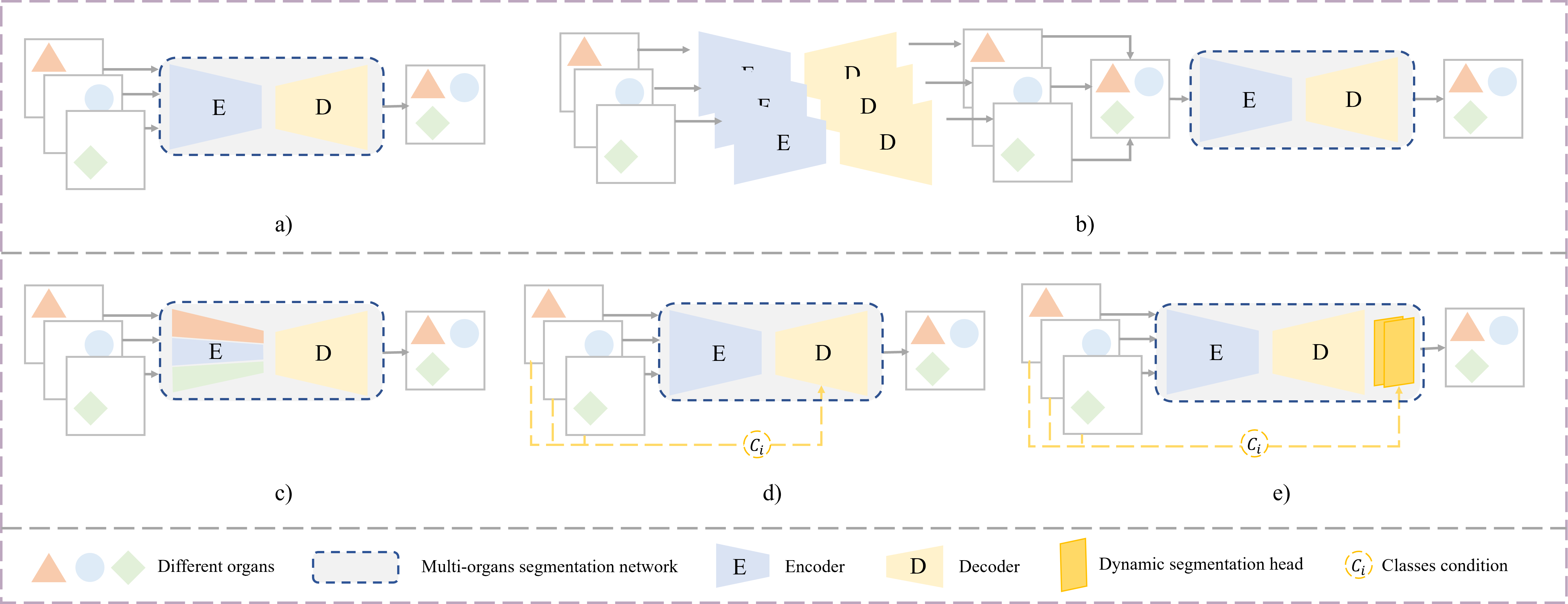
Due to the enormous labor required for multi-organ annotations, partially annotated multi-organ datasets are more readily available than fully annotated multi-organ datasets. For example, KiTS [176] and LiTS [177] datasets only provided labels for a single organ and tumors in the abdomen, while all other organs are marked as background. Learning from multiple datasets containing single or several organs’ annotations together can integrate multiple partial annotations to provide full annotation for desired multiple organs, and such a learning approach is called partially-supervised learning. Partially supervised learning can also be regarded as a kind of the transfer learning or semi-supervised learning, where learning from multiple partially-labeled datasets involves knowledge transfer learning among different datasets, while unlabeled anatomical structures in partially-labeled data may serve as unlabeled data for semi-supervised learning. Partially supervised learning aims to generate a framework universal to multi-organ segmentation from partially-labeled data. The organs of interest may be marked as background in partially-labeled datasets, which may mislead and confuse the multi-organ segmentation. For this reason, how to make better utilization of the partially labeled information without confusing the overall multi-organ segmentation is a key issue for partial supervision. Based on the way to incorporate the partially-labeled datasets in training, we spilt the partial learning methods into unified training and separate training, which are distinguished by the frameworks in Figure 4. The upper row shows the unified training in which the data of different partially-annotated datasets flow through the same multi-organ network while in the separative training showed in the second-row the data flow through the multi-organ network differentially. In the former case, different datasets jointly update the whole network parameters, while in the latter case the optimization of the parameters by different datasets is local or separated. Table 5 lists the partially-supervised learning methods in multi-organ segmentation.
3.3.1 Unified training
To obtain a multi-organ segmentation network, the partially-labeled datasets can be fed into a unified model in which the multiple partially-labeled datasets share the same training architecture. In such unified training, the training with each dataset will benefit from same designed partial supervised loss. Shi et al. [99] exploited the union of partially labeled datasets through two carefully-designed loss functions, where a marginal loss mines the true background from partially-labeled datasets through the boundary probabilities, and an exclusion loss was designed to improve inter-organ discrimination considering the fact that there is no overlap between organs. Both of these loss functions showed apparent improvement to the segmentation performance without introducing any additional computation. With the aim of further utilizing the contextual information, Mao et al.[178] extended these loss functions and imported attention mechanism to collect multi-scale information for fully automatic multi-organ segmentation, which better strikes a trade-off between segmentation accuracy and inference time. Fang et al. [179] proposed a unified U-shape pyramid structure network that aggregates pyramid input and features where an adaptive weighting layer is designed to integrate the deeply-supervised outputs in an automatic way. This method allows a single model adapt to changes in proportion of known labels via a target adaptive loss.
Additionally, using pseudo labels is also a feasible manner for unified training. Zhou et al. [27] proposed PaNN using very little fully-labeled data for training to obtain a prior-aware network, and then optimized it by using the partially-labeled data by the supervised loss computed on labeled organs and the pseudo label-based loss on unlabeled organs. PaNN regularizes the organ sizes by explicitly incorporating anatomical empirical distributions as the penalty of soft constraint with statistical domain-specific knowledge. Huang et al. [180] deployed two coupled networks for few-organ datasets to teach each other and proposed a co-train weight-averaged model to obtain a unified multi-organ segmentation network. The multiple organs’ pseudo labels generated by two coupled networks can be fused to form a fully multi-organ pseudo labels, which can further refine the unified segmentation network. In addition, this work proposes a novel region mask to selectively impose the consistent constraint on the unlabeled organ regions during collaborative teaching, thus delivering further performance improvements.
In addition, Zhou et al. [181] considered that the partially-labeled dataset may not be publicly available in real scenarios, so they unleash the partially annotated and sequentially constructed datasets through incremental learning (IL) without access to previous annotations in each IL stage. Pre-trained on a public dataset of K organs, the model is trained with another single-organ dataset to obtain a segmentation of K+1 organs via IL, and apply a teacher-student framework to transfer knowledge extracted by background remodeling. However, the unified training may be affected by the distribution gap of different datasets resulting a mutual interference.
| Reference | Segmentation mode | Multi-organ dataset | Partially labeled datasets | Integration of partial labelling | Framework |
|---|---|---|---|---|---|
| [27] | Global | BTCV [182] | MSD Spleen [183], NIH [184], LiTS [177] | A priori soft constraints on size | a |
| [99] | Global | BTCV [182] | MSD Spleen [183], KiTS [176] | Marginal and excluded losses | a |
| [178] | Multi-scale | PDDCA[185] | TCIA-CT-scan [186, 187] | Attentional mechanisms integrate multi-scale information | a |
| [179] | Multi-scale | BTCV [182] | LiTS [177], KiTS [176] , MSD Spleen [183] | Adaptive fusion & adaptive loss | a |
| [180] | Nested | LiTS [177], KiTS [176] , NIH [184], MOBA [75] | Pseudo-label generation & Co-training and weight- averaged model | b | |
| [181] | Global | MOBA [75] | MSD Spleen [183], LiTS [177], NIH [184] | Knowledge distillation | a |
| [188] | Associative | LiTS [177], KiTS [176] , MSD Spleen [183], BTCV [182] | Organ-individual encoders and shared Decoder | c | |
| [189] | Global | Sliver07 [190], NIH [184], liver & spleen* | Introduction of categories condition | d | |
| [191] | Associative | LITSC*, MSD LT* | Semi-supervision and conditional strategy | d | |
| [192] | Associative | MOTS [192], BTCV [182] | Task-encoding controller | e |
3.3.2 Separative training
However, influenced by the previous background confusion problem, training partially-labeled data indiscriminately with a simple single model is not an optimal way to fuse different datasets.
Thus, Xu et al. [188] explored a multi-encoding U-Net to extract organ-specific features through different several encoding sub-networks, and implement federated multi-organ segmentation by an auxiliary generic decoder. This approach explores the informative and distinctive information for different organs by multiple encoders but balances the resource with a shared decoder. However, such a design may rise the redundancy and memory usage of the network.
Taking into account the need to reduce redundancy, the use of conditional strategies in the union network allows the introduction of category conditions as auxiliary information for the segmentation of different organs to achieve differentiated segmentation. Inspired by conditional generative networks, Dmitriev et al. [189] utilized a conditional approach to gain a joint multi-organ framework trained from partially-labeled datasets. It is simple yet efficient and shows convincing segmentation performance on multi-organ images. Likewise, Zhang et al. [191] regard two datasets with non-overlapping labeling pairs as mutually unlabeled data and use the classic semi-supervised teacher-student framework to learn from a collection of partially-labeled datasets. The backbone is built on the nnU-Net combined a conditioning strategy to include prior class information into the decoder to better utilize relative unlabeled data, i.e., partially-labeled data.
Going further, Zhang et al. [192] proposed a dynamic on-demand network (DoDNet) by the generated controller, where the majority of the network is a shared encoder-decoder architecture with dynamic segmentation head changed with the class of organ. Different from fixing the kernels after training from the previous works, DoDNet generates the kernels in dynamic head adaptively by the task encoding module, enabling segmentation of multiple organs and tumors in a more efficient and flexible manner. However, the method cannot achieve simultaneous segmentation of multiple organs and can only perform one-by-one inference. Currently, there is still room to explore a flexible, efficient, and low-resource segmentation framework with the integration of partially labeled data in partially supervised tasks.
4 Discussion and future prospects
Current deep learning approaches have already served an important role in multi-organ segmentation, which is significant in helping to reduce annotation consumption and alleviate inter-observer annotation discrepancies. In this paper, we systematically demonstrate the multi-organ segmentation framework and explore the latest works with the data scarcity problem as the entry point. The present annotation-efficient algorithms have shown effective clinical practicability in reducing annotation cost in the field of multi-organ segmentation, however, how to further develop more high-accuracy, low-resource and annotation-efficient segmentation algorithms for multi-organ segmentation still need to be explored.
4.1 Transfer learning
Transfer learning aims to make full use of external annotated data to enhance the performance of multi-organ segmentation, and the key lies in solving domain shift between datasets. Transfer learning currently focuses on the migration of homogeneous data across data sources and adaptation between different modalities, while the cross-modality transfer towards functional imaging with greater differences is still lacking for there is no guarantee to capture sufficient features for cross-modality generation with enormous differences in tissue presentations [117]. Nevertheless, the functional imaging presents lesion region better making the transfer learning with functional images essential for precise segmentation of tumors along with OARs and valuable for further exploration. In addition, embracing artificial interaction in transfer learning can help guarantee the accuracy of target domain segmentation, and using transfer learning to provide a pre-segmentation reference for experts can speed up the efficiency of multi-organ labeling [115]. In the future, scientific research results should concern more about real clinical needs, and more effort should be made to translate the research results into a commercially available online segmentation radiotherapy platform with higher real-time and generalization requirements via transfer learning [193, 194]. Also, the active learning-based approach can be included in transfer learning to select representative samples for annotation thus boosting the adaptation quality and speed [111], which can help construct attempts to build a semi-automatic segmentation platform of high industrial value and clinical feasibility.
4.2 Semi-supervised learning
Semi-supervised learning aims to use unlabeled data to help improve the performance of target multi-organ segmentation. Among the common approaches of semi-supervised methods, the pseudo label-based approaches are more general, but require longer training time in multi-stage optimizations and are more sensitive to the quality of pseudo labels; the consistency regularization-based approaches enable flexible and convenient construction of regularization constraints, but the tight coupling constructed through consistency may cause bottlenecks in accumulated bias correction. And the above issues have been explored by some corresponding solutions [159, 195], however, little work has been done to focus on class imbalance in multi-organ segmentation. So how to effectively improve the quality of pseudo labels of tiny organs or, alternatively, to take into account both global and local segmentation from a consistency perspective are directions that can be investigated in the future. In addition, the few-shot learning [196] approach, similar to semi-supervised learning has also been applied in multi-organ segmentation tasks [197], reflecting a more critical demand for more annotation efficiency. Moreover, similar to the enhancement of representation in unsupervised learning, the use of self-supervised paradigm [198, 199], e.g., contrastive learning to learn the differences between different organs, etc. [59, 200] can help networks better extract valid information from unlabeled data and is likewise worth exploring in semi-supervised learning.
4.3 Partially-supervised learning
Most methods in partially supervised learning tend to focus on how to construct an effective multi-organ segmentation network. To obtain such multi-organ segmentation network, either unified or separative training, it is crucial to exclude interference between different partially labeled datasets, but it is more difficult to effectively establish contextual information links between different organs when full annotations are scarce [178]. Partially supervised learning can also be regarded as a kind of semi-supervised learning or transfer learning, where unlabeled anatomical structures in partially-labeled data can be employed may serve as unlabeled data for semi-supervised learning, while learning from multiple partially-labeled datasets involves knowledge transfer learning among different datasets. And it’s inevitable to face the same challenges of these two learning for the partially-supervised learning. Direct use of global pseudo labels can help achieve a contextual constraint, but it is difficult to balance the delineation performance of organs with various sizes [179]. How to introduce anatomical information constraints in a joint unified multi-organ segmentation framework is still worth exploring. Considering the discrepancies between partially labeled datasets, the existing methods give the assumption of basic consistent data distribution and standardize the data through complex preprocessing. As different datasets continue to be introduced and used, how to adaptively learn knowledge from new samples and preserve most of the previously learned knowledge is also a subsequent trend in fusing partially labeled data [181].
5 Conclusion
In this paper, we provide a systematic summary of multi-organ segmentation from fully supervised to annotation-efficient learning paradigms, and unfold the current state of works in transfer learning, semi-supervised learning, and partially supervised learning. Further, we provide a general review and outlook on the problems and shortcomings of these approaches. The current annotation-efficient multi-organ segmentation approaches are under a period of rapid development, with smaller annotation requirements, wider types of applications, and increasing prediction accuracy. Future exploration of more efficient automatic multi-organ segmentation is also expected to reduce the medical burden in areas with insufficient medical resources [201] and benefit cancer patients, which has far-reaching implications.
References
- [1] L. Liu, Anatomy-aided deep learning for medical image segmentation: a review, Phys. Med. Biol. (2021) 23.
- [2] G. Litjens, T. Kooi, B. E. Bejnordi, A. A. A. Setio, F. Ciompi, M. Ghafoorian, J. A. Van Der Laak, B. Van Ginneken, C. I. Sánchez, A survey on deep learning in medical image analysis, Medical image analysis 42 (2017) 60–88.
- [3] M. A. Vyfhuis, N. Onyeuku, T. Diwanji, S. Mossahebi, N. P. Amin, S. N. Badiyan, P. Mohindra, C. B. Simone, Advances in proton therapy in lung cancer, Therapeutic advances in respiratory disease 12 (2018) 1753466618783878.
- [4] J. K. Molitoris, T. Diwanji, J. W. Snider III, S. Mossahebi, S. Samanta, S. N. Badiyan, C. B. Simone, P. Mohindra, et al., Advances in the use of motion management and image guidance in radiation therapy treatment for lung cancer, Journal of thoracic disease 10 (Suppl 21) (2018) S2437.
- [5] Y. Fu, Y. Lei, T. Wang, W. J. Curran, T. Liu, X. Yang, A review of deep learning based methods for medical image multi-organ segmentation, Physica Medica 85 (2021) 107–122.
- [6] M. Ashok, A. Gupta, A systematic review of the techniques for the automatic segmentation of organs-at-risk in thoracic computed tomography images, Archives of Computational Methods in Engineering 28 (4) (2021) 3245–3267.
- [7] R. Baskar, K. A. Lee, R. Yeo, K.-W. Yeoh, Cancer and radiation therapy: current advances and future directions, International journal of medical sciences 9 (3) (2012) 193.
- [8] B. E. Nelms, W. A. Tomé, G. Robinson, J. Wheeler, Variations in the contouring of organs at risk: test case from a patient with oropharyngeal cancer, International Journal of Radiation Oncology* Biology* Physics 82 (1) (2012) 368–378, publisher: Elsevier.
- [9] T. Zhang, Y. Yang, J. Wang, K. Men, X. Wang, L. Deng, N. Bi, Comparison between atlas and convolutional neural network based automatic segmentation of multiple organs at risk in non-small cell lung cancer, Medicine 99 (34), publisher: Wolters Kluwer Health.
- [10] M. Thor, A. Apte, R. Haq, A. Iyer, E. LoCastro, J. O. Deasy, Using auto-segmentation to reduce contouring and dose inconsistency in clinical trials: the simulated impact on RTOG 0617, International Journal of Radiation Oncology* Biology* Physics 109 (5) (2021) 1619–1626, publisher: Elsevier.
- [11] D. Kawahara, M. Tsuneda, S. Ozawa, H. Okamoto, M. Nakamura, T. Nishio, A. Saito, Y. Nagata, Stepwise deep neural network (stepwise-net) for head and neck auto-segmentation on CT images, Computers in Biology and Medicine 143 (2022) 105295, publisher: Elsevier.
-
[12]
J. J. Cerrolaza, M. L. Picazo, L. Humbert, Y. Sato, D. Rueckert, M. A. G.
Ballester, M. G. Linguraru,
Computational
anatomy for multi-organ analysis in medical imaging: A review, Medical
Image Analysis 56 (2019) 44–67.
doi:10.1016/j.media.2019.04.002.
URL https://linkinghub.elsevier.com/retrieve/pii/S1361841518306273 -
[13]
Y. Lei, Y. Fu, T. Wang, R. L. J. Qiu, W. J. Curran, T. Liu, X. Yang,
Deep Learning in Multi-organ
Segmentation, number: arXiv:2001.10619 arXiv:2001.10619 [physics] (Jan.
2020).
URL http://arxiv.org/abs/2001.10619 -
[14]
Ö. Çiçek, A. Abdulkadir, S. S. Lienkamp, T. Brox, O. Ronneberger,
3D U-Net: Learning Dense
Volumetric Segmentation from Sparse Annotation, arXiv:1606.06650
[cs]ArXiv: 1606.06650.
URL http://arxiv.org/abs/1606.06650 -
[15]
T. D. Bui, J. Shin, T. Moon, 3D
Densely Convolutional Networks for Volumetric Segmentation,
arXiv:1709.03199 [cs]ArXiv: 1709.03199.
URL http://arxiv.org/abs/1709.03199 - [16] F. Milletari, N. Navab, S.-A. Ahmadi, V-net: Fully convolutional neural networks for volumetric medical image segmentation, in: 2016 fourth international conference on 3D vision (3DV), IEEE, 2016, pp. 565–571.
-
[17]
Z. Zhou, M. M. R. Siddiquee, N. Tajbakhsh, J. Liang,
UNet++: Redesigning Skip
Connections to Exploit Multiscale Features in Image
Segmentation, arXiv:1912.05074 [cs, eess]ArXiv: 1912.05074.
URL http://arxiv.org/abs/1912.05074 -
[18]
J. Wang, X. Li, Y. Han, J. Qin, L. Wang, Z. Qichao,
Separated Contrastive Learning for
Organ-at-Risk and Gross-Tumor-Volume Segmentation with Limited
Annotation, arXiv:2112.02743 [cs, eess] (Apr. 2022).
URL http://arxiv.org/abs/2112.02743 -
[19]
S. A. Taghanaki, Y. Zheng, S. Kevin Zhou, B. Georgescu, P. Sharma, D. Xu,
D. Comaniciu, G. Hamarneh,
Combo
loss: Handling input and output imbalance in multi-organ segmentation,
Computerized Medical Imaging and Graphics 75 (2019) 24–33.
doi:10.1016/j.compmedimag.2019.04.005.
URL https://linkinghub.elsevier.com/retrieve/pii/S0895611118305688 -
[20]
S. Cros, E. Vorontsov, S. Kadoury,
Managing Class
Imbalance in Multi-Organ CT Segmentation in Head and Neck
Cancer Patients, in: 2021 IEEE 18th International Symposium on
Biomedical Imaging (ISBI), IEEE, Nice, France, 2021, pp. 1360–1364.
doi:10.1109/ISBI48211.2021.9433991.
URL https://ieeexplore.ieee.org/document/9433991/ -
[21]
D. Guo, D. Jin, Z. Zhu, T.-Y. Ho, A. P. Harrison, C.-H. Chao, J. Xiao, L. Lu,
Organ at Risk
Segmentation for Head and Neck Cancer Using Stratified Learning
and Neural Architecture Search, in: 2020 IEEE/CVF Conference on
Computer Vision and Pattern Recognition (CVPR), IEEE, Seattle, WA,
USA, 2020, pp. 4222–4231.
doi:10.1109/CVPR42600.2020.00428.
URL https://ieeexplore.ieee.org/document/9156960/ -
[22]
H. Tang, X. Chen, Y. Liu, Z. Lu, J. You, M. Yang, S. Yao, G. Zhao, Y. Xu,
T. Chen, Y. Liu, X. Xie,
Clinically applicable
deep learning framework for organs at risk delineation in CT images, Nat
Mach Intell 1 (10) (2019) 480–491.
doi:10.1038/s42256-019-0099-z.
URL http://www.nature.com/articles/s42256-019-0099-z -
[23]
M. H. Hesamian, W. Jia, X. He, P. Kennedy,
Deep Learning
Techniques for Medical Image Segmentation: Achievements and
Challenges, J Digit Imaging 32 (4) (2019) 582–596.
doi:10.1007/s10278-019-00227-x.
URL http://link.springer.com/10.1007/s10278-019-00227-x -
[24]
T. Sadad, A. Rehman, A. Hussain, A. A. Abbasi, M. Q. Khan,
A Review on
Multi-organ Cancer Detection Using Advanced Machine Learning
Techniques, CMIR 17 (6) (2021) 686–694.
doi:10.2174/1573405616666201217112521.
URL https://www.eurekaselect.com/189208/article - [25] D. Karimi, S. K. Warfield, A. Gholipour, Transfer learning in medical image segmentation: New insights from analysis of the dynamics of model parameters and learned representations, Artificial Intelligence in Medicine 116 (2021) 102078.
- [26] O. Chapelle, B. Scholkopf, A. Zien, Semi-supervised learning (chapelle, o. et al., eds.; 2006)[book reviews], IEEE Transactions on Neural Networks 20 (3) (2009) 542–542.
-
[27]
Y. Zhou, Z. Li, S. Bai, X. Chen, M. Han, C. Wang, E. Fishman, A. Yuille,
Prior-Aware Neural
Network for Partially-Supervised Multi-Organ Segmentation, in:
2019 IEEE/CVF International Conference on Computer Vision
(ICCV), IEEE, Seoul, Korea (South), 2019, pp. 10671–10680.
doi:10.1109/ICCV.2019.01077.
URL https://ieeexplore.ieee.org/document/9009566/ -
[28]
A. E. Ilesanmi, T. Ilesanmi, O. P. Idowu, D. A. Torigian, J. K. Udupa,
Organ
segmentation from computed tomography images using the 3D convolutional
neural network: a systematic review, Int J Multimed Info Retr 11 (3) (2022)
315–331.
doi:10.1007/s13735-022-00242-9.
URL https://link.springer.com/10.1007/s13735-022-00242-9 -
[29]
Y. Fu, Y. Lei, T. Wang, W. J. Curran, T. Liu, X. Yang,
A
review of deep learning based methods for medical image multi-organ
segmentation, Physica Medica 85 (2021) 107–122.
doi:10.1016/j.ejmp.2021.05.003.
URL https://linkinghub.elsevier.com/retrieve/pii/S1120179721001848 - [30] V. Cheplygina, M. de Bruijne, J. P. Pluim, Not-so-supervised: a survey of semi-supervised, multi-instance, and transfer learning in medical image analysis, Medical image analysis 54 (2019) 280–296.
- [31] M. Zhang, Y. Zhou, J. Zhao, Y. Man, B. Liu, R. Yao, A survey of semi-and weakly supervised semantic segmentation of images, Artificial Intelligence Review 53 (6) (2020) 4259–4288.
-
[32]
N. Tajbakhsh, L. Jeyaseelan, Q. Li, J. N. Chiang, Z. Wu, X. Ding,
Embracing
imperfect datasets: A review of deep learning solutions for medical image
segmentation, Medical Image Analysis 63 (2020) 101693.
doi:10.1016/j.media.2020.101693.
URL https://linkinghub.elsevier.com/retrieve/pii/S136184152030058X -
[33]
J. Ma, Y. Zhang, S. Gu, C. Zhu, C. Ge, Y. Zhang, X. An, C. Wang, Q. Wang,
X. Liu, S. Cao, Q. Zhang, S. Liu, Y. Wang, Y. Li, J. He, X. Yang,
AbdomenCT-1K: Is
Abdominal Organ Segmentation A Solved Problem, IEEE Trans.
Pattern Anal. Mach. Intell. (2021) 1–1doi:10.1109/TPAMI.2021.3100536.
URL https://ieeexplore.ieee.org/document/9497733/ -
[34]
J.-F. Daisne, A. Blumhofer,
Atlas-based
automatic segmentation of head and neck organs at risk and nodal target
volumes: a clinical validation, Radiat Oncol 8 (1) (2013) 154.
doi:10.1186/1748-717X-8-154.
URL https://ro-journal.biomedcentral.com/articles/10.1186/1748-717X-8-154 -
[35]
J. Y. Lim, M. Leech,
Use
of auto-segmentation in the delineation of target volumes and organs at risk
in head and neck, Acta Oncologica 55 (7) (2016) 799–806.
doi:10.3109/0284186X.2016.1173723.
URL https://www.tandfonline.com/doi/full/10.3109/0284186X.2016.1173723 -
[36]
Q. Li, H. Song, L. Chen, X. Meng, J. Yang, L. Zhang,
An Overview of
Abdominal Multi-organ Segmentation, CBIO 15 (8) (2021) 866–877.
doi:10.2174/1574893615999200425232601.
URL https://www.eurekaselect.com/181340/article - [37] A. Qayyum, A. Lalande, F. Meriaudeau, Automatic segmentation of tumors and affected organs in the abdomen using a 3D hybrid model for computed tomography imaging, Computers in Biology and Medicine 127 (2020) 104097, publisher: Elsevier.
- [38] R. Telford, S. Vattoth, MR anatomy of deep brain nuclei with special reference to specific diseases and deep brain stimulation localization, The neuroradiology journal 27 (1) (2014) 29–43, publisher: SAGE Publications Sage UK: London, England.
-
[39]
P.-H. Conze, A. E. Kavur, E. Cornec-Le Gall, N. S. Gezer, Y. Le Meur, M. A.
Selver, F. Rousseau,
Abdominal
multi-organ segmentation with cascaded convolutional and adversarial deep
networks, Artificial Intelligence in Medicine 117 (2021) 102109.
doi:https://doi.org/10.1016/j.artmed.2021.102109.
URL https://www.sciencedirect.com/science/article/pii/S0933365721001020 -
[40]
R. A. Khan, Y. Luo, F.-X. Wu,
Rms-unet:
Residual multi-scale unet for liver and lesion segmentation, Artificial
Intelligence in Medicine 124 (2022) 102231.
doi:https://doi.org/10.1016/j.artmed.2021.102231.
URL https://www.sciencedirect.com/science/article/pii/S0933365721002244 - [41] J. Long, E. Shelhamer, T. Darrell, Fully convolutional networks for semantic segmentation, in: Proceedings of the IEEE conference on computer vision and pattern recognition, 2015, pp. 3431–3440.
-
[42]
A. K. Mondal, J. Dolz, C. Desrosiers,
Few-shot 3D Multi-modal Medical
Image Segmentation using Generative Adversarial Learning,
arXiv:1810.12241 [cs]ArXiv: 1810.12241.
URL http://arxiv.org/abs/1810.12241 - [43] A. Vaswani, N. Shazeer, N. Parmar, J. Uszkoreit, L. Jones, A. N. Gomez, Ł. Kaiser, I. Polosukhin, Attention is all you need, Advances in neural information processing systems 30.
- [44] H. Cao, Y. Wang, J. Chen, D. Jiang, X. Zhang, Q. Tian, M. Wang, Swin-unet: Unet-like pure transformer for medical image segmentation, arXiv preprint arXiv:2105.05537.
-
[45]
R. Azad, M. T. AL-Antary, M. Heidari, D. Merhof,
TransNorm: Transformer Provides
a Strong Spatial Normalization Mechanism for a Deep Segmentation
Model, arXiv:2207.13415 [cs, eess] (Jul. 2022).
URL http://arxiv.org/abs/2207.13415 -
[46]
O. Ronneberger, P. Fischer, T. Brox,
U-Net: Convolutional Networks
for Biomedical Image Segmentation, arXiv:1505.04597 [cs]ArXiv:
1505.04597.
URL http://arxiv.org/abs/1505.04597 - [47] F. Isensee, P. F. Jaeger, S. A. Kohl, J. Petersen, K. H. Maier-Hein, nnu-net: a self-configuring method for deep learning-based biomedical image segmentation, Nature methods 18 (2) (2021) 203–211.
-
[48]
Y. Wang, L. Zhao, M. Wang, Z. Song,
Organ at Risk
Segmentation in Head and Neck CT Images Using a Two-Stage
Segmentation Framework Based on 3D U-Net, IEEE Access 7 (2019)
144591–144602.
doi:10.1109/ACCESS.2019.2944958.
URL https://ieeexplore.ieee.org/document/8854214/ - [49] R. O. Dogan, H. Dogan, C. Bayrak, T. Kayikcioglu, A two-phase approach using mask R-CNN and 3D U-Net for high-accuracy automatic segmentation of pancreas in CT imaging, Computer Methods and Programs in Biomedicine 207 (2021) 106141, publisher: Elsevier.
- [50] L. Gu, X.-C. Cai, Fusing 2D and 3D convolutional neural networks for the segmentation of aorta and coronary arteries from CT images, Artificial Intelligence in Medicine 121 (2021) 102189, publisher: Elsevier.
- [51] Y. Zhao, H. Li, S. Wan, A. Sekuboyina, X. Hu, G. Tetteh, M. Piraud, B. Menze, Knowledge-Aided Convolutional Neural Network for Small Organ Segmentation, IEEE Journal of Biomedical and Health Informatics 23 (4) (2019) 1363–1373, conference Name: IEEE Journal of Biomedical and Health Informatics. doi:10.1109/JBHI.2019.2891526.
- [52] H. R. Roth, H. Oda, X. Zhou, N. Shimizu, Y. Yang, Y. Hayashi, M. Oda, M. Fujiwara, K. Misawa, K. Mori, An application of cascaded 3D fully convolutional networks for medical image segmentation, Computerized Medical Imaging and Graphics 66 (2018) 90–99, publisher: Elsevier.
- [53] Z. Cao, B. Yu, B. Lei, H. Ying, X. Zhang, D. Z. Chen, J. Wu, Cascaded SE-ResUnet for segmentation of thoracic organs at risk, Neurocomputing 453 (2021) 357–368, publisher: Elsevier.
- [54] J. Cao, H. Lai, J. Zhang, J. Zhang, T. Xie, H. Wang, J. Bu, Q. Feng, M. Huang, 2D–3D Cascade Network for Glioma Segmentation in Multisequence MRI Images Using Multiscale Information, Computer Methods and Programs in Biomedicine (2022) 106894Publisher: Elsevier.
-
[55]
T. Zhong, X. Huang, F. Tang, S. Liang, X. Deng, Y. Zhang,
Boosting‐based
cascaded convolutional neural networks for the segmentation of CT
organs‐at‐risk in nasopharyngeal carcinoma, Med. Phys. 46 (12) (2019)
5602–5611.
doi:10.1002/mp.13825.
URL https://onlinelibrary.wiley.com/doi/10.1002/mp.13825 - [56] Q. Yu, L. Xie, Y. Wang, Y. Zhou, E. K. Fishman, A. L. Yuille, Recurrent saliency transformation network: Incorporating multi-stage visual cues for small organ segmentation, in: Proceedings of the IEEE conference on computer vision and pattern recognition, 2018, pp. 8280–8289.
- [57] M. Larsson, Y. Zhang, F. Kahl, Robust abdominal organ segmentation using regional convolutional neural networks, Applied Soft Computing 70 (2018) 465–471, publisher: Elsevier.
- [58] I. B. Senkyire, Z. Liu, Supervised and semi-supervised methods for abdominal organ segmentation: A review, International Journal of Automation and Computing 18 (6) (2021) 887–914.
- [59] P. Liu, G. Zheng, Context-Aware Voxel-Wise Contrastive Learning for Label Efficient Multi-organ Segmentation, in: International Conference on Medical Image Computing and Computer-Assisted Intervention, Springer, 2022, pp. 653–662.
- [60] H. J. Park, Y. Shin, J. Park, H. Kim, I. S. Lee, D.-W. Seo, J. Huh, T. Y. Lee, T. Park, J. Lee, others, Development and validation of a deep learning system for segmentation of abdominal muscle and fat on computed tomography, Korean journal of radiology 21 (1) (2020) 88–100, publisher: The Korean Society of Radiology.
- [61] M. F. Bobo, S. Bao, Y. Huo, Y. Yao, J. Virostko, A. J. Plassard, I. Lyu, A. Assad, R. G. Abramson, M. A. Hilmes, others, Fully convolutional neural networks improve abdominal organ segmentation, in: Medical Imaging 2018: Image Processing, Vol. 10574, SPIE, 2018, pp. 750–757.
- [62] Y. Chen, D. Ruan, J. Xiao, L. Wang, B. Sun, R. Saouaf, W. Yang, D. Li, Z. Fan, Fully automated multiorgan segmentation in abdominal magnetic resonance imaging with deep neural networks, Medical physics 47 (10) (2020) 4971–4982, publisher: Wiley Online Library.
-
[63]
Z. Zheng, H. Yan, F. C. Setzer, K. J. Shi, M. Mupparapu, J. Li,
Anatomically
Constrained Deep Learning for Automating Dental CBCT
Segmentation and Lesion Detection, IEEE Trans. Automat. Sci. Eng.
18 (2) (2021) 603–614.
doi:10.1109/TASE.2020.3025871.
URL https://ieeexplore.ieee.org/document/9219218/ - [64] M. P. Heinrich, O. Oktay, N. Bouteldja, OBELISK-Net: Fewer layers to solve 3D multi-organ segmentation with sparse deformable convolutions, Medical image analysis 54 (2019) 1–9, publisher: Elsevier.
- [65] D. Bahdanau, K. Cho, Y. Bengio, Neural machine translation by jointly learning to align and translate, arXiv preprint arXiv:1409.0473.
- [66] Z. Chen, C. Li, J. He, J. Ye, D. Song, S. Wang, L. Gu, Y. Qiao, A novel hybrid convolutional neural network for accurate organ segmentation in 3d head and neck CT images, in: International Conference on Medical Image Computing and Computer-Assisted Intervention, Springer, 2021, pp. 569–578.
-
[67]
W. Zhu, Y. Huang, L. Zeng, X. Chen, Y. Liu, Z. Qian, N. Du, W. Fan, X. Xie,
AnatomyNet:
Deep learning for fast and fully automated whole‐volume segmentation of
head and neck anatomy, Med. Phys. 46 (2) (2019) 576–589.
doi:10.1002/mp.13300.
URL https://onlinelibrary.wiley.com/doi/10.1002/mp.13300 - [68] J. Xu, J. Liu, D. Zhang, Z. Zhou, C. Zhang, X. Chen, A 3D segmentation network of mandible from CT scan with combination of multiple convolutional modules and edge supervision in mandibular reconstruction, Computers in Biology and Medicine 138 (2021) 104925, publisher: Elsevier.
-
[69]
S. Gou, N. Tong, S. Qi, S. Yang, R. Chin, K. Sheng,
Self-channel-and-spatial-attention
neural network for automated multi-organ segmentation on head and neck CT
images, Phys. Med. Biol. 65 (24) (2020) 245034.
doi:10.1088/1361-6560/ab79c3.
URL https://iopscience.iop.org/article/10.1088/1361-6560/ab79c3 -
[70]
H. Tang, X. Liu, K. Han, X. Xie, X. Chen, H. Qian, Y. Liu, S. Sun, N. Bai,
Spatial
Context-Aware Self-Attention Model For Multi-Organ
Segmentation, in: 2021 IEEE Winter Conference on Applications of
Computer Vision (WACV), IEEE, Waikoloa, HI, USA, 2021, pp. 938–948.
doi:10.1109/WACV48630.2021.00098.
URL https://ieeexplore.ieee.org/document/9423072/ - [71] F. Zhang, Y. Wang, H. Yang, Efficient context-aware network for abdominal multi-organ segmentation, arXiv preprint arXiv:2109.10601.
-
[72]
H. R. Roth, H. Oda, Y. Hayashi, M. Oda, N. Shimizu, M. Fujiwara, K. Misawa,
K. Mori, Hierarchical 3D fully
convolutional networks for multi-organ segmentation, arXiv:1704.06382 [cs]
(Apr. 2017).
URL http://arxiv.org/abs/1704.06382 - [73] A. D. Weston, P. Korfiatis, T. L. Kline, K. A. Philbrick, P. Kostandy, T. Sakinis, M. Sugimoto, N. Takahashi, B. J. Erickson, Automated abdominal segmentation of CT scans for body composition analysis using deep learning, Radiology 290 (3) (2019) 669–679, publisher: Radiological Society of North America.
- [74] Z. Zhu, Y. Xia, W. Shen, E. Fishman, A. Yuille, A 3D coarse-to-fine framework for volumetric medical image segmentation, in: 2018 International conference on 3D vision (3DV), IEEE, 2018, pp. 682–690.
- [75] E. Gibson, F. Giganti, Y. Hu, E. Bonmati, S. Bandula, K. Gurusamy, B. Davidson, S. P. Pereira, M. J. Clarkson, D. C. Barratt, Automatic multi-organ segmentation on abdominal CT with dense V-networks, IEEE transactions on medical imaging 37 (8) (2018) 1822–1834, publisher: IEEE.
-
[76]
K. Kamnitsas, C. Ledig, V. F. J. Newcombe, J. P. Simpson, A. D. Kane, D. K.
Menon, D. Rueckert, B. Glocker,
Efficient Multi-Scale 3D CNN
with Fully Connected CRF for Accurate Brain Lesion
Segmentation, Medical Image Analysis 36 (2017) 61–78, arXiv: 1603.05959.
doi:10.1016/j.media.2016.10.004.
URL http://arxiv.org/abs/1603.05959 - [77] H. R. Roth, C. Shen, H. Oda, T. Sugino, M. Oda, Y. Hayashi, K. Misawa, K. Mori, A multi-scale pyramid of 3D fully convolutional networks for abdominal multi-organ segmentation, in: International conference on medical image computing and computer-assisted intervention, Springer, 2018, pp. 417–425.
- [78] Y. Xue, T. Xu, H. Zhang, L. R. Long, X. Huang, SegAN: adversarial network with multi-scale L1 loss for medical image segmentation, Neuroinformatics 16 (3) (2018) 383–392, publisher: Springer.
- [79] H. Wang, L. Lin, H. Hu, Q. Chen, Y. Li, Y. Iwamoto, X.-H. Han, Y.-W. Chen, R. Tong, Patch-free 3d medical image segmentation driven by super-resolution technique and self-supervised guidance, in: International Conference on Medical Image Computing and Computer-Assisted Intervention, Springer, 2021, pp. 131–141.
-
[80]
W. Zhao, D. Jiang, J. Peña Queralta, T. Westerlund,
MSS
U-Net: 3D segmentation of kidneys and tumors from CT images with a
multi-scale supervised U-Net, Informatics in Medicine Unlocked 19 (2020)
100357.
doi:10.1016/j.imu.2020.100357.
URL https://linkinghub.elsevier.com/retrieve/pii/S2352914820301969 - [81] C.-Y. Lee, S. Xie, P. Gallagher, Z. Zhang, Z. Tu, Deeply-supervised nets, in: Artificial intelligence and statistics, PMLR, 2015, pp. 562–570.
-
[82]
X. Li, Z. Gong, H. Yin, H. Zhang, Z. Wang, L. Zhuo,
A 3D
deep supervised densely network for small organs of human temporal bone
segmentation in CT images, Neural Networks 124 (2020) 75–85.
doi:10.1016/j.neunet.2020.01.005.
URL https://linkinghub.elsevier.com/retrieve/pii/S0893608020300071 -
[83]
C. Li, Y. Mao, Y. Guo, J. Li, Y. Wang,
Multi-Dimensional
Cascaded Net with Uncertain Probability Reduction for Abdominal
Multi-Organ Segmentation in CT Sequences, Computer Methods and
Programs in Biomedicine 221 (2022) 106887.
doi:10.1016/j.cmpb.2022.106887.
URL https://linkinghub.elsevier.com/retrieve/pii/S0169260722002693 - [84] Y. Wang, Y. Zhou, W. Shen, S. Park, E. K. Fishman, A. L. Yuille, Abdominal multi-organ segmentation with organ-attention networks and statistical fusion, Medical image analysis 55 (2019) 88–102, publisher: Elsevier.
- [85] A. A. A. Setio, F. Ciompi, G. Litjens, P. Gerke, C. Jacobs, S. J. van Riel, M. M. W. Wille, M. Naqibullah, C. I. Sánchez, B. van Ginneken, Pulmonary Nodule Detection in CT Images: False Positive Reduction Using Multi-View Convolutional Networks, IEEE Transactions on Medical Imaging 35 (5) (2016) 1160–1169, conference Name: IEEE Transactions on Medical Imaging. doi:10.1109/TMI.2016.2536809.
- [86] Z. Yang, H. Kuang, X. Zhang, Y. Liu, P. Zhai, L. Chen, L. Zhang, Anatomy-guided Multi-View Fusion Framework for Abdominal CT Multi-Organ Segmentation, in: 2022 the 5th International Conference on Image and Graphics Processing (ICIGP), 2022, pp. 167–173.
- [87] M. Perslev, E. B. Dam, A. Pai, C. Igel, One Network to Segment Them All: A General, Lightweight System for Accurate 3D Medical Image Segmentation, arXiv:1911.01764 [cs, eess] 11765 (2019) 30–38, arXiv: 1911.01764.
-
[88]
X. Ren, L. Xiang, D. Nie, Y. Shao, H. Zhang, D. Shen, Q. Wang,
Interleaved
3D-CNNs for joint segmentation of small-volume structures in head and
neck CT images, Med. Phys. 45 (5) (2018) 2063–2075.
doi:10.1002/mp.12837.
URL https://onlinelibrary.wiley.com/doi/10.1002/mp.12837 - [89] S. Zhang, H. Wang, S. Tian, X. Zhang, J. Li, R. Lei, M. Gao, C. Liu, L. Yang, X. Bi, others, A slice classification model-facilitated 3D encoder–decoder network for segmenting organs at risk in head and neck cancer, Journal of Radiation Research 62 (1) (2021) 94–103, publisher: Oxford University Press.
- [90] T. Zhong, X. Huang, F. Tang, S. Liang, X. Deng, Y. Zhang, Boosting-based cascaded convolutional neural networks for the segmentation of CT organs-at-risk in nasopharyngeal carcinoma (2019).
- [91] Z. Zhao, H. Chen, L. Wang, A Coarse-to-Fine Framework for the 2021 Kidney and Kidney Tumor Segmentation Challenge, in: International Challenge on Kidney and Kidney Tumor Segmentation, Springer, 2022, pp. 53–58.
-
[92]
J. You, P. L. Yu, A. C. Tsang, E. L. Tsui, P. P. Woo, C. S. Lui, G. K. Leung,
N. Mahboobani, C.-y. Chu, W.-h. Chong, W.-l. Poon,
3D
dissimilar-siamese-u-net for hyperdense Middle cerebral artery sign
segmentation, Computerized Medical Imaging and Graphics 90 (2021) 101898.
doi:10.1016/j.compmedimag.2021.101898.
URL https://linkinghub.elsevier.com/retrieve/pii/S089561112100046X -
[93]
J. Harms, Y. Lei, S. Tian, N. S. McCall, K. A. Higgins, J. D. Bradley, W. J.
Curran, T. Liu, X. Yang,
Automatic
delineation of cardiac substructures using a region‐based fully
convolutional network, Med. Phys. 48 (6) (2021) 2867–2876.
doi:10.1002/mp.14810.
URL https://onlinelibrary.wiley.com/doi/10.1002/mp.14810 - [94] S. Fu, Y. Lu, Y. Wang, Y. Zhou, W. Shen, E. Fishman, A. Yuille, Domain adaptive relational reasoning for 3d multi-organ segmentation, in: International Conference on Medical Image Computing and Computer-Assisted Intervention, Springer, 2020, pp. 656–666.
- [95] Z. Li, K. Kamnitsas, B. Glocker, Analyzing Overfitting Under Class Imbalance in Neural Networks for Image Segmentation, IEEE Transactions on Medical Imaging 40 (3) (2021) 1065–1077, conference Name: IEEE Transactions on Medical Imaging. doi:10.1109/TMI.2020.3046692.
-
[96]
Y. Gao, R. Huang, M. Chen, Z. Wang, J. Deng, Y. Chen, Y. Yang, J. Zhang,
C. Tao, H. Li, FocusNet:
Imbalanced Large and Small Organ Segmentation with an
End-to-End Deep Neural Network for Head and Neck CT
Images, arXiv:1907.12056 [cs, eess] version: 1 (Jul. 2019).
URL http://arxiv.org/abs/1907.12056 - [97] Y. Gao, FocusNetv2: Imbalanced large and small organ segmentation with adversarial shape constraint for head and neck CT images, Medical Image Analysis (2021) 14.
- [98] J. M. Johnson, Survey on deep learning with class imbalance (2019) 54.
-
[99]
G. Shi, L. Xiao, Y. Chen, S. K. Zhou,
Marginal
loss and exclusion loss for partially supervised multi-organ segmentation,
Medical Image Analysis 70 (2021) 101979.
doi:10.1016/j.media.2021.101979.
URL https://linkinghub.elsevier.com/retrieve/pii/S1361841521000256 - [100] C. H. Sudre, W. Li, T. Vercauteren, S. Ourselin, M. Jorge Cardoso, Generalised dice overlap as a deep learning loss function for highly unbalanced segmentations, in: Deep learning in medical image analysis and multimodal learning for clinical decision support, Springer, 2017, pp. 240–248.
- [101] T. Wang, Y. Lei, G. Shafai-Erfani, X. Jiang, X. Dong, J. Zhou, T. Liu, W. J. Curran, H.-K. Shu, X. Yang, Learning-based automatic segmentation on arteriovenous malformations from contract-enhanced CT images, in: Medical Imaging 2019: Computer-Aided Diagnosis, Vol. 10950, SPIE, 2019, pp. 1099–1105.
- [102] X.-Y. Zhou, G.-Z. Yang, Normalization in training U-Net for 2-D biomedical semantic segmentation, IEEE Robotics and Automation Letters 4 (2) (2019) 1792–1799, publisher: IEEE.
- [103] Y. Lei, X. Tang, K. Higgins, J. Lin, J. Jeong, T. Liu, A. Dhabaan, T. Wang, X. Dong, R. Press, others, Learning-based CBCT correction using alternating random forest based on auto-context model, Medical physics 46 (2) (2019) 601–618, publisher: Wiley Online Library.
- [104] S. L. Belal, M. Sadik, R. Kaboteh, O. Enqvist, J. Ulén, M. H. Poulsen, J. Simonsen, P. F. Høilund-Carlsen, L. Edenbrandt, E. Trägårdh, Deep learning for segmentation of 49 selected bones in CT scans: first step in automated PET/CT-based 3D quantification of skeletal metastases, European journal of radiology 113 (2019) 89–95, publisher: Elsevier.
-
[105]
S. Ali, J. Li, Y. Pei, R. Khurram, K. u. Rehman, A. B. Rasool,
State-of-the-Art
Challenges and Perspectives in Multi-Organ Cancer Diagnosis via
Deep Learning-Based Methods, Cancers 13 (21) (2021) 5546.
doi:10.3390/cancers13215546.
URL https://www.mdpi.com/2072-6694/13/21/5546 -
[106]
B. Qiu, H. van der Wel, J. Kraeima, H. H. Glas, J. Guo, R. J. H. Borra,
M. J. H. Witjes, P. M. A. van Ooijen,
Automatic Segmentation of
Mandible from Conventional Methods to Deep Learning—A
Review, JPM 11 (7) (2021) 629.
doi:10.3390/jpm11070629.
URL https://www.mdpi.com/2075-4426/11/7/629 -
[107]
C. Chen, C. Qin, H. Qiu, G. Tarroni, J. Duan, W. Bai, D. Rueckert,
Deep
Learning for Cardiac Image Segmentation: A Review, Front.
Cardiovasc. Med. 7 (2020) 25.
doi:10.3389/fcvm.2020.00025.
URL https://www.frontiersin.org/article/10.3389/fcvm.2020.00025/full - [108] J. Bagherzadeh, H. Asil, A review of various semi-supervised learning models with a deep learning and memory approach, Iran Journal of Computer Science 2 (2) (2019) 65–80.
- [109] H. H. Lee, Y. Tang, O. Tang, Y. Xu, Y. Chen, D. Gao, S. Han, R. Gao, M. R. Savona, R. G. Abramson, Y. Huo, B. A. Landman, Semi-supervised multi-organ segmentation through quality assurance supervision, in: B. A. Landman, I. Išgum (Eds.), Medical Imaging 2020: Image Processing, SPIE, Houston, United States, 2020, p. 53. doi:10.1117/12.2549033.
- [110] S. Liu, D. Xu, S. K. Zhou, O. Pauly, S. Grbic, T. Mertelmeier, J. Wicklein, A. Jerebko, W. Cai, D. Comaniciu, 3d anisotropic hybrid network: Transferring convolutional features from 2d images to 3d anisotropic volumes, in: International conference on medical image computing and computer-assisted intervention, Springer, 2018, pp. 851–858.
- [111] H. Zheng, Y. Zhang, L. Yang, C. Wang, D. Z. Chen, An annotation sparsification strategy for 3d medical image segmentation via representative selection and self-training, in: Proceedings of the AAAI Conference on Artificial Intelligence, Vol. 34, 2020, pp. 6925–6932.
- [112] N. Dong, M. Kampffmeyer, X. Liang, Z. Wang, W. Dai, E. Xing, Unsupervised domain adaptation for automatic estimation of cardiothoracic ratio, in: International conference on medical image computing and computer-assisted intervention, Springer, 2018, pp. 544–552.
- [113] C. Ma, Z. Ji, M. Gao, Neural style transfer improves 3d cardiovascular mr image segmentation on inconsistent data, in: International Conference on Medical Image Computing and Computer-Assisted Intervention, Springer, 2019, pp. 128–136.
-
[114]
J. Huang, X. Li, J. Wang, X. Yu, L. Zhu, Y. Zhan, Y. Gao, C. Huang,
Cross-Dataset
Multiple Organ Segmentation From CT Imagery Using
FBP-Derived Domain Adaptation, IEEE Access 9 (2021) 25025–25035.
doi:10.1109/ACCESS.2021.3055803.
URL https://ieeexplore.ieee.org/document/9343249/ -
[115]
W. Ma, S. Zheng, L. Zhang, H. Zhang, Q. Dou,
Rapid Model Transfer
for Medical Image Segmentation Via Iterative Human-in-the-Loop
Update: from Labelled Public to Unlabelled Clinical Datasets for
Multi-Organ Segmentation in CT, in: 2022 IEEE 19th International
Symposium on Biomedical Imaging (ISBI), IEEE, Kolkata, India, 2022,
pp. 1–5.
doi:10.1109/ISBI52829.2022.9761467.
URL https://ieeexplore.ieee.org/document/9761467/ - [116] Y. Zhang, S. Miao, T. Mansi, R. Liao, Task driven generative modeling for unsupervised domain adaptation: Application to x-ray image segmentation, in: International Conference on Medical Image Computing and Computer-Assisted Intervention, Springer, 2018, pp. 599–607.
-
[117]
J. Jiang, A. Rimner, J. O. Deasy, H. Veeraraghavan,
Unpaired
Cross-Modality Educed Distillation (CMEDL) for Medical Image
Segmentation, IEEE Trans. Med. Imaging 41 (5) (2022) 1057–1068.
doi:10.1109/TMI.2021.3132291.
URL https://ieeexplore.ieee.org/document/9632618/ - [118] C. Chen, C. Ouyang, G. Tarroni, J. Schlemper, H. Qiu, W. Bai, D. Rueckert, Unsupervised multi-modal style transfer for cardiac mr segmentation, in: International Workshop on Statistical Atlases and Computational Models of the Heart, Springer, 2020, pp. 209–219.
-
[119]
J. Hong, Y.-D. Zhang, W. Chen,
Source-free
unsupervised domain adaptation for cross-modality abdominal multi-organ
segmentation, Knowledge-Based Systems 250 (2022) 109155.
doi:10.1016/j.knosys.2022.109155.
URL https://linkinghub.elsevier.com/retrieve/pii/S0950705122005743 -
[120]
Q. Wang, Y. Du, H. Fan, C. Ma,
Towards
collaborative appearance and semantic adaptation for medical image
segmentation, Neurocomputing 491 (2022) 633–643.
doi:10.1016/j.neucom.2021.12.040.
URL https://linkinghub.elsevier.com/retrieve/pii/S0925231221018798 - [121] J. Jiang, H. Veeraraghavan, Unified cross-modality feature disentangler for unsupervised multi-domain MRI abdomen organs segmentation, in: International Conference on Medical Image Computing and Computer-Assisted Intervention, Springer, 2020, pp. 347–358.
-
[122]
Q. Xie, Y. Li, N. He, M. Ning, K. Ma, G. Wang, Y. Lian, Y. Zheng,
Unsupervised Domain
Adaptation for Medical Image Segmentation by Disentanglement
Learning and Self-Training, IEEE Trans. Med. Imaging (2022) 1–1doi:10.1109/TMI.2022.3192303.
URL https://ieeexplore.ieee.org/document/9832940/ - [123] V. V. Valindria, N. Pawlowski, M. Rajchl, I. Lavdas, E. O. Aboagye, A. G. Rockall, D. Rueckert, B. Glocker, Multi-modal Learning from Unpaired Images: Application to Multi-organ Segmentation in CT and MRI, in: 2018 IEEE Winter Conference on Applications of Computer Vision (WACV), 2018, pp. 547–556. doi:10.1109/WACV.2018.00066.
-
[124]
Q. Dou, C. Ouyang, C. Chen, H. Chen, B. Glocker, X. Zhuang, P.-A. Heng,
PnP-AdaNet: Plug-and-Play
Adversarial Domain Adaptation Network with a Benchmark at
Cross-modality Cardiac Segmentation, arXiv:1812.07907 [cs] (Dec.
2018).
URL http://arxiv.org/abs/1812.07907 -
[125]
J. Yang, R. Zhang, C. Wang, Z. Li, X. Wan, L. Zhang,
Toward Unpaired Multi-modal
Medical Image Segmentation via Learning Structured Semantic
Consistency, Tech. Rep. arXiv:2206.10571, arXiv, arXiv:2206.10571 [cs]
type: article (Jun. 2022).
URL http://arxiv.org/abs/2206.10571 - [126] M. Sohail, M. N. Riaz, J. Wu, C. Long, S. Li, Unpaired multi-contrast MR image synthesis using generative adversarial networks, in: International Workshop on Simulation and Synthesis in Medical Imaging, Springer, 2019, pp. 22–31.
- [127] H. Zhang, H. Li, J. R. Dillman, N. A. Parikh, L. He, Multi-Contrast MRI Image Synthesis Using Switchable Cycle-Consistent Generative Adversarial Networks, Diagnostics 12 (4) (2022) 816, publisher: MDPI.
- [128] B. Xin, Y. Hu, Y. Zheng, H. Liao, Multi-modality generative adversarial networks with tumor consistency loss for brain mr image synthesis, in: 2020 IEEE 17th international symposium on biomedical imaging (ISBI), IEEE, 2020, pp. 1803–1807.
- [129] K. Bousmalis, N. Silberman, D. Dohan, D. Erhan, D. Krishnan, Unsupervised pixel-level domain adaptation with generative adversarial networks, in: Proceedings of the IEEE conference on computer vision and pattern recognition, 2017, pp. 3722–3731.
- [130] I. Goodfellow, J. Pouget-Abadie, M. Mirza, B. Xu, D. Warde-Farley, S. Ozair, A. Courville, Y. Bengio, Generative adversarial networks, Communications of the ACM 63 (11) (2020) 139–144.
-
[131]
J.-Y. Zhu, T. Park, P. Isola, A. A. Efros,
Unpaired Image-to-Image
Translation using Cycle-Consistent Adversarial Networks,
arXiv:1703.10593 [cs] (Aug. 2020).
URL http://arxiv.org/abs/1703.10593 - [132] S. Albarqouni, J. Fotouhi, N. Navab, X-ray in-depth decomposition: Revealing the latent structures, in: International Conference on Medical Image Computing and Computer-Assisted Intervention, Springer, 2017, pp. 444–452.
- [133] Y.-C. Liu, Y.-Y. Yeh, T.-C. Fu, S.-D. Wang, W.-C. Chiu, Y.-C. F. Wang, Detach and adapt: Learning cross-domain disentangled deep representation, in: Proceedings of the IEEE Conference on Computer Vision and Pattern Recognition, 2018, pp. 8867–8876.
- [134] X. Huang, M.-Y. Liu, S. Belongie, J. Kautz, Multimodal unsupervised image-to-image translation, in: Proceedings of the European conference on computer vision (ECCV), 2018, pp. 172–189.
- [135] H.-Y. Lee, H.-Y. Tseng, J.-B. Huang, M. Singh, M.-H. Yang, Diverse image-to-image translation via disentangled representations, in: Proceedings of the European conference on computer vision (ECCV), 2018, pp. 35–51.
- [136] D. P. Kingma, M. Welling, Auto-encoding variational bayes, arXiv preprint arXiv:1312.6114.
-
[137]
R. Jiao, Y. Zhang, L. Ding, R. Cai, J. Zhang,
Learning with Limited Annotations:
A Survey on Deep Semi-Supervised Learning for Medical Image
Segmentation, arXiv:2207.14191 [cs] (Aug. 2022).
URL http://arxiv.org/abs/2207.14191 -
[138]
A. Meyer, S. Ghosh, D. Schindele, M. Schostak, S. Stober, C. Hansen, M. Rak,
Uncertainty-aware
temporal self-learning (uats): Semi-supervised learning for segmentation of
prostate zones and beyond, Artificial Intelligence in Medicine 116 (2021)
102073.
doi:https://doi.org/10.1016/j.artmed.2021.102073.
URL https://www.sciencedirect.com/science/article/pii/S093336572100066X - [139] D.-H. Lee, et al., Pseudo-label: The simple and efficient semi-supervised learning method for deep neural networks, in: Workshop on challenges in representation learning, ICML, Vol. 3, 2013, p. 896.
-
[140]
Y. Zhou, Y. Wang, P. Tang, S. Bai, W. Shen, E. Fishman, A. Yuille,
Semi-Supervised 3D
Abdominal Multi-Organ Segmentation Via Deep Multi-Planar
Co-Training, in: 2019 IEEE Winter Conference on Applications of
Computer Vision (WACV), IEEE, Waikoloa Village, HI, USA, 2019, pp.
121–140.
doi:10.1109/WACV.2019.00020.
URL https://ieeexplore.ieee.org/document/8658899/ - [141] A. Zhao, G. Balakrishnan, F. Durand, J. V. Guttag, A. V. Dalca, Data augmentation using learned transformations for one-shot medical image segmentation, in: Proceedings of the IEEE/CVF conference on computer vision and pattern recognition, 2019, pp. 8543–8553.
- [142] S. Min, X. Chen, Z.-J. Zha, F. Wu, Y. Zhang, A two-stream mutual attention network for semi-supervised biomedical segmentation with noisy labels, in: Proceedings of the AAAI Conference on Artificial Intelligence, Vol. 33, 2019, pp. 4578–4585, issue: 01.
- [143] D. Nie, Y. Gao, L. Wang, D. Shen, ASDNet: attention based semi-supervised deep networks for medical image segmentation, in: International conference on medical image computing and computer-assisted intervention, Springer, 2018, pp. 370–378.
-
[144]
Y. Xia, D. Yang, Z. Yu, F. Liu, J. Cai, L. Yu, Z. Zhu, D. Xu, A. Yuille,
H. Roth,
Uncertainty-aware
multi-view co-training for semi-supervised medical image segmentation and
domain adaptation, Medical Image Analysis 65 (2020) 101766.
doi:10.1016/j.media.2020.101766.
URL https://linkinghub.elsevier.com/retrieve/pii/S1361841520301304 - [145] M. N. Rizve, K. Duarte, Y. S. Rawat, M. Shah, In defense of pseudo-labeling: An uncertainty-aware pseudo-label selection framework for semi-supervised learning, arXiv preprint arXiv:2101.06329.
-
[146]
H. Yao, X. Hu, X. Li, Enhancing Pseudo
Label Quality for Semi-Supervised Domain-Generalized Medical
Image Segmentation, arXiv:2201.08657 [cs] (Mar. 2022).
URL http://arxiv.org/abs/2201.08657 - [147] Y. Gal, Z. Ghahramani, Dropout as a bayesian approximation: Representing model uncertainty in deep learning, in: international conference on machine learning, PMLR, 2016, pp. 1050–1059.
- [148] Q. Xie, M.-T. Luong, E. Hovy, Q. V. Le, Self-training with noisy student improves imagenet classification, in: Proceedings of the IEEE/CVF conference on computer vision and pattern recognition, 2020, pp. 10687–10698.
- [149] M. Sajjadi, M. Javanmardi, T. Tasdizen, Regularization with stochastic transformations and perturbations for deep semi-supervised learning, Advances in neural information processing systems 29.
- [150] A. Rasmus, M. Berglund, M. Honkala, H. Valpola, T. Raiko, Semi-supervised learning with ladder networks, Advances in neural information processing systems 28.
- [151] S. Laine, T. Aila, Temporal ensembling for semi-supervised learning, arXiv preprint arXiv:1610.02242.
- [152] A. Tarvainen, H. Valpola, Mean teachers are better role models: Weight-averaged consistency targets improve semi-supervised deep learning results, Advances in neural information processing systems 30.
- [153] Q. Xie, Z. Dai, E. Hovy, T. Luong, Q. Le, Unsupervised data augmentation for consistency training, Advances in Neural Information Processing Systems 33 (2020) 6256–6268.
- [154] G. Bortsova, F. Dubost, L. Hogeweg, I. Katramados, M. d. Bruijne, Semi-supervised medical image segmentation via learning consistency under transformations, in: International Conference on Medical Image Computing and Computer-Assisted Intervention, Springer, 2019, pp. 810–818.
-
[155]
H. Basak, R. Bhattacharya, R. Hussain, A. Chatterjee,
An Embarrassingly Simple
Consistency Regularization Method for Semi-Supervised Medical
Image Segmentation, arXiv:2202.00677 [cs, eess] (Feb. 2022).
URL http://arxiv.org/abs/2202.00677 -
[156]
X. Luo, W. Liao, J. Chen, T. Song, Y. Chen, S. Zhang, N. Chen, G. Wang,
S. Zhang, Efficient
Semi-Supervised Gross Target Volume of Nasopharyngeal Carcinoma
Segmentation via Uncertainty Rectified Pyramid Consistency,
arXiv:2012.07042 [cs]ArXiv: 2012.07042.
URL http://arxiv.org/abs/2012.07042 - [157] Y. Meng, X. Chen, H. Zhang, Y. Zhao, D. Gao, B. Hamill, G. Patri, T. Peto, S. Madhusudhan, Y. Zheng, Shape-Aware Weakly/Semi-Supervised Optic Disc and Cup Segmentation with Regional/Marginal Consistency, in: International Conference on Medical Image Computing and Computer-Assisted Intervention, Springer, 2022, pp. 524–534.
-
[158]
J. Peng, M. Pedersoli, C. Desrosiers,
Boosting Semi-supervised Image
Segmentation with Global and Local Mutual Information
Regularization, arXiv:2103.04813 [cs] (Jun. 2021).
URL http://arxiv.org/abs/2103.04813 -
[159]
L. Hu, J. Li, X. Peng, J. Xiao, B. Zhan, C. Zu, X. Wu, J. Zhou, Y. Wang,
Semi-supervised
NPC segmentation with uncertainty and attention guided consistency,
Knowledge-Based Systems 239 (2022) 108021.
doi:10.1016/j.knosys.2021.108021.
URL https://linkinghub.elsevier.com/retrieve/pii/S0950705121011205 -
[160]
X. Li, L. Yu, H. Chen, C.-W. Fu, P.-A. Heng,
Semi-supervised Skin Lesion
Segmentation via Transformation Consistent Self-ensembling Model,
number: arXiv:1808.03887 arXiv:1808.03887 [cs] (Aug. 2018).
URL http://arxiv.org/abs/1808.03887 - [161] X. Li, L. Yu, H. Chen, C.-W. Fu, L. Xing, P.-A. Heng, Transformation Consistent Self-ensembling Model for Semi-supervised Medical Image Segmentation, number: arXiv:1903.00348 arXiv:1903.00348 [cs] (May 2020).
-
[162]
H. Zhang, M. Cisse, Y. N. Dauphin, D. Lopez-Paz,
mixup: Beyond Empirical Risk
Minimization, number: arXiv:1710.09412 arXiv:1710.09412 [cs, stat] (Apr.
2018).
URL http://arxiv.org/abs/1710.09412 - [163] Z.-H. Zhou, A brief introduction to weakly supervised learning, National science review 5 (1) (2018) 44–53, publisher: Oxford University Press.
- [164] X. Luo, J. Chen, T. Song, G. Wang, Semi-supervised medical image segmentation through dual-task consistency, in: Proceedings of the AAAI Conference on Artificial Intelligence, Vol. 35, 2021, pp. 8801–8809, issue: 10.
-
[165]
Y. Wu, Z. Ge, D. Zhang, M. Xu, L. Zhang, Y. Xia, J. Cai,
Mutual Consistency Learning for
Semi-supervised Medical Image Segmentation, arXiv:2109.09960 [cs]
(Jul. 2022).
URL http://arxiv.org/abs/2109.09960 -
[166]
Z. Liu, C. Zhao, Semi-supervised
Medical Image Segmentation via Geometry-aware Consistency
Training, number: arXiv:2202.06104 arXiv:2202.06104 [cs, eess] (Feb.
2022).
URL http://arxiv.org/abs/2202.06104 -
[167]
Y. Wang, H. Wang, Y. Shen, J. Fei, W. Li, G. Jin, L. Wu, R. Zhao, X. Le,
Semi-Supervised Semantic
Segmentation Using Unreliable Pseudo-Labels, number:
arXiv:2203.03884 arXiv:2203.03884 [cs] (Mar. 2022).
URL http://arxiv.org/abs/2203.03884 -
[168]
X. Chen, H.-Y. Zhou, F. Liu, J. Guo, L. Wang, Y. Yu,
MASS:
Modality-collaborative semi-supervised segmentation by exploiting
cross-modal consistency from unpaired CT and MRI images, Medical Image
Analysis 80 (2022) 102506.
doi:10.1016/j.media.2022.102506.
URL https://linkinghub.elsevier.com/retrieve/pii/S1361841522001530 -
[169]
Y. Wu, Z. Wu, Q. Wu, Z. Ge, J. Cai,
Exploring Smoothness and
Class-Separation for Semi-supervised Medical Image Segmentation,
arXiv:2203.01324 [cs, eess] (Jun. 2022).
URL http://arxiv.org/abs/2203.01324 -
[170]
Y. Lin, H. Yao, Z. Li, G. Zheng, X. Li,
Calibrating Label Distribution for
Class-Imbalanced Barely-Supervised Knee Segmentation,
arXiv:2205.03644 [cs] (May 2022).
URL http://arxiv.org/abs/2205.03644 - [171] J. Chen, Uncertainty teacher with dense focal loss for semi-supervised medical image segmentation, Computers in Biology and Medicine (2022) 12.
-
[172]
X. Chen, Y. Yuan, G. Zeng, J. Wang,
Semi-Supervised Semantic
Segmentation with Cross Pseudo Supervision, number: arXiv:2106.01226
arXiv:2106.01226 [cs] (Jun. 2021).
URL http://arxiv.org/abs/2106.01226 -
[173]
A. Dosovitskiy, L. Beyer, A. Kolesnikov, D. Weissenborn, X. Zhai,
T. Unterthiner, M. Dehghani, M. Minderer, G. Heigold, S. Gelly, J. Uszkoreit,
N. Houlsby, An Image is Worth
16x16 Words: Transformers for Image Recognition at Scale, number:
arXiv:2010.11929 arXiv:2010.11929 [cs] (Jun. 2021).
URL http://arxiv.org/abs/2010.11929 - [174] Y. Xue, H. Tang, Z. Qiao, G. Gong, Y. Yin, Z. Qian, C. Huang, W. Fan, X. Huang, Shape-aware organ segmentation by predicting signed distance maps, in: Proceedings of the AAAI Conference on Artificial Intelligence, Vol. 34, 2020, pp. 12565–12572, issue: 07.
- [175] J. Ma, J. Chen, M. Ng, R. Huang, Y. Li, C. Li, X. Yang, A. L. Martel, Loss odyssey in medical image segmentation, Medical Image Analysis 71 (2021) 102035.
- [176] N. Heller, N. Sathianathen, A. Kalapara, E. Walczak, K. Moore, H. Kaluzniak, J. Rosenberg, P. Blake, Z. Rengel, M. Oestreich, others, The kits19 challenge data: 300 kidney tumor cases with clinical context, ct semantic segmentations, and surgical outcomes, arXiv preprint arXiv:1904.00445.
- [177] P. Bilic, P. F. Christ, E. Vorontsov, G. Chlebus, H. Chen, Q. Dou, C.-W. Fu, X. Han, P.-A. Heng, J. Hesser, others, The liver tumor segmentation benchmark (lits), arXiv preprint arXiv:1901.04056.
-
[178]
M. Hongdong, C. Guogang, Z. Shu, L. Shunkun, K. Deqing, L. Sicheng, P. Zeyu,
W. Yan, C. Ying, D. Cuixia,
Multi-scale
organs image segmentation method improved by squeeze-and-attention based on
partially supervised learning, Int J CARS 17 (6) (2022) 1135–1142.
doi:10.1007/s11548-022-02632-0.
URL https://link.springer.com/10.1007/s11548-022-02632-0 -
[179]
X. Fang, P. Yan,
Multi-Organ
Segmentation Over Partially Labeled Datasets With Multi-Scale
Feature Abstraction, IEEE Trans. Med. Imaging 39 (11) (2020) 3619–3629.
doi:10.1109/TMI.2020.3001036.
URL https://ieeexplore.ieee.org/document/9112221/ - [180] R. Huang, Y. Zheng, Z. Hu, S. Zhang, H. Li, Multi-organ segmentation via co-training weight-averaged models from few-organ datasets, in: International Conference on Medical Image Computing and Computer-Assisted Intervention, Springer, 2020, pp. 146–155.
-
[181]
Y. Zhou, X. Zhang, S. Feng, Y. Zhang, Yanfeng,
Uncertainty-aware Incremental
Learning for Multi-organ Segmentation, arXiv:2103.05227 [cs] (Mar.
2021).
URL http://arxiv.org/abs/2103.05227 - [182] B. Landman, Z. Xu, J. Igelsias, M. Styner, T. Langerak, A. Klein, Miccai multi-atlas labeling beyond the cranial vault–workshop and challenge, in: Proc. MICCAI Multi-Atlas Labeling Beyond Cranial Vault—Workshop Challenge, Vol. 5, 2015, p. 12.
- [183] A. L. Simpson, M. Antonelli, S. Bakas, M. Bilello, K. Farahani, B. Van Ginneken, A. Kopp-Schneider, B. A. Landman, G. Litjens, B. Menze, others, A large annotated medical image dataset for the development and evaluation of segmentation algorithms, arXiv preprint arXiv:1902.09063.
- [184] H. R. Roth, A. Farag, E. Turkbey, L. Lu, J. Liu, R. M. Summers, Data from pancreas-ct. the cancer imaging archive, IEEE Transactions on Image Processing.
-
[185]
P. F. Raudaschl, P. Zaffino, G. C. Sharp, M. F. Spadea, A. Chen, B. M. Dawant,
T. Albrecht, T. Gass, C. Langguth, M. Lüthi, F. Jung, O. Knapp, S. Wesarg,
R. Mannion-Haworth, M. Bowes, A. Ashman, G. Guillard, A. Brett, G. Vincent,
M. Orbes-Arteaga, D. Cárdenas-Peña, G. Castellanos-Dominguez, N. Aghdasi,
Y. Li, A. Berens, K. Moe, B. Hannaford, R. Schubert, K. D. Fritscher,
Evaluation of
segmentation methods on head and neck CT: Auto-segmentation challenge
2015, Med. Phys. 44 (5) (2017) 2020–2036.
doi:10.1002/mp.12197.
URL https://onlinelibrary.wiley.com/doi/10.1002/mp.12197 -
[186]
K. Clark, B. Vendt, K. Smith, J. Freymann, J. Kirby, P. Koppel, S. Moore,
S. Phillips, D. Maffitt, M. Pringle, L. Tarbox, F. Prior,
The Cancer
Imaging Archive (TCIA): Maintaining and Operating a Public
Information Repository, J Digit Imaging 26 (6) (2013) 1045–1057.
doi:10.1007/s10278-013-9622-7.
URL http://link.springer.com/10.1007/s10278-013-9622-7 - [187] M. Vallieres, E. Kay-Rivest, L. J. Perrin, X. Liem, C. Furstoss, H. J. Aerts, N. Khaouam, P. F. Nguyen-Tan, C.-S. Wang, K. Sultanem, others, Radiomics strategies for risk assessment of tumour failure in head-and-neck cancer, Scientific reports 7 (1) (2017) 1–14, publisher: Nature Publishing Group.
-
[188]
X. Xu, P. Yan, Federated Multi-organ
Segmentation with Partially Labeled Data, arXiv:2206.07156 [cs,
eess] (Jun. 2022).
URL http://arxiv.org/abs/2206.07156 -
[189]
K. Dmitriev, A. E. Kaufman,
Learning Multi-Class
Segmentations From Single-Class Datasets, in: 2019 IEEE/CVF
Conference on Computer Vision and Pattern Recognition (CVPR),
IEEE, Long Beach, CA, USA, 2019, pp. 9493–9503.
doi:10.1109/CVPR.2019.00973.
URL https://ieeexplore.ieee.org/document/8953428/ - [190] T. Heimann, B. Van Ginneken, M. A. Styner, Y. Arzhaeva, V. Aurich, C. Bauer, A. Beck, C. Becker, R. Beichel, G. Bekes, others, Comparison and evaluation of methods for liver segmentation from CT datasets, IEEE transactions on medical imaging 28 (8) (2009) 1251–1265, publisher: IEEE.
-
[191]
G. Zhang, Z. Yang, B. Huo, S. Chai, S. Jiang,
Automatic
segmentation of organs at risk and tumors in CT images of lung cancer from
partially labelled datasets with a semi-supervised conditional nnU-Net,
Computer Methods and Programs in Biomedicine 211 (2021) 106419.
doi:10.1016/j.cmpb.2021.106419.
URL https://linkinghub.elsevier.com/retrieve/pii/S0169260721004934 -
[192]
J. Zhang, Y. Xie, Y. Xia, C. Shen,
DoDNet: Learning to
Segment Multi-Organ and Tumors from Multiple Partially Labeled
Datasets, in: 2021 IEEE/CVF Conference on Computer Vision and
Pattern Recognition (CVPR), IEEE, Nashville, TN, USA, 2021, pp.
1195–1204.
doi:10.1109/CVPR46437.2021.00125.
URL https://ieeexplore.ieee.org/document/9578768/ - [193] F. Shi, Deep learning empowered volume delineation of whole-body organs-at-risk for accelerated radiotherapy, Nature Communications (2022) 13.
- [194] F. Liang, P. Qian, K.-H. Su, A. Baydoun, A. Leisser, S. Van Hedent, J.-W. Kuo, K. Zhao, P. Parikh, Y. Lu, B. J. Traughber, R. F. Muzic, Abdominal, multi-organ, auto-contouring method for online adaptive magnetic resonance guided radiotherapy: An intelligent, multi-level fusion approach, Artificial Intelligence in Medicine 90 (2018) 34–41. doi:https://doi.org/10.1016/j.artmed.2018.07.001.
- [195] Z. Ke, D. Wang, Q. Yan, J. Ren, R. W. Lau, Dual student: Breaking the limits of the teacher in semi-supervised learning, in: Proceedings of the IEEE/CVF International Conference on Computer Vision, 2019, pp. 6728–6736.
- [196] Y. Wang, Q. Yao, J. T. Kwok, L. M. Ni, Generalizing from a few examples: A survey on few-shot learning, ACM computing surveys (csur) 53 (3) (2020) 1–34, publisher: ACM New York, NY, USA.
- [197] S. Hansen, S. Gautam, R. Jenssen, M. Kampffmeyer, Anomaly detection-inspired few-shot medical image segmentation through self-supervision with supervoxels, Medical Image Analysis 78 (2022) 102385.
- [198] T. Chen, S. Kornblith, M. Norouzi, G. Hinton, A simple framework for contrastive learning of visual representations, in: International conference on machine learning, PMLR, 2020, pp. 1597–1607.
- [199] K. He, H. Fan, Y. Wu, S. Xie, R. Girshick, Momentum contrast for unsupervised visual representation learning, in: Proceedings of the IEEE/CVF conference on computer vision and pattern recognition, 2020, pp. 9729–9738.
- [200] J. Wang, X. Li, Y. Han, J. Qin, L. Wang, Z. Qichao, Separated contrastive learning for organ-at-risk and gross-tumor-volume segmentation with limited annotation, in: Proceedings of the AAAI Conference on Artificial Intelligence, Vol. 36, 2022, pp. 2459–2467.
- [201] K. Men, J. Dai, Y. Li, Automatic segmentation of the clinical target volume and organs at risk in the planning CT for rectal cancer using deep dilated convolutional neural networks, Medical physics 44 (12) (2017) 6377–6389, publisher: Wiley Online Library.