Large Fluorescence Enhancement via Lossless All-Dielectric Spherical Mesocavities
Abstract
Nano- and microparticles are popular media to enhance optical signals, including fluorescence from a dye proximal to the particle. Here we show that homogeneous, lossless, all-dielectric spheres with diameters in the mesoscale range, between nano- ( nm) and micro- ( m) scales, can offer surprisingly large fluorescence enhancements, up to . With the absence of nonradiative Ohmic losses inherent to plasmonic particles, we show that can increase, decrease or even stay the same with increasing intrinsic quantum yield , for suppressed, enhanced or intact radiative decay rates of a fluorophore, respectively. Further, the fluorophore may be located inside or outside the particle, providing additional flexibility and opportunities to design fit for purpose particles. The presented analysis with simple dielectric spheres should spur further interest in this less-explored scale of particles and experimental investigations to realize their potential for applications in imaging, molecular sensing, light coupling, and quantum information processing.
Beckman Institute]Beckman Institute for Advanced Science and Technology, University of Illinois at Urbana-Champaign, Urbana, IL 61801, USA Depts]Departments of Bioengineering, Electrical & Computer Engineering, Mechanical Science & Engineering, Chemical and Biomolecular Engineering and Chemistry, Cancer Center at Illinois, Beckman Institute for Advanced Science and Technology, University of Illinois at Urbana-Champaign, Urbana, IL 61801, USA SunDensity] SunDensity Inc., Rochester, NY 14604, USA
![[Uncaptioned image]](/html/2301.10899/assets/TOC.png)
Keywords: fluorescence, all-dielectric sphere, Mie theory, mesocavity, photonics
1 Introduction
Light-matter interactions are of fundamental importance for advances in a variety of fields, including information technology, sustainable energy, chemical sensing, and spectroscopy. In particular, extensive research has been driven by the quest to control the electromagnetic effects associated with light at the nanoscale over the past few decades. Plasmonic nanoparticles (NPs) made of noble (Au, Ag) 1 and post-transition (Al) 2, 3 metals, transparent conducting oxides (Al:ZnO, Ga:ZnO) 4, 5, and transition-metal nitrides (TiN, ZrN) 5, 6 are well-known to enhance the electromagnetic field on the nanoscale, enabling extraordinary opportunities for boosting various optical effects and for control over electromagnetic radiation in unusual ways. While immensely successful, the efficiency of optical devices based on plasmonic metal NPs is often limited due to the Joule, or Ohmic, losses associated with the (free) electron response of metals. Mitigation of these losses has persisted as a challenge and has spurred the search for alternatives. One such route is to use high-quality resonances within all-dielectric particles 7 for all-dielectric photonics. One of the most exciting and well-developed applications of plasmonic and all-dielectric NPs have been enhanced fluorescence. Fluorescence underlies many modern detection modalities, especially molecularly-specific recognition of species in low abundance in biological systems. Since intrinsic fluorescence emission can be very weak, the development of high-performance devices can be challenging for important applications such as single molecule detection 8, 9, sensitive early diagnostics 10, 11, 12, bioimaging 13, detection in complex food and drug safety backgrounds 14, fingerprint tags 15, advanced light sources, such as micro/nano light-emitting diodes (LEDs) for high-resolution displays 16, 17, and single photon sources for quantum photonics 18, 19. Here we address the problem of enhancing fluorescence in order to improve present possibilities and enable potentially promising applications.
Though large fields around plasmonic NPs are beneficial for fluorescence excitation, unavoidable Ohmic losses reduce the quantum yield. As all-dielectric particles do not suffer from these limiting losses, they are a potentially powerful avenue for fluorescence enhancement. However, as widely believed and revealed quantitatively in recent comparative studies 20, 21, all-dielectric particles cannot enhance fluorescence in a manner comparable to plasmonic NPs due to the relatively smaller locally supported electric fields and, consequently, a low enhancement factor. Giant fluorescence enhancement factors reported, thus, are mostly based on a paradigm of metal-enhanced fluorescence (MEF), where the use of dielectrics is generally limited to acting as a spacer to keep an emitter at an optimal distance from a metal surface. With almost two decades of intensive development of MEF 22, the highest recorded fluorescence enhancement factor of – is considered to be via Ag nanocubes 23, 24, which may well be an upper bound of MEF for single particles since Ag is the most favorable plasmonic material for near-field enhancement 25, 26. Although Mie theory 27 has been known for more than a century, rising interest in resonant optical properties of all-dielectric nanostructures has been triggered by recent advances in the fabrication of individual dielectric particles with a controlled geometry 7. The resonant behavior of high-index (GaAs, Si, Ge, TiO2) particles enables realization of low-loss nonplasmonic metamaterials and metasurfaces with rich optical functionalities, along with enhanced light–matter interactions and advanced linear and nonlinear light manipulations. Despite an impressive number of obvious advantages of all-dielectric particles, the relatively weaker electric field enhancement compared to plasmonic NPs is widely considered to represent their major drawback, seemingly limiting their utilization.
Here we demonstrate that all-dielectric particles with sizes in an intermediate region between the nanoscale ( nm) and microscale ( m), or the mesoscale region, are exceptionally suitable for fluorescence enhancements. The dominant prevalent approaches aim to keep particle sizes as small as possible (e.g. in nanometer range) while accessing higher order resonances. Alas, this leads to conflicting requirements. If the particle size becomes too small, higher order multipole resonances cannot be accessed in the common excitation wavelength ranges for fluorescent species. When particle sizes become too large, in contrast, whispering gallery mode resonances start to dominate that result in extremely small line widths 28, 29. Such particles also become too large for many applications due to steric limitations. By focusing on the mesoscale region, we sought to achieve a compromise between nano- and microworlds while being able to combine the advantages of the both. Our design hypothesis was that mesocavities (MCs) can allow us access to intermediary multipolar resonances () in the visible and near-infrared wavelength ranges. These resonances are well below the typical order of whispering gallery modes () in microparticles 28, 29; however, their quality factors are much higher than of the orders () that can be accessed by nanoparticles. Such mesoparticles can possess resonances with a quality factor up to compared to a quality factor of for a typical dipole resonance within a plasmonic NP. In this manuscript, we use results of our fast numerical open source code Stratify 30 to design and study mesoscale particles and to gain insight into their optical responses with a view to optimize their structure and realize their potential for enhancing fluorescence.
2 Results and Discussion
Consider a fluorescence emitter as an oscillating electric dipole (ED) 31, 32, 33, 34, 35, 36. The enhanced fluorescence can be described in two steps as shown in Figure 1.
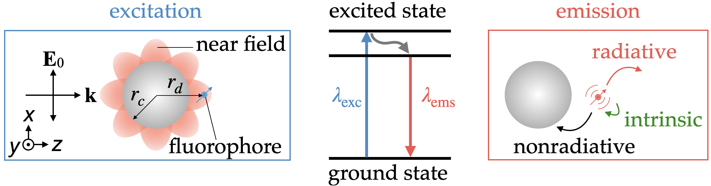
In this model, first, a particle locally enhances an electric field, . This, in turn, amplifies the excitation rate, (), of the emitter. Second, after being excited, the mutual interaction with the particle modifies the radiative decay rate, , of the dipole emitter. The presence of dissipative components also induces changes in the nonradiative decay rate, . Contrary to the widespread misconception that fluorescence enhancement is proportional to radiative decay enhancement, the resulting fluorescence enhancement factor is determined by 34, 37, 20, 38
| (1) |
where the subscript “0” indicates the respective quantity in the free space, the rates with tilde are normalized to the radiative decay rate in free space (e.g. , ), and is the intrinsic quantum yield. , which corresponds to the radiative decay enhancement, is determined by the local density of states, whereas is determined by particle losses. In the special case of and , the last two fractions on the rhs of eq 1 reduce to unity. Any fluorescence enhancement is then determined solely by the first fraction, i.e. by the excitation enhancement, and the fluorescence enhancement in the above particular case is insensitive to any local density of states (LDOS) changes at the emission wavelength. The above illustrates that fluorescence enhancement is a more complex process than radiative decay enhancement and that one should avoid any temptation to characterize fluorescence by either the LDOS or the Purcell factor.
The fluorescence excitation and emission processes are treated independently (i.e., weak coupling) and, in general, there is a Stokes shift between excitation, , and emission, , wavelengths, i.e. . The emitter is assumed to be below saturation 39, 37. According to eq 1, achieving an optimal fluorescence enhancement factor requires a delicate balance of on one hand, and of and on the other hand 39, 37, 40, 41, 42, 43.
Light-matter interaction of a generic process involving an ED characterized by its ED moment is proportional to . Excitation rates are then proportional to . In order to facilitate ensuing calculations, the following common averaging over the ED orientation and its position on a spherical shell of fixed radius are performed (see Supporting Information for details):
-
•
the ED moment in the scalar product is averaged over all possible orientations of at a given fixed , whereby reduces to
-
•
after averaging over all possible orientations of at a given fixed , the resulting scalar product is averaged over the spherical surface of fixed radius .
As a result of this averaging, it is possible to reformulate the scalar product into the respective averages of and 37. After the above averaging, the excitation rate, , becomes proportional to the surface averaged intensity of the electric field, . The latter can be determined by closed-form analytical formulas of ref 44. The averaged is obviously -dependent and does not depend on the spherical angular coordinates. The corresponding decay rates become orientation averaged decay rates 36, 37 after the above averaging, , where superscripts “” and “” denote the radial and tangential orientations of ED, respectively. All the above steps are implemented in freely available MATLAB code, Stratify 30, employed in the present simulations. In essence, surface integrals of the electric field intensity can be performed analytically and the calculation of average intensity costs the same computational time as determining intensity at a given single point. This strategy allows simulating potentially promising designs at a low computational cost, without the need for time-expensive calculations of electric fields at multiple points inside or outside a particle. Once configurations of MCs exhibiting large averaged values of are identified, further detailed calculations of electric fields for these designs are easily implemented (cf. Figure 3).
The rich modal composition of high-index MCs necessitates a delicate search for suitable resonances. This optimization can be complicated to accomplish as both optimal electric field localization and decay rates need to be optimized for fluorescence enhancement, eq 1. The complexity of this problem is a key challenge why previous works 20, 21 did not find the utility of all-dielectric particles for fluorescence enhancement.
The averaged fluorescence enhancement factor (eq 1) in the presence of a lossless () all-dielectric particle reduces to
| (2) |
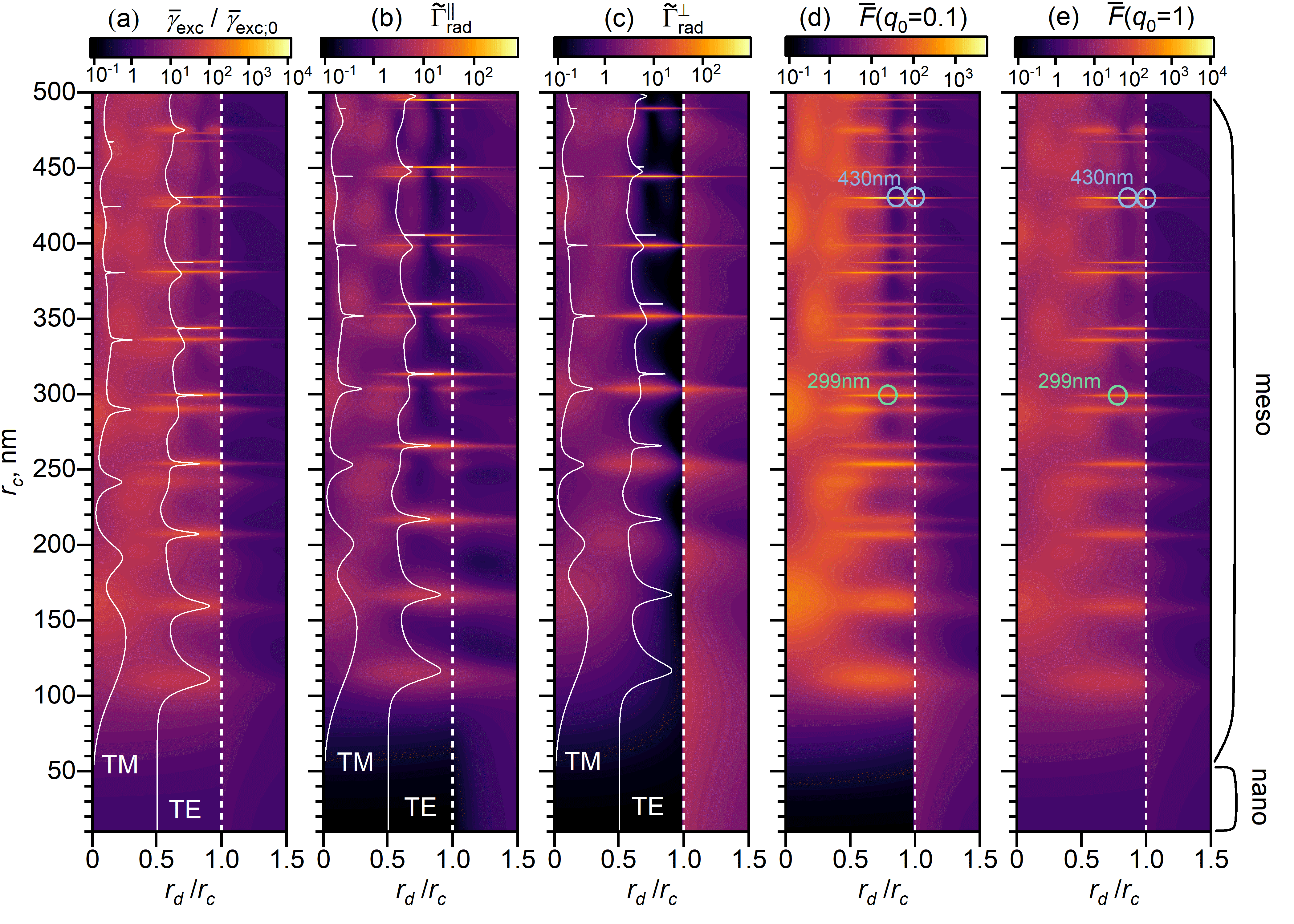
To relate to practical applications, we consider widely used and readily fabricated TiO2 spherical particles with real refractive index , which are lossless in the visible range 7, and vary their radii from to nm, thus sampling both the “nano” (radii from to nm) and “meso” regimes (radii from to nm). For the sake of illustration, either standard Nile Blue dye with nm and nm, or Er3+ with nm and nm is used as ED emitter. It is also worth noting that modern fabrication techniques can embed emitters into spheres with nanometer precision in the radial direction for a number of different materials 45, 46. Controlled positioning of emitters outside the sphere is possible by attaching emitters to (nano)particles via molecular techniques, such as using single-stranded DNA (ssDNA) spacers 47 or DNA origami 48. Therefore, we consider situations with an emitter both inside and outside of a TiO2 sphere. Figures 2(a-c) show excitation and radiative decay rates for two different ED orientations. Extinction spectra for the transverse electric (TE), also called magnetic, and transverse magnetic (TM), also called electric, modes are superimposed on the top of the graphs to show the nature of resonances. The peaks in the radiative decay rate correlate nicely with the peaks in the modes having dominant electric field components along the ED direction, i.e. the TE (TM) mode for the tangential (radial) ED orientation.
Similar to our previous study 49, we observe that it is more beneficial to tune a suitable resonance to an emitter excitation wavelength than to its emission wavelength for enhancing . This is because an enhancement in does not need to have any effect on enhancing fluorescence. In view of eq 2, is proportional to only for intrinsic quantum yield , whereas is independent of for 50.
Tuning a suitable magnetic (TE) MC resonance to the excitation wavelength was found to be the most beneficial. The so-called decay rate engineering advocated within the MEF framework as means of enhancing 51, 22 plays a smaller role in the present case of MCs.
In the interstitial case described by the virtual-cavity model 52, the local field, , felt by emitters inside the particle in the presence of a macroscopic field is , where
| (3) |
is the so-called Lorentz local-field correction 52 (see Supporting Information for its general derivation from the Maxwell’s equations). This correction is particularly interesting for high-index dielectrics because linearly increases with the host dielectric constant, and it can become large. Averaged fluorescence enhancements displayed in Figures 2(d),(e) for ED for two different values of ( and ) are shown with the Lorentz local-field correction included (contributing for the factor of to ).
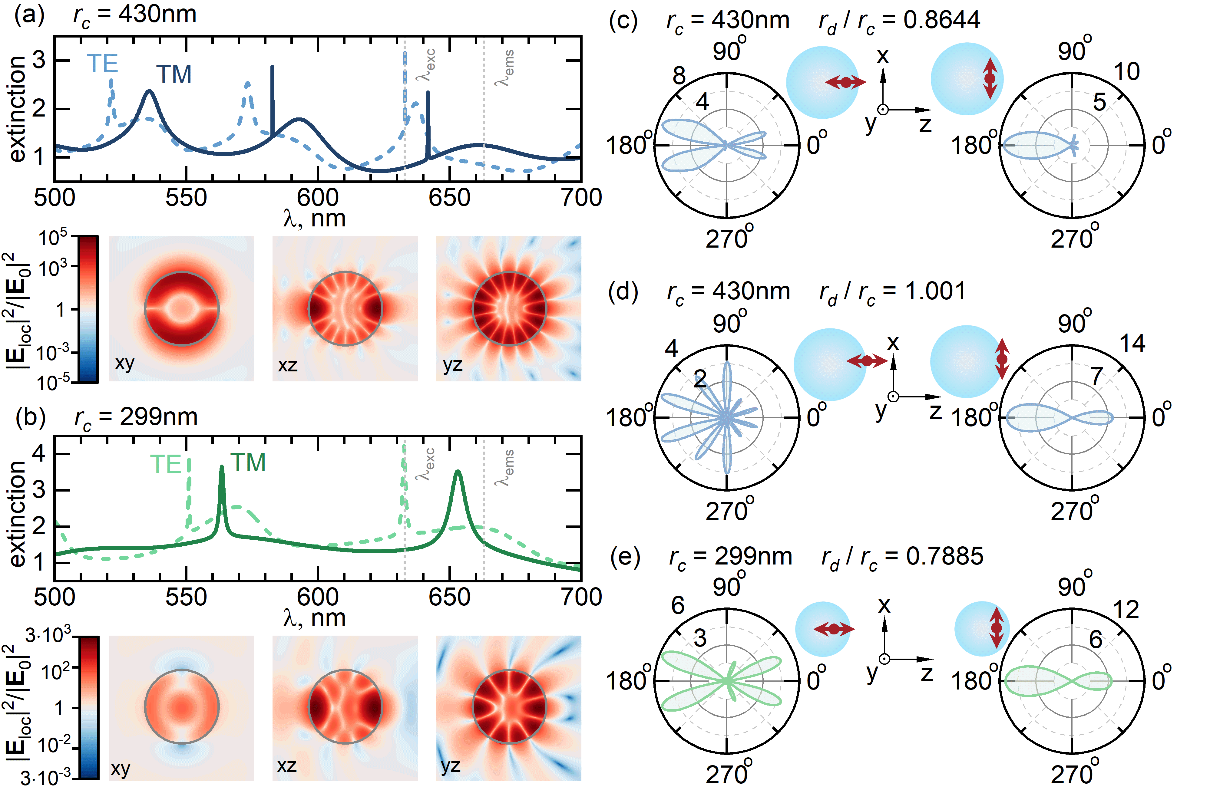
Figure 3a,b shows extinction efficiency and normalized intensity of the electric field, (at ), inside and outside TiO2 sphere with radius (a) nm and (b) nm, on the background of the respective TE and TM resonances. The results correspond to the incident plane-wave amplitude oscillating along the axis and propagation along the axis, as indicated in Figure 1.
Local fields enhancement demonstrates rarely studied patterns of and multipoles induced in MCs with nm and nm, respectively. We notice that optimal arrangement of emitters into hot spots of a mesosphere can further increase the ultimate fluorescence enhancement factor. Interestingly, the spatial extent of these hot spots is quite broad, which makes them suitable for any application benefiting from strong electric field enhancement.
Coupling of ED to a dielectric mesoparticle can result in strong modification of angular emission of ED. To analyze it, we have calculated the radiation directivity (relation of the power emitted into a certain direction to the solid angle averaged emitted power) of ED in the vicinity of TiO2 mesoparticle at nm, which corresponds to the maximum of emission wavelength of Nile Blue dye. Figure 3(c)-(e) shows the angular directivity of radially () and tangentially () oriented ED at specific relative positions highlighted in Figure 2. It can be seen that radially oriented ED doesn’t have large directionality. Tangentially oriented ED, on the other hand, has a highly directional pattern of emission oriented toward mesoparticle (). Interestingly, directivity for ED with tangential orientation embedded inside the mesoparticle with nm (Figure 3(e)) approaches the fundamental Harrington-Chu limit 53, 54 ( for a given sphere size and ).
If in the lossless case described by eq 2: (i) then (the second fraction in eq 2) grows as decreases; (ii) then grows as increases; (iii) then does not depend on . As shown in Figure 4, one can encounter all three cases in practice.
Contrary to the case of plasmonic particles characterized by significant in their proximity due to large Ohmic losses leading to a scaling behavior in 50, we have here and, consequently, there is no scaling in . In fact, the maximum achievable can be increasing (i.e. not decreasing) with increasing as can be seen from Figure 4.
Enhancement mediated by dielectric nanoparticles is a common approach to increasing the intensity of fluorescence signals emitted by fluorescent molecules or materials. When fluorescent molecules are placed near dielectric nanoparticles or embedded in them, the nanoparticles can act as efficient scattering elements, increasing the excitation andor emission rate of these fluorescent molecules through the enhanced local field effect. The choice between dielectric microparticles and nanoparticles for the purpose of fluorescence enhancement depends on the specific requirements of the application, such as the desired enhancement factor, the photostability of the fluorescent molecule, and compatibility with the experimental conditions. In our study, we show that much larger fluorescence enhancement can be achieved with meso-sized particles than with nanoparticles. Contrary to earlier studies involving all-dielectric microparticles 28, 29, our study has been strictly limited to particle sizes between nanoscale ( nm) and microscale ( m) and, consequently, to intermediary multipolar resonances () that is well below typical order of whispering gallery modes () in microparticles. The present proposal seems to be an excellent compromise between nano- and microworlds while being able to combine the advantages of both worlds.
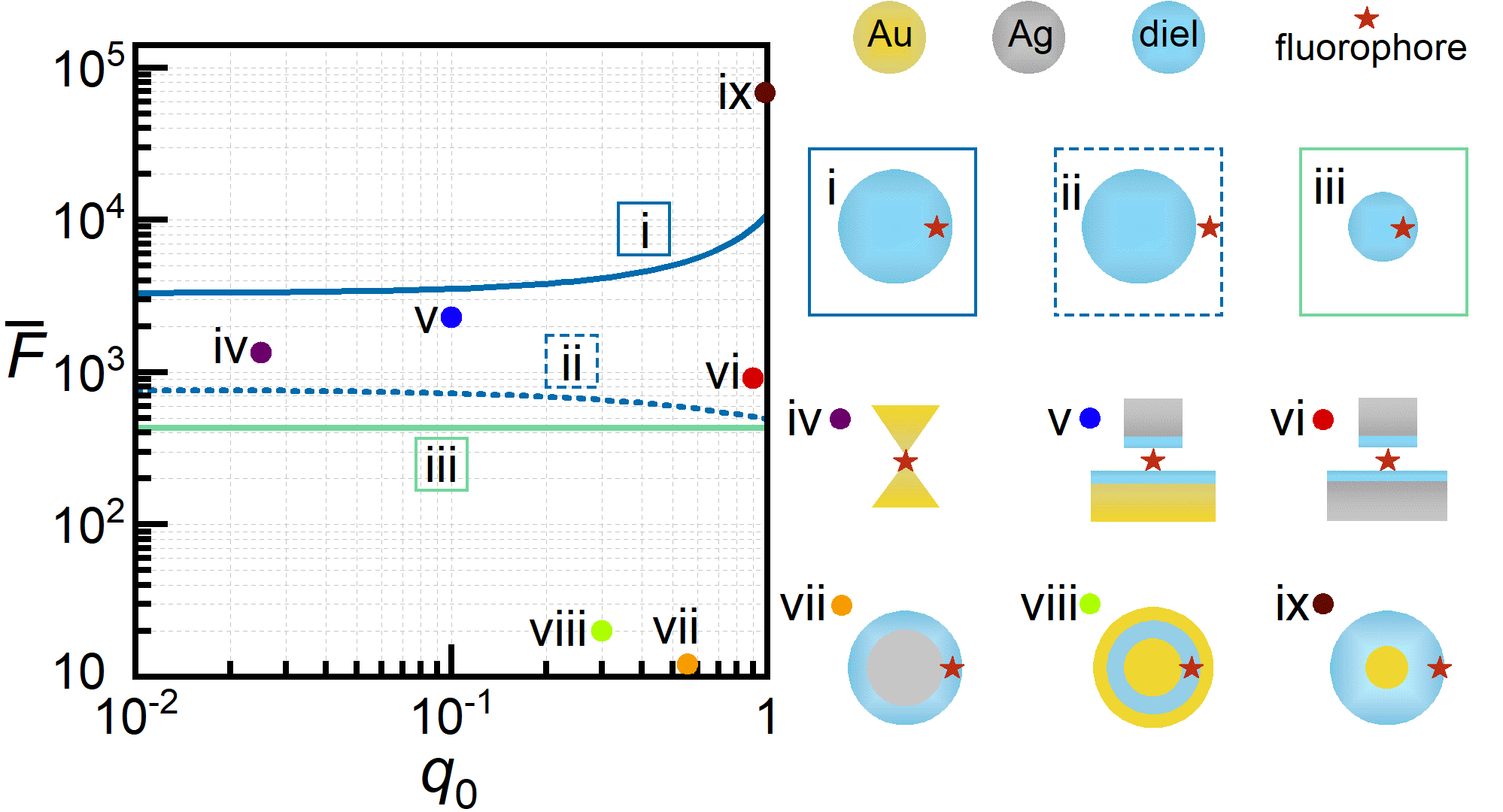
Recently, we have reported that a metal core is essential to get extraordinary fluorescence enhancements exceeding (results presented therein are shown without taking into account the local field corrections) 49. The reported fluorescence enhancement by homogeneous all-dielectric MCs is only slightly smaller than that reported in ref 49 (cf. Figure 4), while bringing significant manufacturing advantages: (i) homogeneous all-dielectric MC are generally much easier to fabricate, (ii) do not require costly noble metals, and (iii) allow one to embed emitters in their entire volume (or realize other options 45) potentially leading to brighter fluorescence sources. Dielectric MCs can provide a tunable and uniform enhancement effect over a wide range of wavelengths. This is because the resonant frequency of the dielectric MCs can be tailored to match the excitation and emission wavelengths of the fluorescent molecule, leading to a more efficient energy transfer.
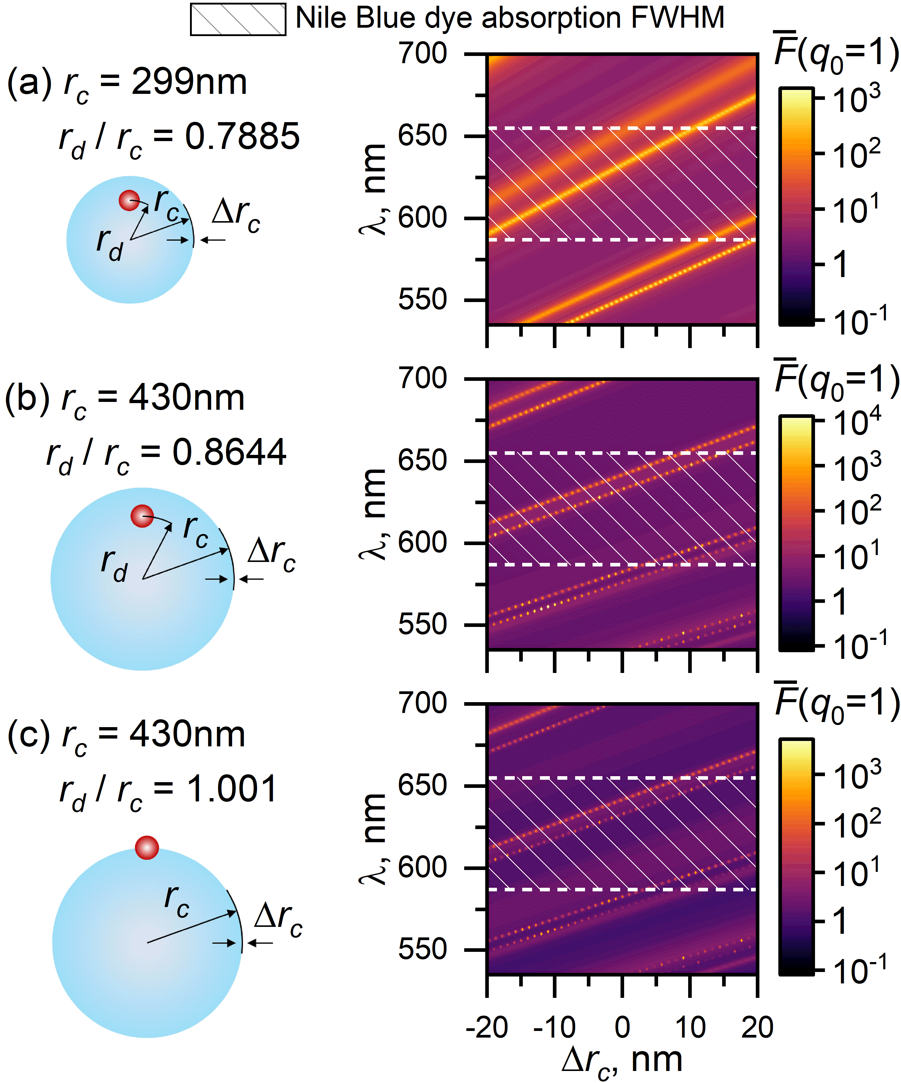
We next discuss practical aspects of utilizing mesoparticles for fluorescence enhancement (Figure 5). We start with examining the effect of particle size polydispersity on the practically achievable fluorescence enhancements. Given that a typical full width at half maximum (fwhm) of organic dyes absorption band is at least nm 58, 59, and assuming as large as nm range of particle size distribution about a target size, the latter size variations can be easily accommodated by a tunable laser diode source, enabling one to excite dyes on the particle resonance, while still remaining within a dye absorption band. Figure 5 shows that the effect of the particle size polydispersity under the above provisions is rather small. As a matter of fact, even as large as nm range of particle size distribution about a target size would be sufficient for enjoying enhanced fluorescence for most applications. We have also observed another interesting result from this figure: high fluorescence enhancement values can be achieved for all possible values of within the range of predefined and . This is enabled by the high multipole order of resonance of the particles, occurring at a wavelength that coincides with the maximum of fluorescence. It is clear that the shaded area between the horizontal dashed lines, which corresponds to the fwhm of the absorption spectrum of Nile Blue dye, will always be enhanced by the presence of th-th order TE or TM resonances of dielectric MC.
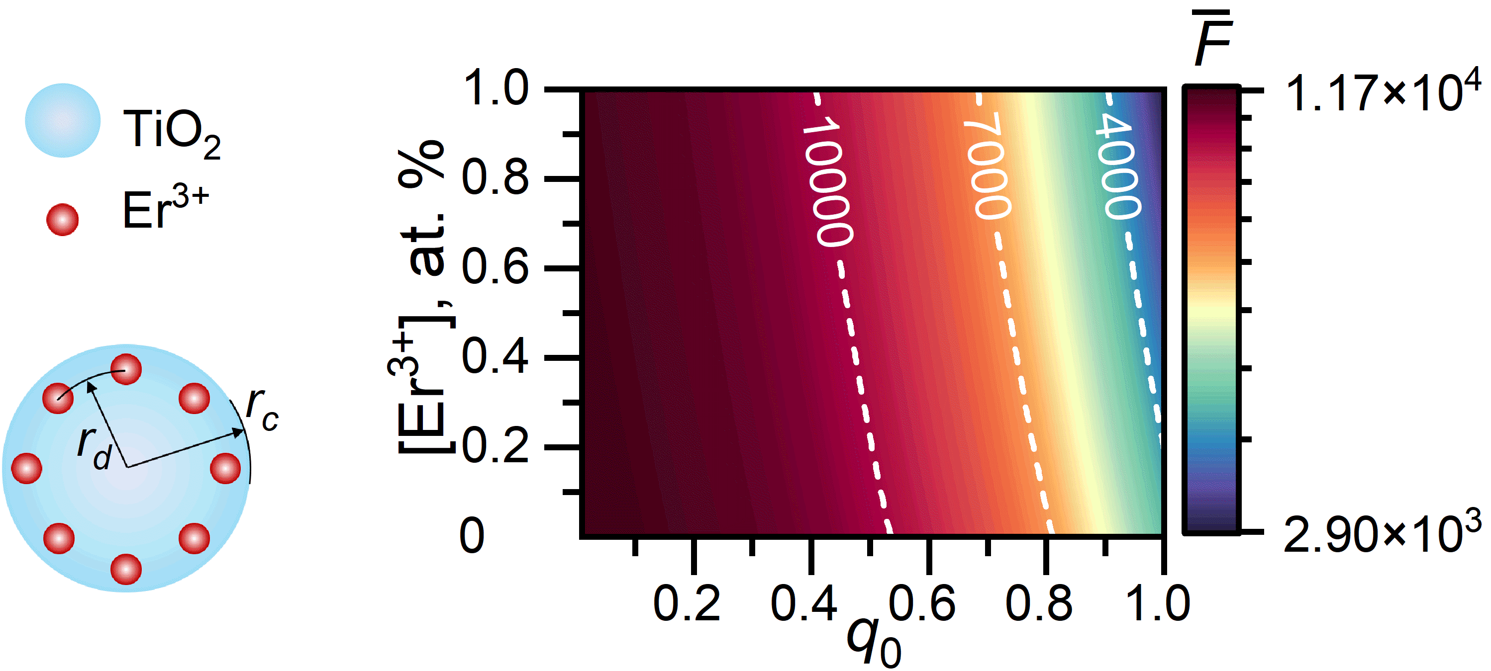
While we have considered a single emitter thus far, other emitters and species may also be proximally present. Let us consider the effect of multiple emitters (Figure 6). Once an emitter is excited, it can transfer its excitation to a nearby emitter via an energy transfer mechanism. With multiple emitters present, this transfer process increases the probability of a decay of emitter excitation. Similarly, if the excitation is transferred to an emitter in a proximity of an impurity, the excitation can be quenched (e.g., OH bonds in silica), whereby the excitation disappears without ever contributing to radiation. This is the essence of the so-called concentration quenching, which does contribute to nonradiative decay rate but whose origin is different from the Ohmic losses of metal particles. In what follows, we consider Er3+ emitters with nm and nm coupled to TiO2 mesoparticle embedded in water with . Concentration quenching in this case can be accounted by 60
| (4) |
where [Er3+] ([Q]) is the erbium (quencher) concentration in at. %, and is a coupling constant. Using a typical coupling value for from the literature ( cm6 s-1), we also assume a quencher concentration of 100 ppm for representative calculations. For example, assuming the dominant quencher in SiO2 is OH, this is a reasonable value for the colloids prepared in a wet–chemical reaction 60. The effect of Er3+ concentration on the averaged fluorescence enhancement is highlighted in Figure 6. Interestingly, even relatively small nonradiative losses have the effect similar to that in the MEF 50, namely larger are attainable with smaller . In the case of non-zero nonradiative losses, utilization of the emitters with the Stokes shift, , smaller than the resonance line width can be beneficial allowing one to achieve large and simultaneously, thus counteracting any emergent quenching induced by .
3 Conclusions
By using our fast calculation approach, modeling showed that size matters: for a given sphere of specific refractive index, much larger fluorescence enhancement can be achieved with meso-sized homogeneous dielectric spheres than with nanospheres. Quite surprisingly, record high averaged fluorescence enhancements for all-dielectric particles of can be achieved with simple homogeneous mesospheres under optimal conditions without requiring the engineering of sophisticated shapes, precise nanogaps, generation of hot spots, or designer metasurfaces. Potential fluorescence enhancements are much larger than those obtained experimentally (Figure 4). Given the importance of fluorescence in numerous practical applications, we find it a very important result beneficial for fluorescence applications to imaging, sensing, strong coupling, and quantum information processing. Only averaged quantities have been discussed so far; it is to be expected that fluorescence enhancements of individual fluorophores can be a further magnitude larger if they are optimally arranged into hot spots of a mesosphere.
4 Methods
Numerical results reported in this manuscript have been obtained via open source code Stratify 30, 61. Detailed derivations and the respective expressions for quantities entering master eq (2) are available as follows: 62, 44 and 31, 33, 36.
Averaged fluorescence enhancement as a proper figure of merit. A general derivation of the Lorentz local-field correction directly from the Maxwell’s equations.
Associated Content: Vadim I. Zakomirnyi; Alexander Moroz; Rohit Bhargava; Ilia L. Rasskazov. Large fluorescence enhancement via lossless all-dielectric spherical mesocavities. 2023. arXiv. https://arxiv.org/abs/2301.10899 (accessed December 1, 2023)
References
- Ostovar et al. 2020 Ostovar, B.; Cai, Y.-Y.; Tauzin, L. J.; Lee, S. A.; Ahmadivand, A.; Zhang, R.; Nordlander, P.; Link, S. Increased intraband transitions in smaller gold nanorods enhance light emission. ACS Nano 2020, 14, 15757–15765
- Knight et al. 2014 Knight, M. W.; King, N. S.; Liu, L.; Everitt, H. O.; Nordlander, P.; Halas, N. J. Aluminum for plasmonics. ACS Nano 2014, 8, 834–840
- Gérard and Gray 2015 Gérard, D.; Gray, S. K. Aluminium plasmonics. Journal of Physics D: Applied Physics 2015, 48, 184001
- West et al. 2010 West, P.; Ishii, S.; Naik, G.; Emani, N.; Shalaev, V.; Boltasseva, A. Searching for better plasmonic materials. Laser & Photonics Reviews 2010, 4, 795–808
- Naik et al. 2011 Naik, G. V.; Kim, J.; Boltasseva, A. Oxides and nitrides as alternative plasmonic materials in the optical range [Invited]. Optical Materials Express 2011, 1, 1090–1099
- Naik et al. 2012 Naik, G. V.; Schroeder, J. L.; Ni, X.; Kildishev, A. V.; Sands, T. D.; Boltasseva, A. Titanium nitride as a plasmonic material for visible and near-infrared wavelengths. Optical Materials Express 2012, 2, 478–489
- Baranov et al. 2017 Baranov, D. G.; Zuev, D. A.; Lepeshov, S. I.; Kotov, O. V.; Krasnok, A. E.; Evlyukhin, A. B.; Chichkov, B. N. All-dielectric nanophotonics: the quest for better materials and fabrication techniques. Optica 2017, 4, 814–825
- Stehr et al. 2019 Stehr, F.; Stein, J.; Schueder, F.; Schwille, P.; Jungmann, R. Flat-top TIRF illumination boosts DNA-PAINT imaging and quantification. Nature Communications 2019, 10, 1268
- Ray et al. 2018 Ray, S.; Widom, J. R.; Walter, N. G. Life under the microscope: Single-molecule fluorescence highlights the RNA world. Chemical Reviews 2018, 118, 4120–4155
- Bower et al. 2018 Bower, A. J.; Li, J.; Chaney, E. J.; Marjanovic, M.; Spillman, D. R.; Boppart, S. A. High-speed imaging of transient metabolic dynamics using two-photon fluorescence lifetime imaging microscopy. Optica 2018, 5, 1290–1296
- Garcia et al. 2018 Garcia, M.; Edmiston, C.; York, T.; Marinov, R.; Mondal, S.; Zhu, N.; Sudlow, G. P.; Akers, W. J.; Margenthaler, J.; Achilefu, S.; Liang, R.; Zayed, M. A.; Pepino, M. Y.; Gruev, V. Bio-inspired imager improves sensitivity in near-infrared fluorescence image-guided surgery. Optica 2018, 5, 413–422
- Park et al. 2019 Park, S.-J.; Kim, B.; Choi, S.; Balasubramaniam, S.; Lee, S.-C.; Lee, J. Y.; Kim, H. S.; Kim, J.-Y.; Kim, J.-J.; Lee, Y.-A.; Kang, N.-Y.; Kim, J.-S.; Chang, Y.-T. Imaging inflammation using an activated macrophage probe with Slc18b1 as the activation-selective gating target. Nature Communications 2019, 10, 1111
- Adachi et al. 2023 Adachi, M.; Sugimoto, H.; Nishimura, Y.; Morita, K.; Ogino, C.; Fujii, M. Fluorophore‐decorated Mie resonant silicon nanosphere for scattering/fluorescence dual‐mode imaging. Small 2023, 2207318
- Andersen and Mortensen 2008 Andersen, C. M.; Mortensen, G. Fluorescence spectroscopy: a rapid tool for analyzing dairy products. Journal of Agricultural and Food Chemistry 2008, 56, 720–729
- Lu et al. 2018 Lu, C.; Zhang, P.; Chen, S.; Zhu, J.; Xu, X.; Huang, H. Fluorescence spectrum photo-bleaching analysis for distinguishing microorganisms (bacteria and fungi) from other particles in air. Optics Express 2018, 26, 28902–28917
- Schmidt et al. 2017 Schmidt, T. D.; Lampe, T.; Sylvinson M. R., D.; Djurovich, P. I.; Thompson, M. E.; Brütting, W. Emitter orientation as a key parameter in organic light-emitting diodes. Physical Review Applied 2017, 8, 037001
- Yang et al. 2015 Yang, Y.; Zheng, Y.; Cao, W.; Titov, A.; Hyvonen, J.; Manders, J. R.; Xue, J.; Holloway, P. H.; Qian, L. High-efficiency light-emitting devices based on quantum dots with tailored nanostructures. Nature Photonics 2015, 9, 259–266
- Reimer and Cher 2019 Reimer, M. E.; Cher, C. The quest for a perfect single-photon source. Nature Photonics 2019, 13, 734–736
- Xu et al. 2019 Xu, L.; Yuan, H.; Zhang, N.; Zhang, J.; Bian, G.; Fan, P.; Li, M.; Zhang, C.; Zhai, Y.; Fang, J. High-efficiency fluorescence collection for NV- center ensembles in diamond. Optics Express 2019, 27, 10787–10797
- Sun et al. 2016 Sun, S.; Wu, L.; Bai, P.; Png, C. E. Fluorescence enhancement in visible light: dielectric or noble metal? Physical Chemistry Chemical Physics 2016, 18, 19324–19335
- Stamatopoulou and Tserkezis 2021 Stamatopoulou, P. E.; Tserkezis, C. Role of emitter position and orientation on silicon nanoparticle-enhanced fluorescence. OSA Continuum 2021, 4, 918–932
- Geddes and Lakowicz 2002 Geddes, C.; Lakowicz, J. Metal-enhanced fluorescence. Journal of Fluorescence 2002, 12, 121–129
- Hoang et al. 2015 Hoang, T. B.; Akselrod, G. M.; Argyropoulos, C.; Huang, J.; Smith, D. R.; Mikkelsen, M. H. Ultrafast spontaneous emission source using plasmonic nanoantennas. Nature Communications 2015, 6, 7788
- Traverso et al. 2021 Traverso, A. J.; Huang, J.; Peyronel, T.; Yang, G.; Tiecke, T. G.; Mikkelsen, M. H. Low-loss, centimeter-scale plasmonic metasurface for ultrafast optoelectronics. Optica 2021, 8, 202–207
- Doiron et al. 2019 Doiron, B.; Mota, M.; Wells, M. P.; Bower, R.; Mihai, A.; Li, Y.; Cohen, L. F.; Alford, N. M.; Petrov, P. K.; Oulton, R. F.; Maier, S. A. Quantifying figures of merit for localized surface plasmon resonance applications: A materials survey. ACS Photonics 2019, 6, 240–259
- Sarychev et al. 2021 Sarychev, A. K.; Ivanov, A. V.; Barbillon, G. Limit possible electric field in plasmon nanogap resonator. 2021, 2111.05511. arXiv. https://arxiv.org/abs/2111.05511 (accessed Dec 14, 2023)
- Mie 1908 Mie, G. Beiträge zur Optik trüber Medien, speziell kolloidaler Metallösungen. Annalen der Physik 1908, 330, 377–445
- Luk’yanchuk et al. 2022 Luk’yanchuk, B. S.; Bekirov, A. R.; Wang, Z. B.; Minin, I. V.; Minin, O. V.; Fedyanin, A. A. Optical Phenomena in Dielectric Spheres Several Light Wavelengths in Size: A Review. Physics of Wave Phenomena 2022, 30, 217–241
- Wang et al. 2022 Wang, Z.; Luk’yanchuk, B.; Wu, B.; Yan, B.; Assel, A.; Yaminsky, I.; Yu, H.; Liu, L. Optical super-resonances in dielectric microsphere particles. Proc. of SPIE. 2022; p 1215205
- Rasskazov et al. 2020 Rasskazov, I. L.; Carney, P. S.; Moroz, A. STRATIFY: a comprehensive and versatile MATLAB code for a multilayered sphere. OSA Continuum 2020, 3, 2290–2309
- Ruppin 1982 Ruppin, R. Decay of an excited molecule near a small metal sphere. Journal of Chemical Physics 1982, 76, 1681–1684
- Ford and Weber 1984 Ford, G.; Weber, W. Electromagnetic interactions of molecules with metal surfaces. Physics Reports 1984, 113, 195–287
- Chew 1987 Chew, H. Transition rates of atoms near spherical surfaces. Journal of Chemical Physics 1987, 87, 1355–1360
- Chew 1988 Chew, H. Radiation and lifetimes of atoms inside dielectric particles. Physical Review A 1988, 38, 3410–3416
- Kim et al. 1988 Kim, Y. S.; Leung, P.; George, T. F. Classical decay rates for molecules in the presence of a spherical surface: A complete treatment. Surface Science 1988, 195, 1–14
- Moroz 2005 Moroz, A. A recursive transfer-matrix solution for a dipole radiating inside and outside a stratified sphere. Ann. Phys. (NY) 2005, 315, 352–418
- Bharadwaj and Novotny 2007 Bharadwaj, P.; Novotny, L. Spectral dependence of single molecule fluorescence enhancement. Optics Express 2007, 15, 14266–14274
- Sun et al. 2019 Sun, S.; Zhang, T.; Liu, Q.; Ma, L.; Du, Q.; Duan, H. Enhanced directional fluorescence emission of randomly oriented emitters via a metal-dielectric hybrid nanoantenna. Journal of Physical Chemistry C 2019, 123, 21150–21160
- Anger et al. 2006 Anger, P.; Bharadwaj, P.; Novotny, L. Enhancement and quenching of single-molecule fluorescence. Physical Review Letters 2006, 96, 113002
- Ringler et al. 2008 Ringler, M.; Schwemer, A.; Wunderlich, M.; Nichtl, A.; Kürzinger, K.; Klar, T. A.; Feldmann, J. Shaping emission spectra of fluorescent molecules with single plasmonic nanoresonators. Physical Review Letters 2008, 100, 203002
- Arruda et al. 2017 Arruda, T. J.; Bachelard, R.; Weiner, J.; Slama, S.; Courteille, P. W. Fano resonances and fluorescence enhancement of a dipole emitter near a plasmonic nanoshell. Physical Review A 2017, 96, 043869
- Sun et al. 2017 Sun, S.; Li, M.; Du, Q.; Png, C. E.; Bai, P. Metal-dielectric hybrid dimer nanoantenna: coupling between surface plasmons and dielectric resonances for fluorescence enhancement. Journal of Physical Chemistry C 2017, 121, 12871–12884
- Sun et al. 2020 Sun, S.; Rasskazov, I. L.; Carney, P. S.; Zhang, T.; Moroz, A. Critical role of shell in enhanced fluorescence of metal-dielectric core-shell nanoparticles. Journal of Physical Chemistry C 2020, 124, 13365–13373
- Rasskazov et al. 2019 Rasskazov, I. L.; Moroz, A.; Carney, P. S. Electromagnetic energy in multilayered spherical particles. Journal of the Optical Society of America A 2019, 36, 1591–1601
- van Blaaderen and Vrij 1992 van Blaaderen, A.; Vrij, A. Synthesis and characterization of colloidal dispersions of fluorescent, monodisperse silica spheres. Langmuir 1992, 8, 2921–2931
- Gritsch et al. 2022 Gritsch, A.; Weiss, L.; Früh, J.; Rinner, S.; Reiserer, A. Narrow optical transitions in erbium-implanted silicon waveguides. Physical Review X 2022, 12, 041009
- Dulkeith et al. 2005 Dulkeith, E.; Ringler, M.; Klar, T. A.; Feldmann, J.; Muñoz Javier, A.; Parak, W. J. Gold nanoparticles quench fluorescence by phase induced radiative rate suppression. Nano Letters 2005, 5, 585–589
- Acuna et al. 2012 Acuna, G. P.; Bucher, M.; Stein, I. H.; Steinhauer, C.; Kuzyk, A.; Holzmeister, P.; Schreiber, R.; Moroz, A.; Stefani, F. D.; Liedl, T.; Simmel, F. C.; Tinnefeld, P. Distance dependence of single-fluorophore quenching by gold nanoparticles studied on DNA origami. ACS Nano 2012, 6, 3189–3195
- Rasskazov et al. 2021 Rasskazov, I. L.; Moroz, A.; Carney, P. S. Extraordinary fluorescence enhancement in metal-dielectric core-shell nanoparticles. Journal of Physical Chemistry Letters 2021, 12, 6425–6430
- Rasskazov and Moroz 2022 Rasskazov, I. L.; Moroz, A. Is there a proper figure of merit for a plasmonic structure involved in metal-enhanced fluorescence? Plasmonics 2022, 17, 1091–1094
- Lakowicz et al. 2002 Lakowicz, J. R.; Shen, Y.; D’Auria, S.; Malicka, J.; Fang, J.; Gryczynski, Z.; Gryczynski, I. Radiative Decay Engineering. Analytical Biochemistry 2002, 301, 261–277
- de Vries and Lagendijk 1998 de Vries, P.; Lagendijk, A. Resonant scattering and spontaneous emission in dielectrics: Microscopic derivation of local-field effects. Physical Review Letters 1998, 81, 1381–1384
- Harrington 1958 Harrington, R. On the gain and beamwidth of directional antennas. IRE Transactions on Antennas and Propagation 1958, 6, 219–225
- Chu 1948 Chu, L. J. Physical limitations of omni-directional antennas. Journal of Applied Physics 1948, 19, 1163–1175
- Kinkhabwala et al. 2009 Kinkhabwala, A.; Yu, Z.; Fan, S.; Avlasevich, Y.; Müllen, K.; Moerner, W. E. Large single-molecule fluorescence enhancements produced by a bowtie nanoantenna. Nature Photonics 2009, 3, 654–657
- Tovmachenko et al. 2006 Tovmachenko, O. G.; Graf, C.; van den Heuvel, D. J.; van Blaaderen, A.; Gerritsen, H. C. Fluorescence enhancement by metal-core/silica-shell nanoparticles. Advanced Materials 2006, 18, 91–95
- Ayala-Orozco et al. 2014 Ayala-Orozco, C.; Liu, J. G.; Knight, M. W.; Wang, Y.; Day, J. K.; Nordlander, P.; Halas, N. J. Fluorescence enhancement of molecules inside a gold nanomatryoshka. Nano Letters 2014, 14, 2926–2933
- Drexhage 1973 Drexhage, K. H. In Dye Lasers; Schäfer, F. P., Ed.; Springer Berlin Heidelberg: Berlin, Heidelberg, 1973; Vol. 1; pp 144–193
- Drexhage 1974 Drexhage, K. H. Progress in Optics; Elsevier, 1974; Vol. 12; pp 163–232
- de Dood et al. 2001 de Dood, M. J. A.; Slooff, L. H.; Polman, A.; Moroz, A.; van Blaaderen, A. Modified spontaneous emission in erbium-doped SiO spherical colloids. Applied Physics Letters 2001, 79, 3585–3587
- 61 Rasskazov, I. L. https://gitlab.com/iliarasskazov/stratify. (accessed Dec 14, 2023)
- Bott and Zdunkowski 1987 Bott, A.; Zdunkowski, W. Electromagnetic energy within dielectric spheres. Journal of the Optical Society of America A 1987, 4, 1361–1365