Raman Scattering Spectra of Boron Imidazolate Frameworks Containing Different Magnetic Ions
Abstract
We present a Raman scattering spectroscopic study of boron imidazolate metal-organic frameworks (BIFs) with three different magnetic metal ions and one non-magnetic in a wide frequency range from 25 to 1700 cm-1, which covers local vibrations of the linkers and well as collective lattice vibrations. We show that the spectral region above 800 cm-1 belongs to the local vibrations of the linkers, which have the same frequencies for the studied BIFs without any dependence on the structure of the BIFs, and are easily interpreted based on the spectra of imidazolate linkers. In contrast, collective lattice vibrations, observed below 100 cm-1, show a distinction between cage and two-dimensional BIFs structures, with a weak dependence on the metal node. We identify the range of vibrations around 200 cm-1, which are distinct for each MOF, depending on a metal node. Our work demonstrates the energy hierarchy in the vibrational response of BIFs.
I Introduction
Much of the study of metal-organic frameworks (MOFs) in recent years has focused on their tunability and porosity. Since MOF structures can include magnetic ions or clusters connected by organic linkers, the possibility of achieving interesting magnetic states in MOFs has been discussed Thorarinsdottir and Harris (2020); Mínguez Espallargas and Coronado (2018). However, most of the recent studies of magnetic properties in MOFs have been limited by basic magnetization and magnetic susceptibility measurements. In order to study magnetism in these materials we will need to employ spectroscopy, such as magnetic Raman scattering. In fact, magnetic Raman scattering has demonstrated its ability to probe the spectrum of magnetic excitations in organic magnetic materials Drichko, Hackl, and Schlueter (2015); Hassan et al. (2018) where magnetic neutron scattering is challenging due to weak signals and presence of hydrogen in the materials. In order to use Raman scattering spectroscopy as a tool to study different MOF structures and their magnetic response, we need to obtain information about the energy scales of all the other excitations; we must identify what part of the acquired spectroscopic information is related to the collective lattice modes, and what part is related to the linker vibrations. Vibrational Raman scattering has been widely used as a characterization tool for MOFs, as the vibrational modes within the organic ligands, and between the ligands and the metal ions, tend to produce strong peaks in the Raman spectra Hadjiivanov et al. (2021). In this manuscript, we present our vibrational Raman scattering studies of a range of MOFs which share the same linkers, but include different transitional metal ions and show three different structures. We aim to understand the hierarchy of the energy scales we observe in the very rich spectra of MOFs, and develop an efficient way to interpret the spectral features.
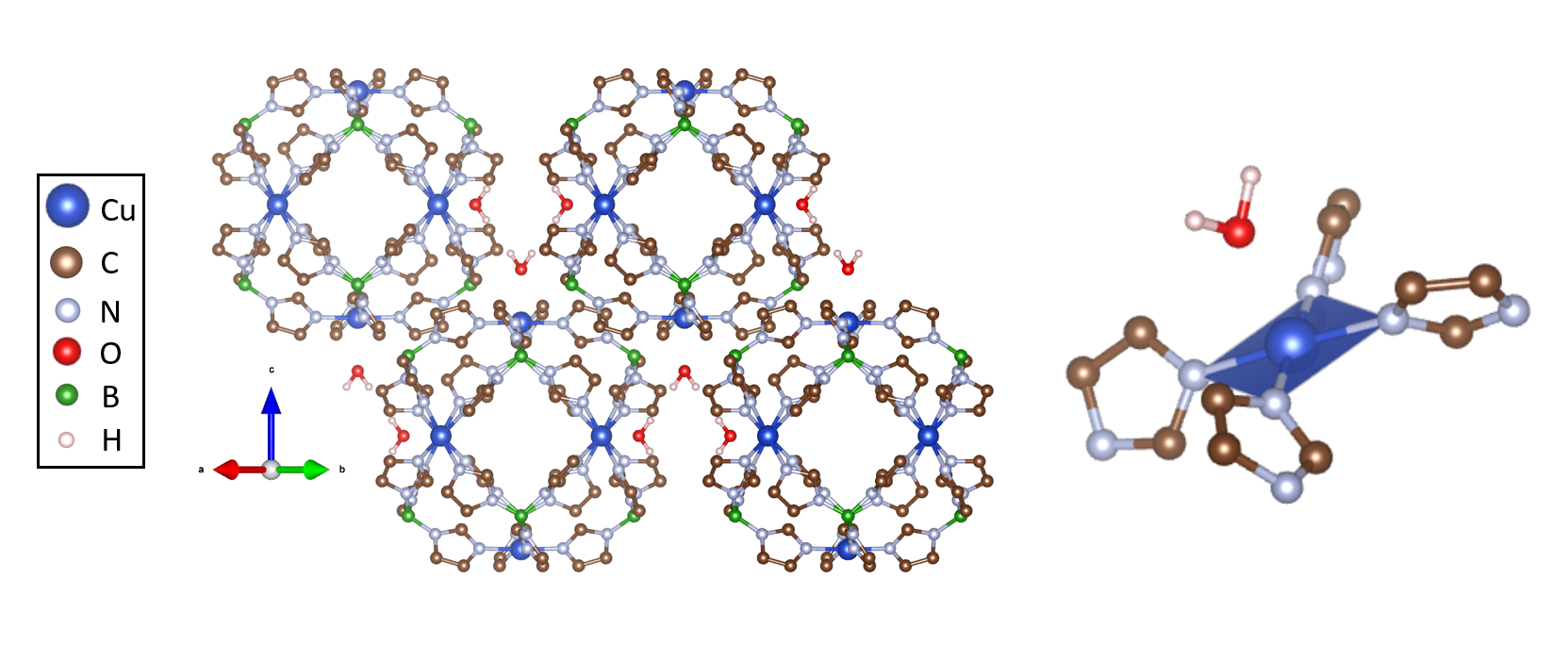
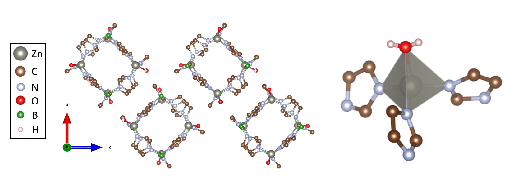
All MOF materials studied here belong to the class of boron imidazolate frameworks (BIFs), which consist of metal ions coordinated by a boron imidazolate ligand. We present Raman spectra of four different BIFs with magnetic ions Cu2+ (S= 1/2), Ni2+ (S=1), Co2+ (S= 3/2) and non-magnetic Zn2+ as the metal cations. These BIFs have three distinct structures: Cu-BIF and Zn-BIF each possesses one of two cage-like structures Zhang et al. (2015); Wen and Zhang (2017), while Co-BIF and Ni-BIF are isostructural, possessing a layered 2D triangular lattice Banerjee et al. (2022a). The cage BIFs have topologies similar to zeolites and zeolitic imidazolate frameworks (ZIFs). In ZIFs, each metal cation bonds to one nitrogen of four different imidazolate rings, creating a tetrahedral metal environment, and these linked tetrahedra form a porous 3D network of connected cages Chen et al. (2014). In cage BIFs, one N on the imidazolate ring binds to a metal cation, while the other binds to a B3+ cation. For instance, in the cage Cu-BIF, pictured in Fig. 1, the metal cations have an environment of four coplanar N atoms from four different imidazolate rings, while one oxygen from one H2O weakly binds to the metal in the out-of-plane direction, creating a low symmetry environment for the metal atom compared to standard inorganic oxides. The Zn-BIF, pictured in Fig. 2, is composed of smaller cages of 4 Zn and 4 boron imidazolate ligands, and exhibits an anisotropic tetrahedral Zn environment.
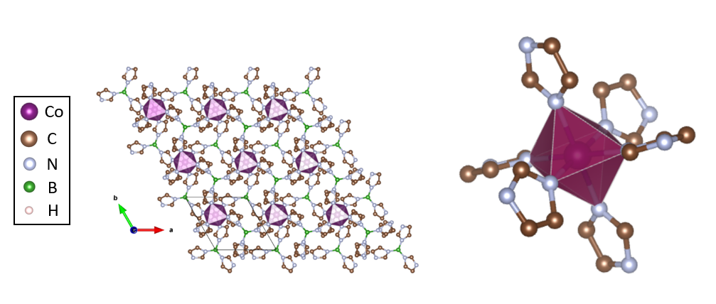
In 2D layered BIFs, no cages are formed at all, and metal ions arrange in a 2D triangular lattice (see Co octahedra in Fig. 3), which is layered along the crystallographic c axis in the bulk. In these BIFs, as illustrated for Co-BIF, metal ions are found in an octahedral environment, as the metal binds to a N atom on 6 imidazolate rings, as shown in Fig. 3.
The boron cations connect three of these metal-imidazolate complexes, as seen in Fig. 1. Because of this, the exchange path between metal centers is through not only one imidazolate ring, as in ZIFs, but follows an extended Me-Im-B-Im-Me path, in both cage and 2D BIFs. This extended exchange path leads to low magnetic exchange between magnetic metal centers, if any at all . While the cage Cu-BIF MOF shows magnetic interactions close to zero, weak magnetic interactions have been detected in 2D layered BIFs Davis et al. (2023).
We show that we can identify the linker vibrations that are independent of the metal ions and their environment, while the vibrations of the metal environment itself are sensitive to the substitution of a metal atom. Despite the differences in crystal structures, the Raman vibrations of the imidazolate linkers observed above 800 cm-1 are similar for these MOFs. In contrast, the lattice vibrations observed below 100 cm-1 are fingerprints of the structure. The intermediate frequencies in the range of 100-300 cm-1 belong to the Raman vibrations of the metal environment and depend on the metal cation. These results demonstrate the distinct energy scales of vibrations of MOFs of different origins. This fact allows for an easy interpretation of the vibrational features of these compounds.
II Experimental
MOFs were synthesized following reported literature procedures for Zn-BIF Wen and Zhang (2017), Cu-BIF Banerjee et al. (2022b), and Co and Ni-BIF Banerjee et al. (2022a).
Raman spectra were measured using a micro-Raman option of a T64000 Horiba-Jobin-Yvon spectrometer equipped with an Olympus microscope and a LN2 cooled CCD. Spectra were excited with the 514.5 nm line of a Coherent Innova 70C laser, with the power kept below 500 W for a probe of 2 m in diameter to avoid heating the sample. Spectra were measured at room temperature with a spectral resolution of 2 cm-1 (low-energy region) and 6 cm-1 (Ni, Zn, and Cu in high energy region).
Intensity of Raman spectra for different materials were normalized on the laser power, grating reflectance, and size of the slit. Background signals of stray laser light were subtracted manually from the low-frequency Co, Cu, and Ni-BIF spectra to highlight the narrow vibrational mode peaks. A Lorentzian peak centered at 0 cm-1 was used to approximately model the background to be subtracted. In Zn-BIF, a broad photoluminescent background was subtracted by comparison to the spectra of pure imidazolate containing an identical background. To compare Raman scattering intensities of measured BIFs vibrational spectra shown in the figures are additionally normalized to the (CN) modes in the 1200-1250 cm-1 range, as a result intensity of Raman spectra were multiplied by a constant , where = 6.5 for Zn-BIF, = 16 for Co-BIF, and = 10 for Cu-BIF. Similarly, low-frequency spectra were multiplied by a constant to highlight lattice modes below 100 cm-1, where = 2.5 for Zn-BIF, = 6.5 for Co-BIF, and = 10 for Cu-BIF.
III Results and Discussion
Measured Raman spectra of Co-BIF, Cu-BIF, Ni-BIF, and Zn-BIF are shown in two different regions in Figs. 4 and 5. The 500-1700 cm-1 region in Fig. 4 contains the expected vibrational modes of imidazolate and allows for preliminary band assignments, which are summarized in Table 1. The 0-500 cm-1 region in Fig. 5 contains low-energy lattice modes below 100 cm-1 and vibrational modes of metal environments above.
The Raman vibrations of imidazolate linkers are expected in the 800-1600 cm-1 spectral range based on assigned Raman spectra of ZIF-8 Hadjiivanov et al. (2021). Raman spectra of the four different BIFs have very similar frequencies of the majority of the molecular vibrations of the imidazolate ligand, despite the fact that they have three different crystal structures and four different metal nodes. This is in agreement with the expectation that the internal structure of the ligand remains unchanged between these MOFs. A comparison to previously reported Raman scattering spectra of ZIF-8 Hadjiivanov et al. (2021) reveals a direct correspondence between molecular vibrations of imidazolate in ZIF and BIF structures and is taken as a basis for our interpretation of the vibrational spectra of BIFs. Table 1 summarizes the frequencies of 8 molecular vibrations, which are consistent with previously reported band assignments for imidazolate ligand. We mark these assignments in the Raman spectra of the BIFs shown in Fig. 4.
The most intense modes in the spectral region of 800-1000 cm-1 are bending vibrations of C-H bonds, which are out-of-plane with respect to the plane of the imidazolate ring. These three bands are found at very similar frequencies for all measured BIFs and close to the reported frequencies in ZIF-8. The C-H bonds are the farthest away from the metals, so one would expect that their frequencies are not strongly dependent on the metal nodes.
The spectral range between 1000 and 1500 cm-1 contains 4 stretching vibrations of C-N bonds as well as a wagging vibration of N-H. Though the positions of the two lowest bands are shifted by 70 cm-1 to higher frequencies compared with the ZIF-8 spectra, the spacing of 40 cm-1 between them and their relative intensities are consistent with reported ZIF spectra. The C-N stretching vibration in the range from 1334-1343 cm-1 displays a shift to lower frequencies of 50 cm-1. These differences between ZIF and BIF C-N vibrations can be attributed to the fact that the two N on an imidazolate ring bind to one metal ion and one B, rather than two Zn ions as in ZIFs. Hence, the molecular vibrations that involve the N are expected to differ qualitatively between ZIF and BIF spectra, though not between BIFs themselves. In contrast, other molecular vibrations (such as C-H bending) display more consistency between ZIF and BIF spectra due to their relative isolation from the environment external to the imidazolate ring.
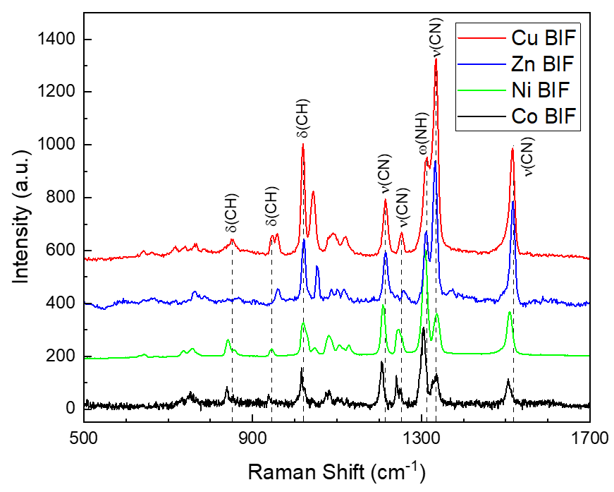
The reported bending vibration of the methyl group in ZIF-8 is absent in BIF spectra since the imidazolate rings in the BIFs have no capping methyl group. The total suppression of the strong imidazolate ring breathing mode, found at 683 cm-1 in ZIF-8, can be a result of the lower symmetry (the absence of mirror symmetry) in the linker in BIF structures, which comes from imidazolate binding to one B and one metal ion, rather than two metal ions as in ZIFs. Alternatively, the suppression of this mode could be a result of a reduction of electronic density on the imidazolate ring.
| Raman Mode | ZIF-8 | Co-BIF | Cu-BIF | Zn-BIF | Ni-BIF |
|---|---|---|---|---|---|
| (CH) | 834 | 831 | 853 | weak | 841 |
| (CH) (C4-C5) | 945 | 939 | 946 | 960 | 945 |
| (CH) (C2) | 1023 | 1018 | 1020 | 1021 | 1021 |
| (C5-N) | 1143 | 1214 | 1215 | 1216 | 1210 |
| (C-N) | 1182 | 1258 | 1252 | 1259 | 1247 |
| (N-H) | 1312 | 1316 | 1314 | 1311 | 1309 |
| (C5-N) | 1385 | 1343 | 1335 | 1333 | 1336 |
| (C2-N) | 1507 | 1505 | 1515 | 1517 | 1510 |
The spectral range of 100-300 cm-1 is the region of metal-ligand vibrations. The frequencies of these vibrations should be dependent both on the metal and on its coordination Andersson et al. (2010). In particular, literature data suggest stretching vibrations of Cu-N in the octahedral environment at 280-290 cm-1, and Zn in a tetrahedral environment at 207 cm-1. Andersson et al. (2010) Modes in this region therefore belong to the N-metal vibration, and have frequencies which depend on the metal ion. Zn-BIF has one mode at 181 cm-1, while Co, Cu, and Ni have four weak modes at frequencies summarized in Table 2. The similarity between Ni and Co-BIF N-metal vibrations is consistent with the identical octahedral environments or Ni and Co, with the small shift in frequencies potentially arising from a change in electronic density on the N atoms in the metal environment. The more significant differences in this region in the Cu and Zn-BIF spectra are consistent with their unique metal environments.
| Co-BIF | Cu-BIF | Ni-BIF |
|---|---|---|
| 136 | 129 | 148 |
| 175 | 151 | 193 |
| 188 | 169 | 205 |
| 248 | 263 | 252 |
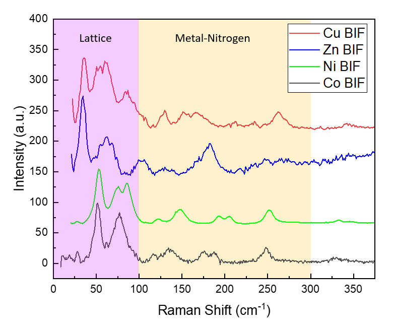
Fig. 5 presents the low frequency spectra of the BIF MOFs. Typically for molecular crystals the region below 100 cm-1 corresponds to lattice vibrations, which would depend on the structure of the MOFs. While to the best of our knowledge, there is no spectroscopic information on the lattice modes of BIFs, the lattice “collective” modes of ZIFs were studied to some extent by THz spectroscopy, neutron scattering, and DFT calculations, and were also found below 100 cm-1.Ryder et al. (2014); Möslein and Tan (2022)
In our experimental data, we find that two low-frequency vibrations of the cage Cu and Zn-BIFs occur at similar frequencies, while 2D Ni and Co-BIFs have similar low-frequency spectra, distinct from the cage BIFs (see Table 3). This demonstrates a distinct dependence of the lattice mode spectra on the structure of the MOFs, showing that the lattice vibrations are fingerprints of a particular lattice structure, and not the chemical environment. We find that two of the lattice modes observed in the spectra for the two cage BIFs, Cu and Zn, are similar to Raman-active THz modes in the calculated DFT spectra of ZIF-8, which is also composed of a porous cage-like structure. These include a strong mode at 33.36 cm-1, assigned to a symmetric 4-membered ring (see Fig. 1,2) gate opening, and a mode at 64.61 cm-1, assigned to a 4-membered ring shearing Ryder et al. (2014) .
| ZIF-8 DFT (cm-1) | Zn-BIF | Cu-BIF | Co-BIF | Ni-BIF |
| ring gate opening (33.36) | 34 | 35 | — | — |
| — | — | — | 52 | 53 |
| ring shearing (64.61) | 62 | 58 | — | — |
| — | — | — | 77 | 74 |
The 2D layered BIFs, on the other hand, do not display any similarity to calculated THz modes of ZIFs, which is consistent with the significant structural differences between the 2D triangular lattice structure and the various cage structures. DFT calculations on the 2D structures are necessary to further interpret these vibrations.
IV Conclusions
In this work we have presented Raman scattering spectra of BIF MOFs with different metal ions in a frequency range between 30 and 2000 cm-1 and demonstrated a separation between the energy scales related to the different types of vibrations of the MOFs. The high frequency region of the spectra contains Raman-active vibrational modes of imidazolate ligands, which are similar between four different BIFs with different structures and metal nodes. They can be assigned based on the literature data of MOFs with the same linker molecules, ZIFs.
The low-energy region of the spectra demonstrates a consistency in lattice modes of isostructural BIFs. The modes of the cage BIFs have frequencies similar to the calculated lattice modes for ZIFs, while 2D BIFs have a distinctly different lattice mode spectrum. These results demonstrate that the lattice vibrations of MOFs are fingerprint of a certain structure.
The spectral region between approximately 100 and 300 cm-1 is dominated by the vibrations of the metal environment. The frequencies of vibrations observed in this region depend on the metal atom, but show some consistency between BIFs with different metals in the same environment.
The consistency of lattice modes and ligand vibrations provides an efficient way to understand and assign the spectra of MOFs without performing full calculations of the vibrational response of each MOF structure.
Acknowledgements.
Acknowledgement is made to the donors of the American Chemical Society Petroleum Research Fund for partial support of this research. We acknowledge the support of NSF Award No. DMR-2004074. S. B., P. B.-V., and V. S. T. acknowledge support by the U.S. Department of Energy (DOE), Office of Science, Office of Basic Energy Sciences, Catalysis Science program, under Award DE-SC0021955. P. B-V. thanks the Dean’s ASPIRE grant from the Office of Undergraduate Research, Scholarly and Creative Activity at Johns Hopkins University. We further acknowledge Professor Tyrel McQueen, Dr. Veronica Stewart (Department of Chemistry, Johns Hopkins University), Chris Lygouras, and Peter Orban (Department of Physics and Astronomy, Johns Hopkins University) for their assistance in obtaining magnetic susceptibility and magnetization data.Data Availability Statement
The data that support the findings of this study are available from the corresponding author upon reasonable request.
Conflict of Interest
The authors have no conflicts to disclose.
References
- Thorarinsdottir and Harris (2020) A. E. Thorarinsdottir and T. D. Harris, “Metal–Organic Framework Magnets,” Chem. Rev. 120, 8716–8789 (2020).
- Mínguez Espallargas and Coronado (2018) G. Mínguez Espallargas and E. Coronado, “Magnetic functionalities in MOFs: From the framework to the pore,” Chem. Soc. Rev. 47, 533–557 (2018).
- Drichko, Hackl, and Schlueter (2015) N. Drichko, R. Hackl, and J. A. Schlueter, “Antiferromagnetic fluctuations in a quasi-two-dimensional organic superconductor detected by raman spectroscopy,” Phys. Rev. B 92, 161112 (2015).
- Hassan et al. (2018) N. Hassan, S. Cunningham, M. Mourigal, E. I. Zhilyaeva, S. A. Torunova, R. N. Lyubovskaya, J. A. Schlueter, and N. Drichko, “Evidence for a quantum dipole liquid state in an organic quasi-two-dimensional material,” Science 360, 1101–1104 (2018).
- Hadjiivanov et al. (2021) K. I. Hadjiivanov, D. A. Panayotov, M. Y. Mihaylov, E. Z. Ivanova, K. K. Chakarova, S. M. Andonova, and N. L. Drenchev, “Power of Infrared and Raman Spectroscopies to Characterize Metal-Organic Frameworks and Investigate Their Interaction with Guest Molecules,” Chem. Rev. 121, 1286–1424 (2021).
- Zhang et al. (2015) D.-X. Zhang, H.-X. Zhang, H.-Y. Li, T. Wen, and J. Zhang, “Self-Assembly of Metal Boron Imidazolate Cages,” Crystal Growth & Design 15, 2433–2436 (2015).
- Wen and Zhang (2017) T. Wen and J. Zhang, “Rational design of metal boron imidazolate cages to frameworks,” Inorganica Chimica Acta Next Generation, 460, 89–92 (2017).
- Banerjee et al. (2022a) S. Banerjee, X. Han, M. A. Siegler, E. M. Miller, N. M. Bedford, B. C. Bukowski, and V. S. Thoi, “Flexible 2D Boron Imidazolate Framework for Polysulfide Adsorption in Lithium–Sulfur Batteries,” Chem. Mater. (2022a), 10.1021/acs.chemmater.2c02324.
- Chen et al. (2014) B. Chen, Z. Yang, Y. Zhu, and Y. Xia, “Zeolitic imidazolate framework materials: Recent progress in synthesis and applications,” J. Mater. Chem. A 2, 16811–16831 (2014).
- Davis et al. (2023) J. Davis et al., “Manuscript in preparation,” (2023).
- Banerjee et al. (2022b) S. Banerjee, J. M. Gorham, P. Beccar-Varela, H. G. Hackbarth, M. A. Siegler, N. Drichko, J. T. Wright, N. M. Bedford, and V. S. Thoi, “Atomically Dispersed CuN x Sites from Thermal Activation of Boron Imidazolate Cages for Electrocatalytic Methane Generation,” ACS Appl. Energy Mater. , acsaem.2c01174 (2022b).
- Momma and Izumi (2011) K. Momma and F. Izumi, “VESTA 3 for three-dimensional visualization of crystal, volumetric and morphology data,” J. Appl. Crystallogr. 44, 1272–1276 (2011).
- Andersson et al. (2010) M. Andersson, J. Hedin, P. Johansson, J. Nordström, and M. Nydén, “Coordination of Imidazoles by Cu(II) and Zn(II) as Studied by NMR Relaxometry, EPR, far-FTIR Vibrational Spectroscopy and Ab Initio Calculations: Effect of Methyl Substitution,” J. Phys. Chem. A 114, 13146–13153 (2010).
- Ryder et al. (2014) M. R. Ryder, B. Civalleri, T. D. Bennett, S. Henke, S. Rudić, G. Cinque, F. Fernandez-Alonso, and J.-C. Tan, “Identifying the Role of Terahertz Vibrations in Metal-Organic Frameworks: From Gate-Opening Phenomenon to Shear-Driven Structural Destabilization,” Phys. Rev. Lett. 113, 215502 (2014).
- Möslein and Tan (2022) A. F. Möslein and J.-C. Tan, “Vibrational Modes and Terahertz Phenomena of the Large-Cage Zeolitic Imidazolate Framework-71,” J. Phys. Chem. Lett. 13, 2838–2844 (2022).
- Isaeva, Papathanasiou, and Kustov (2020) V. I. Isaeva, K. E. Papathanasiou, and L. M. Kustov, “Zeolite-Like Boron Imidazolate Frameworks (BIFs): Synthesis and Application,” Crystals 10, 617 (2020).
- Kumari et al. (2013) G. Kumari, K. Jayaramulu, T. K. Maji, and C. Narayana, “Temperature Induced Structural Transformations and Gas Adsorption in the Zeolitic Imidazolate Framework ZIF-8: A Raman Study,” J. Phys. Chem. A 117, 11006–11012 (2013).
- Salama and Spiro (1978) S. Salama and T. G. Spiro, “Resonance Raman spectra of cobalt(II)-imidazole complexes: Analogs of the binding site of cobalt-substituted zinc proteins,” J. Am. Chem. Soc. 100, 1105–1111 (1978).