Mechanosensitive bonds induced complex cell motility patterns
Abstract
The one-dimensional crawling movement of a cell is considered in this theoretical study. Our active gel model shows that for a cell with weakly mechanosensitive adhesion complexes, as myosin contractility increases, a cell starts to move at a constant velocity. As the mechanosensitivity of the adhesion complexes increases, a cell can exhibit stick-slip motion. Finally, a cell with highly mechanosensitive adhesion complexes exhibits periodic back-and-forth migration. A simplified model which assumes that the cell crawling dynamics are controlled by the evolution of the myosin density dipole and the asymmetry of adhesion complex distribution captures the motility behaviors of crawling cells qualitatively. It suggests that the complex cell crawling behaviors observed in the experiments could result from the interplay between the distribution of contractile force and mechanosensitive bonds.
pacs:
87.17.Jj,87.16.UvIntroduction.– The crawling motion of eukaryotic cells is ubiquitous in biology as it plays important roles in processes such as embryogenesis, wound healing, cancer metastasis, and immunology [1]. Common if not universal features of a crawling cell include myosin motors distributed mainly behind the center, dominant actin polymerization in the leading edge, and higher density of adhesion complexes in the leading region [2]. Such polarized molecular distribution enables protrusion in the leading edge due to actin polymerization, treadmilling of actomyosin cytoskeleton due to contractility, and traction force pulling the cell body. These features, therefore, are included in many theoretical models for crawling cells [3].
Interestingly, besides non-motile resting and steady-moving behaviors, cells crawling along a one-dimensional track either on a substrate or in a three-dimensional environment also exhibit moving patterns that are non-stationary in time. For example, stick-slip crawling motion due to slip between integrin and the extracellular matrix in focal adhesions under the contractility provided by myosin II has been observed in human osteosarcoma cells [4]. Periodic back-and-forth migration has been observed in crawling zyxin-depleted cells in a collagen matrix [5] and dendritic cells crawling along microfabricated channels [6].
Several theoretical models have been proposed to explain some of these deterministic complex moving patterns. A model that includes the mechanochemical coupling of actin promotor dynamics and actin polymerization to myosin kinetics was shown to produce periodic back-and-forth migration [7]. On the other hand, a purely mechanical model emphasizing the interplay between mechanosensitive bonds and membrane tension exhibited stick-slip motion even for slip bonds [8]. Interestingly, it has also been shown that stick-slip can result from the interplay between mechanosensitive bonds, contractility, and a force that tends to restore a cell’s preferred length [9].
In this letter, we present a theoretical study to show that the coupling between mechanosensitive adhesion complexes and myosin contractility is sufficient to generate deterministic complex cell crawling behaviors, including stick-slip, periodic back-and-forth movements, and other complex moving patterns. We first construct an active gel model with mechanosensitive adhesion complexes and show that the distribution of myosin motors and adhesion complexes computed from our model agree with experimentally observed features. By exploring the motility behavior with different strengths of contractility and mechanosensitivity, we show that this model can lead to complex motility behaviors other than rest and constant-velocity moving states. Among these complex motility patterns, unidirectional stick-slip motion and periodic back-and-forth movement are the most common. When the adhesion complexes are less mechanosensitive, as myosin contractility increases, the cell performs constant velocity motion. On the other hand, for cells with highly mechanosensitive adhesion complexes, as myosin contractility becomes sufficiently strong, periodic back-and-forth crawling motion can be observed. Finally, stick-slip and other complex motility patterns can be observed by increasing the mechanosensitivity of the adhesion complexes for a cell moving at constant velocity.
To understand the physical mechanisms that produce these complex motility patterns, a simplified model inspired by the active gel model is constructed. The simplified model assumes that the dynamics of a sufficiently slow-moving cell are dominated by the dipole moment of myosin density and the difference in the total number of adhesion complexes near the two cell ends. Remarkably, the motility behavior predicted by this simple model agrees qualitatively with the motility behaviors predicted by the active gel model. These results suggest that, in general, diverse complex motility behaviors can result from the interplay between mechanosensitive adhesions and the dynamical organization of contractile myosin motors.
Model summary.– To focus on the contribution of cell mechanics to motility behaviors, chemical signaling is not included in our model. The cytoplasm of the cell is modeled as an active gel [10, 11] enclosed by the cell membrane, the adhesion complexes are treated as reversible bonds with specific binding-unbinding rates, and actin polymerization is assumed to happen only at the cell ends. The forces acting on the cell include the stress in the cytoskeleton, the drag force from the substrate, and the force due to the adhesion complexes.
Our model only considers one spatial direction, i.e., the cell’s moving direction, and the stress in the cytoplasm obeys the constitutive equation
| (1) |
where is the effective one-dimensional viscosity of the cytoplasm, is the flow field, is the strength of contractility provided by myosin motors (), and is the concentration of myosin attached to the actin network. For simplicity, compressibility is not included [11]. Thus pressure does not appear in the constitutive relation. The force exerted by the substrate is
| (2) |
The first term on the right hand is the drag provided by the adhesion complexes [12], is a constant that characterizes the resistance of the adhesion complexes to cell movement, and is the number density of adhesion complexes. The second term comes from the viscous drag of the fluid between the cell and the substrate, and is the drag coefficient. Putting Eqs. (1)(2) together, the resulting force balance equation, , takes the following form
| (3) |
Myosin motors attached to actin filaments move with the cytoplasm, while those detached from actin filaments diffuse freely. The attachment/detachment of motors is reversible. On long-time scales, the density of the motors can be effectively described by an advection-diffusion equation [13]
| (4) |
where is the effective diffusion coefficient of myosin motors.
Adhesion complexes providing anchorage to the extracellular matrix are also physically coupled to the contractile cytoplasm. As a result, they are pulled when the cytoplasm moves [8]. Once an adhesion complex is formed, the adhesion site does not move, but the dissociation rate of the adhesion complex is affected by the motion of the cytoskeleton because the bond is stretched or compressed. In our model, the evolution of the density of adhesion complexes is assumed to obey
| (5) |
where is the binding rate, is the unbinding rate at , and tells us how unbinding rate is affected by the cytoplasmic flow. Our model assumes that when the strain rate is dilating, giving more space for the adhesion complexes, the unbinding rate decreases.
Actin polymerization at the cell ends depends on the distribution of actin activators [15][16]. In the presence of environmental cues, a gradient of actin activator concentration within the cell is established, and actin polymerization is polarized due to this concentration gradient. In the absence of such external influence, the cell can nevertheless polarize itself by spontaneous symmetry breaking, and the net actin polymerization rate becomes asymmetric. In our model, we consider a homogeneous environment, and the net actin polymerization rate at the end of the cell is assumed to be
| (6) |
where () is the net rate of extension due to actin polymerization at the cell end located at , comes from the base polymerization rate, in the exponent of the numerator comes from the effect of free energy cost for polymerization when the cell length is different from its natural length ( is the natural length of the cell, and ), and the term with makes the net polymerization rate in a moving cell at both ends different, with more polymerization events in the leading end than the trailing end. It will become clear that the qualitative results of cell motility behavior do not depend on the specific form we assumed for the dissociation rate of the adhesion complexes and .
The evolution of cell-end positions is determined by the velocity of cytoplasm and actin polymerization,
| (7) |
where () is the velocity of cytoplasm at the end.
Experimentally it has been shown that a cell tends to restore its length to a preferred magnitude [17]. We model this effect by the following force balance condition at cell ends
| (8) |
Here is a constant associated with the restoring force that brings the cell length to .
Because no myosin motors can leave or enter the cell, the total flux of myosin motors across a cell end should vanish. This leads to
| (9) |
The first two terms on the left-hand side are the advective flux relative to the moving cell end, and the last term is the diffusive flux at the cell end.
It is convenient to introduce effective drag coefficient and choose as the unit length, as the unit time, as the unit stress, as the unit density for adhesion complexes, and as the unit myosin concentration, where is the total number of myosin motors in the cell. Therefore the dimensionless drag coefficient , contractility , and cell elastic constant are used in the following discussion.
Simulation of motility behaviors.– From the point of view of nonequilibrium thermodynamics, the drag force between the cell and the substrate, the viscous force in the cytoplasm, and the diffusion of myosin are passive processes against cell movement. On the other hand, active processes such as actin polymerization and myosin contractility drive the movement of the cell, and the binding/unbinding dynamics of the adhesion complexes modulate cell movement. As the myosin motors provide contractility against viscous and substrate drag, the contractility-induced cytoplasmic flow drifts the motors to aggregate and also affects the distribution of adhesion complexes. Once the cell is in motion, the feedback in the actin polymerization rate further enhances cell polarization. The balance between these processes determines the state of the cell. In general, there is no analytical solution when all these effects are included. Therefore we numerically integrate the equations of motion by a finite difference method. The details of our numerical methods and our choice of parameters are discussed in [20].
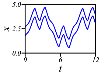
Figure 1 shows the motility behaviors for a cell with parameters chosen to be compatible with typical cells [20] and a range of adhesion complex mechanosensitivity and contractility strengths. The following motility behaviors are found: rest, moving at a constant velocity, unidirectional stick-slip movement, back-and-forth motion with stick-slip, and periodic back-and-forth movement. For a cell with weakly mechanosensitive adhesion complexes, as contractility increases, a cell at rest starts to move at constant velocity. As the adhesion complexes become more mechanosensitive, a moving cell shows other complex motility behaviors. For example, stick-slip motion and (at high contractivity) back-and-forth motion with stick-slip, and finally, the cell performs periodic back-and-forth motion when the adhesion complexes are highly mechanosensitive. Another motility phase diagram in the Supplement Materials [20] shows that, within our model, a cell with a high actin polymerization rate can exhibit other complex motility behaviors between stick-slip and periodic back-and-forth movements.
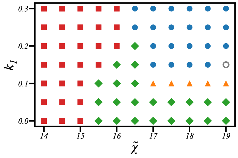
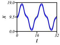
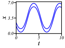
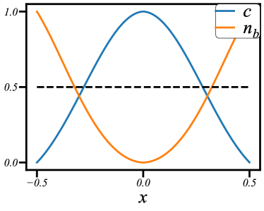
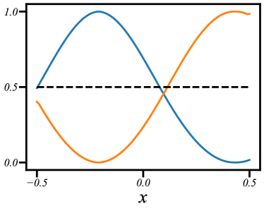
Figure 2 shows that when the cell is at rest, myosin motor distribution is symmetric around the center of the cell, and the number of adhesion complexes near both cell ends is the same; on the other hand, for a cell moving at constant velocity, myosin motors aggregate close to the trailing end and adhesion complexes are mainly close to the leading end. The distribution of myosin motors and adhesion complexes for a cell undergoes stick-slip, and periodic back-and-forth movements are shown in Fig. S2 of [20]. It is clear that whenever the cell has a definite moving direction, myosin motors aggregate in a regime behind the center of the cell, and more adhesion complexes form near the leading end than the trailing end. Indeed, the adhesion complex binding/unbinding rates Eq.(5) and net actin polymerization at the cell ends Eq.(6) in our model lead to reasonable molecular distributions in a cell.
Reduction to the simplified model.– To obtain an intuitive physical picture of the complex motility behaviors, especially the transitions from the rest state to the constant-velocity movement and periodic back-and-forth movement, we construct a simplified model from the active gel model. First, we consider a limiting situation in which adhesion complexes only appear in a small region close to the cell ends. In this regime, it is convenient to introduce , the total number of adhesion complexes close to , and , to describe the distribution of adhesion complexes. We also introduce , the dipole moment of myosin motors density relative to the center of the cell [20], to characterize the spatial distribution of myosin motors. The net actin polymerization velocity at the cell ends , where in the limit of small cell velocity. Therefore we write . In this regime, straightforward calculation shows that the velocity of the cell is [20]
| (10) |
where and are positivet coefficients that depend on and . Note that, from the definition, cannot be greater than unity. Therefore a cell with and has positive . This is in agreement with experimental observations.
We further simplify the analysis by considering the limit of large , i.e., . The following equations are constructed to describe the dynamics of , , and . First, the evolution equations for and are
| (11) |
where , , and are positive constants. Next, in the spirit of Landau-type approximation, the evolution of is assumed to obey the following equation,
| (12) |
where and are treated as -independent constants for simplicity.
The simple model equations (11)(12) have a solution with constant and . This corresponds to a cell at rest. Solutions with nonzero constant and correspond to a cell moving at a constant velocity; solutions with time-periodic and are stick-slip (periodic back-and-forth) movement if the time-average of and are nonzero (zero). The linear stability analysis of the rest state shows that as the contractility increases, a cell at rest starts to move as the system undergoes a bifurcation, the moving state is the constant-velocity state when is small, i.e., the adhesion complexes are less mechanosensitive. When is sufficiently large, the rest-to-moving transition leads to a periodic back-and-forth moving state [20].
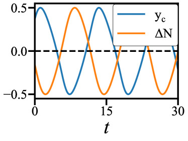
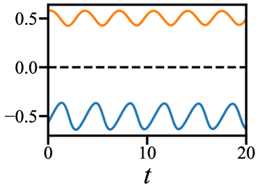
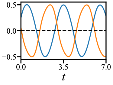
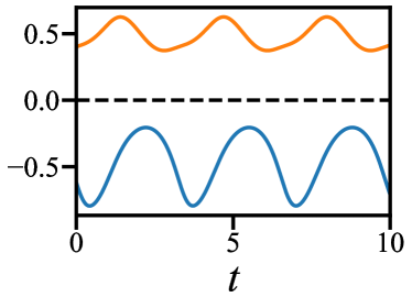
As shown in Fig. S3 [20], the model Eqs. (11) (12) exhibit a motility phase diagram qualitatively the same as the numerical solutions of our active gel model. The minor differences come from those simplifications made when constructing the simplified model. Furthermore, from the simplified model, it is easy to see how the symmetry properties and the couplings of the key driving variables lead to the observed cell motion. For example, and in Fig. 3(a) for a cell performing periodic back-and-forth movement suggests the following physical picture about how the coupling terms in the simplified model lead to this motion. According to Eq. (11), a small tend to increase when is sufficiently negative, and Eq. (12) states that when , tends to move toward the center of the cell when the magnitude of is sufficiently large. The result is that at sufficiently large , the number of adhesion complexes in the leading end of a moving cell increases sufficiently fast such that at some point, the myosins are pulled to the other half of the cell, reversing the sign of , then reversing the sign of , and eventually the direction of cell motion is reversed. This is how rest/constant-velocity transition becomes rest/back-and-forth transition as the adhesion complexes are more mechanosensitive. In between the constant velocity and periodic back-and-forth movement, complex motility patterns with stick-slip can be observed. As shown in Fig. 3(b), and in a cell that undergoes stick-slip movement have nonzero time-average values, and they oscillate with similar phase-relations as a cell undergoes periodic back-and-forth movement. This is because the mechanosensitivity of the adhesion complexes is sufficiently strong to induce an oscillation of and , but not sufficiently strong to change their signs. Figure 3(c) and Fig. 3(d) show that and (defined as the difference of the total number of adhesion complexes in the leading and trailing halves of the cell) in the numerical simulations of the active gel model behave similarly, suggesting that the physical picture obtained from studying the simplified model can be applied to more detailed models.
Discussion.– Within our active gel model, a cell with highly mechanosensitive adhesion complexes can exhibit periodic back-and-forth movement similar to what was observed in zyxin-depleted cells in a collagen matrix. Since zyxin proteins act as mechanosensors in mature adhesion complexes [21], our study suggests that the difference in the mechanosensitivity of the adhesion complexes in zyxin-depleted and wild-type cells could be the origin of the periodic back-and-forth movement observed in [5]. Future experiments can be designed to examine this prediction.
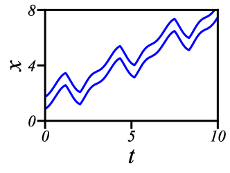
The physical picture suggested by our simplified model also implies the possibility that, in general, when some of the simplifications are lifted, more complex one-dimensional cell motility behaviors can be found. This is indeed the case, as we explore the behavior of our active gel model for a broader range of actin polymerization rates, complex trajectories which come from further bifurcations are found. This is shown in Fig. S1 in [20] and Fig. 4. Further study of the physical mechanisms for these behaviors will be our future work [22].
Finally, although the physical mechanisms for symmetry-breaking transitions, such as rest/periodic back-and-forth transition and rest/constant velocity transition, can be understood from the dynamics of and , it is interesting to study how other important physical observables, such as the multipoles of the traction force [23][24], behave in cells with different moving patterns. It is also important to check if the basic features of these physical observables in different moving patterns depend on the details of the binding/unbinding dynamics of adhesion complexes, as it plays a significant role in our understanding of many interesting features of cell motility.
Acknowledgments –
H.-Y. C. thanks Prof. Jasnow (University of Pittsburgh) for stimulating discussions and encouragement in the early stage of this work. H.-Y. C. is supported by the Ministry of Science and Technology, Taiwan (MOST 108-2112-M-008-016 ). The authors also acknowledge the support from National Center for Theoretical Sciences, Taiwan.
References
- [1] D. Bray, Cell Movements, 2nd ed. (Talyor and Francis, New York, 2001).
- [2] P.T. Yam, C.A. Wilson, L. Ji, B. Hebert, E.L. Barnhart, N.A. Dye, P.W. Wiseman, G. Danuser, and J.A. Theriot, Actin–myosin network reorganization breaks symmetry at the cell rear to spontaneously initiate polarized cell motility, J. Cell Biol., 178, 1207 (2007).
- [3] I.S. Aranson ed., Physical Models of Cell Motility, Springer, 2016.
- [4] Y. Aratyn-Schaus and M. L. Gardel, Transient frictional slip between integrin and the ECM in focal adhesions under myosin II tension, Curr. Biol. 20, 1145 (2010).
- [5] S. I. Fraley, Y. Feng, A. Giri, G. D. Longmore, and D.Wirtz, Dimensional and temporal controls of three-dimensional cell migration by zyxin and binding partners, Nat. Commun. 3, 719 (2012).
- [6] Chabaud, M. et al. Cell migration and antigen capture are antagonistic processes coupled by myosin II in dendritic cells. Nature Commun. 6, 7526 (2015).
- [7] B.A. Camley, Y. Zhao, B. Li, H. Levine, and W.-J. Rappel, Periodic migration in a physical model of cells on micropatterns, Phys. Rev. Lett., 111, 158102 (2013).
- [8] P. Sens, Stick-slip model for actin-driven cell protrusions, cell protrusions, cell polarization, and crawling, Proc. Natl. Acad. Sci. 117, 24670 (2020).
- [9] J.E. Ron, P. Monzo, M.C. Gauthier, R. Voituriez, and N.S. Gov, One-dimensional cell motility patterns, Phys. Rev. Res., 2, 033237 (2020).
- [10] F. Jülicher, K. Kruse, J. Prost, and J.-F. Joanny, Active behavior of the Cytoskeleton, Phys. Rep. 449, 3 (2007).
- [11] P. Recho, T. Putelat, and L. Truskinovsky, Contraction-driven cell motility, Phys. Rev. Lett. 111, 108102 (2013).
- [12] K. Tawada, K. Sekimoto, Protein friction exerted by motor enzymes through a weak-binding interaction’, J. Theo. Biol., 150, 193 (1991).
- [13] P. Recho and L. Truskinovsky, “Cell locomotion in one dimension,” in Physical Models of Cell Motility, p. 135, Springer, 2016.
- [14] T. Putelat, P. Recho, and L. Truskinovsky, Mechanical stress as a regulator of cell motility,’ Phys. Rev. E, 97, 012410 (2018).
- [15] R. Meili and R.A. Firtel, Two poles and a compass, Cell, 114, 153 (2003).
- [16] Y. T. Maeda, J. Inose, M. Y. Matsuo, S. Iwaya, and M. Sano, Ordered patterns of cell shape and orientational correlation during spontaneous cell migration, PLoS one, 3, e3734 (2008).
- [17] T. Y.-C. Tsai, S.R. Collins, C.K. Chan, A. Hadjitheororou, P.-Y. Lam, S.S. Lou, H.W. Yang, J. Jorgensen, F. Ellett, D. Irimia, M.W. Davidson, R.S. Fischer, A. Huttenlocher, T. Meyer, J.E. Ferrell Jr, and J.A. Theriot, Efficient front-rear coupling in neutrophil chemotaxis by dynamic myosin II localization, Dev. Cell 49, 189 (2019).
- [18] M. Nickaeen, I. L. Novak, S. Pulford, A. Rumack, J. Brandon, B. M. Slepchenko, and A. Mogilner, A free-boundary model of a motile cell explains turning behavior, PLoS Comp. Biol, 13, e1005862 (2017).
- [19] G. Horton and S. Vandewalle, A space-time multigrid method for parabolic partial differential equations, SIAM Journal on Scientific Computing, 16, 848 (1995).
- [20] See our Supplementary Material.
- [21] T.P. Lele, J. Pendse, S. Kumar, M. Salanga, J. Karavitis, and D.E. Ingber, Mechanical forces alter zyxin unbinding kinetics within focal adhesions of living cells, J. Cell Physiol., 207, 187 (2006).
- [22] J.-Y. Lo and H.-Y. Chen, manuscript in preparation.
- [23] H. Tanimoto and M. Sano, A simple force-motion relation for migrating cells revealed by multipole analysis of traction stress, Biophys. J., 106, 16 (2014).
- [24] T. Ohta, M. Tarama, and M. SanoA simple model of cell crawling, Physica D, 318, 3 (2016).