Manipulation of the magnetic state of a porphyrin-based molecule on gold: From Kondo to quantum nanomagnet via the charge fluctuation regime
Résumé
By moving individual Fe-Porphyrin-based molecules with the tip of Scanning Tunneling Microscope in the vicinity of a Br-atom containing elbow of the herringbone-reconstructed Au(111), we reversibly and continuously control their magnetic state. Several regimes are obtained experimentally and explored theoretically: from the integer spin limit, through intermediate magnetic states with renormalized magnetic anisotropy, until the Kondo-screened regime, corresponding to a progressive increase of charge fluctuations and mixed valency due to an increase in the interaction of the molecular Fe states with the substrate Fermi sea. Our results open a route for the realization, tuning and experimental studies of novel quantum magnetic states in molecule-surface hybrids.
I Keywords
spin-flip excitation, spin states, magnetic anisotropy, Kondo, charge fluctuation, mixed-valence states, scanning tunneling microscopy, transition metal complexes
II Introduction
Addressing and manipulating the spin state of molecular species at interfaces is a challenge that could greatly benefit spintronics Khajetoorians et al. (2015), nanoelectronics Kumar and Ruben (2017); Molnár et al. (2018) and quantum electronics Heinrich et al. (2013) in the near future. When the valence of a magnetic molecule deposited on a surface is integer, the description of spin-polarized molecular orbitals can be done in the framework of the atomic limit in terms of crystal field and spin-orbit coupling: the spin state is simply interpreted as a quantum magnet for which the Hund’s rule determines the fundamental magnetic state. However, the atomic limit is no longer valid in the mixed-valence regime which is the most general behavior of an interacting magnetic impurity with an electron bath.
Among the most studied classes of magnetic molecules are the transition metal phthalocyanine (Pc) and porphyrin (P) families which are easily deposited at surface in vacuum and studied by various local probe microscopies and spectroscopies. In these molecules, the spin state is mainly given by the spin polarization of the central transition metal ion d-states Tao et al. (2022); Zhang et al. (2011). Several works have focused on the influence of external parameters on the magnetic ground state such as the influence of charge transfer to the orbitals of the molecule Zhao et al. (2008); Chen et al. (2008); Krull et al. (2013); Garnica et al. (2014); Zhang et al. (2015); Karan et al. (2016); Ormaza et al. (2017); Karan et al. (2018); Granet et al. (2020), the effect of surface spin-orbit coupling and magnetic anisotropy Hiraoka et al. (2017); Rubio-Verdú et al. (2018), the coupling to the substrate, Hiraoka et al. (2017); Girovsky et al. (2017); Kügel et al. (2018); Baljozović et al. (2021), the interaction with attached and neighboring molecules Isvoranu et al. (2011); Wang et al. (2017), the structural deformation Parks et al. (2010); Li et al. (2018) or the chemical substitution of ligands Hou et al. (2020). For all these studies, a systematic understanding of the effect of mixed valence and charge fluctuations is still missing, although they have a strong influence on the effect of magnetic anisotropy.
Here we show that charge fluctuations in Fe 5,15-di-4-pyridyl-10,20-di-4-bromophenyl porphyrin () molecules adsorbed on the Br decorated Au(111) surface allow the magnetic state of the molecule to be driven between high-spin (S=1) and Kondo-screened states in a reversible and continuous manner through the intermediate valence regimes.
III Results and discussion
III.1 Structural and spectroscopic properties of monomers
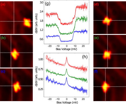
were deposited in ultrahigh vacuum on Au(111) and studied in situ by scanning tunneling microscopy (STM) and spectroscopy (STS) at 1.3K (see Methods section for more details). The molecules are randomly located on hcp domains, fcc domains or stacking faults lines of the herringbone reconstruction Barth et al. (1990) of Au(111) and appear as bright spots in constant-current topographic STM images in figures 1 (a-f). The molecules do not exhibit a planar configuration on the Au(111) surface because two pyrrole rings bend toward the vacuum and exhibit a higher height in STM images; the other two pyrrole rings bend toward the substrate, resulting in a lower height in images. Such deformation corresponds to the saddling distortion of porphyrin-based molecules Ishizuka et al. (2022) that is quite commonly observed Kim et al. (2009); Wang et al. (2015); Rubio-Verdú et al. (2018); Chang et al. (2019); Meng et al. (2023). Thus, the shape of the molecules in STM images allows us to determine the central position of the Fe-atom, the porphine macrocycle and the bromophenyl and pyridyl ligands attached to the macrocycle, as well as their location relative to the surface reconstruction. Importantly, during deposition and annealing, some Br-atoms detach from the molecules Grill et al. (2007), migrate at the surface, and get trapped by highly reactive elbows of the Au(111) herringbone reconstruction Meyer et al. (1996); Clair et al. (2006). The intersection of the dotted lines in figures 1 (a-c) points to the position of these elbows decorated by Br-adatoms.
Depending on their position on the surface, the molecules exhibit different spectral signatures. When a molecule is located on the hcp and fcc domains (figures 1 (a-c)), the tunneling spectra show conductance steps characteristic of a spin = 1 quantum magnet affected by the presence of magnetic anisotropy Lambe and Jaklevic (1968); Heinrich et al. (2004); Tsukahara et al. (2009); Rubio-Verdú et al. (2018); Meng et al. (2023), figure 1 (g). The steps of conductance are due to the opening of additional spin-flip tunneling channels trough inelastic excitations. This is in agreement with the known behavior of Fe-Pc and Fe-P which behave as S=1 nanomagnets once deposited on the Au(111) surface Minamitani et al. (2012a); Hiraoka et al. (2017); Wang et al. (2017); Li et al. (2018); Rubio-Verdú et al. (2018); Meng et al. (2023). The presence of single step or double step is a quite common phenomenon, e.g. ref. Tsukahara et al. (2009); Rubio-Verdú et al. (2018); Meng et al. (2023) and supplementary information of ref. Rubio-Verdú et al. (2018). The step-like spin-flip signatures are recorded at both upper pyrrole and Fe-atom locations, providing evidence for hybridization of the molecular states of the pyrrole with the Fe magnetic d-states Rubio-Verdú et al. (2018); Meng et al. (2023).
When the molecule is located on the elbow of the reconstruction, it behaves differently and exhibits a spectral resonance at the Fermi level. The molecule rotation induced by the microscope tip over the elbow site barely affects the shape and amplitude of the spectral resonance, figures 1 (d-f, h).
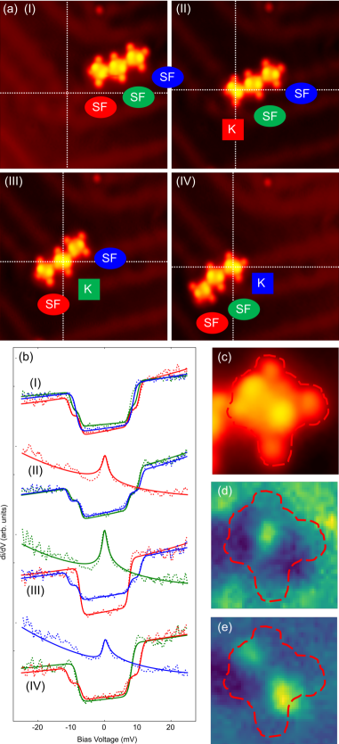
In the following we show that the two distinct spectral signatures are fully controlled by the adsorption site of the molecule. To this end we have prepared chains of 3 covalently bonded molecules by Ulmann’s coupling (see Methods section). In figure 2, a trimer chain is moved by the tip of the microscope to various positions of the reconstructed surface. The targeted locations are the hcp and fcc domains and the Br-decorated elbow of the reconstruction. In the first panel (I) of figure 2, the 3 molecules are located inside a fcc domain. In panels (II-IV), the molecular chain is sequentially repositioned with the microscope tip to move the iron center of each molecule over the Br-decorated site. When the molecules are located inside fcc and hcp domains, the spectra exhibit inelastic excitations of independent spin 1 nanomagnets in presence of magnetic anisotropy, similar to which was already measured for monomers in figure 1. This observation means that, here, neither the nature of the covalent bonds nor the substrate mediated interaction are efficient enough for coupling the molecules together. The spectra usually display a symmetric double-step structure. Following the standard analysis Heinrich et al. (2004), the characteristic voltages of the steps is interpreted to be related to the out of plane and in plane magnetic anisotropy energies. Depending on adsorption sites, the out of plane and in plane magnetic anisotropy energies can vary from to meV and from to meV respectively. The out of plane anisotropy parameters correspond to typical values of Fe porphyrin and phthalocyanine based magnetic molecules adsorbed on gold Tsukahara et al. (2009); Li et al. (2018); Hiraoka et al. (2017); Rubio-Verdú et al. (2018); Meng et al. (2023). The variation of the measured transverse anisotropy can be explained by the many possible configurations of the two fold symmetry molecules on the highly inhomogeneous Au(111) surface.
In panels (II-IV), the molecules are moved one by one above a Br-decorated elbow. In each case, the molecule exhibits a spectral resonance at 0 bias similarly to the monomer in figure 1. In figure 2, the resonance is found to be reversible once the molecule is removed from the elbow site and independent of the selected molecule. We identify the spectral peak as an Abrikosov-Suhl resonance Abrikosov (1965); Suhl (1965); Nagaoka (1965) (also named Kondo peak) due to the many body Kondo interactions of the Fe d states with the substrate electron bath. The characteristic Kondo temperature, K, evaluated by fitting the lineshape with a Frota function Frota and Oliveira (1986); Frota (1992), is found to be independent of the molecular orientation with respect to the surface atomic lattice. We therefore expect that magnetic anisotropy plays no role.
The Kondo effect originating from degenerate triplet ground state is extremely sensitive to magnetic anisotropy which tends to lift the degeneracy Parks et al. (2010). In the present case, meV is much smaller than the measured anisotropy. Three possible phenomena can explain the robustness of the Kondo effect on the magnetic anisotropy occurring at the Br-decorated elbow site: 1. The charge fluctuations due to the coupling of the Fe states with the substrate states Jacob (2018); Žitko et al. (2021); Blesio and Aligia overwhelm the magnetic anisotropy energy at this location, effectively restoring the degeneracy and screening the magnetic S=1 moment; 2. A sizable charge transfer from the substrate or the ligands onto the Fe atom induces a spin reduction from S = 1 to S = 1/2, resulting in a spin 1/2 Kondo effect which is naturally immune against magnetic anisotropy Jacob (2018); 3. The deformation of the molecule at this position induces the suppression of the magnetic anisotropy energy Parks et al. (2010). This last explanation is not in agreement with our tunneling topography studies which did not reveal a significant deformation of the molecule whose shape is preserved on the Br-site. It has also been shown that the anisotropy energy is not significantly affected when a similar molecule is pressed onto the surface with the tip of a microscope while staying away from the tip-molecule contact Hiraoka et al. (2017); Karan et al. (2018); Meng et al. (2023). Scenario 2, which assumes that charge transfer from the substrate gives rise to a molecular doublet, is the natural interpretation and has been analyzed in previous studies exploring different configurations Ormaza et al. (2017); Karan et al. (2018). The experimental data, DFT calculations and theoretical interpretation that follow lean more towards the first scenario in the present case.
III.2 Density Functional Theory analysis
DFT was used to study the electronic properties as a function of the distance of the molecule from the substrate. The latter is not easily determined by standard state-of-the-art DFT calculations describing the electronic properties accurately because it involves a delicate equilibrium with Van der Waals forces Hermann et al. . We have therefore used this distance as a parameter.
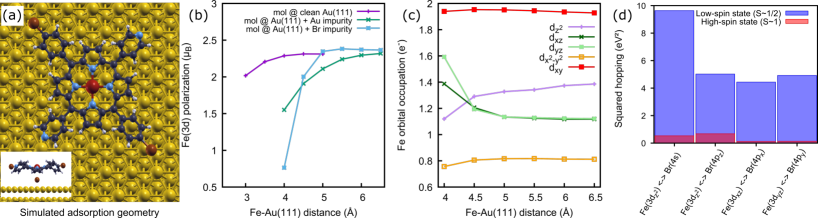
In order to rationalize the effect of the Br-adatom on the electronic properties of , two sets of situations were simulated: 1. as a function of the distance to the genuine Au surface 2. as a function of the distance to a Br (or Au) adatom on Au slabs positioned below the Fe atom of the molecule .
The Fe 3d states are dominant in the coupling with the substrate through the Br states. Fe dxz, dyz states are mainly hybridized with the pyrroles molecular orbitals. Therefore, further analysis will be discussed on the basis of the Fe 3d hybrids with molecular orbital. In particular the Fe dxz, dyz and d hybrid states were proven to be at the origin of the observed phenomena.
The DFT simulation shows that the magnetic polarization (contrary to the total charge) of the Fe atom, figure 3 (b), depends strongly on the molecule-surface distance when the surface is decorated with a Br (or Au) adatom, whereas it is much less sensitive when approaching the clean surface. When the molecule is away from the Br site, the total occupancy and magnetic moment are about 6.4 and which corresponds to a configuration close to the spin 1 state (high spin state) and the hybridization of the Fe d states with the substrate is small. This is in good agreement with the measured spectroscopic signature of a spin 1 quantum magnet when the molecule is above fcc or hcp domains. Indeed, the lack of a Kondo signature may be the result of weak hopping integrals from Fe orbitals to surface orbitals when the molecule is far away. Hybridization with the substrate cannot compete with the magnetic anisotropy and the Kondo effect is prevented. The magnetic anisotropy energy dominates the physics and the experimental spectroscopic signature is that of a spin 1 subjected to an anisotropy of a few meV.
Moving the molecule of 1 Å towards the Br adatom, from 5 to 4 Å, leaves the occupancy of the Fe states roughly unchanged (only a minor reshuffling of the charge distribution between the orbitals is observed (figure 3 (c)) - but induces a strong reduction of the magnetic polarization from about to about . The approximately constant charge suggests that charge fluctuations are the cause of this reduction, which is confirmed by the fact that the hybridization of the Fe and Br states increases exponentially as the molecule is moved closer to the surface (the squared hoppings that enters in this form in the hybridization function between the impurity and the substrate are plotted in the figure 3 (d)). The squared hopping integrals of the Fe states to the surface are indeed one order of magnitude smaller when the molecule is far away. The Fe 3d hybrids and Br 4s states, which have a almost perfect rotational symmetry, are supposed to dominate the physics when the molecule is closer (figure 3 (d)). This configuration is favorable to the emergence of a charge transport that is insensitive to magnetic anisotropy and independent of the relative orientation between the molecule and the surface once the iron atom is located just above the adsorbed Br atom, in perfect agreement with observations.
The cases presented above are perfectly consistent with the scenario in which the molecule evolves from a quasi-atomic moment (spin 1) under the influence of magnetic anisotropy to an Fe atom carrying a screened magnetic moment resulting from a strong hybridization with the substrate. Finally, the DFT calculations clearly indicate that the strength of the charge fluctuations should vary with the distance to the Br atom. Indeed the transition from spin 1 nanomagnet to Kondo screening, through the mixed-valence regime has been experimentally probed in a reversible manner and summarized in figure 4.
III.3 Crossover from spin 1 nanomagnet to Kondo-screened ground state through the mixed-valence regime
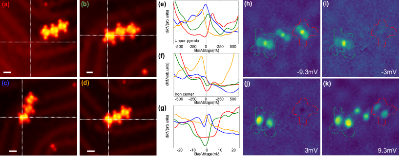
It was possible to finely tune the electronic configuration of the iron atom by using a chain of molecules in order to stabilize positions for which a molecule deviates slightly from the Br site. The studied position of the molecules are presented in topography images (a-d) of figure 4 and the density of states close to the Fermi energy are measured by STS spectra figure 4 (g)). The spin 1 signature is observed when the molecule is far from the elbow site as already mentioned above, figure 4 (a). When the molecule is positioned in such a way that the Br site is located below the upper pyrrole ring, image 4 (b), the LDOS shows a broadened and asymmetric step like shape originating from spin flip inelastic excitations but with a lower characteristic energy, indicating a renormalization of the anisotropy energy. At intermediate positions where the Br adsorption-site is located in between the Fe site and the upper pyrrole location, 4 (c), the spectroscopy shows a small anti-resonance at the Fermi level with a Fano-like shape which we believe is the extrapolation of the inelastic spin flip signature but for stronger charge fluctuations. When the molecule is strictly coinciding with the Br-adsorption site, figure 4 (d), it is exhibiting a Kondo peak. Therefore, the situation of images 4 (b) & (c) corresponds to two intermediate mixed-valence regimes between spin 1 nanomagnet and Kondo-screened ground states. Indeed as shown by D. Jacob in Ref. Jacob (2018) the gradual increase of charge fluctuations alters the spectroscopic signatures of the spin 1 nanomagnet by renormalizing the magnetic excitations to lower energy. This gradually closes the gap and finally restores a Kondo peak as in the case of image 4 (d). In this process, the absence of particle-hole symmetry causes a typical Fano resonance to appear. The renormalization of the magnetic excitations is evidenced in pannels 4 (h-k) where differential conductance is used to map the inelastic spin-flip probability density in the real space in the same configuration as in (b). This situation corresponds to the occurrence of one molecule in the intermediate state and 2 molecules in the regime of the spin 1 nanomagnet. At mV, spin-flip channels are open in all 3 molecules and the probability to induce a spin flip follows a two-lobes spatial pattern in all three molecules. At mV, panels (i) & (j) the spin-flip excitations are hindered by the magnetic anisotropy in the molecules far from the Br-site. On the contrary, the molecule in the intermediate regime shows spin-flip processes already being developed at mV, indicating the renormalization of the magnetic excitation energy to a smaller scale.
This transition through a mixed-valence regime might be due to a net increase of Fe charge while remaining still in a regime of weak coupling with the substrate (our previously outlined scenario 2). Our DFT calculations rather suggest that the predominant effect is the drastic increase of the hopping from/to the substrate (scenario 1), instead. Accordingly, the spin crossover was found to be accompanied with an energy shift of the frontier orbitals which are localized at the upper pyrrole rings (frontier orbital A, FOA) and at the iron atom position (frontier orbital B, FOB). A carefully analysis of figures 4 (e) & (f) allows to follow the evolution of FOA and FOB energies when moving the molecule. FOA shows a continuous evolution of density of states from the unoccupied states towards occupied states when approaching the molecule to the Br-site, indicating an increase of the orbital occupation. FOB shows a reverse behavior with a shift from below the Fermi level towards the unoccupied states. The gap between the frontier orbitals is reducing when approaching the Br site so the d level splitting tends to be weakened and could be favoring a decrease of orbital momentum and a renormalization of the magnetic anisotropy in agreement with the closing of the steps of conductance due to spin-flip excitations in the measured data of panel 4 (g) and figures 4 (h-k).
Following the work in ref.Rubio-Verdú et al. (2018), the conductance images presented figure 2 (c-e) support the hybrid Fe 3dxz, dyz character of FOA and the strong Fe 3d character of FOB in agreement with the orbital occupations in the DFT calculations (figure 3 (c)). It is interesting to note that according to conductance images of figure 4 (h-k), the spin-flip probability density is following the 3dxz, dyz symmetry at energies above and below the Fermi energy (two-lobes shape). On the contrary, the 3d character of the excitations (bright localized spot at the Fe site) is only visible at negative bias, so for occupied states. This difference is explaining the breaking of the electron-hole symmetry observed in inelastic spectra.
The fano-like shape of the blue spectrum in figure 4 (g) occurs when the frontier orbitals are overlapping at the Fermi level, as visible in figure 4 (e) & (f) and when the charge fluctuations between orbitals are expected to be strong. The hypothesis of intermediate regimes driven by charge fluctuations is well described by prediction in ref. Jacob (2018) as well as the shape of the spectrum. However in the present case the frontier orbitals, could also recover their degeneracy when overlapping in the intermediate regimes, implying a possible Kondo SU(4) effect Minamitani et al. (2012a, b), although this is less likely in a molecular system Kügel et al. (2018) and cannot be easily distinguished from the simple effect of strong charge fluctuations Fernández et al. (2015). Finally, when the iron atom is located above the Br site, FOA migrates below the Fermi level and FOB is washed out towards incoherent excitation and only the Kondo resonance survives at .
IV Conclusion
We have revealed in experiments and DFT calculations that the spin state switching is caused by the strong hybridization potential at Br-decorated elbow sites which induce charge fluctuation and a renormalization of the magnetic anisotropy when molecules are adsorbed on these sites.
The direct consequence of charge fluctuations is to tune the energies of two molecular frontier orbitals which are respectively closest to the Fermi level in positive and negative energies. The energetic overlapping of these two frontier orbitals gives rise to low-energy spin excitation channels near Fermi level. Under the condition of relative small charge fluctuations, the two frontier orbitals are far from Fermi level and the overlapping is small, hence the molecule will have high spin (S = 1) state, while under strong charge fluctuation these two orbitals can cross Fermi level, resulting a renormalization of the magnetic anisotropy and ultimately the Kondo screened ground state.
Our work provides a new approach for tuning not only the spin state but also the intermediate valence character/charge fluctuation in hybrids made of molecules on reconstructed surfaces.
V Methods
Iron(III) 5,15-(di-4-bromophenyl)-10,20-(di-4-pyridyl)porphyrin chloride molecules molecules were ordered from PorphyChem company (95% purity, CAS not assigned). Samples were prepared under ultrahigh vacuum (base pressure mbar) just before their scanning tunneling studies. The gold substrate was prepared by repeated cycles of Ar ion sputtering followed by annealing at 900 K. The molecules were deposited by organic molecular beam epitaxy (OMBE) when the sample was kept at Room Temperature (Tcrucible=575K). It was observed that the molecule source is also depositing Br atoms on the surface of gold. We have attributed this concomitant deposition of Br with to the stochastic Br detaching from molecules inside the crucible when heated at evaporation temperature. In-crucible Br detaching form similar molecules was already reported in ref. Grill et al. (2007) at 590K. In our case, the low amount of deposited Br atoms was seen to be enough for occupying all the elbow sites of the herringbone reconstruction. Once the molecules deposited, the sample temperature was quenched at liquid nitrogen temperature then at helium liquid temperature in less than 20 minutes. For low coverage this procedure produces single monomers scattered on the surface. A post annealing at 400 K for 10 minutes was used for producing molecular chains by Ullman’s coupling at the surface following recipes in ref. Grill et al. (2007); Krasnikov et al. ; DiLullo et al. (2012); Lafferentz et al. (2012); Clair and de Oteyza (2019). As for other porphyrins deposited on surfaces, the chlorine atom is detached from the molecule during the deposition or the post-annealing.
STM/STS measurements were achieved in-situ in ultrahigh vacuum by means of a modified "Tyto SPM" from SPECSTM at a base temperature of 1.3 K; the tunneling bias was applied to the sample. Mechanically cut PtIr tips were used. Soft lateral manipulation as follows was used for moving the molecules 1) positioning the tip above the molecule to move 2) Turning off the microscope feedback loop and approaching the tip towards molecule in order to increase tip-molecule interaction 2) Moving the tip to the destination in constant height mode 3) Retracting the tip and engaging the feedback loop. The analyses of the microscopy and spectroscopy data were done with WSxM software Horcas et al. (2007) and homemade python procedures respectively Gao (2023).
The fit of spectra from figure 1 & 2 involved a large Gaussian background mimicking the frontier orbital DOS plus a specific function associated to spin-flips (step functions introduced in Lambe and Jaklevic (1968)) or Kondo ground states (Frota function). Marginally, a convolution with a Gaussian function was used to clean up the incoherent noise of all conductance spectra presented here.
The spin-polarized Density-Functional Theory (DFT) calculations have been carried out with v.6.8 of the Quantum ESPRESSO package Giannozzi et al. (2009, 2017) within the Projector Augmented-Wave (PAW) scheme Blöchl (1994) and using the Perdew-Burke-Ernzerhof (PBE) functional Perdew et al. (1996). PAW pseudopotentials have been taken from the PSlibrary Dal Corso (2014); PSl ; QEp .
The Au(111) surface has been modeled by a finite slab consisting of 3 gold layers with Åof vacuum between periodic replicas along the direction. The Au-Au nearest-neighbor distance has been set to Å, the PBE equilibrium distance of the corresponding fcc bulk structure, which is larger than the experimental room-temperature value Davey (1925).
Given the sizeable extension of the molecule, a supercell of Au atoms on the plane has been used in order to avoid interaction between its periodic replicas, for a total of 300 Au atoms in the simulation cell. The molecule geometry has been relaxed in the vacuum until all components of the forces were smaller than Ry/, with being the Bohr radius. Fe Orbital polarizations and Density of states projected over the atomic states of the Fe pseudopotential can be found in supporting information. The projected density of states (pDoS) and the Mulliken orbital occupations and polarizations have been evaluated by projecting the Kohn-Sham orbitals on the atomic states of the pseudopotentials. They have been computed by considering different adsorption geometries between the molecule and three different substrates: the clean Au(111) surface, the Au(111) surface with a Au impurity and the Au(111) surface with a Br impurity. The impurities have been placed at the preferred PBE adsorption site at the equilibrium distance, which has resulted in the hollow-hcp site for both the Au and the Br impurity, respectively at Åand Åfrom the Au(111) surface. All calculations have been performed with and a plane-wave cutoff of Ry on the wavefunctions and 320 Ry on the density. Brillouin zone integrals have been evaluated at the point using a gaussian smearing of Ry. The hopping parameters have been computed via the projection method as implemented in the Wannier90 package Pizzi et al. (2020).
VI Acknowledgement
Cheryl Feuillet-Palma is acknowledged for her constructive discussions. TG thanks Paolo Restuccia for fruitful discussions. LdM acknowledges Giorgio Sangiovanni for useful discussions. SV, DR and SP acknowledge the French National Research Agency for the support of the SHOGUN project, Re. ANR-22-CE09-0028-01. TG and LdM are supported by the European Commission through the ERC-CoG2016, StrongCoPhy4Energy, GA No724177.
Références
- Khajetoorians et al. (2015) A. A. Khajetoorians, M. Valentyuk, M. Steinbrecher, T. Schlenk, A. Shick, J. Kolorenc, A. I. Lichtenstein, T. O. Wehling, R. Wiesendanger, and J. Wiebe, Nature Nanotechnology 10, 958 (2015), ISSN 1748-3395, URL https://doi.org/10.1038/nnano.2015.193.
- Kumar and Ruben (2017) K. S. Kumar and M. Ruben, Coordination Chemistry Reviews 346, 176 (2017).
- Molnár et al. (2018) G. Molnár, S. Rat, L. Salmon, W. Nicolazzi, and A. Bousseksou, Advanced Materials 30, 1703862 (2018).
- Heinrich et al. (2013) B. W. Heinrich, G. Ahmadi, V. L. Müller, L. Braun, J. I. Pascual, and K. J. Franke, Nano Lett. 13, 4840 (2013), ISSN 1530-6984, URL https://doi.org/10.1021/nl402575c.
- Tao et al. (2022) L. Tao, Y.-y. Zhang, and S. Du, Wiley Interdisciplinary Reviews: Computational Molecular Science 12, e1591 (2022).
- Zhang et al. (2011) Y. Y. Zhang, S. X. Du, and H.-J. Gao, Physical Review B 84, 125446 (2011), URL https://link.aps.org/doi/10.1103/PhysRevB.84.125446.
- Zhao et al. (2008) A. Zhao, Z. Hu, B. Wang, X. Xiao, J. Yang, and J. G. Hou, J. Chem. Phys. 128, 234705 (2008), ISSN 0021-9606, URL https://doi.org/10.1063/1.2940338.
- Chen et al. (2008) X. Chen, Y.-S. Fu, S.-H. Ji, T. Zhang, P. Cheng, X.-C. Ma, X.-L. Zou, W.-H. Duan, J.-F. Jia, and Q.-K. Xue, Physical Review Letters 101, 197208 (2008), URL https://link.aps.org/doi/10.1103/PhysRevLett.101.197208.
- Krull et al. (2013) C. Krull, R. Robles, A. Mugarza, and P. Gambardella, Nature Materials 12, 337 (2013), ISSN 1476-4660, URL https://doi.org/10.1038/nmat3547.
- Garnica et al. (2014) M. Garnica, D. Stradi, F. Calleja, S. Barja, C. Díaz, M. Alcamí, A. Arnau, A. L. Vázquez de Parga, F. Martín, and R. Miranda, Nano Lett. 14, 4560 (2014), ISSN 1530-6984, URL https://doi.org/10.1021/nl501584v.
- Zhang et al. (2015) Q. Zhang, G. Kuang, R. Pang, X. Shi, and N. Lin, ACS Nano 9, 12521 (2015), ISSN 1936-0851, URL https://doi.org/10.1021/acsnano.5b06120.
- Karan et al. (2016) S. Karan, N. Li, Y. Zhang, Y. He, I.-P. Hong, H. Song, J.-T. Lü, Y. Wang, L. Peng, K. Wu, et al., Physical Review Letters 116, 027201 (2016), URL https://link.aps.org/doi/10.1103/PhysRevLett.116.027201.
- Ormaza et al. (2017) M. Ormaza, P. Abufager, B. Verlhac, N. Bachellier, M.-L. Bocquet, N. Lorente, and L. Limot, Nature Communications 8, 1974 (2017), ISSN 2041-1723, URL https://doi.org/10.1038/s41467-017-02151-6.
- Karan et al. (2018) S. Karan, C. García, M. Karolak, D. Jacob, N. Lorente, and R. Berndt, Nano Lett. 18, 88 (2018), ISSN 1530-6984, URL https://doi.org/10.1021/acs.nanolett.7b03411.
- Granet et al. (2020) J. Granet, M. Sicot, I. C. Gerber, G. Kremer, T. Pierron, B. Kierren, L. Moreau, Y. Fagot-Revurat, S. Lamare, F. Chérioux, et al., J. Phys. Chem. C 124, 10441 (2020), ISSN 1932-7447, URL https://doi.org/10.1021/acs.jpcc.9b11141.
- Hiraoka et al. (2017) R. Hiraoka, E. Minamitani, R. Arafune, N. Tsukahara, S. Watanabe, M. Kawai, and N. Takagi, Nature Communications 8, 16012 (2017), ISSN 2041-1723, URL https://doi.org/10.1038/ncomms16012.
- Rubio-Verdú et al. (2018) C. Rubio-Verdú, A. Sarasola, D.-J. Choi, Z. Majzik, R. Ebeling, M. R. Calvo, M. M. Ugeda, A. Garcia-Lekue, D. Sánchez-Portal, and J. I. Pascual, Communications Physics 1, 15 (2018), ISSN 2399-3650, URL https://doi.org/10.1038/s42005-018-0015-6.
- Girovsky et al. (2017) J. Girovsky, J. Nowakowski, M. E. Ali, M. Baljozovic, H. R. Rossmann, T. Nijs, E. A. Aeby, S. Nowakowska, D. Siewert, G. Srivastava, et al., Nature Communications 8, 15388 (2017), ISSN 2041-1723, URL https://doi.org/10.1038/ncomms15388.
- Kügel et al. (2018) J. Kügel, M. Karolak, A. Krönlein, D. Serrate, M. Bode, and G. Sangiovanni, npj Quantum Materials 3, 53 (2018), ISSN 2397-4648, URL https://doi.org/10.1038/s41535-018-0126-z.
- Baljozović et al. (2021) M. Baljozović, X. Liu, O. Popova, J. Girovsky, J. Nowakowski, H. Rossmann, T. Nijs, M. Moradi, S. F. Mousavi, N. C. Plumb, et al., Magnetochemistry 7 (2021), ISSN 2312-7481, URL https://www.mdpi.com/2312-7481/7/8/119.
- Isvoranu et al. (2011) C. Isvoranu, B. Wang, E. Ataman, K. Schulte, J. Knudsen, J. N. Andersen, M.-L. Bocquet, and J. Schnadt, The Journal of Chemical Physics 134, 114710 (2011), eprint https://doi.org/10.1063/1.3563635, URL https://doi.org/10.1063/1.3563635.
- Wang et al. (2017) Y. Wang, X. Li, X. Zheng, and J. Yang, J. Chem. Phys. 147, 134701 (2017), ISSN 0021-9606, URL https://doi.org/10.1063/1.4996970.
- Parks et al. (2010) J. J. Parks, A. R. Champagne, T. A. Costi, W. W. Shum, A. N. Pasupathy, E. Neuscamman, S. Flores-Torres, P. S. Cornaglia, A. A. Aligia, C. A. Balseiro, et al., Science 328, 1370 (2010), ISSN 0036-8075, eprint https://science.sciencemag.org/content/328/5984/1370.full.pdf, URL https://science.sciencemag.org/content/328/5984/1370.
- Li et al. (2018) R. Li, N. Li, H. Wang, A. Weismann, Y. Zhang, S. Hou, K. Wu, and Y. Wang, Chem. Commun. 54, 9135 (2018), URL http://dx.doi.org/10.1039/C8CC02994F.
- Hou et al. (2020) J. Hou, Y. Wang, K. Eguchi, C. Nanjo, T. Takaoka, Y. Sainoo, R. Arafune, K. Awaga, and T. Komeda, Communications Chemistry 3, 36 (2020), ISSN 2399-3669, URL https://doi.org/10.1038/s42004-020-0282-5.
- Barth et al. (1990) J. V. Barth, H. Brune, G. Ertl, and R. J. Behm, Phys. Rev. B 42, 9307 (1990), URL https://link.aps.org/doi/10.1103/PhysRevB.42.9307.
- Ishizuka et al. (2022) T. Ishizuka, N. Grover, C. J. Kingsbury, H. Kotani, M. O. Senge, and T. Kojima, Chem. Soc. Rev. 51, 7560 (2022), URL http://dx.doi.org/10.1039/D2CS00391K.
- Kim et al. (2009) H. Kim, W.-j. Son, W. J. Jang, J. K. Yoon, S. Han, and S.-J. Kahng, Physical Review B 80, 245402 (2009), URL https://link.aps.org/doi/10.1103/PhysRevB.80.245402.
- Wang et al. (2015) W. Wang, R. Pang, G. Kuang, X. Shi, X. Shang, P. N. Liu, and N. Lin, Physical Review B 91, 045440 (2015), URL https://link.aps.org/doi/10.1103/PhysRevB.91.045440.
- Chang et al. (2019) M. H. Chang, Y. H. Chang, N.-Y. Kim, H. Kim, S.-H. Lee, M.-S. Choi, Y.-H. Kim, and S.-J. Kahng, Physical Review B 100, 245406 (2019), URL https://link.aps.org/doi/10.1103/PhysRevB.100.245406.
- Meng et al. (2023) X. Meng, J. Möller, M. Mansouri, D. Sánchez-Portal, A. Garcia-Lekue, A. Weismann, C. Li, R. Herges, and R. Berndt, ACS Nano 17, 1268 (2023), ISSN 1936-0851, URL https://doi.org/10.1021/acsnano.2c09310.
- Grill et al. (2007) L. Grill, M. Dyer, L. Lafferentz, M. Persson, M. V. Peters, and S. Hecht, Nature nanotechnology 2, 687 (2007).
- Meyer et al. (1996) J. Meyer, I. Baikie, E. Kopatzki, and R. Behm, Surface science 365, L647 (1996).
- Clair et al. (2006) S. Clair, S. Pons, S. Fabris, S. Baroni, H. Brune, K. Kern, and J. V. Barth, J. Phys. Chem. B 110, 5627 (2006), ISSN 1520-6106, URL https://doi.org/10.1021/jp057239s.
- Lambe and Jaklevic (1968) J. Lambe and R. Jaklevic, Physical Review 165, 821 (1968).
- Heinrich et al. (2004) A. J. Heinrich, J. A. Gupta, L. C. P., and E. D. M., Science 306, 466 (2004), URL https://doi.org/10.1126/science.1101077.
- Tsukahara et al. (2009) N. Tsukahara, K.-i. Noto, M. Ohara, S. Shiraki, N. Takagi, Y. Takata, J. Miyawaki, M. Taguchi, A. Chainani, S. Shin, et al., Physical Review Letters 102, 167203 (2009), URL https://link.aps.org/doi/10.1103/PhysRevLett.102.167203.
- Minamitani et al. (2012a) E. Minamitani, N. Tsukahara, D. Matsunaka, Y. Kim, N. Takagi, and M. Kawai, Physical Review Letters 109, 086602 (2012a), URL https://link.aps.org/doi/10.1103/PhysRevLett.109.086602.
- Abrikosov (1965) A. A. Abrikosov, Physics Physique Fizika 2, 5 (1965), URL https://link.aps.org/doi/10.1103/PhysicsPhysiqueFizika.2.5.
- Suhl (1965) H. Suhl, Physical Review 138, A515 (1965), URL https://link.aps.org/doi/10.1103/PhysRev.138.A515.
- Nagaoka (1965) Y. Nagaoka, Physical Review 138, A1112 (1965), URL https://link.aps.org/doi/10.1103/PhysRev.138.A1112.
- Frota and Oliveira (1986) H. Frota and L. Oliveira, Physical Review B 33, 7871 (1986).
- Frota (1992) H. Frota, Physical Review B 45, 1096 (1992).
- Jacob (2018) D. Jacob, Physical Review B 97, 075428 (2018), URL https://link.aps.org/doi/10.1103/PhysRevB.97.075428.
- Žitko et al. (2021) R. Žitko, G. G. Blesio, L. O. Manuel, and A. A. Aligia, Nature Communications 12, 6027 (2021), ISSN 2041-1723, URL https://doi.org/10.1038/s41467-021-26339-z.
- (46) G. G. Blesio and A. A. Aligia (????), URL https://doi.org/10.48550/arXiv.2301.08159.
- (47) J. Hermann, R. A. J. DiStasio, and A. Tkatchenko, 117, 4714 (????), ISSN 0009-2665, URL https://doi.org/10.1021/acs.chemrev.6b00446.
- Minamitani et al. (2012b) E. Minamitani, N. Tsukahara, D. Matsunaka, Y. Kim, N. Takagi, and M. Kawai, Physical Review Letters 109, 086602 (2012b), URL https://link.aps.org/doi/10.1103/PhysRevLett.109.086602.
- Kügel et al. (2018) J. Kügel, P.-J. Hsu, M. Böhme, K. Schneider, J. Senkpiel, D. Serrate, M. Bode, and N. Lorente, Physical Review Letters 121, 226402 (2018), URL https://link.aps.org/doi/10.1103/PhysRevLett.121.226402.
- Fernández et al. (2015) J. Fernández, A. A. Aligia, and A. M. Lobos, EPL (Europhysics Letters) 109, 37011 (2015), URL https://doi.org/10.1209/0295-5075/109/37011.
- (51) S. A. Krasnikov, C. M. Doyle, N. N. Sergeeva, A. B. Preobrajenski, N. A. Vinogradov, Y. N. Sergeeva, A. A. Zakharov, M. O. Senge, and A. A. Cafolla, 4, 376 (????), ISSN 1998-0000, URL https://doi.org/10.1007/s12274-010-0092-7.
- DiLullo et al. (2012) A. DiLullo, S.-H. Chang, N. Baadji, K. Clark, J.-P. Klöckner, M.-H. Prosenc, S. Sanvito, R. Wiesendanger, G. Hoffmann, and S.-W. Hla, Nano Lett. 12, 3174 (2012), ISSN 1530-6984, URL https://doi.org/10.1021/nl301149d.
- Lafferentz et al. (2012) L. Lafferentz, V. Eberhardt, C. Dri, C. Africh, G. Comelli, F. Esch, S. Hecht, and L. Grill, 4, 215 (2012), ISSN 1755-4349, URL https://doi.org/10.1038/nchem.1242.
- Clair and de Oteyza (2019) S. Clair and D. G. de Oteyza, Chem. Rev. 119, 4717 (2019), ISSN 0009-2665, URL https://doi.org/10.1021/acs.chemrev.8b00601.
- Horcas et al. (2007) I. Horcas, R. Fernández, J. Gomez-Rodriguez, J. Colchero, J. Gómez-Herrero, and A. Baro, Review of scientific instruments 78, 013705 (2007).
- Gao (2023) Y. Z. Gao, Lattices of Shiba states in supramolecular architechtures (PSL Research University, 2023).
- Giannozzi et al. (2009) P. Giannozzi, S. Baroni, N. Bonini, M. Calandra, R. Car, C. Cavazzoni, D. Ceresoli, G. Chiarotti, M. Cococcioni, I. Dabo, et al., J. Phys.: Condens. Matter 21, 395502 (2009), URL http://www.quantum-espresso.org.
- Giannozzi et al. (2017) P. Giannozzi, O. Andreussi, T. Brumme, O. Bunau, M. Buongiorno Nardelli, M. Calandra, R. Car, C. Cavazzoni, D. Ceresoli, M. Cococcioni, et al., J. Phys.: Condens. Matter 29, 465901 (2017).
- Blöchl (1994) P. E. Blöchl, Physical review B 50, 17953 (1994).
- Perdew et al. (1996) J. P. Perdew, K. Burke, and M. Ernzerhof, Phys. Rev. Lett. 77, 3865 (1996), URL https://link.aps.org/doi/10.1103/PhysRevLett.77.3865.
- Dal Corso (2014) A. Dal Corso, Computational Materials Science 95, 337 (2014).
- (62) PSlibrary on github, https://dalcorso.github.io/pslibrary/.
- (63) Quantum ESPRESSO pseudopotential tables, http://pseudopotentials.quantum-espresso.org/legacy_tables.
- Davey (1925) W. P. Davey, Physical Review 25, 753 (1925).
- Pizzi et al. (2020) G. Pizzi, V. Vitale, R. Arita, S. Blügel, F. Freimuth, G. Géranton, M. Gibertini, D. Gresch, C. Johnson, T. Koretsune, et al., Journal of Physics: Condensed Matter 32, 165902 (2020).