:
\theoremsep
A Machine Learning Case Study for AI-empowered echocardiography of Intensive Care Unit Patients in low- and middle-income countries
Abstract
We present a Machine Learning (ML) study case to illustrate the challenges of clinical translation for a real-time AI-empowered echocardiography system with data of ICU patients in LMICs. Such ML case study includes data preparation, curation and labelling from 2D Ultrasound videos of 31 ICU patients in LMICs and model selection, validation and deployment of three thinner neural networks to classify apical four-chamber view. Results of the ML heuristics showed the promising implementation, validation and application of thinner networks to classify 4CV with limited datasets. We conclude this work mentioning the need for (a) datasets to improve diversity of demographics, diseases, and (b) the need of further investigations of thinner models to be run and implemented in low-cost hardware to be clinically translated in the ICU in LMICs.
keywords:
Machine Learning, Artificial Intelligence, Echocardiography.1 Introduction
Echocardiography is an important clinical procedure in Intensive Care Units (ICUs) because of the features of Ultrasound (US) image modality such as portability, low cost, non-ionising radiation and its real-time capabilities to visualise cardiac anatomy (Vieillard-Baron et al., 2008; Campbell et al., 2018). Typically, the identification of cardiac abnormalities from 2D US views (Apical 4-Chamber (A4C), Parastemal Long-Axis and other six views) is achieved by specialist clinicians in echocardigraphy following the Focused Intensive Care Echo protocol (Hall et al., 2017). However, the application of point-of-care echocardiography of ICU patients in low- and middle-income countries (LMICs) faces two challenges: (1) intra-view variability of echocardiograms (physiological variations of patients and acquisition parameters) and inter-observer variability of expertise for sonographer and radiologist (Feigenbaum, 1996; Khamis et al., 2017; Field et al., 2011), and (2) limited number of specialist clinicians to perform US imaging analysis and to provide accurate diagnosis, and the limited equipment and hospitalisation requirements (Becker et al., 2016; Hao et al., 2021; Tran et al., 2021). One promising approach to address such challenges is the application of Machine Learing (ML) and Artificial Intelligence (AI) methods to echocardiography (Asch et al., 2022). AI-empowered echocardiography has been successful for detection of different apical views, inter-observer variability of sonographer’s expertise, implementation of one-stop AI models with multimodal imaging (US, MRI and clinical data), detection of high/low risk of heart failure, detection of endocardial borders and automatic left ventricle assessment in 2D echocardiography videos (Tromp et al., 2022; Zhang et al., 2022; Behnami et al., 2020; Ono et al., 2022). In spite of the success in applying AI and ML methods to support echocardiography, there are still important challenges for these methods to be integrated as clinical system and translated to clinical practice:
-
1.
inter-view similarity of echocardiograms (similar views of valve motion, wall motion, left ventricle, etc) and transducer position during acquisition when performing serial echoes (Zhang et al., 2018),
- 2.
-
3.
internal and external validation of AI-based models, data patient privacy to train commercial algorithms, and regulations of software as medical devices (Stewart et al., 2021).
The first and second challenges are important because of 2D US video data requires to appropriately be collected, validated and managed to apply AL and ML methods, and the third challenge because AI-based medical devices require to be aligned to standards to then be ready for clinical translation. Hence, the adoption of good machine learning practices (data curation, open-source code implementation, model selection, training and tuning; model validation and inference) might help to address such challenges.
This work presents a scoping review for (a) AI-empowered echocardiography for ICU in LMICs and (b) classification US images with thinner neural networks. We present a ML case study to classify A4C US image considering three thinner neural networks trained with curated data from ICU patients in LMICs. We present training results from SqueezeNet for different datasize, number of frames and clips batches. Further details for datsets, results of thinner networks are also added in the appendixes. We conclude with needs for clinical translation and add future work with validation of AI-based models.
2 Scoping review
2.1 AI-empowered echocardiography for ICU in LMICs
William and Marshall (2001) reviewed various AI-based applications in the ICU where real-time analysis of waveforms of electrocardiograms and electroencephalograms with neural network were used to identify cardiac ischemia and diagnosis of myocardial ischemia. Ghorbani et al. (2020) reported how deep learning models predicts systematic phenotypes from echocardiogram images which are difficult for human interpreters. Cheema et al. (2021) reported five patients with covid-19 in the ICU to illustrate ”how decision making affect in patient care” and how the use of AI-enabled tools provided real-time guidance to acquire desired cardiac 2D US views. Recently, Hong et al. (2022) reviewed 673 papers that apply ML methods to help making clinical decision in the ICU, of these studies the majority used supervised learning (91%) and few of them applied unsupervised learning and reinforcement learning methods. Similarly, Hong et al. (2022) identified 20 of the most frequent variables in ML pipelines for ICU patients, being the top five (age, sex, heart rate, respiratory rate, and pH), and mentionted that the most studied diseases are sepsis, infection and kidney injury. Despite such advances, there is little research on AI-empowered echocardiography used by clinicians in the ICU, specifically in LMICs. For instance, Tran et al. (2021) reported challenges in resourced limited ICUs including: infrastructure, education, personnel, data pipelines, regulation and trust in AI. Kerdegari et al. (2021b, a); Nhat et al. (2021) presented a deep-learning pipelines to classify lung US pathologies for ICU patients in LMICs, stating the challenges of data imbalance, integration of technology and the limited IT infrastructure.
2.2 Classification of US images with thinner neural networks
Classifying echochardiograms has been sucessfully done in the previous years (\sectionrefsubsec:nns_echochardiograms), however real-time deployment of AI-models suggest to require thinner networks. For instance, Baumgartner et al. (2017) proposed SonoNet which is a VGG-based architecture, having the same first 13 layers of VGG16, and SmallNet, loosely inspired by AlexNet, for real-time detection and bounding box localisation of standard views in freehand fetal US. Toussaint et al. (2018) applied four feature extraction networks couple with batchnormalization and soft proposal layer (VGG13-SP, VGG16-SP, ResNet18-SP, ResNet34-SP), resulting in real-time performance at inference time of 40 m per image (20Hz) Al-Dhabyani et al. (2019) applied AlexNet and transfer learning of four architectures (VGG16, Inception, ResNet, and NASNet) with and without augmentation techniques to perform tumor classification of breast US imaging. Authors stated that transfer learning with NASNet presented the best accuracy (99%) using BUSI+B datasets with DAGAN augmentation. Xie et al. (2020) proposed a dual-sampling convolutional neural network for US image breast cancer classification, being their network more efficient for such task than AlexNet, VGG16, ResNet18, GoogleNet and EfficientNet. Recently, Snider et al. (2022) reported summaries of CNN heuristics to detect shrapnel in US images, including layer activators, 2D CNN layer architectures, model optimisers dense nodes, and the effect of image augmentation and dropout rate and epoch number. Similarly, Boice et al. (2022) proposed ShrapML, a CNN model to detect shrapnel in US imaging. Authors compared ShrapML against DarkNet19, GoogleNet, MobileNetV2 and SqueezeNet, being ShrapML (8layers–6CNN and 2FC, making a network of 0.43 million parameters) 10x faster than MobileNetV2 and offering the highest accuracy.
3 Machine learning case study
fig:main-figure

3.1 Dataset
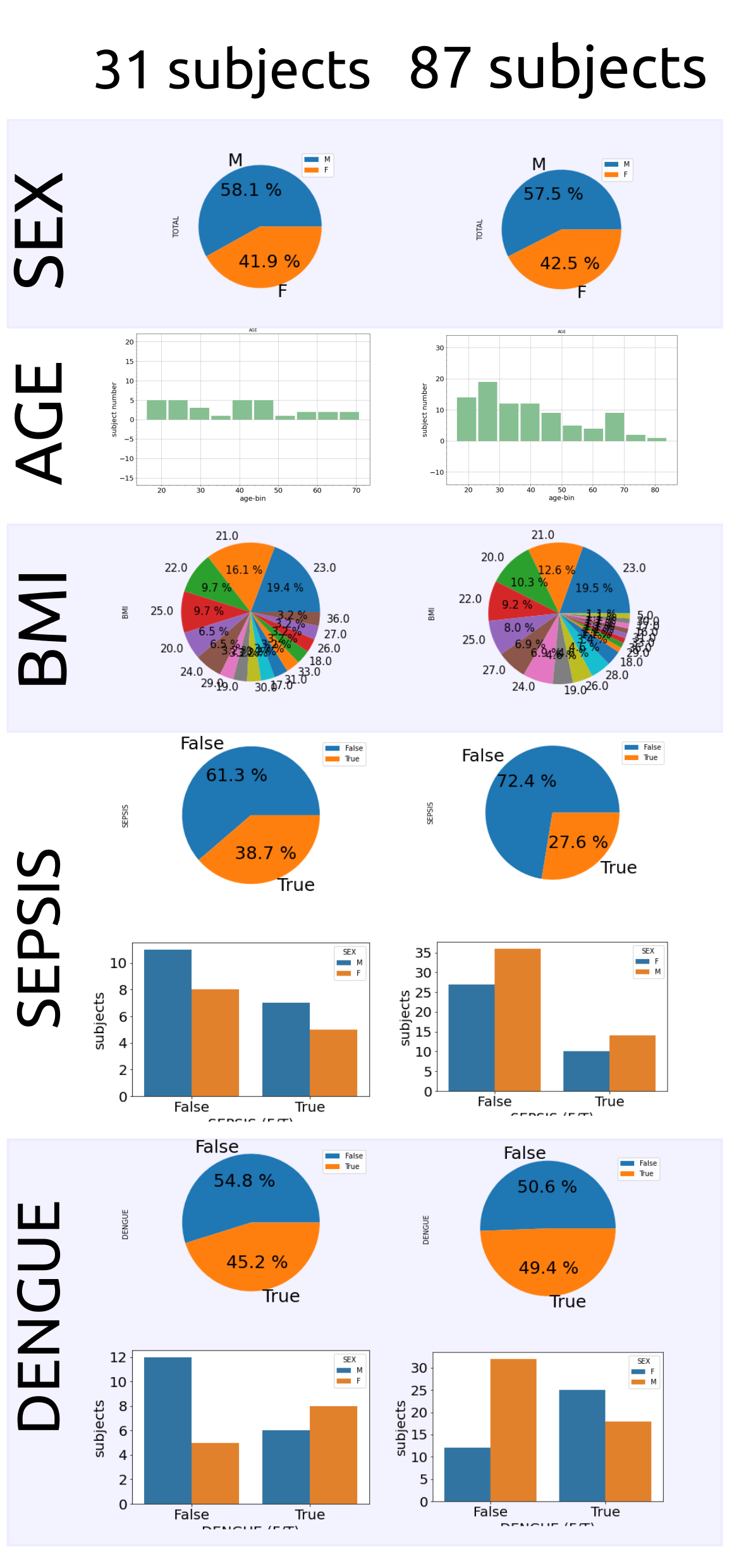
Echocardiography videos of 31 anonymised patients in the ICU were considered for this work which were collected by four radiologists using the clinical US devices: GE Venue Go machine with GE convex probe C1-5-D. The 31 patients had the following demographics: Sex: % (Male): 58.1%; Age: 38.70 (16.08) years; Weight: 61.51 (15.06) Kg; Height: 1.62 (0.07) m, and BMI: 23.80 (4.30). Values represent mean(std). See \appendixrefapd:datasets for further details on the demographics of the dataset, including the complete dataset of 87 patients.
3.1.1 Ethics statement
This study was approved by the anonymised entity one and anonymised entity two. All participants gave written informed consent to participate in the data collection before enrollment.
3.1.2 Data annotation, validation and management
Apical 4 Chamber view (A4C) is considered as an important view to compute heart failure measurements from 2D US echocardiography (Hall et al., 2017). Hence, for this work, timestamps in the video files for A4C were annotated by one research clinician of 10 years of experience using VGG Image Annotator (VIA). Then the same clinician and one researcher validated timestamps annotations where few filenames and timestamps were fixed (\figurereffig:main-figure(a)).
3.2 Model selection and heuristics
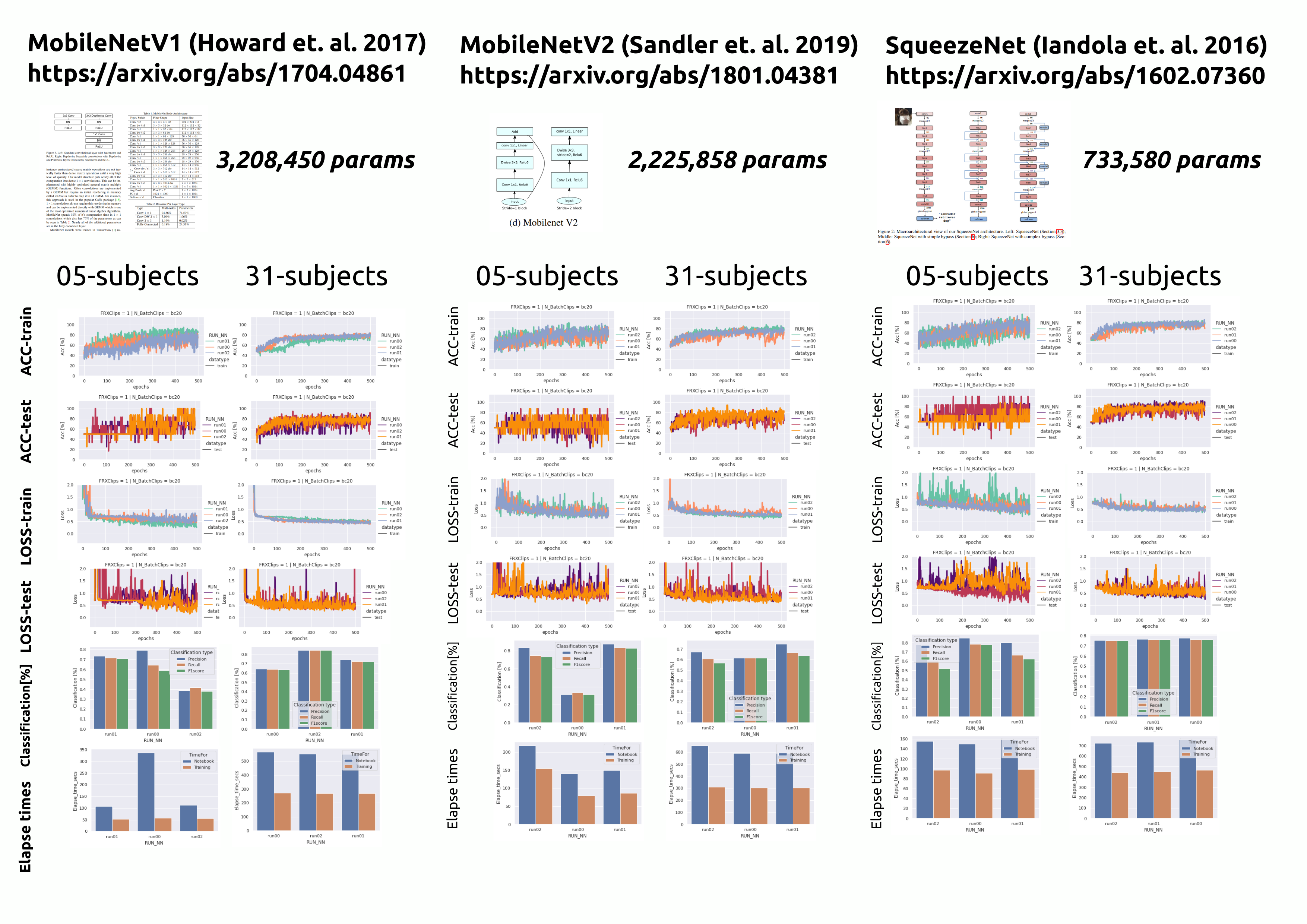
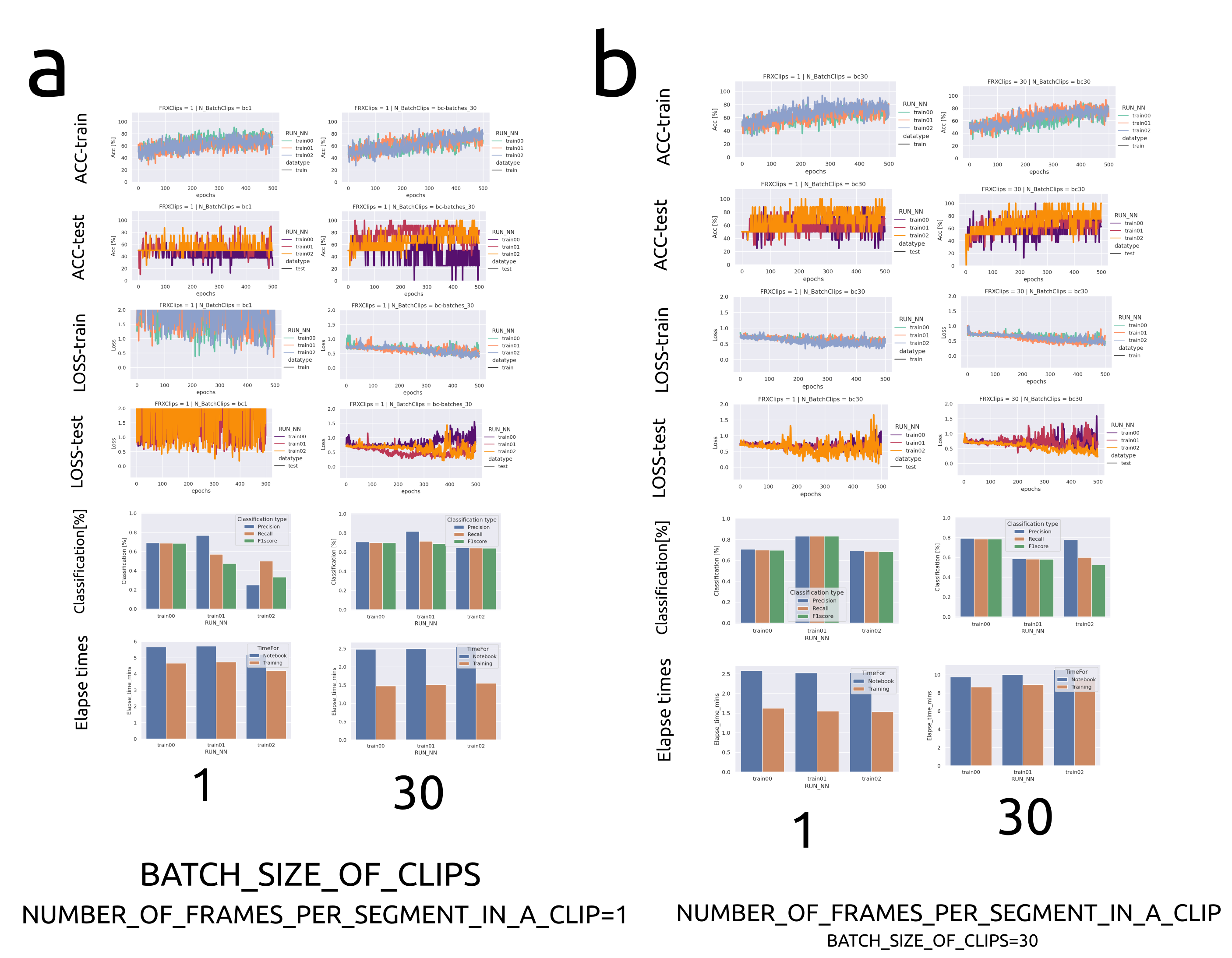
Considering different datasets characteristic (number of frames, clips, pixel image, clinical equipment, etc) and the number of parameters of different networks, we selected three neural networks for this work: MobileNetV1 (Howard et al., 2017) with 3,208,450 parameters, MobileNetV2 (Sandler et al., 2018) with 2,225,858 parameters, and SqueezeNet (Iandola et al., 2017) with 733,580 parameters. We then performed heuristics for each model to understand the impact of their performance for different hyperparameters (dataset size, frames numbers and clip length). MobileNetV1, MobileNetV2 and SqueezeNet were trained with data of 5 and 31 subjects (\appendixrefapd:heuristics) in which SqueezeNet showed constant training metrics. Hence, \figurereffig:heuristics_results_SqueezeNet_source0 presents the impact of different datasize, batch size of clips, and number of frames per segment in a clip for SqueezeNet.
fig:heuristics_results_SqueezeNet_source0
4 Conclusions and Future Work
We presented a ML case study that includes data selection, validation and management, model selection and validation. Our results show the promising use of thinner neural networks to address scarcity of datasets and model deployment in low-cost hardware. Future work will include real-time inference and deployment of thinner classification and segmentation models, leading to clinical translation of cardiac outputs of ICU patiens in LMICs. The code, data and other resources to reproduce this work will be available upon acceptance of this work.
References
- Al-Dhabyani et al. (2019) Walid Al-Dhabyani, Mohammed Gomaa, Hussien Khaled, and Aly Fahmy. Deep learning approaches for data augmentation and classification of breast masses using ultrasound images. International Journal of Advanced Computer Science and Applications, 10(5), 2019. 10.14569/IJACSA.2019.0100579. URL http://dx.doi.org/10.14569/IJACSA.2019.0100579.
- Asch et al. (2022) Federico M. Asch, Tine Descamps, Rizwan Sarwar, Ilya Karagodin, Cristiane Carvalho Singulane, Mingxing Xie, Edwin S. Tucay, Ana C. Tude Rodrigues, Zuilma Y. Vasquez-Ortiz, Mark J. Monaghan, Bayardo A. Ordonez Salazar, Laurie Soulat-Dufour, Azin Alizadehasl, Atoosa Mostafavi, Antonella Moreo, Rodolfo Citro, Akhil Narang, Chun Wu, Karima Addetia, Ross Upton, Gary M. Woodward, Roberto M. Lang, Vince Ryan V. Munoz, Rafael Porto De Marchi, Sergio M. Alday-Ramirez, Consuelo Orihuela, Anita Sadeghpour, Jonathan Breeze, Amy Hoare, Carlos Ixcanparij Rosales, Ariel Cohen, Martina Milani, Ilaria Trolese, Oriana Belli, Benedetta De Chiara, Michele Bellino, Giuseppe Iuliano, and Yun Yang. Human versus artificial intelligence–based echocardiographic analysis as a predictor of outcomes: An analysis from the world alliance societies of echocardiography covid study. Journal of the American Society of Echocardiography, 2022. ISSN 0894-7317. https://doi.org/10.1016/j.echo.2022.07.004. URL https://www.sciencedirect.com/science/article/pii/S0894731722003510.
- Baumgartner et al. (2017) Christian F. Baumgartner, Konstantinos Kamnitsas, Jacqueline Matthew, Tara P. Fletcher, Sandra Smith, Lisa M. Koch, Bernhard Kainz, and Daniel Rueckert. Sononet: Real-time detection and localisation of fetal standard scan planes in freehand ultrasound. IEEE Transactions on Medical Imaging, 36(11):2204–2215, 2017. 10.1109/TMI.2017.2712367.
- Becker et al. (2016) Dawn M. Becker, Chelsea A. Tafoya, Sören L. Becker, Grant H. Kruger, Matthew J. Tafoya, and Torben K. Becker. The use of portable ultrasound devices in low- and middle-income countries: a systematic review of the literature. Tropical Medicine & International Health, 21(3):294–311, 2016. https://doi.org/10.1111/tmi.12657.
- Behnami et al. (2020) Delaram Behnami, Christina Luong, Hooman Vaseli, Hany Girgis, Amir Abdi, Dale Hawley, Ken Gin, Robert Rohling, Purang Abolmaesumi, and Teresa Tsang. Automatic cine-based detection of patients at high risk of heart failure with reduced ejection fraction in echocardiograms. Computer Methods in Biomechanics and Biomedical Engineering: Imaging & Visualization, 8(5):502–508, 2020. 10.1080/21681163.2019.1650398. URL https://doi.org/10.1080/21681163.2019.1650398.
- Boice et al. (2022) Emily N. Boice, Sofia I. Hernandez-Torres, and Eric J. Snider. Comparison of ultrasound image classifier deep learning algorithms for shrapnel detection. Journal of Imaging, 8(5), 2022. ISSN 2313-433X. 10.3390/jimaging8050140. URL https://www.mdpi.com/2313-433X/8/5/140.
- Brindise et al. (2020) Melissa C. Brindise, Brett A. Meyers, Shelby Kutty, and Pavlos P. Vlachos. Unsupervised segmentation of b-mode echocardiograms, 2020.
- Campbell et al. (2018) Steven J. Campbell, Rabih Bechara, and Shaheen Islam. Point-of-care ultrasound in the intensive care unit. Clinics in Chest Medicine, 39(1):79–97, 2018. ISSN 0272-5231. https://doi.org/10.1016/j.ccm.2017.11.005. URL https://www.sciencedirect.com/science/article/pii/S0272523117301168. Interventional Pulmonology: An Update.
- Cheema et al. (2021) Baljash S. Cheema, James Walter, Akhil Narang, and James D. Thomas. Artificial intelligence–enabled pocus in the covid-19 icu: A new spin on cardiac ultrasound. JACC: Case Reports, 3(2):258–263, 2021. ISSN 2666-0849. https://doi.org/10.1016/j.jaccas.2020.12.013. URL https://www.sciencedirect.com/science/article/pii/S2666084920314637.
- Feigenbaum (1996) Harvey Feigenbaum. Evolution of echocardiography. Circulation, 93(7):1321–1327, 1996. 10.1161/01.CIR.93.7.1321. URL https://www.ahajournals.org/doi/abs/10.1161/01.CIR.93.7.1321.
- Field et al. (2011) Larry C. Field, George J. Guldan, and Alan C. Finley. Echocardiography in the intensive care unit. Seminars in Cardiothoracic and Vascular Anesthesia, 15(1-2):25–39, 2011. 10.1177/1089253211411734. URL https://doi.org/10.1177/1089253211411734. PMID: 21719547.
- Ghorbani et al. (2020) Amirata Ghorbani, David Ouyang, Abubakar Abid, Bryan He, Jonathan H. Chen, Robert A. Harrington, David H. Liang, Euan A. Ashley, and James Y. Zou. Deep learning interpretation of echocardiograms. npj Digital Medicine, 3(1):10, Jan 2020. ISSN 2398-6352. 10.1038/s41746-019-0216-8. URL https://doi.org/10.1038/s41746-019-0216-8.
- Hall et al. (2017) David P Hall, Helen Jordan, Shirjel Alam, and Michael A Gillies. The impact of focused echocardiography using the focused intensive care echo protocol on the management of critically ill patients, and comparison with full echocardiographic studies by bse-accredited sonographers. Journal of the Intensive Care Society, 18(3):206–211, 2017. 10.1177/1751143717700911. URL https://doi.org/10.1177/1751143717700911.
- Hao et al. (2021) NV Hao, LM Yen, R Davies-Foote, TN Trung, NVT Duoc, VTN Trang, PTH Nhat, DH Duc, NTK Anh, PT Lieu, TTD Thuy, DB Thuy, NT Phong, NT Truong, PB Thanh, DTH Tam, Z Puthucheary, and CL Thwaites. The management of tetanus in adults in an intensive care unit in southern vietnam [version 2; peer review: 3 approved]. Wellcome Open Research, 6(107), 2021. 10.12688/wellcomeopenres.16731.2.
- Hong et al. (2022) Na Hong, Chun Liu, Jianwei Gao, Lin Han, Fengxiang Chang, Mengchun Gong, and Longxiang Su. State of the art of machine learning–enabled clinical decision support in intensive care units: Literature review. JMIR Med Inform, 10(3):e28781, Mar 2022. ISSN 2291-9694. 10.2196/28781. URL https://medinform.jmir.org/2022/3/e28781.
- Howard et al. (2017) Andrew G. Howard, Menglong Zhu, Bo Chen, Dmitry Kalenichenko, Weijun Wang, Tobias Weyand, Marco Andreetto, and Hartwig Adam. Mobilenets: Efficient convolutional neural networks for mobile vision applications. CoRR, abs/1704.04861, 2017. URL http://arxiv.org/abs/1704.04861.
- Iandola et al. (2017) Forrest N. Iandola, Song Han, Matthew W. Moskewicz, Khalid Ashraf, William J. Dally, and Kurt Keutzer. Squeezenet: Alexnet-level accuracy with 50x fewer parameters and 0.5MB model size, 2017. URL https://openreview.net/forum?id=S1xh5sYgx.
- Kerdegari et al. (2021a) Hamideh Kerdegari, Phung Tran Huy Nhat, Angela McBride, Reza Razavi, Nguyen Van Hao, Louise Thwaites, Sophie Yacoub, and Alberto Gomez. Automatic detection of b-lines in lung ultrasound videos from severe dengue patients. In 2021 IEEE 18th International Symposium on Biomedical Imaging (ISBI), pages 989–993, 2021a. 10.1109/ISBI48211.2021.9434006.
- Kerdegari et al. (2021b) Hamideh Kerdegari, Nhat Tran Huy Phung, Angela McBride, Luigi Pisani, Hao Van Nguyen, Thuy Bich Duong, Reza Razavi, Louise Thwaites, Sophie Yacoub, Alberto Gomez, and VITAL Consortium. B-line detection and localization in lung ultrasound videos using spatiotemporal attention. Applied Sciences, 11(24), 2021b. ISSN 2076-3417. 10.3390/app112411697. URL https://www.mdpi.com/2076-3417/11/24/11697.
- Khamis et al. (2017) Hanan Khamis, Grigoriy Zurakhov, Vered Azar, Adi Raz, Zvi Friedman, and Dan Adam. Automatic apical view classification of echocardiograms using a discriminative learning dictionary. Medical Image Analysis, 36:15–21, 2017. ISSN 1361-8415. https://doi.org/10.1016/j.media.2016.10.007. URL https://www.sciencedirect.com/science/article/pii/S1361841516301876.
- Nhat et al. (2021) Phung Tran Huy Nhat, Hamideh Kerdegari, Angela McBride, Luigi Pisani, Nguyen Van Hao, Le Dinh Van Khoa, Shujie Deng, Le Ngoc Minh Thu, Duong Bich Thuy, VITAL Consortium, Marcus J. Schultz, Reza Razavi, Andrew P. King, Louise Thwaites, Sophie Yacoub, and Alberto Gomez. Lung ultrasound pathology classification for icu patient management in lmic. White paper, September 2021.
- Ono et al. (2022) Shunzaburo Ono, Masaaki Komatsu, Akira Sakai, Hideki Arima, Mie Ochida, Rina Aoyama, Suguru Yasutomi, Ken Asada, Syuzo Kaneko, Tetsuo Sasano, and Ryuji Hamamoto. Automated endocardial border detection and left ventricular functional assessment in echocardiography using deep learning. Biomedicines, 10(5), 2022. ISSN 2227-9059. 10.3390/biomedicines10051082. URL https://www.mdpi.com/2227-9059/10/5/1082.
- Sandler et al. (2018) Mark Sandler, Andrew Howard, Menglong Zhu, Andrey Zhmoginov, and Liang-Chieh Chen. Mobilenetv2: Inverted residuals and linear bottlenecks. In Proceedings of the IEEE Conference on Computer Vision and Pattern Recognition (CVPR), June 2018.
- Snider et al. (2022) Eric J. Snider, Sofia I. Hernandez-Torres, and Emily N. Boice. An image classification deep-learning algorithm for shrapnel detection from ultrasound images. Scientific Reports, 12(1):8427, May 2022. ISSN 2045-2322. 10.1038/s41598-022-12367-2. URL https://doi.org/10.1038/s41598-022-12367-2.
- Stewart et al. (2021) Jonathon E Stewart, Adrian Goudie, Ashes Mukherjee, and Girish Dwivedi. Artificial intelligence-enhanced echocardiography in the emergency department. Emergency Medicine Australasia, 33(6):1117–1120, 2021. https://doi.org/10.1111/1742-6723.13847. URL https://onlinelibrary.wiley.com/doi/abs/10.1111/1742-6723.13847.
- Toussaint et al. (2018) Nicolas Toussaint, Bishesh Khanal, Matthew Sinclair, Alberto Gomez, Emily Skelton, Jacqueline Matthew, and Julia A. Schnabel. Weakly supervised localisation for fetal ultrasound images. In Deep Learning in Medical Image Analysis and Multimodal Learning for Clinical Decision Support, pages 192–200, Cham, 2018. Springer International Publishing. ISBN 978-3-030-00889-5.
- Tran et al. (2021) Huy Nhat Phung Tran, Nguyen Van Hao, Luigi Pisani, Hamideh Kerdegari, Duong Bich Thuy, Le Ngoc Minh Thu, Truong Thi Phuong Thao, Le Thi Mai Thao, Ha Thi Hai Duong, MarcusJ. Schultz, Reza Razavi, Andrew P. King, Louise Thwaites, Sophie Yacoub, and Alberto Gomez. Role of ai-enabled ultrasound imaging in a resource limited intensive care unit. White paper, September 2021.
- Tromp et al. (2022) Jasper Tromp, Paul J. Seekings, Chung-Lieh Hung, Mathias Bøtcher Iversen, Matthew James Frost, Wouter Ouwerkerk, Zhubo Jiang, Frank Eisenhaber, Rick S. M. Goh, Heng Zhao, Weimin Huang, Lieng-Hsi Ling, David Sim, Patrick Cozzone, A. Mark Richards, Hwee Kuan Lee, Scott D. Solomon, Carolyn S. P. Lam, and Justin A. Ezekowitz. Automated interpretation of systolic and diastolic function on the echocardiogram: a multicohort study. The Lancet Digital Health, 4(1):e46–e54, Jan 2022. ISSN 2589-7500. 10.1016/S2589-7500(21)00235-1. URL https://doi.org/10.1016/S2589-7500(21)00235-1.
- Van Woudenberg et al. (2018) Nathan Van Woudenberg, Zhibin Liao, Amir H. Abdi, Hani Girgis, Christina Luong, Hooman Vaseli, Delaram Behnami, Haotian Zhang, Kenneth Gin, Robert Rohling, Teresa Tsang, and Purang Abolmaesumi. Quantitative echocardiography: Real-time quality estimation and view classification implemented on a mobile android device. In Danail Stoyanov, Zeike Taylor, Stephen Aylward, João Manuel R.S. Tavares, Yiming Xiao, Amber Simpson, Anne Martel, Lena Maier-Hein, Shuo Li, Hassan Rivaz, Ingerid Reinertsen, Matthieu Chabanas, and Keyvan Farahani, editors, Simulation, Image Processing, and Ultrasound Systems for Assisted Diagnosis and Navigation, pages 74–81, Cham, 2018. Springer International Publishing. ISBN 978-3-030-01045-4.
- Vieillard-Baron et al. (2008) Antoine Vieillard-Baron, Michel Slama, Bernard Cholley, Gérard Janvier, and Philippe Vignon. Echocardiography in the intensive care unit: from evolution to revolution? Intensive Care Medicine, 34(2):243–249, Feb 2008. ISSN 1432-1238. 10.1007/s00134-007-0923-5. URL https://doi.org/10.1007/s00134-007-0923-5.
- William and Marshall (2001) Hanson C. William and Bryan E. Marshall. Artificial intelligence applications in the intensive care unit. Critical Care Medicine, 29(2), 2001. ISSN 0090-3493. URL https://journals.lww.com/ccmjournal/Fulltext/2001/02000/Artificial_intelligence_applications_in_the.38.aspx.
- Xie et al. (2020) Jiang Xie, Xiangshuai Song, Wu Zhang, Qi Dong, Yan Wang, Fenghua Li, and Caifeng Wan. A novel approach with dual-sampling convolutional neural network for ultrasound image classification of breast tumors. Physics in Medicine and Biology, 65(24):245001, dec 2020. 10.1088/1361-6560/abc5c7. URL https://doi.org/10.1088/1361-6560/abc5c7.
- Zhang et al. (2018) Jeffrey Zhang, Sravani Gajjala, Pulkit Agrawal, Geoffrey H. Tison, Laura A. Hallock, Lauren Beussink-Nelson, Mats H. Lassen, Eugene Fan, Mandar A. Aras, ChaRandle Jordan, Kirsten E. Fleischmann, Michelle Melisko, Atif Qasim, Sanjiv J. Shah, Ruzena Bajcsy, and Rahul C. Deo. Fully automated echocardiogram interpretation in clinical practice. Circulation, 138(16):1623–1635, 2018. 10.1161/CIRCULATIONAHA.118.034338. URL https://www.ahajournals.org/doi/abs/10.1161/CIRCULATIONAHA.118.034338.
- Zhang et al. (2022) Zisang Zhang, Ye Zhu, Manwei Liu, Ziming Zhang, Yang Zhao, Xin Yang, Mingxing Xie, and Li Zhang. Artificial intelligence-enhanced echocardiography for systolic function assessment. Journal of Clinical Medicine, 11(10), 2022. ISSN 2077-0383. 10.3390/jcm11102893. URL https://www.mdpi.com/2077-0383/11/10/2893.
Appendix A Classification of echochardiograms
Khamis et al. (2017) considered 309 clinical echocardiograms of apical views which were visually classified and labelled by two experts into three classes: 103 A2C views, 103 A4C views and 103 ALX views to then applied spatio-temporal feature extraction (Cuboic Detector) and supervised learning dictionary (LC-KSVD) resulting in an overall recognition rate of 95%. Van Woudenberg et al. (2018) applied DenseNet and LSTM to extract temporal information on sequences of 16000 echo cine frames to classify 14 heart views with an average accuracy of 92.35%. Van Woudenberg et al. (2018) also presents timing diagrams to quantify frame arrival and real-time performance to operate at 30 frames per second, while providing feedback with a mean latency of 352.91 ± 38.27 m when measured from the middle of the ten-frame sequence. Zhang et al. (2018) performed view classification with 277 echocardiograms to create a 23-class models (including A4C no occlusions, A4C occluded LA, A4C occluded LV, etc) using 13-layer CNN with 5-fold cross-validation for accuracy assessment and resulting in 84% for overall accuracy where challenges for partial obscured LVs for A2C, A3C and A4C. Similarly, Zhang et al. (2018) applied U-net to segment 5 views (A2C, A3C, A4C, PSAX, PLAX) and CNN model for 3 cardiac diseases with the use of A4C capturing most of the information for the diseases.
Appendix B Datasets
fig:demographics illustrates demographics for sex, age, BMI, sepsis and dengue for the complete dataset and the 31 subjects considered for this work.
fig:demographics
Appendix C Heuristics of model selection
fig:ThinnerNeuralNetsResults illustratres heristics for accuraty, train, classification and elapse times of 5 and 31 subjects.
fig:ThinnerNeuralNetsResults