Contrast inversion in neutral atom microscopy using atomic cluster beams
Abstract
Abstract: This work explores the possibility of atomic cluster beams as a probe for neutral atom microscopy (NAM) measurements. Using a beam of Kr clusters with mean size 104 atoms/cluster we demonstrate that topographical contrast can be obtained, similar to that in the case of monoatomic beams. Further, using atomically thin films of grown on substrate we show that NAM imaging using Kr clusters is also possible in domains where topographical contrast is not expected. Surprisingly, these images show an inverted contrast pattern when compared to the case of monoatomic beams. We attempt to understand these observations on the basis of angular distributions resulting from cluster-surface scattering. Finally, we discuss the implications of these results towards achieving a high lateral resolution neutral atom microscope using atomic cluster beams.
I Introduction
Using beams of neutral atoms as a probe to image surfaces, also known as Neutral Atom Microscopy (NAM) or Scanning Helium Microscopy (SHeM, in case of He atoms), is an emerging microscopy technique that holds the promise of probing surfaces in a soft manner allison_2003 ; witham_NAM_2014 . Here, similar to charged particle based methods (like scanning electron microscopy), an atomic beam typically with incident kinetic energy in the range of 10 - 500 meV is made incident on the sample of interest and the scattered atoms are detected in a position-sensitive manner to generate a contrast map (image) of the surface. One of the major questions in this area of research is to understand contrast generation mechanisms, which is intimately connected to the underlying atom - surface collision dynamics. Another major challenge is to achieve high a lateral resolution. This is largely constrained by the limited ability to manipulate and control a beam of slow moving neutral atoms.
In the context of microscopy, several schemes to manipulate neutral atom beams have been put forward in the past. One approach is to focus atomic beams using precisely prepared surfaces having high reflectivity and appropriate shape, acting as mirrors for atomic beams holst_mirror1997 ; holst_ellipsoidalAtomMirror2010 ; farias_focussing2017 ; farias_graphenemirror_2011 ; farias_QuantumStab_AtomMirror2008 . Another approach has been to use the wave nature of atoms and focus atomic beams using zone plate structures toennies_zoneplate1999 ; allison_2003 ; koch_imaging_2008 ; holst_ZonePlate2008 ; holst_theoretical_model_zoneplate2017 . Despite several promising developments in these areas, focusing of atomic beams well beyond 1 m spot size remains a scientific and technological challenge.
Arguably, the most successful strategy to date is based on the pinhole design, where a series of apertures are used to collimate the incident atomic beam, consequently reaching a high lateral resolution. This design is relatively easier to build and has been widely adopted by several groups witham ; dastoor_NAM_design_2014 ; dastoor_NAM_Design_RSI2015 ; bhardwaj2022neutral . At present, some of the best images in terms of signal-to-noise ratio dastoor_NAM_Design_RSI2015 and resolution (sub-micron) witham_NAM_2014 have been achieved using such pinhole designs. An excellent overview of different NAM designs, following its systematic development and current state of the art is provided in a recent review article by Palau and coworkers holst_NAM_reviev2021 .
Despite the promising developments in pinhole based NAM, several challenges still remain to be overcome. Highest achievable lateral resolution is dictated largely by the dimensions of the final collimation aperture (pinhole). Using a smaller pinhole to increase the resolution is necessarily accompanied by a loss of signal-to-noise ratio, as the number of incident particles (and the corresponding scattered signal) decreases. Therefore NAM imaging becomes difficult in the region of sub-micron resolution. Additionally, with pinhole sizes less than 1 m, diffraction of incident atoms from the aperture starts becoming significant. This causes a lateral spread of the beam, thereby limiting the obtainable resolution for a given working distance. A detailed analysis of this situation using numerical simulations has been carried out by Palau and coworkers holst_theoretical_pinhole . By optimizing the positions of collimating apertures, working distance and accounting for lateral spread of the incident atomic beam caused by diffraction, they estimate that a resolution of 40 nm is achievable under realistic measurement conditions. It should be noted that such performance remains to be experimentally demonstrated yet.
In this regard, using beams of atomic clusters as a probe offers some interesting possibilities. Firstly, large atomic density of clusters can compensate for the loss of incident intensity, enabling the use of much smaller pinhole sizes. At the same time, heavier mass of individual clusters means negligible diffraction effects, even in case of small aperture sizes ( 1 m). It is worth pointing out that a necessary prerequisite to evaluate the potential of these possibilities is to understand whether the scattering of atomic clusters from surfaces can give rise to contrast maps or not. If in case contrast is observed, what is its nature and how does it compare with the usual scenario of monoatomic beams? These questions form the subject of our present study.
In the forthcoming sections, we describe the experimental methods used to produce and characterize atomic cluster beams. Following this we discuss the results of NAM measurements in topographical and beyond topographical contrast regime, where a contrast inversion is seen. We discuss the possible origins of this unusual contrast inversion and implications of these findings towards developing a high-resolution NAM.
II Experimental setup
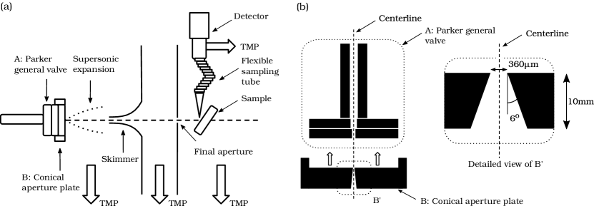
Experimental setup and sample preparation procedure used in the present work is largely the same as in our previous work bhardwaj2022neutral . Only specific features essential to understand the present work are described in detail below. Fig. 1(a) shows a schematic diagram of the experimental setup used in this work. A pulsed atomic beam source comprising of the following components was used: (A) pulsed solenoid valve (Parker 009-1643-900, orifice diameter = 0.5 mm) and (B) a custom-built aperture plate with a 10 mm long conical opening with the diameter of the smaller orifice being 360 m and a half opening angle of 6∘ (Fig. 1(b)). This aperture plate was mounted on the front plate of the pulsed valve while keeping their centers aligned. He or Kr gas, was allowed to expand supersonically from this atomic beam source into the source chamber. A 200 m skimmer was used to extract the center-line intensity from the gas expansion forming a beam in the first differential chamber. Finally a 50 m aperture, placed inline, was used to obtain a collimated beam. The width (full width half maximum, FWHM) of He and Kr beams, measured at a distance of 15 mm (sample plane) from the final collimation aperture using a knife edge scanning method, were observed to be 60 m and 56 m, respectively. These correspond to an angular divergence of 1 mrad (see SI-1). For He and Kr beams, based on the pressure changes observed in the detection chamber with the molecular beam on and off (without the sample), we estimate that each gas pulse consists of approximately 1010 atoms being incident on the sample. This corresponds to a flux of 1016 atoms/(sec str). The estimated incidence energies of the pure He and 50% Kr in He beams are 65 meV and 124 meV, respectively. The collimated beam scatters from the sample placed on a movable platform (XY) comprised of two piezoelectric stages stacked over each other, housed in the detection chamber.
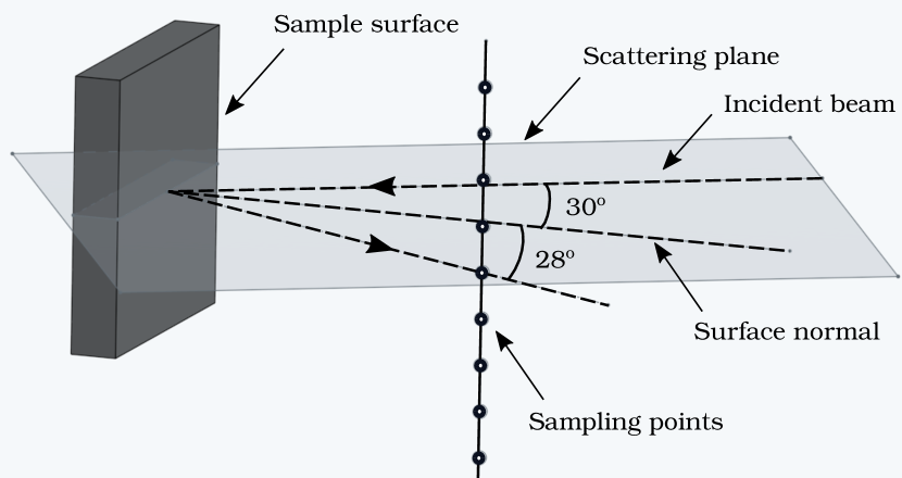
A 180 mm long flexible stainless steel bellow (inner diameter approximately 3.6 mm) was used as a sampling tube with an orifice of diameter 1 mm drilled at its end. One end of the sampling tube was mounted on a single axis manipulator, enabling measurement of the scattered signal at different angular positions. At present, in this set up the angular distribution measurements, were limited to a plane perpendicular to the scattering plane (Fig. 2). A significant distortion was observed in our attempts (results not shown here) in measuring in-plane angular distributions. Hence, we restrict ourselves to report and discuss only the out-of-plane scattered distributions in this work.
For NAM measurements, the pulsed valve was driven by a pulse valve driver (IOTA ONE, 060-0001-900, Parker). Opening time was set to 25 msec and the repetition rate at 2 Hz. Under these conditions, the steady state pressures in the source, first differential and detection chamber were 310-4 mbar, 510-6 mbar, and 310-7 mbar respectively. Mass spectrometer (SRS RGA 200) sampling rate was set to 40 Hz and the resulting signal corresponded to pulses of 250-300 ms being detected. Channeltron voltage was set to -1560 V corresponding to a nominal gain of 1.2104. Dwell time at each sample position (pixel) was set to 2.5 - 3.5 seconds depending on the signal-to-noise ratio and the overall duration of each measurement.
The additional aperture plate with a long conical orifice was used to aid in the formation of large clusters. The relation among average cluster size produced by a nozzle is described empirically by determining the scaling parameter () as given below hagena1972cluster ; hagena_clusters_ZphysD1987 ; hagena_ClusterIonSoures_RSI1992 ; ditmire_laserCluster_1996 ; ditmire_laserCluster_1998 :
Here, k is gas dependent condensation parameter wormer_k_parameter_1989 ; ditmire_laserCluster_1996 ; ditmire_laserCluster_1998 , 4 for He, 1700 for Ar, 2900 for Kr, is the half-angle of the conical aperture (in degrees), d is the orifice diameter (in m), is the backing pressure (in mbar), and is the initial gas temperature (in K).
In the present experiments a mixture of 50% Kr in He was used for NAM measurements with clusters as it resulted in the highest signal observed (see SI-2). In this case 1.6 (at 6 bar backing pressure). Under these conditions, based on the previously reported scaling relations among , average cluster size and condensation fraction hagena_clusters_ZphysD1987 ; wormer_k_parameter_1989 ; dorchies_ClusterSizeScaling_2003 , nearly all Kr atoms are expected to be in cluster form with a mean size of 104 atoms/cluster. On the other hand, for pure He beams under similar conditions, is 17 at 2 bar and 86 at 10 bar. Therefore, He beams are expected to be largely monoatomic in nature with negligible cluster formation. Additional measurements to characterize cluster formation were carried out by means of measuring the change in center-line intensity as a function of backing pressure (see SI-2) and X-ray generation by intense femtosecond laser ionization (see SI-3 and SI-4).
Samples of grown on substrate used in the present study were prepared using chemical vapor deposition method and characterized using optical microscopy and Raman spectroscopy as described previously bhardwaj2022neutral . Features on a typical sample consist of bare substrate, thin (one to three layers) and thick (more than 6 layers). Here one monolayer corresponds to a thickness of 0.65 nm changgu_MoS2Thickness_raman_2010 . These features correspond to the purple, blue and light-blue colors in the optical microscopy images obtained using white light illumination, respectively (see Figs. 4 and 5).
III Results and discussions
III.1 Generation of topographical contrast with Kr clusters
Topographic contrast is the most commonly observed contrast in NAM images koch_imaging_2008 . It arises in the regime where incident atoms undergo diffuse scattering upon impact with the surface and spatial features of interest are much larger than the beam spot size. Under these conditions NAM images closely resemble the geometric features of the sample. As far as clusters are concerned, a priori it is not obvious whether simple topographical contrast, commonly seen with monoatomic beams, can be observed or not. This ambiguity stems from the fact that extent of thermalization of Kr atoms, in the form of large clusters, upon impact with the surface is unknown.
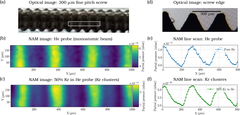
In order to understand if topographic contrast is generated or not, we first image a microscopically rough object (a fine-pitched stainless steel screw, pitch = 300 m) using both monoatomic Helium and Krypton cluster beam. Features being imaged in this case are much larger than the incident beam size and we can expect to see images largely governed by topographical contrast. Figure 3(a) shows an optical image of the screw. Panels (b) and (c) show NAM images of a small portion of the screw (marked by a white rectangle in panel (a)), obtained using a beam of Helium and Krypton clusters respectively. Panel (d) shows an optical image depicting the side view of the screw edge. Figures (e) and (f) depict the line profiles corresponding to the optical image shown in (d), measured using He atoms and Kr clusters, respectively. Quite clearly, the NAM images obtained using a beam of He (monoatomic) and Kr (clusters) show a one-to-one correspondence with the optical images. We conclude that large atomic cluster beams are well capable of generating topographical contrast. We also infer that Kr atoms get thermalized on the surface to a large extent.
These results are of potential interest towards developing a high resolution NAM. In measurements with monoatomic beams such as He, diffraction of incident atoms from the final aperture sets the ultimate limit to the highest achievable lateral resolution. For He atoms, using numerical modelling , this limit has been estimated to be approximately 40 nm holst_theoretical_pinhole . Large atomic clusters such as those used in our experiments, owing to their much higher mass ( 104 times compared to Helium atoms), are expected to behave like classical particles. Consequently, the diffraction effects will be negligible even for very small pinhole sizes, providing a route to achieve a higher lateral resolutions compared to monoatomic beams. In addition, the higher density offered by atomic clusters can lead to much higher incident and scattered signals even with smaller aperture sizes. We believe that the ultimate limit in this case is likely to be set by the interaction of incident atomic clusters with the edge of small pinhole they are sent through. At distances where inter-atomic binding energy of the cluster becomes comparable to the interaction energy with atoms constituting the edge of the pinhole, one can expect the clusters to fragment as they travel across the aperture. Given that these interactions are of Van der Waals type, such forces will be significant only at nm length scales. In principle, this can allow nm-sized pinholes to be used for collimation.
III.2 Beyond topographical contrast: Inverted contrast with Kr clusters
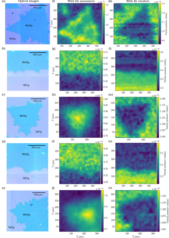
Regimes beyond topographic contrast correspond to situations where specific details of atom-surface collision play an important role in contrast generation. This is unlike the case of topographic contrast resulting from diffuse scattering, where there is no correlation among the incident and final momentum of scattered particles. As an example, chemical contrast barr_NatComm2016 has been hypothesized to arise from surface specific inelastic scattering with phonons, leading to a contrast dependent on the surface chemical composition. Contrast arising due to diffraction of the incident atomic beam as a result of scattering from surfaces with local crystalline order has also been reported recently jardine_diffraction . In our previous work, it was shown that using atom scattering based microscopy, thin films up to a single monolayer of on can be successfully imaged using a 20-30 m sized beam of He and/or Kr atoms as an incident probe. Further it was also observed that contrast was degrading with incident energy bhardwaj2022neutral . These results point towards the fact that contrast mechanisms beyond simple topographical in nature are at play. Here, we investigate whether NAM imaging of atomically thin layers of MoS2, in the beyond topographic contrast regime, is possible with a beam of Kr clusters or not.
Measurements with monoatomic beam of He and Kr atoms, produced with a 20 m continuous nozzle, can be seen in Fig. 4. Panel (a) shows an optical image of a small portion of grown on /Si substrate and the corresponding NAM images obtained with He and Kr beams are depicted in (b) and (c), respectively. The scattered flux of He and Kr from is consistently higher as compared to in both cases and a clear one-to-one correspondence is seen. Such kind of contrast generation with the underlying possible reasons have been discussed in our previous work bhardwaj2022neutral .
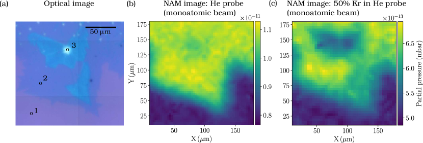
Measurements performed with a beam of Kr clusters and a monoatomic beam of He are shown in Fig. 5. Panels 5(a - e) show optical images of small portions of independently prepared samples of films on substrate. NAM measurements using He atom beam are shown in Fig. 5(f - j). Here we observe the expected contrast pattern as seen previously with monoatomic beams (Fig. 4), i.e. higher scattered flux from surface as compared to . Figs. 5(g - i) show the images obtained using Kr clusters. Again, a one-to-one correspondence with the optical images is seen, but interestingly, contrast patterns appear inverted. Here, a larger signal for scattered Kr is obtained from substrate as compared to regions covered with . Quite clearly, scattering of large Kr clusters does not simply lead to diffuse scattering alone and also seems to be sensitive to the surface characteristics. Observation of such a contrast inversion merits further discussion.
Previous studies on scattering of atomic and molecular clusters from surfaces, using experimental gspann_ClusterReflectionSS_1974 ; bernasek_Cluster_Fe_scattering_1988 ; vach_ArgonCluster_graphite_1992 ; vach_N2Clusters_Graphite_1999 and computational methods tully_ArClusters_Pt_1989 ; pettersson_ArClusters_Pt_1997 , offer valuable insights. These studies span a range of atomic and molecular clusters (such as He, H2, N2 and Ar) and different surfaces ranging from microscopically rough to atomically flat single crystals. A common feature arising in all these works is that for large clusters, angular distributions of the scattered particles show a prominent peak at angles much greater than specular direction (supraspecular). Further, it has also been reported that on rough surfaces (prepared by ion sputtering of atomically flat, clean single crystal surfaces) this pronounced supraspecular scattering becomes weaker and the angular distributions tend towards diffuse scattering (peaking at surface normal) bernasek_Cluster_Fe_scattering_1988 .
This general aspect of cluster scattering from surfaces seems significant as far as our observation of contrast inversion is concerned. For monoatomic beams, the scattered flux is expected to peak close to the specular direction in case of elastic scattering or towards the surface normal, in case of a large diffuse scattering component. On the other hand, for large Kr clusters (n 104), the majority of the scattered flux is likely to exit at large angles from surface normal, in supraspecular direction. Given that our sampling aperture is placed near specular direction, a large fraction of the supraspecular scattered atoms will not be captured by the detector. Our previous study has shown that at an atomic scale, surfaces are generally smoother than the substrate bhardwaj2022neutral . Given the above points, we hypothesize that on surface, supraspecular scattering leads to relatively fewer atoms entering the collection aperture placed along the specular direction. At the same time, the increased roughness on substrate can lead to more diffuse scattering like behavior, resulting in a relatively higher scattered signal being detected.
Besides the above possibility, other systematic differences could also be at play here. films are bound to the lower layers or substrate by relatively weak Van der Waals interaction. As a result, surface acts as a relatively softer landing site for Kr clusters. Large Kr clusters having sizable incidence energy compared to its monoatomic counterparts, when scattered from a relatively softer surface (compared to ) can lead to a broad scattered distribution. Consequently, a lower flux will be measured by the detector placed in specular direction compared to scattering from a relatively rigid surface, consistent with the inverted contrast observed in our measurements. The effect of rigidity of thin films on the scattered angular distributions have been studied by Taleb and co-workers farias_interlayerForces_2018 by studying the heavy vs. light atom scattering from graphene surface in the case of weak and strong interaction with the substrate. They observed that in case of weak interaction with substrate (Gr/Ir(111)), neon atoms scattered largely inelastically leading to a broad angular distribution. On the other hand, scattering from a more strongly bound substrate such as Gr/Ni(111), sharp diffraction peaks were obtained.
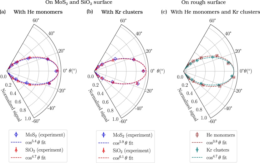
In order to understand these points better, we resort to angle-resolved measurements. Figs. 6(a) and (b) show a comparison of the angular distributions resulting from scattering on and surfaces, obtained using He (monoatomic) and Kr (clusters), respectively. Angular distributions from a microscopically rough surface (stainless steel screw, shown in Fig. 3 are also shown in (c) for the sake of comparison. It should be noted that these distributions were measured for an out-of-plane scattering configuration (see Fig. 2) and are not corrected for any distortions resulting from the measurement configuration. Nonetheless, a systematic comparison of the relative changes based on these results is still possible.
A common feature observed in the case of and is that angular distributions obtained using monoatomic He are rather broad and only slightly narrower than that observed from a rough surface. This indicates that a substantial fraction of the atoms undergo diffuse scattering. This is expected since our samples were placed in a vacuum chamber operating at a base pressure of 310-7 mbar and not true ultrahigh vacuum conditions. Further, no in-situ sample cleaning was done. Under these conditions, a significant amount of adsorbates will be present on the surface, leading to a large diffuse scattering component. For He beam, width of the distribution in case of surface, is somewhat narrower than substrate, consistent with the contrast observed in NAM images. Angular distributions obtained with Kr clusters appear very similar for and . Given that these distributions almost look same and the fact that we see an inverted contrast pattern, we infer that the changes are largely occurring in the in-plane scattering distributions. This is also expected from previous studies showing supraspecular scattering. A clear answer to these questions can be obtained by in-plane angle-resolved measurements, which is currently unavailable in our setup. An upgraded version with provision for measuring both, in-plane and out-of-plane angular distributions with the aid of a combined rotation and linear manipulator along with sample heating capability (for removing weakly bound adsorbates) is being designed in our lab for future studies.
IV Concluding Remarks
In this work we have demonstrated that NAM imaging is possible using a beam of Kr atom clusters and the well-known topographic contrast can be obtained for rough surfaces. Using samples of atomically thin films of grown on substrate, we have shown that NAM imaging with Kr clusters is possible even in the regime of beyond simple topographical contrast. Interestingly, here we observe a contrast inversion compared to the similar measurements made with monoatomic beams.
Importantly, the results presented here clearly establish that NAM imaging can be done with atomic clusters as well. To the best of our knowledge, this possibility has not been explored previously. These results point toward two interesting possibilities in the direction of developing a high lateral resolution NAM. Firstly, the higher atomic density of clusters can be exploited to obtain high incident beam flux which can in turn allow the use of smaller pinholes leading to higher lateral resolution. Secondly, in the case of atomic clusters, owing to their much higher mass compared to their monoatomic counterparts, the problem of lateral spread caused by diffraction from small pinholes is expected to be negligible. A direct consequence of both the above points is that smaller pinhole sizes of the order of cluster sizes (few nm) can be used, providing a possible route towards realizing a high lateral resolution (few nm) neutral atom microscope.
A systematic exploration of these possibilities will be needed especially to understand the maximum centerline intensity obtainable in case of atomic cluster beams korobeishchikov_MeanCluster_2017 . Here, the role of mass focusing effect knuth_massFocussing_1976 towards enhancing the density of clusters along the centerline and the counter-acting effect of warming up of the beam to reduce the centerline intensity need to be understood well. Further, these results also suggest an interesting possibility of developing a guided negatively charged cluster ion source with subsequent neutralization. Using this approach, a tightly focused beam of low-energy neutral atomic clusters can be generated. Such focused neutral atomic cluster beam sources can be of interest for developing a high lateral resolution NAM.
Supplementary Information
-
•
SI-1: Incident beam width estimation
-
•
SI-2: Variation in scattered signal with backing pressure
-
•
SI-3: Experimental setup to verify cluster formation using X-ray generation
-
•
SI-4: X-ray spectra
Data Availability
All relevant data related to the current study are available from the corresponding author upon reasonable request.
Acknowledgements
This work was partly supported by intramural funds at TIFR Hyderabad from the Department of Atomic Energy and Scientific and Engineering Research Board, Department of Science and Technology (grant numbers: CRG/2020/003877 and ECR/2018/001127). We thank T. N. Narayanan and Dipak Maity for sample preparation and characterization. Vandana Sharma (IIT-Hyderabad) for providing vacuum manipulator, M. Krishnamurthy and lab members for providing a turbo molecular pump and help with the femtosecond laser ionization and X-ray emission measurements, Rakesh Moodika for fabricating the sampling aperture and parts for the movable sampling tube assembly.
Author Contributions
GB designed and characterized the NAM and the cluster beam experimental setup with inputs from PRS. GB performed the measurements and analyzed the data. PRS conceived the idea and provided conceptual inputs. GB and PRS discussed the results and prepared the manuscript.
References
- [1] D. A. MacLaren, B. Holst, D. J. Riley, and W. Allison. Focusing elements and design considerations for a scanning Helium microscope (SHeM). Surface Review and Letters, 10(02n03):249–255, 2003.
- [2] P. Witham and E. Sánchez. Exploring neutral atom microscopy: Exploring neutral atom microscopy. Crystal Research and Technology, 49(9):690–698, September 2014.
- [3] B. Holst and W. Allison. An atom-focusing mirror. Nature, 390(6657):244–244, 1997.
- [4] K. Fladischer, H. Reingruber, T. Reisinger, V. Mayrhofer, W. E. Ernst, A. E. Ross, D. A. MacLaren, W. Allison, D. Litwin, J. Galas, et al. An ellipsoidal mirror for focusing neutral atomic and molecular beams. New journal of Physics, 12(3):033018, 2010.
- [5] Gloria Anemone, Amjad Al Taleb, Sabrina D. Eder, Bodil Holst, and Daniel Farías. Flexible thin metal crystals as focusing mirrors for neutral atomic beams. Physical Review B, 95(20):205428, May 2017.
- [6] P. Sutter, M. Minniti, P. Albrecht, D. Farías, R. Miranda, and E. Sutter. A high-reflectivity, ambient-stable graphene mirror for neutral atomic and molecular beams. Applied Physics Letters, 99(21):211907, November 2011.
- [7] D. Barredo, F. Calleja, P. Nieto, J. J. Hinarejos, G. Laurent, A. L. Vázquez de Parga, D. Farías, and R. Miranda. A quantum-stabilized mirror for atoms. Advanced materials, 20(18):3492–3497, 2008.
- [8] R. B. Doak, R. E. Grisenti, S. Rehbein, G. Schmahl, J. P. Toennies, and C. Wöll. Towards realization of an atomic de Broglie microscope: Helium atom focusing using fresnel zone plates. Physical review letters, 83(21):4229, 1999.
- [9] M. Koch, S. Rehbein, G. Schmahl, T. Reisinger, G. Bracco, W. E. Ernst, and B. Holst. Imaging with neutral atoms—a new matter-wave microscope. Journal of Microscopy, 229(1):1–5, January 2008.
- [10] T. Reisinger and B. Holst. Neutral atom and molecule focusing using a fresnel zone plate. Journal of Vacuum Science & Technology B: Microelectronics and Nanometer Structures Processing, Measurement, and Phenomena, 26(6):2374–2379, 2008.
- [11] A. S. Palau, G. Bracco, and B. Holst. Theoretical model of the Helium zone plate microscope. Physical Review A, 95(1):013611, 2017.
- [12] P. Witham and E. Sánchez. A simple approach to neutral atom microscopy. Review of Scientific Instruments, 82(10):103705, 2011.
- [13] M. Barr, A. Fahy, A. Jardine, J. Ellis, D. Ward, D.A. MacLaren, W. Allison, and P.C. Dastoor. A design for a pinhole scanning helium microscope. Nuclear Instruments and Methods in Physics Research Section B: Beam Interactions with Materials and Atoms, 340:76–80, December 2014.
- [14] A. Fahy, M. Barr, J. Martens, and P. C. Dastoor. A highly contrasting scanning helium microscope. Review of Scientific Instruments, 86(2):023704, February 2015.
- [15] Geetika Bhardwaj, Krishna Rani Sahoo, Rahul Sharma, Parswa Nath, and Pranav R Shirhatti. Neutral-atom-scattering-based mapping of atomically thin layers. Physical Review A, 105(2):022828, 2022.
- [16] Adrià Salvador Palau, Sabrina Daniela Eder, Gianangelo Bracco, and Bodil Holst. Neutral Helium Microscopy (SHeM): A Review. arXiv:2111.12582 [physics], December 2021. arXiv: 2111.12582.
- [17] A. S. Palau, G. Bracco, and B. Holst. Theoretical model of the Helium pinhole microscope. Physical Review A, 94(6):063624, 2016.
- [18] O. F. Hagena and W. Obert. Cluster formation in expanding supersonic jets: Effect of pressure, temperature, nozzle size, and test gas. The Journal of Chemical Physics, 56(5):1793–1802, 1972.
- [19] O. F. Hagena. Condensation in free jets: Comparison of rare gases and metals. Zeitschrift für Physik D Atoms, Molecules and Clusters, 4(3):291–299, September 1987.
- [20] O. F. Hagena. Cluster ion sources. Review of scientific instruments, 63(4):2374–2379, 1992.
- [21] T. Ditmire, T. Donnelly, A. M. Rubenchik, R. W. Falcone, and M. D. Perry. Interaction of intense laser pulses with atomic clusters. Physical Review A, 53(5):3379–3402, May 1996.
- [22] T. Ditmire, E. Springate, J. W. G. Tisch, Y. L. Shao, M. B. Mason, N. Hay, J. P. Marangos, and M. H. R. Hutchinson. Explosion of atomic clusters heated by high-intensity femtosecond laser pulses. page 14, 1998.
- [23] J. Wörmer, V. Guzielski, J. Stapelfeldt, and T. Möller. Fluorescence excitation spectroscopy of xenon clusters in the VUV. Chemical Physics Letters, 159(4):321–326, July 1989.
- [24] F. Dorchies, F. Blasco, T. Caillaud, J. Stevefelt, C. Stenz, A. S. Boldarev, and V. A. Gasilov. Spatial distribution of cluster size and density in supersonic jets as targets for intense laser pulses. Physical Review A, 68(2):023201, August 2003.
- [25] C. Lee, H. Yan, L. E. Brus, T. F Heinz, J. Hone, and S. Ryu. Anomalous lattice vibrations of single-and few-layer MoS2. ACS nano, 4(5):2695–2700, 2010.
- [26] R. F. Frindt. Single crystals of mos2 several molecular layers thick. Journal of Applied Physics, 37(4):1928–1929, 1966.
- [27] M. Barr, A. Fahy, J. Martens, A. P. Jardine, D. J. Ward, J. Ellis, W. Allison, and P. C. Dastoor. Unlocking new contrast in a scanning Helium microscope. Nature communications, 7(1):1–5, 2016.
- [28] M. Bergin, S. M. Lambrick, H. Sleath, D. J. Ward, J. Ellis, and A. P. Jardine. Observation of diffraction contrast in scanning Helium microscopy. Scientific reports, 10(1):1–8, 2020.
- [29] J. Gspann and G. Krieg. Reflection of clusters of helium, hydrogen, and nitrogen as function of the reflector temperature. The Journal of Chemical Physics, 61(10):4037–4047, November 1974.
- [30] R. J. Holland, G. Q. Xu, J. Levkoff, A. Robertson, and S. L. Bernasek. Experimental studies of the dynamics of nitrogen van der Waals cluster scattering from metal surfaces. The Journal of Chemical Physics, 88(12):7952–7963, June 1988.
- [31] M. Châtelet, A. De Martino, J. Pettersson, F. Pradère, and H. Vach. Argon cluster scattering from a graphite surface. Chemical Physics Letters, 196(6):563–568, August 1992.
- [32] A. De Martino, M. Châtelet, F. Pradère, E. Fort, and H. Vach. Experimental investigation of large nitrogen cluster scattering from graphite: Translational and rotational distributions of evaporated N2 molecules. The Journal of Chemical Physics, 111(15):7038–7046, October 1999.
- [33] Guo-Qin Xu, R. J. Holland, and Steven L. Bernasek. Dynamics of cluster scattering from surfaces. J. Chem. Phys., 90:8, 1989.
- [34] Marcus Svanberg, Nikola Marković, and Jan B.C. Pettersson. Scattering of large argon clusters from a Pt(111) surface with low collision velocities. Chemical Physics, 220(1-2):137–153, July 1997.
- [35] Amjad Al Taleb, Gloria Anemone, Rodolfo Miranda, and Daniel Farías. Characterization of interlayer forces in 2D heterostructures using neutral atom scattering. 2D Materials, 5(4):045002, July 2018.
- [36] N. G. Korobeishchikov, M. A. Roenko, and G. I. Tarantsev. Mean Gas Cluster Size Determination from Cluster Beam Cross-Section. Journal of Cluster Science, 28(5):2529–2547, September 2017.
- [37] P. K. Sharma, E. L. Knuth, and W. S. Young. Species enrichment due to Mach‐number focusing in a molecular‐beam mass‐spectrometer sampling system. The Journal of Chemical Physics, 64(11):4345–4351, June 1976.