\ul
Domain Adaptation and Generalization on Functional Medical Images: A Systematic Survey
**footnotetext: Equal Contribution****footnotetext: Corresponding AuthorAbstract– Machine learning algorithms have revolutionized different fields, including natural language processing, computer vision, signal processing, and medical data processing. Despite the excellent capabilities of machine learning algorithms in various tasks and areas, the performance of these models mainly deteriorates when there is a shift in the test and training data distributions. This gap occurs due to the violation of the fundamental assumption that the training and test data are independent and identically distributed (i.i.d). In real-world scenarios where collecting data from all possible domains for training is costly and even impossible, the i.i.d assumption can hardly be satisfied. The problem is even more severe in the case of medical images and signals because it requires either expensive equipment or a meticulous experimentation setup to collect data, even for a single domain. Additionally, the decrease in performance may have severe consequences in the analysis of medical records. As a result of such problems, the ability to generalize and adapt under distribution shifts (domain generalization (DG) and domain adaptation (DA)) is essential for the analysis of medical data. This paper provides the first systematic review of DG and DA on functional brain signals to fill the gap of the absence of a comprehensive study in this era. We provide detailed explanations and categorizations of datasets, approaches, and architectures used in DG and DA on functional brain images. We further address the attention-worthy future tracks in this field.
1 Introduction
Machine Learning (ML) is the process of guiding a computer system on how to make accurate predictions for a specific task when fed with data. Given the popularity of previous machine learning approaches, the main challenge in using them is how to choose features that fit more information and overlap less before learning. Deep Learning (DL) is a subset of machine learning techniques that achieve precise performance and flexibility in several learning tasks, such as medical image analysis, without the need to specify features before the learning process; these models are trained based on the assumption that the training and testing data are identically and independently distributed (i.i.d.) [1]. Due to the i.i.d assumption and the emergence of a variety of datasets in every machine learning task, models trained on a particular domain data work poorly on the data from other domains and cannot function well on new data samples. The lack of domain generalization, which is the ability of the model to work well on new data samples from different domains, makes many deep neural networks and traditional ML models impractical and unusable for real-world applications.
The generalization issue is even more apparent in medical image analysis. On the one hand, because of the vast range of conditions, priors, and affecting factors for each data sample, expecting the model to work on data measured in a different situation or from a different subject is often impracticable. On the other hand, considering that the study of medical images directly concerns people’s health, even small mistakes are unacceptable and can lead to severe consequences. Hence, in these tasks, the ability to adapt the model trained on single or multiple source domains into a new target domain, known as domain adaptation (DA), and train generalizable models, known as domain generalization (DG), is crucial.
In this work, we present the first comprehensive review of methods for establishing domain generalization/adaptation for medical images, focusing on functional brain data.
In this survey, each model is categorized by:
-
1.
Approach: main idea for DG/DA,
-
2.
Architecture: the building block using which the model is trained to make a DG/DA system,
-
3.
Domain: the type of domain defined in the generalization/adaptation task,
-
4.
Task: the main task that the model is required to solve in a DG/DA fashion,
-
5.
Multi/single source: whether the work tackles the situation in which we have multiple sources (multi-source) or not (single-source).
We also collect the popular and mainly used datasets in the literature and provide a brief explanation of each as well as a comparison by different properties, such as the number of subjects and size of the dataset.
There are several review papers on domain generalization or adaptation methods in general concept [2], [1], [3], [4], and one survey paper specialty on domain adaptation in medical images [5] which mainly focuses on models built for structural brain data such as MRI. Nevertheless, this work focuses on functional brain data, which is more inclusive and vital, and it also investigates recent models more thoroughly and systematically.
This paper is organized as follows: In section 2, we briefly review the concepts, notations, and fields related to DG/DA and medical images and signals analysis. Section 3 describes the applications and studied tasks of domain adaptation and generalization in medical image analysis. In the following, the most widely adopted architectures in the methods reviewed in this study are presented in Section 4. Next, Section 5 reviews remarkable recent DA and DG methods used to process medical image data. In this section, we provide a comprehensive hierarchy of the approaches followed in the literature that semantically categorizes recent studies in this field. In Section 6, we go through the popular public datasets used as benchmarks for domain adaptation or generalization of medical image data. Lastly, in Section 7, we propose potential future works that are suggested to be followed according to our studies, and in Section 8 we conclude the article.
2 Background
In this section, we briefly describe issues, notations, and categories in domain adaptation and domain generalization. In addition, we also describe tasks and problems associated with medical image analysis and signal analysis.
2.1 Domain Adaptation
With an increasing amount of massive data, deep learning models are being pushed to get the most accurate results in a wide range of fields and applications. However, a significant portion of the available data is unlabeled, and preparing and labeling proper data for deep neural networks is challenging and time-consuming [6]. Furthermore, in some fields, like medical image analysis, acquiring data is challenging, and tagging the data requires the collaboration of several experts.
In order to address this problem, deep models may be trained using labeled datasets and used directly on the target dataset for inference (called direct transfer). In spite of this, direct transfer cannot effectively transfer knowledge between datasets. It has been demonstrated by the [7] that direct transfer in digit recognition and semantic segmentation does not perform as well as traditional supervised learning methods. In direct transfer, the model’s performance deteriorates due to the domain shift between the source and target datasets.
Alternatively, transfer learning can be used to resolve the problem. Transfer learning involves transferring a well-trained model from a dataset with large numbers of labeled samples to a target dataset with fewer or sparse labels.
Figure 1 illustrates the different subcategories of transfer learning.
Domain adaptation is referred to as a group of machine learning methods that can transform learned information from one or several fully labeled source datasets to a target dataset, defined on the same task, considering the existence of domain shift. As a result, DA methods are effective tools for learning general knowledge from the source domain(s) and transferring it to target domains.
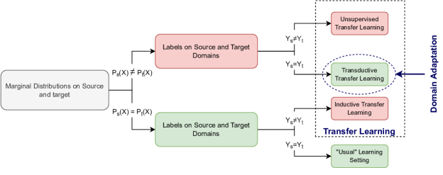
Based on [8], DA is, in fact, a transfer learning method. In DA, the label space, or tasks, are mostly the same between source and target domains, but the marginal distributions of domains are different. Domain differences may exist between the source and target domains as well as between the source domains themselves.
2.1.1 Notation
To define the DA problem, we should explain the source and target domains. Suppose a shared space where is the space of feature values and is the space of label values. A domain is a collection of paired data and labels sampled from a distribution. The samples of training or test data come from their corresponding domains. Assume that is a domain where is sampled from joint distribution defined on .
Consider there are source domains , where , and one target domain - note that in some scenarios, we can have more than one target domain, but for simplicity, we consider a single-target domain problem. Each source domain is denoted by where is drawn from the joint distribution on . We consider the target domain denoted as , drawn from distribution on . For the task of unsupervised domain adaptation which is the focus of this study, we only have the unlabeled target domain .
In the DA problem, unlike other categories of transfer learning, the conditional distribution of source and target domain is the same, that is , but the marginal distribution, for and are different; in other words, . This discrepancy is known as domain shift. Note that this discrepancy also exists among each pair of source domains: for and .
The goal of domain adaptation is to reduce the negative effects caused by domain shifts between source and target domains. In other words, given a domain adaption algorithm proposes a generalizable and robust function that gets the minimum prediction error on unseen samples which are drawn from the target domain:
2.2 Domain Generalization
In the previous subsection, the issue of domain adaptation was discussed. As with domain adaptation techniques, domain generalization approaches attempt to address the challenging problem where test data distributions differ from training ones. Differentiating these two categories, DG cases have an unknown distribution of the test data while DA cases have a known distribution of the target data.
2.2.1 Notation
The same notation as the previous section applies to domain generalization. The only difference is that the target domain is unknown and can be drawn from an arbitrary distribution on . So here the domain generalization algorithm uses only the set of source domains to estimate a generalizable robust function which minimizes the prediction error on any arbitrary target domain:
2.3 Domain Adaptation and Domain Generalization Categories
Based on several factors, scenarios, limitations, and algorithms, DG/DA methods can be categorized into independent groups. The following section discusses the most significant categories related to our issue, based on three different settings: the availability of labeled data, the number and distribution of source domains, and the distribution of label space:
2.3.1 Labeled Data Availability
According to the availability of labeled target data, we can have three classes of DG/DA methods:
-
•
Supervised DG/DA: All data from the target domain have labels.
-
•
Semi-supervised DG/DA: A small number of target domain samples have labels.
-
•
Unsupervised DG/DA: There is no labeled data in the target domain.
This paper refers to DG/DA as unsupervised DG/DA.
2.3.2 Number of Source Domains
DG/DA methods can be divided into two varieties based on the number of source domains: single-source and multi-source.
-
•
Single-Source DG/DA
The single-source DG/DA setting assumes that the training data are selected from a single distribution. This category includes data collected from one or more domains but does not require domain labels, so it can be applied to multi-source scenarios. Despite the fact that single-source techniques are generally less complicated than multi-source ideas, they may not be efficient when there are multiple sources from different domains available [9].
-
•
Multi-Source DG/DA Multi-source setting assumes that multiple distinct relevant domains are available. The corresponding methods are based on the assumption that labeled training data collected from multiple sources will have different distributions. As a trivial matter, it is possible to combine the sources into a single source and discard the differences between them. However, source-combined DG/DA often results in lower performance than simply using one or a collection of suitable sources and discarding the others, as this approach neglects the fundamental variations among multiple sources that affect the DG/DA algorithm. In addition, by selecting a small subset of domains and learning the relationship between them, a model can learn patterns that are more stable across source domains. This helps it generalize more effectively to unknown domains. Domain shifts among different sources can be addressed in a variety of ways; for example, latent space transformation can be used to align and transform source domains and target domains into latent spaces. An alternative approach to handling inter-source domain shifts is to align each source domain separately with the target domain, and then aggregate the adapted sources.[9]
2.3.3 Label Distribution
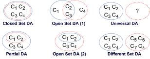
Based on [10], labels of source and target domains can consist of the same or different classes. As illustrated in Figure 2, the variability of this difference creates several scenarios for DG/DA methods:
-
•
Closed set DG/DA: Labels of source and target domains come from the same classes,
-
•
Partial DG/DA: Target domain’s classes are a subset of source domains’ classes,
- •
-
•
Universal DG/DA: Target domain’s label set is unknown and might have a number of common classes with the source domains’ label set [13].
-
•
Different set DG/DA: Source and target domains have completely different classes.
2.4 Medical Images and Signals Analysis
The automatic analysis of medical images and signals involves the study of medical measurements of different body parts, whether in the form of images or electromagnetic signals. This is done in order to provide an intelligent diagnosis of various medical conditions. Due to the development of systems based on Artificial Intelligence (AI) and the rapid increase in computational power, it has become more and more common to process high-resolution medical images intelligently. By using ML systems trained on large datasets of medical recordings, the analysis of medical images can be made faster and more accurate. These models can assist physicians when they have doubts about their diagnosis or miss a critical clue in the recordings.
There are two types of medical images and signals: structural and functional. Structural medical images are the ones that only record the state of the body in a single unit of time. They focus on the spatial structure of the body part in the form of an image. They include Computed Tomography (CT), Magnetic Resonance Imaging (MRI), Pathology, Endoscopy, Colonoscopy, Automated Breast Volume Scan (ABVS), Gastroscopy, Cytology, and X-Ray images. Functional medical images and signals contain spatial and temporal information, capturing measurements of body processes rather than body states. The majority of these measures are captured from the brain, such as EEG, fMRI, MEG, and fNIRS. Other functional medical signals are not measured from the brain, such as ECG (from the heart) and EOG (from the eyes). Structural medical images have been used to diagnose cancers and abnormalities in body organs, including brain tumors.
([14], [15], [16], [17], [18], [19]), breast tumors ([20], [21], [22], [23], [24]), lung, liver and kidney diseases ([25],
[26], [27],
[28],
[29],
[30],
[31]).
This review focuses on functional medical images and signals.
Although not as popular, recordings from non-brain areas of the body have also been studied in the literature. For example, ECG signals are analyzed to detect heart problems ([32], [33], [34], [35], [36]), or EOG signals.
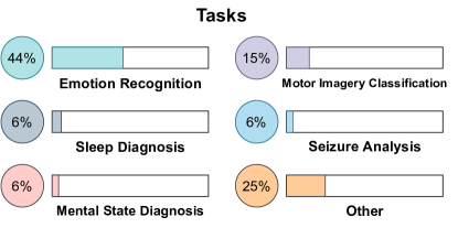
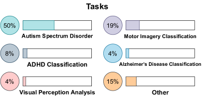
Brain-Computer-Interface (BCI) models are the systems that analyze and use brain-related medical images, which constitute the majority of models based on functional medical images. We have categorized different tasks in brain signal analysis as follows:
2.4.1 Motor Imagery (MI)
2.4.2 Bain-related Disease Diagnosis (BDD)
Another application of BCI systems is the detection of brain-related diseases and conditions such as Parkinson’s Disease ([43], [44], [45], [46]), Alzheimer ([47], [48], [49], [50]), Schizophrenia ([51], [52], [53], [54]), and Autism Spectrum Disorder (ASD) ([55], [56], [57], [58]). Effective and precise detection of such diseases can be very helpful for early detection and diagnosis of them, leading to more effective treatments.
2.4.3 Emotion Recognition (ER)
2.4.4 Seizure Analysis (SA) and Mental State Diagnosis (MSD)
Monitoring people’s mental state also has many applications such as seizure and epilepsy prediction and detection ([65], [66], [67], [68], [69], [70], [71], [72]). Other popular applications include mental workload classification and assessment, mental state prediction, and diagnosis of some mental diseases such as Tinnitus.
2.4.5 Awareness Monitoring (AM)
2.4.6 Sleep Diagnosis (SD)
2.4.7 Visual Perception Analysis (VPA)
The analysis of human visual imagination and understanding of the surroundings using brain signals is an interesting field of research in brain signal analysis. It includes the study of Steady-State Visual-Evoked Potentials (SSVEP) ([85], [86], [87], [88], [89], [90], [91]) and visual recognition ([92], [93], [94], [95], [96]).
2.4.8 Human Thought Analysis (HTA)
There are also interesting ongoing studies based on human thought prediction, such as creative drawing ([97]) and imagined speech recognition ([98], [99], [100]). However, these areas of study are still at a very primitive stage.
It is worth mentioning that the distribution of the most frequently used tasks in recent DG/DA-related EEG and fMRI papers is shown in Figure 3 and Figure 4 respectively.
3 Domain Adaptation and Generalization for Medical Image Analysis
Machine Learning techniques in medical field analysis usually suffer from domain shift. This problem can arise from various factors, such as different centers, different devices in the same center, different subject populations, or different experimental conditions. Moreover, gathering substantial medical data can be very time-consuming and expensive. Some medical signals require costly measurement devices (e.g., MEG), while others need a meticulous and stable experimentation setup (e.g., EEG). Hence, gathering a reasonable amount of data on every new site or for every new subject is not affordable in many cases. Thus, the challenge of domain shift is unavoidable in the case of medical image analysis.
Due to the direct impact of this problem on some crucial medical diagnoses, the use of domain adaptation in the medical field is undisputed. Additionally, there is a common issue in most of the ML models that they should have high performance on newly gathered data. This will be more relevant in the medical field because of the higher frequency of facing data from a new domain, such as a new patient. Therefore, domain generalization is also of great importance in this field.
3.1 DG and DA Tasks in Medical Image Analysis
Different adaptation or generalization tasks can be defined between different types of domains, such as subjects, datasets, sessions, etc. The cross-subject task is the most common in DA or DG on medical data, which considers the variability of data across subjects and tries to eliminate shifts between different subjects. Cross-dataset is another common DG/DA task on the medical data related to existing domain shifts between datasets. This task aims to learn various aspects of these differences across medical image datasets. Cross-session task is also frequent in medical image analysis and is defined when the goal of DA or DG is to consider intra-subject data variabilities that emerged during different experimental circumstances. Some other DA or DG tasks are less common than those mentioned above; for instance, the cross-day task is analogous to the cross-session task. Also, it is worth noting that the cross-device task may also be studied, which considers the data variability caused by different devices used to measure the subject’s signals.
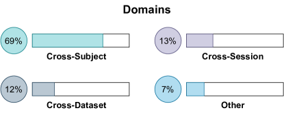
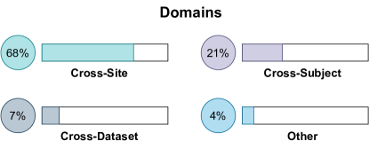
Furthermore, in Figures 5 and 6, we show the distribution of the most frequently used medical domains in recent DG/DA-related EEG and fMRI studies respectively.
3.2 DG vs. DA in Medical Image Analysis
There are fundamental differences between domain adaptation and domain generalization, which cause different applications. As mentioned before, good performance for unseen medical data is almost vital; it is very time-consuming to learn a different model for a new subject or patient. Nevertheless, generalization is not always the desired function in the medical field; sometimes, we face specific domains, such as data from the same organ acquired by different devices or from different subjects. In these situations, what is most needed is the minimization of domain shift between these related but different domains, so domain adaptation is the key. Sometimes in adaptation, there are a few seen target data; sometimes, all the target data is unseen. The goal is to use this knowledge to learn about the target domain. To conclude, the main difference between adaptation and generalization is the access to target data during the training process; in other words, in adaptation, we aim to complete our diagnosis for a related domain, and to that end, we take advantage of our current knowledge on source data, and the structure of target data. In contrast, in generalization, we are only allowed to use our knowledge of accessed source data and expand it for an unseen domain of data whose structure is even unknown.
Generalization of the model is usually an essential desire of researchers, especially in medical image analysis. As mentioned, the better our model performs on new and unseen data, the stronger it is and will become more valuable and practical. So the importance of generalization is inevitable even when the main task is adaptation. To handle this, some papers with the main task of adaptation exploit generalization ideas as well. Therefore, we will explain both their adaptation task and the different ideas used for generalization.
4 Architectures
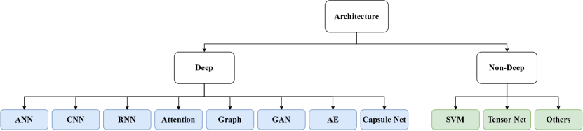
In this study, we have thoroughly investigated the most commonly used architectures in the works on DG/DA of functional medical data. These architectures are categorized and illustrated in Figure 7 as follows:
Deep Architectures: This category includes most of the recent works in the literature.
-
•
Artificial Neural Network (ANN): Some methods are implemented as simple (feed-forward) neural networks.
-
•
Convolutional Neural Network (CNN): The majority of the methods exploit CNNs for processing multi-dimensional functional medical data.
-
•
Recurrent Neural Network (RNN): These architectures enable the use of temporal dependencies in the data sequences.
-
•
Attention-based Models: Attention ideas are sometimes performed to introduce weights for different channels or dimensions (and sometimes timesteps) in the data
-
•
Graph-based Architectues: Using graphs for modeling data helps model interactions among different devices or electrodes, especially in EEG data.
-
•
Generative Adversarial Network (GAN): GANs may also be used for augmenting data in medical use cases.
-
•
Auto-encoder (AE): Some methods use the latent representations of the medical data extracted by an auto-encoder in an unsupervised manner.
-
•
Capsule Networks [101]: The recently-introduced architectures of capsule networks are implemented in recent models.
Non-Deep Architectures: Some non-deep models are still widely used in the proposed models for DG/DA on functional medical data.
-
•
Support Vector Machine (SVM): SVM is one of the most famous and broadly adopted non-deep models in this era.
-
•
Tensor Network: Some works adopt tensor networks that have the advantage of graph-based data modeling.
-
•
Others: There are other non-deep models, such as KNNs, random forests, etc., also used in the literature.
The architectures used in each studied model are depicted in Table 1 and Table 2 for EEG and fMRI data, respectively.
5 Methods
The most recent methods used for the adaptation and generalization of tasks on functional medical images are categorized and introduced in this section. These methods are explained based on two perspectives, DA and DG, in the following.
5.1 DA Approaches
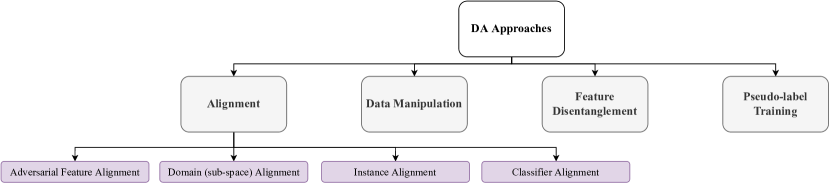
We have studied the latest research seeking domain adaptation in the context of functional medical images and based on their design ideas, these methods are classified as in the hierarchy depicted in Figure 8, including alignment, data manipulation, feature disentanglement, and pseudo-label training. Summarized information about the methods discussed in this section can be found in Table 1. In the following parts of this section, these approaches and works following their ideas are described.
5.1.1 Alignment
One of the most common domain adaptation strategies arises from aligning the model’s input at test time with previously seen data or features. A majority of approaches rely on these techniques so that the inputs (or secondary features) to the model are kept aligned with a fixed network architecture. Consequently, the same architecture can yield relatively similar performance for source and target data. Alignment-based methods consist of adversarial alignment (alignment using an adversarial objective), domain alignment (aligning the distribution of target and source data), instance alignment (aligning source and target sample by sample), and classifier alignment (adapting the classifier model to the target domain).
5.1.1.1 Adversarial Feature Alignment
This approach is implemented in a substantial number of papers focusing on aligning features between source and target domains. The objective of these methods is to extract features that are similar between target and source data using an adversarial training setup. Inspired by the Domain Adversarial Neural Network (DANN) [102], in most of them, a common feature encoder is trained in a min-max game with a domain classifier. Essentially, the feature encoder learns to extract features such that the domain classifier is unable to distinguish between source and target data. This procedure results in achieving a common feature space between data from different domains.
In [103] and [104], the idea of DANN is applied by training a domain classifier whose loss is inverted by a Gradient Reversal Layer (GRL) [105] and forcing the feature extractor to remove domain-variant features while improving the classification accuracy of the main task. In [106], Su et al. employ an adversarial discriminator that is trained to be challenged by a pre-trained feature extraction for brain anomaly detection on fMRI data. Heremans et al. adopt an akin approach in [107] to enhance the performance of common neural networks used for sleep stage classification by using an adversarial domain classifier on the feature extraction backbone. In [108], multi-view features are extracted in the time and frequency domain and then, combined with the original data, are used in an adversarial learning module with two generators for separating patient and seizure features alongside discriminators ensuring this separation. Also, in [109], an Adversarial Discriminative Temporal Convolutional Network (AD-TCN) is proposed, where initially, an encoder and classification layer are trained on the source data. Secondly, the adversarial loss is employed via a domain classifier applied to the source encoded features and a distinct target encoder, making the target encoder able to be combined with this new classifier for target inference. In [110], in one branch, features obtained from a pure-info encoder are fed into a classifier and an adversarial side discriminator so that data from the two ears are aligned and processed efficiently together in the classifier. In another branch, after applying a domain-variance encoder, the resulting features plus the ones from the first branch are combined to reconstruct the data, where a domain discriminator is further adversarially trained. [111] proposes adversarial adaptation in multi-source setup by first selecting the source samples most correlated with the target sample, and then mapping their corresponding features in a common space, with the aid of a discriminator intended to not be able to differentiate domains. In [112], the Fader network method [113] is used for domain adaptation and removing task-irrelevant features in fMRI data. In this method, an auto-encoder is utilized whose output encoding is used for the final classification task, as well as the domain classification in an adversarial manner. Furthermore, Li et al. use an adversarial subject classifier in [114] to ensure the subject independence of the extracted features for emotion recognition. Moreover, two different RNNs are also employed, for the right and left brain hemispheres, in each of the two vertical and horizontal streams over the electrodes to maintain structural information. Eldele et al. [115] utilize adversarial training along with self-attention and self-training in their method where the extracted features are passed through unshared attention-based modules to retain domain-specific features as well as task-related ones, as domain-specific features may also be helpful for label prediction. The adversarial domain classifier further encourages data alignment while preserving domain-related features. In [116], Bao et al., in addition to Maximum Mean Discrepancy (MMD) [117] minimization, further use a domain classifier so that it fails to separate source and target domains, as they mention that merely MMD will not guarantee multi-source DA. In [118], separate branches are designed for extracting features from EEG data and the music used for data collection. The representation from these two branches is further aligned by a modality classifier, inserted with a GRL, enabling adversarial alignment among modalities as in DANN. [119] also introduces a domain and a subject classifier implemented with GRL layers, that perform domain and subject classification and are trained adversarially so that features extracted from topological maps obtained from Power Spectral Density (PSD) features are free of dataset or subject priors.
Multiple works have further added modifications to DANN to make it more applicable for their purpose. Ding et al. extend DANN in [120] by designing two label predictors instead of one. Using pre-trained label predictors on the source data, the two fully connected classifiers are tuned by Maximum Classifier Discrepancy (MCD) criterion [121]. First, target outliers are detected and MCD is maximized to achieve broader classification boundaries. After relocating target samples to new boundaries, MCD is minimized for better adaptation. [122] extends the concept of DANN by applying the domain discriminator to the conditional features, i.e. multiplying the features by the softmax output, thereby capturing a 2-D matrix (outer product) that can be inputted into a GRL and a domain discriminator. Likewise, in [123], Huang et al. explore the same idea, combined with an entropy-based regularizer that adjusts the informativeness of different samples, on fMRI data for ADHD classification. A similar approach is taken in [124], where both global (marginal) and local (conditional) domain discriminators are adversarially trained against the main classifier, with a dynamic weight adjusting their importance. Some works also perform DANN on the shallower representation of the data. In [125], features from shallow layers are used for domain discrimination, which is trained adversarially to align the marginal distributions, while deeper features are fed into two different classifiers whose prediction difference is aimed to be maximized to detect target samples close to the decision boundary. Similarly, [126] benefits from an adversarial adaptation by feeding shallower representations to a domain discriminator, as earlier layers typically produce more task-invariant features that reflect the difference in data domains. Additionally, the association strategy computes the probability of transition between source and target domain based on their features in each batch and introduces a loss in these transitions to encourage them to return to the same class. [127] integrates the idea of a DANN with an attention mechanism. A graph convolutional neural network (GCNN) is exploited with numerous stacked CNN layers, creating multi-level features from the GCNN and CNNs. The concatenated representations are inputs to separate adversarial domain classifiers, which help extract more domain-invariant features. For the final label predictor, the feature regions are multiplied by attention weights indicating how difficult it was for the classifier to classify the domains in each region. [128] combines a GAN with a DANN to seek domain adaptation. First, a GAN is trained to achieve a robust target data generator and an accurate target data discriminator. Then, the closest source samples to the target distribution, as specified by the discriminator, are further augmented with fake target data and are used in the DANN to adversarially train the final fatigue prediction network against a domain classifier. In [129], the idea of the adversarial domain discriminator is integrated into a federated learning framework that has pre-trained site-specific feature generators that are further trained to confuse the discriminator.
In some cases, a min-max game is performed to separate features in the data. Zhu et al. adversarially train two classifiers on features from an auto-encoder in [130]. After training the auto-encoder and classifiers, their prediction discrepancy is maximized. Following that, in a min-max game, the auto-encoder is optimized to decrease this discrepancy. In [131], Jeon et al. design a common point-wise convolutional encoder producing class-relevant and class-irrelevant features and a network estimating the mutual information of these two features that are optimized in a min-max manner to guarantee the omission of subject-specific features from the input of the classification network.
An adversarial scheme may also be used to fit the data to certain priors or prototypes. In [132], a Manifold Adversarial Auto-Encoder (MAAE) is developed to fit a manifold prior distribution to the distribution of the auto-encoder latent space. [133] also discards the data specific to patients by presuming a Laplace prior distribution on different patients and considering them as real data. Inspired by GANs, the VAE outputs are regarded as fake data and are fed alongside the real data to a discriminator, aiming to deceive the discriminator. In [134], Wang et al. create source and target prototypes and classify samples based on distance from these prototypes using a domain classifier trained in a min-max game with the generator (the symmetric and positive definite matrix network applied to the data covariance matrix).
Some works consider private encoders per domain in adversarial domain alignment. In [135], Luo et al. propose two variants of Wasserstein-distance-based Multi-source Adversarial Domain Adaptation (wMADA) for domain adaptation in vigilance estimation and emotion recognition. The first variant, wMADA-, adversarially trains different private discriminators on the Wasserstein distance between source and target outputs. In the second variant, wMADA-, source features are inputs to a public discriminator as well. Also, Qu et al. utilize private and common feature extractors in source and target domains plus a domain classifier (with GRL unit) in [136] to separate sleep-related features from unrelated ones for insomnia detection. A difference loss also forces the two networks to obtain orthogonal features. In order to improve accuracy, reconstruction losses are embedded in the network and the target common classifier’s features are fed into an LSTM and then the final classifier.
Adversarial training may also be applied to transform source data into the target distribution. In [137], Huang et al. propose a generator network that attempts to generate samples similar to target data, from source samples by using an adversarial domain discriminator, as in GANs. Overall, the sample data is first transformed to have the target distribution. Finally, an emotion classifier is trained on this data, allowing the target data to be used directly at test time.
Adversarial domain alignment is broadly used in a great number of works, due to its general framework that can be combined with various feature encoding modules; as in arenas other than medical data analysis, adversarial training is a powerful tool for making a backbone feature encoding model more robust. Moreover, another benefit that adversarial methods bring about is that they may be applied to unlabeled source data as well, in an unsupervised manner. Despite the numerous improvements adversarial approaches bring about, using them can also be challenging. First of all, training them can be unstable, as finding an equilibrium between the two modules adversarially trained against each other may not be practical. In other words, these two modules may end up with a suboptimal solution where their performance is not satisfying for the final task. The performance of an adversarial model might be limited by the mode collapse issue, i.e. if there is no proper alignment between features and classes in different domains, the separate design of the task classifier and domain discriminator may degrade the model’s performance [138]. Additionally, adversarial scenarios demand a larger number of data samples to provide meaningful results in comparison with other methodologies. This issue can be problematic in medical data analysis, where the volume of data and providing neat data is labor-intensive and expensive. Also, adversarial methods have the problem of being time-consuming at the training stage.
5.1.1.2 Domain Alignment
A non-adversarial source and target features alignment can be achieved using domain alignment techniques. In this regard, subspace alignment is one of the most straightforward methods. In the context of subspace alignment, the initial concept is to learn a common intermediate representation shared between domains. It has been found that most adaptation approaches in this category start by creating a low-dimensional representation of original data using a variety of deep or non-deep methods and then use distinct objectives such as Kullback–Leibler Divergence (KLD) and MMD to reduce the discrepancy between marginal and conditional distributions in a new subspace, as in [139], [140], [116], and [141]. In order to achieve this goal, various deep neural network models are used. For example, Hao Chen et al.[142] propose a model consisting of an ANN-based common feature extractor and multiple domain-specific feature extractors, with one network per pair of source and target, which is designed to minimize MMD in order to transform each pair into a different subspace. Likewise, Zhao et al.[143], allocate a model for each pair of source and target. However, they consider the importance of each class as they minimize MMD. Additionally, the paper [143] provides domain-invariant feature extraction modules built on a Common EEGNet-based Network (C-EEGNet)[144] as well as domain-specific feature extraction in each pair of sources and targets by using CNN-based subnets (S-CNNs). A novel alignment algorithm called Local Label-based MMD (LLMMD) is proposed in this paper to diminish the discrepancy between source and target domains, which explores local label-based fine-grained structure information across all domains and extracts label-based domain-invariant features. And In [145], the Dempster-Shafer (D-S)[146] evidence theory, and rough adjoint inconsistency are applied to derive weight coefficients for each domain. Afterward, the target domain class proportion and optimal coupling distribution set are solved iteratively. Lastly, each source domain is aligned with the target domain and is used to train the final classifier.
In [147], a Subspace Alignment Auto-Encoder-based model (SAAE) is proposed to embed extracted Differential Entropy (DE) features from EEG signals and minimize MMD in an infinite-dimensional Reproducing Kernel Hilbert Space (RKHS). Moreover, Peizhen Peng et al. [133] use auto-encoder and MMD on time-frequency images for the mentioned purpose.
In the paper [148], to extract features, Kee et al. use a single-layer Gated Recurrent Unit (GRU) embedded in a semantic manifold and used Multi-Kernel MMD (MK-MMD) as a divergence metric.
Feature extraction can also be conducted using tensors; Mu Shen et al. propose a tensor-based alignment model in [149]. This model uses Tucker decomposition to tensorize EEG channel data. As a result of tensor network summation, features of training and testing tensor samples are derived from corresponding subspace matrices. A Deep Domain Adaptation Network (DDAN) is proposed by Wenlong Hang et al. [150] that employs a CNN to automatically detect features, MMD to minimize distribution discrepancy, and a Center-based Discriminative Feature Learning (CDFL) method to force the deep features closer to their respective class centers and to make the inter-class centers more distinguishable. As well, in [151], a novel domain adaptation method, Deep Subdomain Associate Adaptation Network (DSAAN), is described that combines the advantages of both subdomain adaptation and associate loop calculation. This model uses a ResNet [152] to extract features.
It is also possible to extract meaning from EEG signals by using GNNs; Feng Kuang et al. [153] offer a Multi-Spatial Domain Adaptive Network (MSDAN). Through MSDAN, the original EEG data is mapped into multiple graph-based spaces, and the distribution of the source and target domains in those spaces is narrowed by the use of MMD.
Attention mechanisms can also be useful to solve excessive alignment problems as well. As an example, in [154], a CBAM-based module was designed to extract the common features of the source and target. The MMD in RKHS is also used to align the distribution of the two domains. In this article, to overcome the excessive alignment problem in which the samples of the two domains are mixed, and the categories within each domain cannot be distinguished well, the few-shot learning module is introduced to retain the domain-specific information. And, [155] suggests CS-DASA, which learns the common features from multi-frame EEG images using the convLSTM. Also, to adapt the source and target domains, the model uses a subject-specific module using 2D-CNN with MK-MMD loss in the RKHS. Furthermore, a subject-to-subject spatial attention mechanism focused on the discriminative spatial features from the target image data is used.
Other classic machine learning techniques are similarly useful for reducing dimension and finding shared subspaces. As a dimensionality reduction technique, Transfer Component Analysis (TCA) [156] aims to minimize distribution discrepancies by learning a set of transfer components. In [157] and [158], a transfer learning-enabled classifier consisting of a TCA is implemented to mitigate the mismatch among distributions. It anticipates a projection to a latent subspace where the projected source and target data achieve a reduced MMD in RKHS. Similarly, Yueying Zhou et al. [159] use TCA, Joint Distribution Adaptation (JDA)[160], Balanced Domain Adaptation (BDA)[161], and Transfer Joint Matching (TJM) [162] with MMD distance measure to adapt the domains. [163] uses a JDA-based adaptation module that joints the marginal distribution alignment and conditional distribution alignment to minimize the data distance between the source and the target domains with MMD measure. Very similarly, Transport-Based Joint Distribution Alignment (T-JDA) blocks are proposed in [164] that can propagate features or labels from source to target by minimizing the global transportation cost between the empirical joint distribution of a pair of source and target domains. An Independent Component Analysis (ICA) [165] method is employed to determine the independent components of unlabeled and labeled EEG signals in [166]. In this work, the energy features of ICs are extracted as the source and target domains. As a final step, the marginal distributions of the source subspace base vectors are aligned with the base vectors of the target subspace using linear mappings.
Alternatively, another category of methods assumes that there exists a manifold of transformations between the source and target domains; this manifold consists of a space of parameters where each point generates a possible domain. For instance, Wen Zhang and Dongrui Wu [167] propose a Manifold Embedded Knowledge Transfer (MEKT) approach by aligning the covariance matrices of the EEG trials in the Riemannian manifold, extracting tangent features, and then performing domain adaptation by minimizing the joint probability distribution shift between the source and target domains. Jiang et al. [168] have also proposed a Kernel-based Riemannian Manifold Domain Adaptation technique (KMDA) in which the covariance matrices are aligned in the Riemannian manifold and then mapped to a high dimensional space by a log-Euclidean metric Gaussian kernel, which is then reduced by MMD.
Despite the fact that domain alignment techniques are powerful and widely used for domain adaptation issues, these techniques need a significant amount of parameters for adaption [169] and the constraints they are subject to could cause a distortion of semantic feature structures and a loss of class discriminability [170].
5.1.1.3 Instance Alignment
Target domains can also be aligned with source domains at the instance level. In some studies, pairs of source and target data samples are directly guided to become closer to each other.
Chambon et al. [171] find the optimal transport from the source domain to the target domain and train the model on transported source samples. From a representation learning aspect, Lee et al. [172] and Wang et al. [173] try to represent each sample of every source domain by a shared transformation and a low-rank transformation of target samples and use it as the new representation for source samples. The shared transformation further generates a new representation for target samples. Similarly, in [174], Lee et al. follow a contrastive approach by decreasing the distance between pairs of samples in the same class and different subjects compared to samples with different classes and the same subject.
It is also common to represent target samples or predictions based on their similarity to source samples. For instance, in [175], after training source-shared and source-specific encoders and decoders and a target-specific encoder, the final prediction on target data results as a combination of target model prediction and source model predictions, weighted by their feature similarity to the target sample. Likewise, Li et al. [176] duplicate the batch normalization layer for each source. In the test phase, for new target samples, the average of batch normalization branches is computed and further weighted by layer statistics similarities of the target and each one of the source domains. From a slightly different viewpoint, Lin et al. [177], train task and subject predictor networks, and select samples from most similar subjects to train the model on a new domain. Moreover, in [111], using direct transfer accuracy, Wang et al. select only related source domains (subjects) to be used for the main adaptation module.
There are also studies conducted on aligning source and target samples based on their discriminative statistics. For example, Tao et al. [178] align kernel-based classifiers for each domain to match the label structure of the samples and minimize the distribution of source and target domains. In a similar manner, Shen et al. [179], learn a transformation on samples for each source domain, so that their covariance matrix becomes as close as possible to the covariance matrix of the target domain samples.
As a more comprehensive form of domain alignment, instance alignment attempts to align domains at the sample level. Although this extension allows for more accurate adaptation when it is successful, it can also fail more frequently. Instance alignment requires that the target domain data can be represented by the source domain or vice versa and can be adversely affected by outliers. Additionally, similarity-based methods are not able to adapt to target spaces that differ significantly from source spaces.
5.1.1.4 Classifier Alignment
Classifiers trained on features extracted from different sources may result in misaligned predictions for target samples close to the domain boundaries in a multi-source setting. By minimizing specific classifier costs, the classifiers can be better aligned, resulting in more accurate and generalized models. As an example, Zhang et al. [164] adapt multi-source domains to a single target and penalize decision inconsistency among diverse classifiers trained on paired joint distribution aligned features by minimizing the consistency loss between classifiers trained on source-target domain pairs. In addition, in [142], a discrepancy loss metric is introduced to achieve convergence of predictions from classifiers trained on domain-adapted sources. Moreover, [143] integrates the probability distribution from classifiers by the weighted mechanism. Each classifier’s prediction probability distribution is used to calculate the weights; the global optimization strategy can also remove the negative impact of significant individual differences.
Unlike the described methods, classifier alignment is used with a different purpose by Xia et al. in [180]. In their presented model, to have a more robust target classifier, different perturbation of target data is fed into some auxiliary classifiers, which are aligned to each other and to the fixed source classifier using consistency regularization loss.
Classifier alignment techniques are practical, especially when the target domain samples are at the decision boundary. In this case, the variance of predictions will be high, which will significantly impact the results. While these techniques can provide high-accuracy results, they require the development of a module to extract source and target common features for the classifier’s input.
5.1.2 Data Manipulation
In this group of methods, data is changed and manipulated for adaptation purposes. The major sub-type in these approaches is preprocessing, given that solutions to domain adaptation can be injected into data preprocessing steps.
For example, in [181], the authors showed that feature normalization had a relevant effect on the conditional shift, and by performing z-score normalization as preprocessing, the conditional and marginal shifts could be reduced. Another approach that can be categorized as preprocessing is subject clustering. Concerning this, Li et al. [182] propose Domain Adaptation with the Subject Clustering (DASC) which clusters the subjects according to their inter-subject similarity of emotion-specific EEG activities and only uses the source cluster that matched the target better for adaptation to the target. Moreover, the optimal subjects from the selected cluster, with positive transfer impacts, are used to classify the emotional situation of the target subject.
Applying this approach as a data manipulation method provides independence from the necessity of training and makes it directly applicable. Also, preprocessing is integrable with other DA methods and this increases our options. On the contrary, information loss and error propagation through the whole pipeline are disadvantages of this approach.
5.1.3 Feature Disentanglement
One of the recently popular techniques in DA is to disentangle input data into domain-specific and domain-invariant features. By defining appropriate objectives for each disentangled part, domain-specific information can be removed from the data, and thus, domain-invariant features can be used in a new domain for prediction.
Jeon et al. [131] force class-invariant and class-relevant features to contain the least common information by minimizing their mutual information. In [110], aside from guiding each part to predict its own information, Liu et al. use adversarial training to prevent them from estimating each other’s information. Furthermore, the separated parts are used to reconstruct the original data in order to ensure that there is a minimum loss of information during disentanglement.
In the context of multi-source domain adaptation, Zhao et al. [175] disentangle data from each source into domain-private and shared features and then, reconstruct the original data via a shared decoder. The new target domain uses a private encoder that is trained with the reconstruction objective. During the inference phase, data from the target domain can be classified using both private and shared encoders.
When data samples are inherently a combination of multiple parts mixed together, the disentanglement technique is useful. Brain signals are a complex mixture of the brain’s response to various stimuli in the environment. Usually, only one of them (class-relevant response) is intended to be analyzed in DA for medical image analysis. Consequently, disentanglement is a very intuitive and natural way of dealing with complex brain data. Nonetheless, It is important to note that disentanglement requires a significant amount of data to work well, due to the need to extract irrelevant features as well.
5.1.4 Pseudo-label training
The generation of pseudo-labels from the source model is a common DA approach in medical image analysis. In this approach, a model is trained on source domain data, then its predictions on target domain data are considered pseudo-labels for the model, and they are exploited to adjust the model to the target domain.
Since pseudo-labels are noisy and not completely accurate, some studies apply them iteratively, and as the model produces better predictions, it uses its more accurate pseudo-labels from the previous iteration. This strategy can be used to transform any transfer learning method that uses target domain labels into an unsupervised domain adaptation one.
Some examples of this type of iterative self-supervision are the works of Zhang et al. [167], Eldele et al. [115], Shi et al. [183], Jiang et al. [168], Shen et al. [139], and Wang et al. [163]. These works, while aligning source and target samples, generate pseudo-labels at each iteration that is used for the next step. Moreover, Edele et al. tackle the cold start problem by ignoring the target classification loss in the initial iterations. Shi et al. perform JDA iteratively and enhance pseudo labels using label propagation algorithm proposed in [184]. The idea to enhance the pseudo-labels is also proposed by Han et al. [185], where they apply a single step of K-Means algorithm to the features of each pseudo-label to form more coherent and less uncertain labels. Wang et al. come up with the aforementioned iterative self-supervision from an objective that is optimized with respect to both representation parameters and target domain labels.
In some studies, pseudo-labels are directly treated as target data labels, along with other objectives imposed to guide the model in the correct direction. Guarneros et al. [186] retrain their source model on target data using pseudo-labels with an additional loss term controlling the uncertainty and increasing the diversity of predictions. Zhao et al. [103] and Heremans et al. [107] use pseudo-labels for target classification alongside their adversarial objective. Tao et al. [178] also generate pseudo-labels and consider the target domain as one of the sources and adapt all of them together.
Pseudo-labels may also be used indirectly, for an objective other than classification primarily. In [124], Hong et al. use pseudo-labels to approximate the class probability of target samples for conditional discrimination between samples of source and target. Additionally, in [159], Zhou et al. utilize JDA as one of their experimented methods which requires target data labels (derived from pseudo-labels) to calculate class conditional distributions. Moreover, Meng et al. [151] use pseudo-labels to partition target domain samples into subdomains and estimate the similarity of samples in source and target domains.
As a result of pseudo-label-based DA, useful information held by the source model is retained during the transition to the target domain. Despite its ability to prevent the forgetting of source domain information, it may not be able to eliminate the bias associated with the source domain. In particular, the performance of this approach is relatively weak when the difference between the source and target domains is so significant that iterative fine-tuning or additional objectives are not capable of correctly adapting the model. Moreover, convergence is a critical concern when using iterative methods, since negative feedback in the self-supervision process may prevent the model from eventually reaching its optimal state.
5.1.5 Hybrid Methods
There are some studies that combine multiple domain adaptation approaches. Particularly, as pseudo-labeling can be conducted independently, it can be easily integrated with other adaptation methods. Thus, as discovered in this study, the most common combination is pseudo-labeling and domain alignment ([168], [151], [159], [186], [163], [139], [167], [185]). Pseudo-labeling is also used alongside adversarial feature alignment ([107], [124], [115], [103]). In these combinations, the pseudo-labels generated from the source model help align the source and target domains more accurately.
Furthermore, classifier alignment is mostly accompanied by domain alignment ([142], [143], [164]). There are also other combinations in the literature. Bao et al. [116] and Peng et al. [133] try to adapt their model using adversarial feature alignment and domain distribution alignment together. Liu et al. [110] use adversarial training to train their disentangled feature extraction model. Zhao et al. [175] incorporate disentanglement along with instance alignment. In [111], Wang et al. utilize adversarial training in conjunction with instance alignment. Additionally, in [178] pseudo-labels are used to employ instance alignment.
5.2 DG Approaches
Various DG methods have been suggested to process functional brain signals. These approaches include representation learning, data manipulation & preprocessing, learning scenarios, and embedded architectures, which can also be merged to enhance performance on various tasks. Our designed approach hierarchy for these methods based on their approaches and the motivations behind their design is shown in Figure 9. In the remainder of this section, we explain DG-specific approaches as well as approaches presented as part of a domain adaptation method where adaptation techniques are improved via generalization ideas. A precise summary of these explanations is also provided in Table 2.
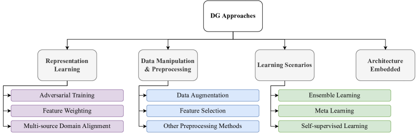
5.2.1 Representation Learning
An overall strategy for domain generalization is representation learning, aiming to ensure that the learned representations are domain-invariant, meaning that they do not contain much knowledge about the domain from which they originate. In other words, feature extraction is guaranteed to be generalizable to all domains as long as they include similar semantics about the studied task. In the following, the representation learning methods are grouped into adversarial training, domain Alignment, and feature weighting. Most of these ideas are designed for multi-source DG scenarios.
5.2.1.1 Adversarial Training
Learning a generalizable representation needed for domain generalization may be achieved through adversarial training. It should be noted that most methods that use an adversarial setting for such a purpose have multi-source setups. Derived from DANN, in most of these methods, common feature extraction is applied to different sources; afterward, a domain classifier, as a discriminator, tries to distinguish domains. By min-max training, the discriminator will fail to identify the domain from the extracted features, meaning that features are global among different source domains. Hence, these features can make the main model generalizable.
Many works employ variations of the above strategy for gaining a domain-generalizable feature extraction model. [187] and [188] propose an adversary network that tries to output the source domain identity (i.e., the given dataset or subject) from the encoded features. Training this domain discriminator adversarially with the main classifier results in a generalized pipeline where the learned features are domain-invariant and, hence, accurately classifiable. Similarly, in [189], a domain discriminator is used and is designed to fail at distinguishing the incoming domain while maintaining task-relevant parts in the extracted features obtained via spatial and temporal graph convolutions. In [190], Hagad et al. employ a DANN, consisting of a domain and emotion classifier, alongside a beta-VAE [191], treating each of the multiple sources as a single domain and feeding the DANN with outputs of bi-lateral convolutional on the concatenated VAE outputs of the two hemispheres. [192] proposes a generalization approach that theoretically guarantees a generalization bound on unseen domains. From the practical viewpoint, they implement this method by using one-vs-all classifiers, each of which is responsible for computing a divergence score between every source and all other sources. By adversarially training these classifiers with a feature encoder and a task classifier, they demonstrate the generalizability of their given method for EEG emotion recognition. In [193], a domain classifier is used to force encoders in two different branches to extract features that diminish the distinguishability of domains. In [194], Ma et al. suggest that biased network weights in source feature extractors can be regarded as domain-relevant clues and, therefore, incorporate separate encoders for each domain, having shared unbiased weights and specific biased weights for each domain. They further use the encoded features as inputs to a label predictor and an adversarial domain classifier.
Adversarial training may also be useful for enforcing a low-dimensional distribution of the data. Inspired by GANs, Ming et al. propose an adversarial scheme in [195] to align multiple sources with an artificial empirical distribution in low dimensions. To this end, they design a discriminator considering samples from the artificial domain as “real” data and the encoder’s output as “fake”. These two networks are further adversarially trained so that eventually, data from all sources is mapped in a coherent low-dimensional representation space. In order to avoid a lack of information for further classification tasks due to the artificial distribution, the generator is split into an adapter and a mapper, and the intermediate output from the adapter is used for the final task.
Although adversarial training can remarkably help generalize the representation learning step and may be implemented in integration with various feature encoding methods, they still face challenges such as time-consuming training and mode collapse, as pointed out in the DA section. Also, most notably, being data-hungry, these methods may be hard to obtain their best performance, especially in DG in medical use cases, where data collection is tricky.
5.2.1.2 Feature Weighting
Some works apply feature weighting to obtain domain-invariant features from different sources. These methods’ features are associated with some learnable weights during representation learning. Fusing the features concerning these weights will result in a more general representation of features. It is critical to keep in mind that some features might be weighted to zero during the learning process, resulting in some features being omitted.
As an example of this category, [196] presents Feature Weighted Episodic Training (FWET) which consists of feature weighting to determine the importance of various features and episodic training for domain generalization. Feature weighting, regarding the different significance of various brain areas, assigns a weight to each feature. Episodic training is also converted from classification to regression and combined with FW for better generalization.
Feature weighting provides the ability to determine different levels of importance for various features. By doing so, we can improve the value of more generalizable features. However, in some cases, it might be better to use a combination of features instead of setting different weights for features.
5.2.1.3 Multi-source Domain Alignment
Similar to feature alignment, mentioned in the domain adaptation section, extracted features from multiple source domains can be aligned to remove the shift between various sources, thereby generalizing the final model.
To reach a shared space, some works apply TCA algorithms, RKHS-based approaches, or MMD-based losses. For example, in [197], Ayodele et al. utilize a modified version of the Multi TCA algorithm based on an RKHS approach to extract a common subspace of the datasets. Moreover, Bethge et al. [198] design a multi-source learning framework for domain-invariant representation learning, including a private feature encoder per domain and a cross-domain shared classifier, for which an MMD-based domain alignment loss is leveraged across private feature encoders to decrease domain-specific deficiency within the learned representations.
Musellim et al. [199] propose a prototype-based framework that forces cross-instance style in-variance in the same domain by using subject labels. Also, they use Convolutional Prototype Learning (CPL) as the open-set recognition method for subject classification.
Zhang et al. present a novel Convolutional Recurrent Attention Model (CRAM) in [200] that encodes timepieces extracting the Spatio-temporal information. They apply a recurrent-attention network to explore the temporal dynamics among various time portions and focus on the most discriminative ones.
Common feature extraction may also be done in two steps to align source features. For example, Yousefnezhad et al. [201], propose a Shared Space Transfer Learning (SSTL) that first finds common features for all subjects in each site and maps them to a site-independent shared space. Next, it uses a scalable optimization procedure that uses a single iteration multi-view approach to extract the common features for each site and then maps them to the site-independent shared space. There are also some ideas in this arena that take advantage of graph structures to align sources. For instance, Li et al. [202], propose a graph decoding model in which a cross-subject graph showing the similarities across subjects is used. By further regularization, developing a kernel-based optimization, which enables the extraction of nonlinear features, would be possible.
In this category, some models attempt to extract features from the intermediate space that can be reconstructed from the original data. It is pertinent to note that in this intermediate space, no information is removed. Accordingly, a Discriminant Autoencoder Network with Sparsity constraint (DANS) is presented in [203], which is developed to learn domain-shared features. The model introduces an optimized discriminant item based on the correlation function in the cost function at the pre-training stage to generate discriminating features for binary classification. Furthermore, Huang et al.[204] present Manifold-Regularized Multiple Decoders AutoEncoder (MRMD-AE) network that extracts common latent space representations from multiple sources while respecting the individual data geometry by a pre-computed PHATE embedding while maintaining the ability to decode individual raw fMRI signals. Also, the authors of [205] have proposed a low-rank subspace built on low-rank representation theory using the multi-source RS-fMRI dataset. They initially encode all domains in a common lower-dimension space. The graph-based data is then loaded into the graph convolution network module, followed by a classification head for autism spectrum disorder diagnosis. In a similar approach, in [206], Wang et al. reduce the feature space dimension to align source data in a lower-dimension space. To this end, they apply plural discrete wavelet transforms and nonlinear analysis to extract representative features. Moreover, they employ a PCA algorithm, along with the feature ranking method of the analysis of variance (ANOVA), to extract features further in five frequency sub-bands based on clinical interest and omit irrelevant features.
Although multi-source domain alignment is an approach that increases model generalization by concentrating on more common features between various sources and constructing shared subspaces, it could also cause a scarcity of discriminative features among different classes.
5.2.2 Learning Scenarios
Some methods use learning-based approaches for domain generalization. These methods are categorized into ensemble learning, meta-learning, and self-supervised learning.
5.2.2.1 Ensemble Learning
One learning-based idea for generalization is ensemble learning, boosting the final model’s performance and accuracy by combining various networks and specifying the main output by majority voting. For example, Li et al. [207] propose a novel decomposition-based ensemble CNN framework. The outputs are integrated with an ensemble architecture employed in two modes, Train CNNs Together (TT), in which the score after the fully connected layer and before the Softmax layer is averaged and backpropagation is executed on the entire ensemble network, and, Output Fusion (OF), in which the outputs of the Softmax layer are averaged directly. Moreover, in the test phase of [175], predictions of the shared classifier integrated with those of individual classifiers are ensembled after modulation by similarity weights. As a result, this idea helps increase the generalizability of emotion decoding.
In [208], however, Zhu et al. first evaluate the feasibility of utilizing EEGNet models [209] with various kernel numbers to decode SSVEP in ear-EEG signals. Then, due to the difficulty of separating useful information from background noise caused by weak SSVEP in ear-EEG, they use an ensemble learning strategy to combine EEGNet models with different kernel numbers to enhance the classification of ear-EEG signals. Roots et al. [210] propose a model called EEGNet Fusion, a multi-branch 2D CNN that utilizes various hyperparameter values for each branch and is more flexible to data from different subjects.
As multiple networks are capable of extracting a wider range of features and processing them in a more varied manner, ensemble learning can significantly improve domain-invariant results. However, this approach cannot reveal the unknown differences between various samples and populations. Also, such models are not easy to interpret.
5.2.2.2 Meta-Learning
The main goal of meta-learning is learning to learn, meaning that the model observes how different machine learning methods perform various tasks and uses their meta-data to learn how the learning procedure is performed. For example, Luo et al. propose Pseudo Domain Adaptation via Meta-Learning (PDAML) in [211] to reduce the time, cost, and storage usage of their emotion predictor model in the test phase. Firstly, they introduce Pseudo Domain Adaptation (PDA). Also, they use an additive decomposable structure, known as a domain shift governor, and a meta-learning-based approach to make the model fast to generalize to a new domain using the target data.
Some works utilize Model Agnostic Meta-Learning (MAML) [212] framework. For instance, Lemkhenter et al. [213] introduce a meta-learning method for sleep scoring built on top of MAML, where the model is trained on many subjects with the goal of generalizing to unseen subjects by zero-shot learning. Also, in [214], Duan et al. propose Meta-Learning on Constrained transfer Learning (MLCL). The transfer process is quickened by utilizing the MAML algorithm, performed under a novel constrained setting, which preserves adequate flexibility to adapt to a new subject where the number of must-transfer parameters is decreased substantially.
Furthermore, Lee et al. [172] try to learn the adaptation of feature representations within a meta-learning framework by using an episodic-learning strategy. Regarding episodes of the target task to simulate differences between sites, the modulation network learns different patterns that can cope with various domain shifts.
Using meta-learning leads to more generalization in the model, together with a faster and cheaper training process; because fewer experiments are used in learning and unnecessary ones are removed. However, the rule set utilized in this approach may be incomplete; also, in some of its approaches, there is a limit to the volume of information that meta-features can capture, due to the fact that these features might only capture relations between two attributes or a class and an attribute.
5.2.2.3 Self-Supervised Learning
Self-supervised learning is a machine learning method used to extract useful information from data that has not been labeled. Therefore, it is reasonable to address the lack of sufficient labeled data in medical domains using this method. In this area, two general types of self-supervised methods are contrastive and non-contrastive methods. In contrastive methods, the similarity between two augmented versions of a data sample is maximized in a positive pair, whereas the difference between each of these two samples and samples in negative pairs is minimized. On the other hand, in non-contrastive methods, there are no negative pairs, and self-supervised learning is performed only within positive pairs [215].
Self-supervised contrastive learning can be used to solve domain shift problems, and several novel works have been proposed to perform domain generalization. In [216], Shen et al. propose Contrastive Learning for Inter-Subject Alignment (CLISA), a self-supervised contrastive learning method to address the issue of inter-subject variability. CLISA is grounded on a neuroscientific inspection which assumes that the neural activity state of subjects is similar when they receive indistinguishable stimuli.
Cheng et al. present a subject-aware learning method in [217], which combines adversarial training with self-supervised contrastive learning to reduce the inter-subject variability in bio-signals such as EEG and ECG. With this method, they manage to achieve competitive results in varied kinds of downstream tasks. In [218], Wagh et al. propose three novel self-supervised pre-text tasks, which exploit known patterns in scalp EEG signals and enable the learning of features that could be transferred to other domains and tasks. In their method, pre-text tasks are designed to examine the spatial similarities between the left and right hemispheres of the brain, the behavior of the brain, and changes related to brain activity. In [219], sleep stage classification (sleep scoring) is performed using MetaSleepLearner (MAML), a meta-learning method based on few-shot domain adaptations. The MAML model, however, is vulnerable to overfitting even on datasets with many samples. A self-supervised stage was introduced to MAML by Lemkhenter et al. in [213] to solve the over-fitting problem without using newly labeled target data (zero-shot learning).
A similar idea to contrastive self-supervised learning is applied in [180], in which features extracted by an encoder from unlabeled target data were perturbed and then were used as input of a number of classifiers to train a robust and adaptable model for motor imagery classification task in the cross-subject setting. The proposed approach is different from other mentioned self-supervised DG methods, as the pre-training has been done on target data instead of source data.
Self-supervised learning methods have the advantage of reducing the need for labeled data. These types of methods have also shown considerably high performance in different areas. One of the limitations of this method is that it takes time to prepare a proper pre-trained model, and the model also might need additional data sources for pre-training.
5.2.3 Data Manipulation
By processing and manipulating the input data, a number of studies have been able to increase the generalizability of their models. Some attempt to augment the input data, mostly through adversarial approaches, while others attempt to eliminate unimportant or redundant data. Furthermore, data normalization has been shown to reduce domain bias in some studies.
5.2.3.1 Data Augmentation
In general, more data results in more generalizability because the model can explore a greater proportion of the data space. Consequently, adding new data samples to the available dataset can enhance the generalization capability of the model. In [217], the authors define augmentations such as channel dropout and temporal cutout and extract features based on contrastive learning. Additionally, subject-invariant features are extracted using adversarial training. Similarly, the authors of [180] enhance the generalization of the source model by using channel dropout in source model training. Additionally, Han et al. [193] equip their model with a set of augmentation functions. Aside from Gaussian noise, scaling, and temporal cutout, they shift the signal’s amplitude, roll it in time, and upsample intervals in the signal.
One of the common methods to add new data samples is through adversarial training. In [220], adding new data samples to the dataset is accomplished this way. Additionally, they add constraints, such as covariance matrix alignment, to the training objective to ensure that generated data is similar to the original ones.
A consistency-diversity trade-off always exists in the context of data augmentation. On one hand, it is more likely that augmented samples will be inconsistent with the real distribution of the data if they are too different from the original data samples. On the other hand, if the augmented data are too similar to the original data, it will not allow the model to explore much more of the data space, which makes them ineffective. Despite the fact that appropriate augmentation methods can increase diversity while maintaining consistency, the evaluation of such methods is not straightforward as the actual data space is unknown.
5.2.3.2 Feature Selection
Prior to the advent of deep learning, selecting important and essential features from data was one of the most common strategies. This approach is useful even when there are deep models present, as it prevents the model from over-fitting to the data. In some studies, feature selection is applied as a preprocessing step. In [221], graph pooling is used to select important nodes in the brain network. Graph pooling eliminates redundant data by discarding nodes that are close to the average of their neighbors. In [222], to preserve only the important brain regions, Wang et al. used standard brain atlases and significantly reduced the number of features by averaging values in these important brain regions.
Statistical selection of important features has also been addressed in the literature. Harrison et al. [223] conduct a t-test for feature selection. As an alternative feature selection method, the method also solves a regularized classification (also called ElasticNet) and discards features with zero weights. Similarly, Harrison et al. [224] calculate a statistical comparison between the distribution of the values for each of the features, separately for positive and negative data (binary classification), and only retain the features with significant differences.
Manual feature selection has the ability to insert prior or expert knowledge into the system, though in many cases is not available or is very limited. Contrary to this, automatic (learning-based or statistical) feature selection methods can be used in most cases, but their associated criteria are very simple and can only be applied to discard obviously useless features. Information redundancy and sharing are two of the major challenges in feature selection as all of the features may contain some useful information.
5.2.3.3 Other Preprocessing Methods
As another data-driven approach for DG, the normalization of data values can help reduce domain biases in the data. Every successful medical image analysis study includes a series of preprocessing steps, so here we only focus on those which are directly aimed at DG.
In [225] and [110], each subject’s data values were normalized into the interval to remove subject-specific information that affected the scale and position of the data. Chen et al. [155] use the preprocessing proposed in EEGNet in order to start with generalizable features, considering the good generalization power of EEGNet.
Numerous preprocessing methods have been employed to remove data noise and various artifacts available in functional brain signals. When it comes to EEG signals, preprocessing becomes more important since they have a low signal-to-noise ratio, meaning there is a lot of trial-specific, unwanted information. Preprocessing is essential for any kind of adaptation model since the datasets in this field are typically too small for the model to detect and remove data artifacts by itself. Although preprocessing is essential, its performance is always dependent on the prediction model and usually needs other methods as well.
5.2.4 Architecture Embedded
Some methods exploit architectures that are naturally capable of more generalizable learning of the task. Also, using particular layers inside the network, such as batch normalization layers, can help reduce the unwanted variability of data within the network. Such ideas have been investigated in multiple works. In this category, some methods try to bring extra values to conventional CNNs used for feature extraction. Two different CNN architectures were proposed by Dissanayake et al. in [226] for predicting epileptic seizures from EEG signals. As a result of the use of a customized convolutional architecture, it is possible to learn major features from data much more efficiently and robustly. On the CHB-MIT-EEG[227] dataset, the proposed models performed well when compared to the existing models. In addition, they use interpretability methods to understand how these models work. In [228], Cui et al. developed InterpretableCNN, a CNN architecture for driver drowsiness recognition in cross-subject settings, and an interpretation technique to uncover what happens inside the model. Compared with other models, the model achieved competitive results by incorporating separable convolutions to process spatial-temporal aspects of EEG signals. Additionally, the proposed interpretation technique can provide meaningful insights into the model and input data. [229] takes advantage of the generalizability of a novel Separated Channel Attention Convolutional Neural network (SC-CNN-Attention), which enables a good performance in the leave-one-site-out scenario. To this end, a separated channel CNN yields temporal features of brain regions, followed by an attention-based network learning temporal dependencies and a fully connected classifier for ADHD diagnosis. Jiang et al.[230] proposed 4DResNet, an architecture that combines 4D convolution with 3D attention modules to extract temporo-spatial information from fMRI signals. The attention mechanism improved the framework’s ability to recognize distinct features and enhanced its performance. They used their proposed model in different settings, including cross-task and cross-dataset.
In a study published by Jana et al. in [231], the capsule network [232] is utilized to recognize emotional states across subjects by exploiting spatial and temporal information from EEG signals. To create a spatio-temporal frame group for EEG recordings, spatial frames were stacked with time frames (temporal frames). A particular data-splitting method was also used to make the model perform better on unseen data.
Some methods exploit graph structures to provide a generalizable representation. Self-organizing graph neural networks (SOGNNs) were introduced by Li et al. in [233] for cross-subject emotion recognition on EEG signals. With the help of a self-organized graph construction module, their proposed architecture can dynamically generate specific graph structures for each signal. Cross-subject performance of the model is enhanced by aggregating connections between channels and temporal features. In another work, [234], Cao et al. introduced a framework consisting of a 16-layer deep graph convolutional neural network (DeepGCN) with ResNet and DropEdge[235] units for the task of Autism Spectrum Disorder (ASD) diagnosis in a cross-site scenario on the ABIDE I dataset. Based on their experiments, their proposed method is robust to vanishing gradients, over-fitting, and over-smoothing.
In [236], Joshi et al. trained several machine learning methods with a cross-dataset scenario on SEED, DEAP, and IDEA[237] datasets, using different types of EEG signal features like PSD, Hjorth parameters, and Linear Formulation of Differential Entropy (LF-DE). They found that the bidirectional LSTM model with LF-DE features performed best in inter-dataset mode.
The Sub-epoch-wise Feature Encoder (SEFE), developed by Lee et al. in [238], can be added to well-known deep models for EEG signals to extract temporal information from input data. By using SEFE in DeepConvNet[239], ShallowConvNet[239], and EEGNet, the performance of the model improves in the task of visual imagery classification in a subject-independent setting.
Some works benefit from the generalization obtained by combining multiple known structures. To detect emotion in Parkinson’s disease patients, Dar et al. combined a one-dimensional Convolutional Recurrent Neural Network (CRNN) with an Extreme Learning Machine (ELM) classifier and used several preprocessing methods in [240]. They demonstrated that their proposed framework is reliable in cross-subject and cross-dataset scenarios by testing it on cross-dataset data. In [241], a convolutional-recurrent neural network is proposed that is applied in subject-level cross-validation. First, BOLD signals, calculated from covariance and standard deviation from fMRI time series, are calculated and further passed through a network with spatial and temporal convolutions followed by an LSTM, which captures relations between consecutive neighboring windows. Giving this output to the final classifier leads to a generalizable model for Alzheimer’s disease classification.
In order to reduce inter-subject variability in EEG signals, Li et al.[207] employ the Component-Specific Batch Normalization (CSBN) layer in their proposed ensemble model. In [186], Jiménez-Guarneros et al. use Adaptive Batch Normalization (AdaBN) to reduce cross-subject variability and normalize extracted features from different domains.
Huang et al.[204] propose a Manifold-Regularized Multiple Decoder, AutoEncoder (MRMD-AE) that can extract shared features from a number of different subjects’ fMRI data and reconstruct specific data for each subject with its numerous decoders. Furthermore, a special kind of regularization and penalties have been used to extract more precise shared representations. Harrison et al.[242] proposed the PROFUMO framework, which can be used to model rfMRI properties in spatial and temporal domains. Furthermore, it can capture differences in levels of activity and generate additional summaries of this kind of data. Zeng et al.[203] introduced Discriminant Autoencoder Network with Sparsity Constraint (DANS) to extract site-shared information from functional connectivity MRI (fcMRI) data in the task of schizophrenia classification. The model is capable of learning reliable patterns in the multi-site setting.
DG methods based on an architecture-embedded approach perform well on specific tasks and datasets because of their specialized structure and architecture. However, one of their disadvantages is that they cannot be used for multiple downstream tasks and settings. In the case of having a single specified task for the final performance of the model is the goal, these type of methods is optimal. However, if a general framework is needed for several tasks and datasets, it might be better to use another approach.
5.2.5 Feature Disentanglement
Learning disentangled feature representations can also contribute to model generalization. In [132], similarities between subjects are computed based on their samples’ feature correlation. Based on these similarities, subjects are then clustered. Lastly, Singular Value Decomposition (SVD) is performed on each cluster separately and then on the entire data to reduce feature dimension. As discussed in the DA section, the idea of disentanglement is very intuitive due to the mixed nature of brain data. Still, in DG, the same issue of disentanglement exists as in DA, meaning that the disentanglement process requires a lot of data to produce appropriate results.
5.2.6 Hybrid Methods
DG approaches can be fused together to produce a more generalizable model. Among different combinations, data augmentation and self-supervised learning have been tried together ([180], [217]). In addition, in [204] and [203] the authors not only attempt to match source domain distributions but also account for domain generalization when training their models through regularization. Also, Li et al, [207] carry out ensemble learning while embedding adaptive batch normalization layers in their model architecture and Lemkhenter et al. [213] integrate meta-learning with self-supervised learning.
| DA Approaches | DG Approaches | Architecture | ||||||||||||||||||||||||||||||||||||||
| Alignment | Representation Learning | Data Manipulation | Learning Scenarios | Dataset | Deep | Non-Deep | ||||||||||||||||||||||||||||||||||
| Reference | Year | Code |
Adversarial Feature Alignment |
Domain Alignment |
Instance Alignment |
Classifier Alignment |
Data Manipulation |
Feature Disentanglement |
Pseudo-label Training |
Adversarial Training |
Feature Weighting |
MS Domain Alignment |
Augmentation |
Feature Selection |
Other Preprocessing Methods |
Ensemble Learning |
Meta Learning |
Self-Supervised Learning |
Architecture Emb. |
SEED |
DEAP |
SEED IV |
SEED VIG |
DREAMER |
BCI |
Others |
ANN |
CNN |
RNN |
Attention |
Graph |
GAN |
Auto-Encoder |
Capsule Net |
SVM |
Tensor Net |
Others |
Multi/Single Source | Task Categories | Domain (Cross-X) |
| [228] | 2022 | link | SS | AM | Subject | |||||||||||||||||||||||||||||||||||
| [198] | 2022 | - | MS | ER | Subject | |||||||||||||||||||||||||||||||||||
| [231] | 2022 | - | SS | AM | Subject | |||||||||||||||||||||||||||||||||||
| [180] | 2022 | link | SS | MI | Subject | |||||||||||||||||||||||||||||||||||
| [236] | 2022 | - | SS | ER | Dataset | |||||||||||||||||||||||||||||||||||
| [174] | 2022 | link | MS | VPA | Subject | |||||||||||||||||||||||||||||||||||
| [168] | 2022 | - | SS | MI | Subject | |||||||||||||||||||||||||||||||||||
| [240] | 2022 | - | MS | ER | Dataset | |||||||||||||||||||||||||||||||||||
| [132] | 2022 | - | MS | SA | Subject | |||||||||||||||||||||||||||||||||||
| [110] | 2022 | - | MS | MSD | Dataset | |||||||||||||||||||||||||||||||||||
| [187] | 2022 | link | MS | MSD | Dataset | |||||||||||||||||||||||||||||||||||
| [137] | 2022 | - | SS | ER | Subject | |||||||||||||||||||||||||||||||||||
| [207] | 2022 | - | SS | AM | Subject | |||||||||||||||||||||||||||||||||||
| [151] | 2022 | - | SS | ER | Subject/Session | |||||||||||||||||||||||||||||||||||
| [166] | 2022 | - | MS | MSD | Session | |||||||||||||||||||||||||||||||||||
| [213] | 2022 | - | SS | SD | Subject/Dataset | |||||||||||||||||||||||||||||||||||
| [107] | 2022 | - | SS | SD | Dataset | |||||||||||||||||||||||||||||||||||
| [199] | 2022 | - | SS | MI | Subject | |||||||||||||||||||||||||||||||||||
| [118] | 2022 | link | SS | ER | Subject | |||||||||||||||||||||||||||||||||||
| [159] | 2022 | - | SS | MSD | Task/Subject | |||||||||||||||||||||||||||||||||||
| [130] | 2022 | - | SS | ER | Session | |||||||||||||||||||||||||||||||||||
| [158] | 2022 | - | SS | ER | Day | |||||||||||||||||||||||||||||||||||
| [108] | 2022 | - | SS | SA | Subject | |||||||||||||||||||||||||||||||||||
| [225] | 2021 | - | MS | ER | Subject | |||||||||||||||||||||||||||||||||||
| [189] | 2021 | link | SS | SD | Subject | |||||||||||||||||||||||||||||||||||
| [134] | 2021 | - | MS | ER | Subject | |||||||||||||||||||||||||||||||||||
| [226] | 2021 | - | SS | SA | Subject | |||||||||||||||||||||||||||||||||||
| [175] | 2021 | - | MS | ER | Subject | |||||||||||||||||||||||||||||||||||
| [116] | 2021 | - | MS | ER | Day/Subject | |||||||||||||||||||||||||||||||||||
| [233] | 2021 | link | SS | ER | Subject | |||||||||||||||||||||||||||||||||||
| [186] | 2021 | - | SS | HTA | Subject | |||||||||||||||||||||||||||||||||||
| [128] | 2021 | - | SS | FAM | Subject | |||||||||||||||||||||||||||||||||||
| [208] | 2021 | - | SS | VPA | Session | |||||||||||||||||||||||||||||||||||
| [124] | 2021 | - | SS | MI | Session | |||||||||||||||||||||||||||||||||||
| [119] | 2021 | link | SS | ER | Subject/Dataset | |||||||||||||||||||||||||||||||||||
| [142] | 2021 | link | MS | ER | Subject/Session | |||||||||||||||||||||||||||||||||||
| [176] | 2021 | - | SS | AM | Subject | |||||||||||||||||||||||||||||||||||
| [111] | 2021 | - | SS | ER | Subject | |||||||||||||||||||||||||||||||||||
| [178] | 2021 | - | MS | ER | Subject/Dataset | |||||||||||||||||||||||||||||||||||
| [143] | 2021 | - | MS | MSD | Subject | |||||||||||||||||||||||||||||||||||
| [179] | 2021 | link | MS | AM | Subject | |||||||||||||||||||||||||||||||||||
| [109] | 2021 | - | SS | ER | Subject/Dataset | |||||||||||||||||||||||||||||||||||
| [182] | 2021 | - | MS | ER | Subject | |||||||||||||||||||||||||||||||||||
| [193] | 2021 | - | SS | MI | Subject/Session | |||||||||||||||||||||||||||||||||||
| [190] | 2021 | - | SS | ER | Subject | |||||||||||||||||||||||||||||||||||
| [120] | 2021 | - | SS | ER | Subject/Phase | |||||||||||||||||||||||||||||||||||
| [216] | 2021 | - | MS | ER | Subject | |||||||||||||||||||||||||||||||||||
| [163] | 2021 | - | SS | ER | Session | |||||||||||||||||||||||||||||||||||
| [148] | 2021 | link | MS | VPA | Subject | |||||||||||||||||||||||||||||||||||
| [153] | 2021 | - | SS | ER | Subject/Device | |||||||||||||||||||||||||||||||||||
| [133] | 2021 | - | SS | SA | Subject | |||||||||||||||||||||||||||||||||||
| [154] | 2021 | - | SS | ER | Subject | |||||||||||||||||||||||||||||||||||
| [127] | 2021 | - | MS | ER | Subject | |||||||||||||||||||||||||||||||||||
| [125] | 2021 | - | SS | ER | Subject/Session | |||||||||||||||||||||||||||||||||||
| [238] | 2021 | - | SS | ND | Subject | |||||||||||||||||||||||||||||||||||
| [218] | 2021 | - | SS | BEE | Subject/Session/Dataset | |||||||||||||||||||||||||||||||||||
| [220] | 2021 | - | MS | MI | Subject | |||||||||||||||||||||||||||||||||||
| [136] | 2021 | - | MS | SD | Dataset | |||||||||||||||||||||||||||||||||||
| [115] | 2021 | link | SS | SD | Dataset | |||||||||||||||||||||||||||||||||||
| [177] | 2021 | - | SS | ER | Subject | |||||||||||||||||||||||||||||||||||
| [139] | 2021 | - | SS | ER | Session/Trial | |||||||||||||||||||||||||||||||||||
| [211] | 2021 | - | SS | ER | Subject | |||||||||||||||||||||||||||||||||||
| [135] | 2021 | - | SS | AM | Subject | |||||||||||||||||||||||||||||||||||
| [155] | 2021 | - | SS | MSD | Subject | |||||||||||||||||||||||||||||||||||
| [188] | 2020 | - | SS | MI | Subject | |||||||||||||||||||||||||||||||||||
| [217] | 2020 | - | SS | ND | Subject | |||||||||||||||||||||||||||||||||||
| [210] | 2020 | - | SS | MI | Subject | |||||||||||||||||||||||||||||||||||
| [103] | 2020 | - | SS | MI | Subject | |||||||||||||||||||||||||||||||||||
| [122] | 2020 | - | SS | MI | Subject | |||||||||||||||||||||||||||||||||||
| [214] | 2020 | - | MS | MI | Subject | |||||||||||||||||||||||||||||||||||
| [197] | 2020 | - | MS | SA | Dataset | |||||||||||||||||||||||||||||||||||
| [149] | 2020 | - | SS | AM | Session | |||||||||||||||||||||||||||||||||||
| [104] | 2020 | - | SS | ER | Subject/Session/Dataset | |||||||||||||||||||||||||||||||||||
| [126] | 2019 | - | SS | ER | Subject/Session | |||||||||||||||||||||||||||||||||||
| [200] | 2019 | link | SS | MI | Subject | |||||||||||||||||||||||||||||||||||
| [192] | 2019 | link | MS | ER | Subject | |||||||||||||||||||||||||||||||||||
| [114] | 2019 | - | SS | ER | Subject | |||||||||||||||||||||||||||||||||||
| [224] | 2019 | - | SS | ER | Subject | |||||||||||||||||||||||||||||||||||
| [167] | 2019 | link | SS | MI | Subject | |||||||||||||||||||||||||||||||||||
| [196] | 2019 | - | MS | AM | Subject | |||||||||||||||||||||||||||||||||||
| [150] | 2019 | - | SS | MI | Subject | |||||||||||||||||||||||||||||||||||
| [194] | 2019 | - | MS | ER | Subject | |||||||||||||||||||||||||||||||||||
| [157] | 2019 | - | SS | AM | Subject | |||||||||||||||||||||||||||||||||||
| [195] | 2019 | - | SS | AM | Subject/Session | |||||||||||||||||||||||||||||||||||
| [131] | 2019 | link | SS | MI | Subject | |||||||||||||||||||||||||||||||||||
| [181] | 2019 | - | MS | MSD | Subject | |||||||||||||||||||||||||||||||||||
| [171] | 2018 | - | SS | SD | Dataset | |||||||||||||||||||||||||||||||||||
| [206] | 2017 | - | SS | SA | Subject | |||||||||||||||||||||||||||||||||||
| [147] | 2016 | - | SS | ER | Subject/Session | |||||||||||||||||||||||||||||||||||
| DA Approaches | DG Approaches | Architecture | ||||||||||||||||||||||||||||||
| Alignment | Representation Learning | Preprocessing | Learning scenarios | Dataset | Deep | Non-Deep | ||||||||||||||||||||||||||
| Reference | Year | Code |
Adversarial Feature Alignment |
Domain Alignment |
Instance Alignment |
Classifier Alignment |
Pseudo Label Training |
Feature Disentanglement |
Adversarial Training |
MS Domain Alignment |
Feature Selection |
Other Preprocessing Methods |
Meta Learning |
Architecture Emb. |
ABIDE |
HCP |
ADHD-200 |
OpenfMRI |
ADNI |
ABCD |
Others |
ANN |
CNN |
RNN |
Attention |
Graph |
GAN |
Auto-Encoder |
SVM |
kNN |
Task | Domain (Cross-X) |
| [243] | 2022 | - | ASD-D | Site | ||||||||||||||||||||||||||||
| [145] | 2022 | - | ASD-D | Site | ||||||||||||||||||||||||||||
| [185] | 2022 | - | ASD-D | Site | ||||||||||||||||||||||||||||
| [234] | 2021 | - | ASD-D | Site | ||||||||||||||||||||||||||||
| [222] | 2021 | - | ASD-D | Site | ||||||||||||||||||||||||||||
| [112] | 2021 | link | ASD-D | Site | ||||||||||||||||||||||||||||
| [221] | 2021 | link | ASD-D | Site | ||||||||||||||||||||||||||||
| [183] | 2021 | - | ASD-D | Site | ||||||||||||||||||||||||||||
| [204] | 2021 | - | VPA | Subject | ||||||||||||||||||||||||||||
| [106] | 2021 | - | AD | Site | ||||||||||||||||||||||||||||
| [230] | 2021 | - | HTA | Subject/Task/Dataset | ||||||||||||||||||||||||||||
| [205] | 2021 | - | ASD-D | Site | ||||||||||||||||||||||||||||
| [241] | 2021 | - | AD-D | Subject | ||||||||||||||||||||||||||||
| [172] | 2021 | - | ASD-D | Site | ||||||||||||||||||||||||||||
| [129] | 2020 | link | ASD-D | Site | ||||||||||||||||||||||||||||
| [173] | 2020 | - | ASD-D | Site | ||||||||||||||||||||||||||||
| [242] | 2020 | link | MSV | Subject | ||||||||||||||||||||||||||||
| [229] | 2020 | - | ADHD-D | Site | ||||||||||||||||||||||||||||
| [164] | 2020 | - | ASD-D | Site | ||||||||||||||||||||||||||||
| [201] | 2020 | - | HTA | Site | ||||||||||||||||||||||||||||
| [141] | 2020 | - | WM / HTA | Subject | ||||||||||||||||||||||||||||
| [123] | 2020 | - | ADHD-D | Site | ||||||||||||||||||||||||||||
| [202] | 2020 | - | HTA | Subject | ||||||||||||||||||||||||||||
| [140] | 2019 | link | HTA | Dataset | ||||||||||||||||||||||||||||
| [203] | 2018 | - | Sc-D | Site | ||||||||||||||||||||||||||||
| [223] | 2018 | link | ASD-D | Site | ||||||||||||||||||||||||||||
6 Datasets
In this section, we provide some general information, including attributes and experiment procedures, about the most common datasets used in the papers. Furthermore, we mention some other datasets that have the same data types, which are well-known in other fields.
6.1 Main EEG Datasets
This section introduces the most commonly used EEG datasets, which are organized according to their tasks. In Table 3, a summary of these datasets is presented. Also, it is worth mentioning that for the most frequent ones, including SEED, DEAP, SEED-IV, SEED-VIG, BCI Competition IV: Dataset 2a, BCI Competition IV: Dataset 2b, and DREAMER, the state-of-the-art papers are marked in Table 1.
6.1.1 Emotion Recognition (ER)
DEAP[244] is a multi-modal emotion recognition dataset consisting of 32 subjects (16 males and 16 females with ages between 19 and 37, with an average of 26.9). This dataset includes 32-channel EEG signals alongside 13 peripheral physiological signals. Participants rated several music video clips from five different aspects: arousal, valence, like/dislike, dominance, and familiarity.
DREAMER[245] is a multi-modal dataset including EEG and electrocardiogram (ECG) signals from 23 healthy participants. Each subject watched 18 emotional film clips and rated their emotional response based on three concepts: valence, arousal, and dominance. Participants were between 23 and 33 years old (the mean age is 26.6).
SEED[246] is a dataset consisting of two main sections: SEED_EEG, which includes EEG data, and SEED_Multimodal, which consists of EEG and eye movement data. The EEG signals were recorded as 62-channel samples. Generally, there are 15 Chinese subjects in this dataset consisting of seven males and eight females who are 23.27 years old on average. Each subject had been assigned a number from 1 to 15, and only subjects 6, 7, and 15 did not have eye movement data. All subjects underwent three recording sessions with two-week breaks between successive sessions. There were 15 trials per subject in each session, and during each of the trials, 4-minute movie excerpts were used to induce positive, negative, and neutral emotions.
SEED-IV[247] is an expansion of the original SEED dataset. This dataset has four emotion classes: happy, neutral, sad, and fear. The dataset’s subject population is almost identical to the original. Each subject participated in three recording sessions which were held on different days. There were 24 trials per subject in each session, and during each of these trials, the subject watched a 2 minute clip, and their 62-channel EEG signals and eye movement data were collected.
THU-EP[248] is a dataset consisting of 32-channel EEG signals of 80 subjects. This experiment includes 28 video clips made up of 12 clips related to negative feelings (such as anger, disgust, fear, and sadness), 12 clips related to positive emotions (such as amusement, joy, inspiration, and tenderness), and four clips related to neutral feelings. Overall, there are nine emotion classes, and while all other emotions have three clips, neutral has four video clips. These 28 clips were divided into seven 4-clip blocks, and each participant was required to solve 20 arithmetic problems between two successive blocks.
MAHNOB-HCI[249] is a multi-modal dataset including 32-channel EEG and ECG signals of 30 subjects. Experiments of this dataset had two parts; in the first part, participants watched movie fragments, then they were asked to determine their emotional state after each fragment on a valence and arousal scale. Then, in the second part, movie fragments or images are shown to participants while a tag is displayed at the bottom of the screen. After watching each item, subjects must press a button, green if they agree with the tag; otherwise, they must press the red button.
6.1.2 Motor Imagery (MI)
BCI Competition IV111https://www.bbci.de/competition/iv/ provides five datasets, two of which are used frequently in the papers: dataset 2a [250], and dataset 2b [251]. In dataset 2a, 22-channel EEG signals were recorded from nine subjects in two sessions on different days. This dataset has four classes, including the imagination of movement of the left hand, right hand, both feet, and tongue. Dataset 2b contains three bipolar-channel EEG signals gathered from 9 subjects with two classes: left hand and right hand.
Korea University Dataset(KU) [252] is a multi-modal dataset comprising 62-channel EEG and 4-channel EMG signals gathered from 54 healthy subjects, 25 females and 29 males, with an age range of 24-35. This dataset has three tasks: ERP, MI, and SSVEP. In ERP, participants had to copy-spell a given sentence; for the MI task, they completed an imagery task of taking the arrow with the right hand, and in SSVEP, four stimuli were presented in four positions, and the subjects were instructed to gaze in the direction of highlighted stimuli. Each of these tasks had both training and test phases. In the training phase, the experimenters recorded EEG data, then used it to build a classifier. During the test phase, they acquired real-time EEG data and decoded it according to the classifier.
Gwangju Institute of Science and Technology EEG dataset(GIST)[253] is a multi-modal dataset consisting of 62-channel EEG and 2-channel EMG signals acquired from 52 subjects, 19 females and 33 males, 24.8 years old on average. During the MI task in this dataset, the subjects must imagine moving their fingers from the index finger to the little finger. They must imagine a kinesthetic experience rather than a visual experience of movement.
6.1.3 Sleep Stage Classification (SSC)
Montreal Archive of Sleep Studies(MASS)[254] includes whole-night recordings of EEG, EOG, EMG, and ECG signals from 200 healthy subjects (97 males and 103 females) with sleep stage labels.
ISRUC-Sleep[255] is a multi-modal dataset containing 6-channel EEG, 2-channel EOG, 3-channel EMG, and 1-channel ECG signals acquired from human adults made up of 3 main groups. The first group consists of 100 subjects, with one recording session for each subject. The second has eight subjects, consisting of two recording sessions for each subject. The third one includes ten healthy subjects who participated in a single recording session. In these experiments, sleep-wake cycles are classified into awake, non-rapid eye movement (NREM), and rapid eye movement (REM) sleep stages. Furthermore, NREM is divided into stages such as N1 (drowsiness sleep), N2 (light sleep), and N3 (deep sleep).
6.1.4 Situation Awareness Recognition (SAR)
Taiwan Driving Dataset[256] is a collection of 32-channel EEG data recorded from 27 subjects who drove on a four-lane highway in a driving simulator. The participant’s task was to keep the car moving in the lane, considering that random events induced the car to move out of the lane.
6.1.5 Seizure Prediction (SP)
CHB-MIT[227] is a dataset including EEG signals from subjects with intractable seizures. The dataset consists of 22 subjects (5 males and 17 females) and 969 hours of scalp EEG recordings with 173 seizures of various types (clonic, atonic, and tonic).
6.1.6 Vigilance Estimation (VE)
SEED-VIG[257] is a multi-modal dataset consisting of 12-channel EEG, 6-channel EEG, 4-channel forehead EEG, and EOG signals of 23 subjects that are made up of 11 males and 12 females with an average age of 23.3 years old. A four-lane highway was shown to subjects controlling the wheel as well as the gas pedal of a vehicle in front of an LCD screen. Some unique situations were also provided for better inducing fatigue. Most experiments were conducted immediately after lunch. The simulated road was straight and monotonous, and the experiment duration was about 2 hours. Finally, from eye tracking data, they acquired labels ranging from 0 for drowsy to 1 for wakeful.
6.2 Other EEG Datasets
There are also datasets like Kermanshah University of Medical Sciences Dataset [258], WAUC[259], University Hospital Bonn Dataset[260], MPED[261], TUSZ[262], HGD[263], Spanish Words Dataset[264], Long Words Dataset[265], Sleep-EDF[266], SHHS[267], and Freiburg Hospital Dataset[268] which are not employed as frequently as the main EEG datasets.
Dataset Name Subject # Total Sample Count Channel # Task Class # Year Link + Availability Article References SEED [246] 15 675 62 ER 3 2015 Access Request [269] [198] [175] [142] [178] [190] [216] [211] [126] [192] [194] [147] [111] [120] [135] [114] [187] [130] [158] [270] [154] [127] [116] [224] [225] [214] [177] [151] [233] [125] [236] DEAP [244] 32 1280 32 ER 5 2012 Access Request [269] [198] [178] [190] [109] [126] [118] [187] [158] [137] [154] [134] [224] [214] [231] [236] [104] [182] DREAMER [245] 32 414 14 ER 3 2017 Access Request [198] [109] [187] [134] SEED-IV [247] 15 1080 62 ER 4 2019 Access Request [198] [139] [142] [163] [114] [187] [233] [240] CHB-MIT [227] 22 664 Mostly 23 or 24-26 SP 4 2010 Available [132] [197] [226] [133] [108] ISRUC-Sleep [255] 100, 8, 10 126 6 SSC 3 2015 Available [189] [213] MASS [254] 200 200 4, 17, 19, 20 SSC 5 2014 Access Request [189] [136] [171] [107] Taiwan Driving Dataset [256] 27 81576 32 SAR 3 2019 Raw Preprocessed [176] [207] [228] BCI Competition IV: dataset 2a [250] 9 5184 22 MI 4 2008 Available [193] [220] [103] [200] [150] [214] [168] [124] BCI Competition IV: dataset 2b [251] 9 6480 3 bipolar MI 2 2008 Available [103] [124] THU-EP [248] 80 2240 32 ER 9 2022 Available [216] MAHNOB-HCI [249] 27 540 32 ER 2 2011 Access Request [119] [158] SEED-VIG [257] 23 885 6, 12 VE 2 2017 Access Request [194] [135] KU [252] 54 5400 62 MI 2 2019 Access Request [131] GIST [253] 52 5200 6240 62 MI 2 2017 Available [131]
6.3 Main fMRI Datasets
Here are some of the most commonly used fMRI datasets, categorized by their usage in different tasks. An overview of these datasets can be found in Table 4. In addition, the state-of-the-art papers are marked in Table 2 for the most frequent ones, including ABIDE, HCP, ADNI, ADHD-200, and OpenfMRI: The Ballon analog risk-taking task dataset.
6.3.1 Autism Spectrum Disorders Identification
Autism Brain Imaging Data Exchange (ABIDE)222https://fcon_1000.projects.nitrc.org/indi/abide/ contains two main groups: ABIDE I[271] and ABIDE II[272]. Each group involves several sites sharing datasets with resting-state functional magnetic resonance imaging (R-fMRI), anatomical, and phenotypic data types. These datasets have the same labels, including Autism Spectrum Disorder (ASD) and Typical Controls (TC); it is worth mentioning that the scanning procedure is almost identical in two groups of ABIDE across different sites. Although it varies a bit in various places, subjects were told to relax and move a bit in most cases. At some sites, subjects are instructed to look at a plain picture; at other sites, subjects are instructed to close their eyes. To recognize each ABIDE group more clearly, it is necessary to consider their differences. While ABIDE I data is gathered from 17 international sites and yields an 1112-member dataset comprising 539 subjects with ASD and 573 subjects with TC problems aged from 7 to 64 years and 14.7 years old as median, ABIDE II data is assembled from 19 sites and yields a 1114-member dataset including 521 subjects with ASD and 593 subjects with TC problems aged between 5 and 64 years old.
6.3.2 Decoding Cognitive States
Human Connectome Project (HCP)[273] is a dataset consisting of 1206 adult subjects who are healthy and aged between 22 and 35 years old from families with and without siblings. This dataset has five data types: structural MRI, R-fMRI, task fMRI (T-fMRI), diffusion MRI (dMRI), and MEG. During the experiments, participants were asked to follow these instructions. Firstly, process different types of information like words, images, voices, or letters. Secondly, utilize various thinking skills like memory, language generation, and decision-making. Finally, respond in various ways, such as shouting the answer or pressing some buttons. Exploration of the heritability of different brain circuits is possible in this dataset because its subjects consist of twins and non-twin siblings. Besides, genotyping of all subjects will provide genome-wide association studies (GWAS) to assess genetic influences on brain circuitry.
OpenfMRI333https://openfmri.org/ database is a repository for human brain imaging data gathered using MRI and EEG techniques. This database provides several datasets, one of which is used frequently in the papers: Balloon analog risk-taking task dataset[274]. The Ballon analog risk-taking task dataset contains fMRI data from 16 right-handed and healthy subjects. During the experiment, subjects must inflate simulated balloons, and for each successive pump during a particular trial, monetary rewards were assigned to them. The number of trials in this experiment varied for each subject because the task was self-paced. There were 10-minute scanning runs unless the subject ran out of balloons.
6.3.3 ADHD Classification
ADHD-200[275] is a dataset with the R-fMRI type of data acquired from 8 independent sites and composed of 973 subjects, including 176 participants as the test dataset and 776 participants as the training dataset, which is comprised of 491 ordinary individuals and 285 participants with ADHD aged between 7 and 21 years old. This dataset has some adjoining phenotypic data, including diagnostic status, dimensional ADHD symptom measures, age, sex, intelligence quotient (IQ), and lifetime medication status.
6.3.4 Alzheimer’s disease (AD) classification
Alzheimer’s Disease Neuroimaging Initiative (ADNI)444https://adni.loni.usc.edu/ provides two primary datasets: ADNI 1[276] and ADNI 2[277]. In ADNI 1, 1.5T T1-weighted structural MRI data were acquired from 819 subjects, including 192 AD, 229 cognitively normal (CN), and 398 MCI participants. However, ADNI 2 added 782 participants to the 819 recruited by ADNI 1. Also, ADNI-2 added a cohort clinically evaluated as cognitively normal but with subjective memory complaints (SMC).
6.4 Other fMRI Datasets
There are also datasets like BOLD5000[278], the Sherlock movie-watching dataset[279], Auditory oddball task to the FBIRN phase II dataset[280], Individual Brain Charting (IBC) project[281], and some other OpenfMRI datasets such as Probabilistic classification, Deterministic classification, Rhyme judgment, Mixed-gambles task, Stop signal task: Letter classification, Stop signal task: Letter naming, Stop signal task: Pseudoword naming, Simon task, Flanker task, DS105WB, DS105ROI, DS011, DS001, DS232, Raider movie, and Raider image which are not frequently used in papers like primary fMRI datasets.
Dataset Name Subject # Task Class # Year Link + Availablity Extra Information Article References ABIDE I[271] 1112 ASDI 2 2012 Access Request Site #: 17 [243] [145] [185] [234] [222] [221] [183] [205] [129] [173] [164] [172] [223] ABIDE II[272] 1114 ASDI 2 2017 Access Request Site #: 19 [243][112] HCP[273] 1206 DCS 2 2013 Available – [106] [230] [242] [141] OpenfMRI: Balloon Analog Risk-Taking Task[274] 16 DCS 2 2016 Available – [202][140] ADHD-200[275] 973 ADHDC 3 2012 Access Request Site #: 8 [229] [123] ADNI I[276] 819 ADC 3 2010 Access Request – [241] ADNI II[277] 1601 ADC 4 2015 Access Request – [241]
7 Future Directions
In this section, we address the attention-worthy tracks that deserve to be investigated more deeply in the literature of domain adaptation and generalization in functional medical data in the future. These topics are presented as follows:
7.1 Interpretation
Interpretability of deep neural networks, specifically in the health and medical domains, has always been a limiting factor for use cases requiring trust in the obtained results. This has attracted the attention of experts to factors and causalities that are unknown. Interpretability and visualization play a critical role in the generalization and adaptation of methods for health-related data analysis. In spite of the fact that only a few interpretable DG/DA models have been proposed for the analysis of medical data, most existing approaches remain black boxes. By making a DG/DA model interpretable, it may be possible to determine the influential aspects and features for each domain, select common elements, eliminate noise, and finally, improve its reliability for health professionals. Hence, developing effective strategies in this field is essential, as they have significant real-world implications.
7.2 Multi-Source Processing
Using a multi-source approach, one assumes that the source data originates from multiple domains, and a model learns more stable patterns across the source domains when it distinguishes between source domains (such as subjects or sites), resulting in a more stable pattern across source domains and a better ability to generalize to previously unseen target spaces. For example, in real-world scenarios involving medical image analysis, we are not able to access the target domain during training, so we need to extract shared information from multiple sources to achieve more general results and perform better. Although there are a variety of models in this category, such as in [142] and [269], it remains difficult to determine how to effectively incorporate differences between source domains during the training phase of DG/DA approaches proposed for medical data.
7.3 Incremental and Online Learning
A limited number of data sources are available during the training process for real-world issues, which means that when a new source, sample, or target becomes available over time, the model needs to be trained based on limited data and then be improved accordingly; as an example, brain data processing can take on data from a new subject, a new center, or a new trial. As a result, if the planned model is able to learn from consistently added data over time, the model will be more accurate and provide significant benefits, such as the ability to adapt to time-related changes. Taking these observations into account in practical use cases, it is necessary to establish online and incremental strategies to address the mentioned scenario in a timely manner.
7.4 Privacy Preserving
A noticeable amount of work has recently focused on preserving information privacy for DG/DA in deep neural networks, in which the network is adapted or generalized without any prior knowledge about the data sources used for the training of the model. In such cases, only the architecture of the network (or, in a more extreme case, a black-box query system) is available, so many common DG/DA approaches that rely on moderating data from the source domains fail. Although these privacy-preserving ideas have been widely applied to various fields, such as computer vision and natural language processing, they have not yet been used in medical data analysis to the greatest extent possible. It is worth noting that the importance of privacy in medical fields of study is undeniable. There are serious concerns in some cases about the confidentiality of patients’ information that necessitates keeping the signals recorded from people private. Accessing people’s clinical information also has the risk of identifying population traits. Furthermore, many medical centers might not be willing to share their data with others so as to keep the methodologies and sources safe from potential rivals. Lastly, the exclusion of data from adaptation and generalization approaches should be carefully considered so that the performance of the models in medical tasks is not severely compromised since medical tasks are more vital than usual vision-based tasks because they involve people’s lives and health conditions. Still, to the best of our knowledge, so far, only three works, one on EEG data ([180]) and two on fMRI data ([185] and [129]), have studied source-free domain adaptation on EEG data. Thus, there are increasing demands for research dedicated to pursuing DG/DA while preserving data privacy.
7.5 Disentangled Representation Learning
Disentanglement in representation learning refers to the process of learning different features from deep networks, each one being narrowly separable from the other, and often supervised by specific semantics within the data. These techniques have been extensively used for disentangling features in images and videos. Evidently, this area can also be examined in the context of medical data, in which a variety of clues relating to subjects and tasks are intertwined with the given data signals. Particularly, suppose that task-specific and subject-specific features can be appropriately separated from each other in functional images. In that case, DG/DA can be achieved efficiently by focusing on task-specific features that can be obtained by designing approaches for omitting the influence of features varying among subjects. The disentanglement of features in brain data, however, is more complex than the disentanglement of features in images or videos, which are relatively understandable and easily visualizable, making it possible to distinguish features more meaningfully. The amount of knowledge we have about functional brain data, however, is limited, and more research is necessary to understand them. Therefore, defining separate features in them is much more challenging, and they cannot be validated effectively. In this study, we have noted several works that address disentanglement for DG/DA on functional medical data([131], [110], [175], and [132]) [282]. Nevertheless, a more profound investigation of disentanglement for solving the problem of domain shift in functional medical data remains a challenging problem that needs to be addressed in the future.
7.6 Multi-Modal Medical Data Processing
Medical data, especially brain signals, can be processed multi-modally so that the model’s performance improves. We can gain a more substantial amount of information if we process multiple types of brain signals at the same time. In addition, we can extract useful information from each of them since each type of brain signal has its own properties, advantages, and disadvantages. Because brain signals contain noise and artifacts, they are inherently a mixture of unwanted information. Although EEG signals have a high temporal resolution, they lack spatial resolution. However, fMRI signals have a high spatial resolution but cannot accurately determine the timestamp of observations. Consequently, processing EEG/fMRI data at the same time allows us to utilize EEG’s superior spatial resolution and fMRI’s high temporal resolution. This allows us to make more accurate predictions. The dual-accurate property can assist in tasks such as human thought detection and processing, which require both temporal accuracy (people’s thoughts change instantly, so immediate detection is sometimes essential) and spatial accuracy (thinking is a complex process and spatial information is required to cover all aspects).
Furthermore, different types of signals are produced by different physical and biological processes. In contrast to common pairs of modality such as image and text, which contain much more common information (e.g. you can estimate text based on its corresponding image), using medical images together can provide more information.
There are challenges with the pairwise disparity between different types of brain signals. Due to their completely different structures, the efficient fusion of these signals requires carefully designed architectures.
There are also measurement costs associated with multi-modal medical data processing. Each signal has its own limitations and costs, so if we measure two signals together, we are complying with all of those limitations and costs. As an example, in contrast to fMRI data, EEG signals can be measured while moving under controlled conditions, like driving. Adding fMRI to EEG limits our ability to study driving-related tasks and limits our experiments on steady-state scenarios.
7.7 Federated Learning of Medical Data
There are numerous medical datasets available for research, but the amount of data generated by medical institutions and hospitals is well beyond what academics can access. Even regardless of privacy issues, the resources required to process, store, maintain, and update the dataset make it impractical to gather all data continuously measured at medical sites. A high-performance system that works well in real-world applications can be obtained by processing this data using federated learning. Local sources (e.g., hospitals) will not be concerned about their private patient information or statistics being exposed since a federated learning framework denies direct access to local data. Furthermore, each source can train and prepare its model using a cheap computer system, update its data and model according to its schedule, and use a high-performing model trained based on information from multiple sources at virtually no extra cost.
Using a federated learning approach to analyze medical data is also motivated by the diversity of signals and images generated by different physical and biological measurement processes and devices. Brand-new measurement technologies and more accurate devices are continuously being developed in order to improve the usable information extracted from medical data. Consequently, it’s challenging to design a centralized system that handles all types and variations of medical data.
Federated learning architectures, with flexible source updates and inter-source information flow, can significantly help with efficient and high-performance medical data analysis.
7.8 Cross-Modality Generalization and Adaptation
As previously explained in detail, numerous definitions of domains, identified by subjects, sessions, datasets, and many more, are studied in generalization and adaptation. However, presuming domains as modalities and investigating cross-modality cases have not yet been explored in this era. In this case, it is worth noting that the data recorded via different neuroimaging techniques may differ from each other since they reflect the behavior and functionality of different sections of the brain. The incomplete knowledge of the actual brain mechanisms implies that sometimes, one type of data might be a better choice than the other to be used in a fixed manner for a specific task; for example, a model trained on EEG data might need to be applied to fMRI data for the same task. Additionally, it should be noted that operating imaging equipment is typically time-consuming and costly, and that the data collected from multiple centers might inevitably have varying data types. It might not be affordable for them to change the equipment to meet the requirements of the data type specified for the model being used. Consequently, domain shift within different modalities is a worthy and useful problem that has to be explored deeply in medical data analysis. Of course, serious challenges in aligning data representations from different modalities need to be tackled for cross-modality studies. For this purpose to be achieved in the future, it will be necessary to develop a more precise understanding of the meaning behind captured signals, along with more interpretable and disentangled representation learning techniques.
8 Conclusion
Several methods have been developed in the literature to allow medical image analysis models trained on one or more source domains to adapt to unidentified Out-Of-Distribution (OOD) target data and train generalizable models. There have been many methods developed covering various applications. This area of research has recently received considerable attention and is an essential component of the deployment of many machine-learning algorithms. We presented a systematic and comprehensive review of domain adaptation and generalization methods for functional medical images, particularly functional brain images. In addition to the fundamentals of OOD generalization and adaptation, we presented the major approaches, architectures, and essential datasets used in the field of medical image analysis. Our analysis addresses a number of potential outstanding issues and promising directions for future research to advance the field, which was discussed in the section 7.
9 Acknowledgements
This paper and the research behind it would not have been possible without the help of Saba Hashemi and Amirreza Soleymanbeigi, who helped us with the preparation and review of fMRI-related studies.
References
- Wang et al. [2022a] Jindong Wang, Cuiling Lan, Chang Liu, Yidong Ouyang, Tao Qin, Wang Lu, Yiqiang Chen, Wenjun Zeng, and Philip Yu. Generalizing to unseen domains: A survey on domain generalization. IEEE Transactions on Knowledge and Data Engineering, 2022a.
- Zhou et al. [2022a] Kaiyang Zhou, Ziwei Liu, Yu Qiao, Tao Xiang, and Chen Change Loy. Domain generalization: A survey. IEEE Transactions on Pattern Analysis and Machine Intelligence, 2022a.
- Farahani et al. [2021] Abolfazl Farahani, Sahar Voghoei, Khaled Rasheed, and Hamid R Arabnia. A brief review of domain adaptation. Advances in data science and information engineering, pages 877–894, 2021.
- Liu et al. [2022a] Xiaofeng Liu, Chaehwa Yoo, Fangxu Xing, Hyejin Oh, Georges El Fakhri, Je-Won Kang, Jonghye Woo, et al. Deep unsupervised domain adaptation: a review of recent advances and perspectives. APSIPA Transactions on Signal and Information Processing, 11(1), 2022a.
- Guan and Liu [2021] Hao Guan and Mingxia Liu. Domain adaptation for medical image analysis: a survey. IEEE Transactions on Biomedical Engineering, 69(3):1173–1185, 2021.
- Wu et al. [2019] Bichen Wu, Xuanyu Zhou, Sicheng Zhao, Xiangyu Yue, and Kurt Keutzer. Squeezesegv2: Improved model structure and unsupervised domain adaptation for road-object segmentation from a lidar point cloud. 2019 International Conference on Robotics and Automation (ICRA), pages 4376–4382, 2019.
- Zhao et al. [2020a] Sicheng Zhao, Bo Li, Colorado Reed, Pengfei Xu, and Kurt Keutzer. Multi-source domain adaptation in the deep learning era: A systematic survey. ArXiv, abs/2002.12169, 2020a.
- Pan and Yang [2010] Sinno Jialin Pan and Qiang Yang. A survey on transfer learning. IEEE Transactions on Knowledge and Data Engineering, 22:1345–1359, 2010.
- Zhao et al. [2020b] Sicheng Zhao, Bo Li, Pengfei Xu, and Kurt Keutzer. Multi-source domain adaptation in the deep learning era: A systematic survey. arXiv preprint arXiv:2002.12169, 2020b.
- He and Wu [2020] He He and Dongrui Wu. Different set domain adaptation for brain-computer interfaces: A label alignment approach. IEEE Transactions on Neural Systems and Rehabilitation Engineering, 28:1091–1108, 2020.
- Busto and Gall [2017] Pau Panareda Busto and Juergen Gall. Open set domain adaptation. In 2017 IEEE International Conference on Computer Vision (ICCV), pages 754–763, 2017. doi:10.1109/ICCV.2017.88.
- Saito et al. [2018a] Kuniaki Saito, Shohei Yamamoto, Y. Ushiku, and Tatsuya Harada. Open set domain adaptation by backpropagation. ArXiv, abs/1804.10427, 2018a.
- You et al. [2019] Kaichao You, Mingsheng Long, Zhangjie Cao, Jianmin Wang, and Michael I. Jordan. Universal domain adaptation. 2019 IEEE/CVF Conference on Computer Vision and Pattern Recognition (CVPR), pages 2715–2724, 2019.
- Havaei et al. [2017] Mohammad Havaei, Axel Davy, David Warde-Farley, Antoine Biard, Aaron Courville, Yoshua Bengio, Chris Pal, Pierre-Marc Jodoin, and Hugo Larochelle. Brain tumor segmentation with Deep Neural Networks. Medical Image Analysis, 35:18–31, January 2017. ISSN 13618415. doi:10.1016/j.media.2016.05.004. URL https://linkinghub.elsevier.com/retrieve/pii/S1361841516300330.
- Ottom et al. [2022] Mohammad Ashraf Ottom, Hanif Abdul Rahman, and Ivo D. Dinov. Znet: Deep Learning Approach for 2D MRI Brain Tumor Segmentation. IEEE Journal of Translational Engineering in Health and Medicine, 10:1–8, 2022. ISSN 2168-2372. doi:10.1109/JTEHM.2022.3176737. URL https://ieeexplore.ieee.org/document/9779760/.
- Jiang et al. [2021a] Min Jiang, Fuhao Zhai, and Jun Kong. A novel deep learning model DDU-net using edge features to enhance brain tumor segmentation on MR images. Artificial Intelligence in Medicine, 121:102180, November 2021a. ISSN 09333657. doi:10.1016/j.artmed.2021.102180. URL https://linkinghub.elsevier.com/retrieve/pii/S0933365721001731.
- Khan et al. [2021] Amjad Rehman Khan, Siraj Khan, Majid Harouni, Rashid Abbasi, Sajid Iqbal, and Zahid Mehmood. Brain tumor segmentation using K-means clustering and deep learning with synthetic data augmentation for classification. Microscopy Research and Technique, 84(7):1389–1399, July 2021. ISSN 1059-910X, 1097-0029. doi:10.1002/jemt.23694. URL https://onlinelibrary.wiley.com/doi/10.1002/jemt.23694.
- Ullah et al. [2022] Zahid Ullah, Muhammad Usman, Moongu Jeon, and Jeonghwan Gwak. Cascade multiscale residual attention CNNs with adaptive ROI for automatic brain tumor segmentation. Information Sciences, 608:1541–1556, August 2022. ISSN 00200255. doi:10.1016/j.ins.2022.07.044. URL https://linkinghub.elsevier.com/retrieve/pii/S0020025522007332.
- Jiang et al. [2022a] Yun Jiang, Yuan Zhang, Xin Lin, Jinkun Dong, Tongtong Cheng, and Jing Liang. SwinBTS: A Method for 3D Multimodal Brain Tumor Segmentation Using Swin Transformer. Brain Sciences, 12(6):797, June 2022a. ISSN 2076-3425. doi:10.3390/brainsci12060797. URL https://www.mdpi.com/2076-3425/12/6/797.
- Han et al. [2017] Seokmin Han, Ho-Kyung Kang, Ja-Yeon Jeong, Moon-Ho Park, Wonsik Kim, Won-Chul Bang, and Yeong-Kyeong Seong. A deep learning framework for supporting the classification of breast lesions in ultrasound images. Physics in Medicine & Biology, 62(19):7714–7728, September 2017. ISSN 1361-6560. doi:10.1088/1361-6560/aa82ec. URL https://iopscience.iop.org/article/10.1088/1361-6560/aa82ec.
- Laar et al. [2013a] van de Laar, Bram, Ivo Brugman, Femke Nijboer, Mannes Poel, Anton Nijholt, and Leslie Miller. BrainBrush, a multimodal application for creative expressivity. IARIA XPS Press, February 2013a. ISBN 9781612082509. OCLC: 6893370982.
- Nahid et al. [2018] Abdullah-Al Nahid, Mohamad Ali Mehrabi, and Yinan Kong. Histopathological Breast Cancer Image Classification by Deep Neural Network Techniques Guided by Local Clustering. BioMed Research International, 2018:1–20, 2018. ISSN 2314-6133, 2314-6141. doi:10.1155/2018/2362108. URL https://www.hindawi.com/journals/bmri/2018/2362108/.
- Rasti et al. [2017] Reza Rasti, Mohammad Teshnehlab, and Son Lam Phung. Breast cancer diagnosis in DCE-MRI using mixture ensemble of convolutional neural networks. Pattern Recognition, 72:381–390, December 2017. ISSN 00313203. doi:10.1016/j.patcog.2017.08.004. URL https://linkinghub.elsevier.com/retrieve/pii/S0031320317303084.
- Samala et al. [2018] Ravi K Samala, Heang-Ping Chan, Lubomir M Hadjiiski, Mark A Helvie, Caleb Richter, and Kenny Cha. Evolutionary pruning of transfer learned deep convolutional neural network for breast cancer diagnosis in digital breast tomosynthesis. Physics in Medicine & Biology, 63(9):095005, May 2018. ISSN 1361-6560. doi:10.1088/1361-6560/aabb5b. URL https://iopscience.iop.org/article/10.1088/1361-6560/aabb5b.
- Ben-Cohen et al. [2018] Avi Ben-Cohen, Eyal Klang, Ariel Kerpel, Eli Konen, Michal Marianne Amitai, and Hayit Greenspan. Fully convolutional network and sparsity-based dictionary learning for liver lesion detection in CT examinations. Neurocomputing, 275:1585–1594, January 2018. ISSN 09252312. doi:10.1016/j.neucom.2017.10.001. URL https://linkinghub.elsevier.com/retrieve/pii/S0925231217316259.
- Causey et al. [2021] Jason Causey, Jonathan Stubblefield, Jake Qualls, Jennifer Fowler, Lingrui Cai, Karl Walker, Yuanfang Guan, and Xiuzhen Huang. An Ensemble of U-Net Models for Kidney Tumor Segmentation with CT images. IEEE/ACM Transactions on Computational Biology and Bioinformatics, pages 1–1, 2021. ISSN 1545-5963, 1557-9964, 2374-0043. doi:10.1109/TCBB.2021.3085608. URL https://ieeexplore.ieee.org/document/9444778/.
- Hou et al. [2020] Xiaoshuai Hou, Chunmei Xie, Fengyi Li, Jiaping Wang, Chuanfeng Lv, Guotong Xie, and Yang Nan. A Triple-Stage Self-Guided Network for Kidney Tumor Segmentation. In 2020 IEEE 17th International Symposium on Biomedical Imaging (ISBI), pages 341–344, Iowa City, IA, USA, April 2020. IEEE. ISBN 9781538693308. doi:10.1109/ISBI45749.2020.9098609. URL https://ieeexplore.ieee.org/document/9098609/.
- Anirudh et al. [2016] Rushil Anirudh, Jayaraman J. Thiagarajan, Timo Bremer, and Hyojin Kim. Lung nodule detection using 3D convolutional neural networks trained on weakly labeled data. page 978532, San Diego, California, United States, March 2016. doi:10.1117/12.2214876. URL http://proceedings.spiedigitallibrary.org/proceeding.aspx?doi=10.1117/12.2214876.
- Sarker et al. [2017] Prionjit Sarker, Md. Maruf Hossain Shuvo, Zakir Hossain, and Sabbir Hasan. Segmentation and classification of lung tumor from 3D CT image using K-means clustering algorithm. In 2017 4th International Conference on Advances in Electrical Engineering (ICAEE), pages 731–736, Dhaka, September 2017. IEEE. ISBN 9781538608692. doi:10.1109/ICAEE.2017.8255451. URL http://ieeexplore.ieee.org/document/8255451/.
-
Wei et al. [2019]
Hao Wei, Qin Wang, Weibing Zhao, Minqing Zhang, Kun Yuan, and Zhen Li.
Two-phase Framework for Automatic Kidney and Kidney Tumor
Segmentation.
In Submissions to the 2019 Kidney Tumor Segmentation
Challenge: KiTS19. University of Minnesota Libraries Publishing, 2019.
doi:10.24926/548719.043.
URL
https://kits.lib.umn.edu/two-phase-framework-for-automatic-kidn
ey-and-kidney-tumor-segmentation/. - Wu et al. [2022] Jiaqi Wu, Muki Furuzuki, Guangxu Li, Tohru Kamiya, Shingo Mabu, Masahiro Tanabe, Katsuyoshi Ito, and Shoji Kido. Segmentation of liver tumors in multiphase computed tomography images using hybrid method. Computers & Electrical Engineering, 97:107626, January 2022. ISSN 00457906. doi:10.1016/j.compeleceng.2021.107626. URL https://linkinghub.elsevier.com/retrieve/pii/S0045790621005565.
- Kiranyaz et al. [2016] Serkan Kiranyaz, Turker Ince, and Moncef Gabbouj. Real-Time Patient-Specific ECG Classification by 1-D Convolutional Neural Networks. IEEE Transactions on Biomedical Engineering, 63(3):664–675, March 2016. ISSN 0018-9294, 1558-2531. doi:10.1109/TBME.2015.2468589. URL http://ieeexplore.ieee.org/document/7202837/.
- Kutlu et al. [2016] Yakup Kutlu, Novruz Allahverdi, and Gokhan Altan. A Multistage Deep Belief Networks Application on Arrhythmia Classification. International Journal of Intelligent Systems and Applications in Engineering, 4(Special Issue-1):222–228, December 2016. ISSN 2147-6799. doi:10.18201/ijisae.2016SpecialIssue-146978. URL https://ijisae.org/IJISAE/article/view/964.
- Lih et al. [2020] Oh Shu Lih, V Jahmunah, Tan Ru San, Edward J Ciaccio, Toshitaka Yamakawa, Masayuki Tanabe, Makiko Kobayashi, Oliver Faust, and U Rajendra Acharya. Comprehensive electrocardiographic diagnosis based on deep learning. Artificial Intelligence in Medicine, 103:101789, March 2020. ISSN 09333657. doi:10.1016/j.artmed.2019.101789. URL https://linkinghub.elsevier.com/retrieve/pii/S0933365719309030.
- Liu et al. [2018] Wenhan Liu, Qijun Huang, Sheng Chang, Hao Wang, and Jin He. Multiple-feature-branch convolutional neural network for myocardial infarction diagnosis using electrocardiogram. Biomedical Signal Processing and Control, 45:22–32, August 2018. ISSN 17468094. doi:10.1016/j.bspc.2018.05.013. URL https://linkinghub.elsevier.com/retrieve/pii/S1746809418301150.
- Tan et al. [2018] Jen Hong Tan, Yuki Hagiwara, Winnie Pang, Ivy Lim, Shu Lih Oh, Muhammad Adam, Ru San Tan, Ming Chen, and U. Rajendra Acharya. Application of stacked convolutional and long short-term memory network for accurate identification of CAD ECG signals. Computers in Biology and Medicine, 94:19–26, March 2018. ISSN 00104825. doi:10.1016/j.compbiomed.2017.12.023. URL https://linkinghub.elsevier.com/retrieve/pii/S0010482517304201.
- Amin et al. [2019] Syed Umar Amin, Mansour Alsulaiman, Ghulam Muhammad, Mohamed Amine Mekhtiche, and M. Shamim Hossain. Deep Learning for EEG motor imagery classification based on multi-layer CNNs feature fusion. Future Generation Computer Systems, 101:542–554, December 2019. ISSN 0167739X. doi:10.1016/j.future.2019.06.027. URL https://linkinghub.elsevier.com/retrieve/pii/S0167739X19306077.
- Hassanpour et al. [2019] Ahmad Hassanpour, Majid Moradikia, Hojjat Adeli, Seyed Raouf Khayami, and Pirooz Shamsinejadbabaki. A novel end-to-end deep learning scheme for classifying multi-class motor imagery electroencephalography signals. Expert Systems, 36(6), December 2019. ISSN 0266-4720, 1468-0394. doi:10.1111/exsy.12494. URL https://onlinelibrary.wiley.com/doi/10.1111/exsy.12494.
- Li et al. [2019] Yang Li, Xian-Rui Zhang, Bin Zhang, Meng-Ying Lei, Wei-Gang Cui, and Yu-Zhu Guo. A Channel-Projection Mixed-Scale Convolutional Neural Network for Motor Imagery EEG Decoding. IEEE Transactions on Neural Systems and Rehabilitation Engineering, 27(6):1170–1180, June 2019. ISSN 1534-4320, 1558-0210. doi:10.1109/TNSRE.2019.2915621. URL https://ieeexplore.ieee.org/document/8709723/.
- Ortiz-Echeverri et al. [2019] César J. Ortiz-Echeverri, Sebastián Salazar-Colores, Juvenal Rodríguez-Reséndiz, and Roberto A. Gómez-Loenzo. A New Approach for Motor Imagery Classification Based on Sorted Blind Source Separation, Continuous Wavelet Transform, and Convolutional Neural Network. Sensors, 19(20):4541, October 2019. ISSN 1424-8220. doi:10.3390/s19204541. URL https://www.mdpi.com/1424-8220/19/20/4541.
- Zhang et al. [2020a] Dalin Zhang, Lina Yao, Kaixuan Chen, Sen Wang, Xiaojun Chang, and Yunhao Liu. Making Sense of Spatio-Temporal Preserving Representations for EEG-Based Human Intention Recognition. IEEE Transactions on Cybernetics, 50(7):3033–3044, July 2020a. ISSN 2168-2267, 2168-2275. doi:10.1109/TCYB.2019.2905157. URL https://ieeexplore.ieee.org/document/8698218/.
- Zhang et al. [2019a] Zhiwen Zhang, Feng Duan, Jordi Sole-Casals, Josep Dinares-Ferran, Andrzej Cichocki, Zhenglu Yang, and Zhe Sun. A Novel Deep Learning Approach With Data Augmentation to Classify Motor Imagery Signals. IEEE Access, 7:15945–15954, 2019a. ISSN 2169-3536. doi:10.1109/ACCESS.2019.2895133. URL https://ieeexplore.ieee.org/document/8630915/.
- Arasteh et al. [2021] Emad Arasteh, Ailar Mahdizadeh, Maryam Mirian, Soojin Lee, and Martin McKeown. Deep Transfer Learning for Parkinson’s Disease Monitoring by Image-Based Representation of Resting-State EEG Using Directional Connectivity. Algorithms, 15(1):5, December 2021. ISSN 1999-4893. doi:10.3390/a15010005. URL https://www.mdpi.com/1999-4893/15/1/5.
- Hassin-Baer et al. [2022] Sharon Hassin-Baer, Oren S. Cohen, Simon Israeli-Korn, Gilad Yahalom, Sandra Benizri, Daniel Sand, Gil Issachar, Amir B. Geva, Revital Shani-Hershkovich, and Ziv Peremen. Identification of an early-stage Parkinson’s disease neuromarker using event-related potentials, brain network analytics and machine-learning. PLOS ONE, 17(1):e0261947, January 2022. ISSN 1932-6203. doi:10.1371/journal.pone.0261947. URL https://dx.plos.org/10.1371/journal.pone.0261947.
- Lee et al. [2021a] Soojin Lee, Ramy Hussein, Rabab Ward, Z. Jane Wang, and Martin J. McKeown. A convolutional-recurrent neural network approach to resting-state EEG classification in Parkinson’s disease. Journal of Neuroscience Methods, 361:109282, September 2021a. ISSN 01650270. doi:10.1016/j.jneumeth.2021.109282. URL https://linkinghub.elsevier.com/retrieve/pii/S016502702100217X.
- Oh et al. [2020] Shu Lih Oh, Yuki Hagiwara, U. Raghavendra, Rajamanickam Yuvaraj, N. Arunkumar, M. Murugappan, and U. Rajendra Acharya. A deep learning approach for Parkinson’s disease diagnosis from EEG signals. Neural Computing and Applications, 32(15):10927–10933, August 2020. ISSN 0941-0643, 1433-3058. doi:10.1007/s00521-018-3689-5. URL http://link.springer.com/10.1007/s00521-018-3689-5.
- Bi et al. [2018a] Xia-an Bi, Qing Shu, Qi Sun, and Qian Xu. Random support vector machine cluster analysis of resting-state fMRI in Alzheimer’s disease. PLOS ONE, 13(3):e0194479, March 2018a. ISSN 1932-6203. doi:10.1371/journal.pone.0194479. URL https://dx.plos.org/10.1371/journal.pone.0194479.
- Ieracitano et al. [2019] Cosimo Ieracitano, Nadia Mammone, Alessia Bramanti, Amir Hussain, and Francesco C. Morabito. A Convolutional Neural Network approach for classification of dementia stages based on 2D-spectral representation of EEG recordings. Neurocomputing, 323:96–107, January 2019. ISSN 09252312. doi:10.1016/j.neucom.2018.09.071. URL https://linkinghub.elsevier.com/retrieve/pii/S0925231218311524.
- Nakamura et al. [2018] Akinori Nakamura, Pablo Cuesta, Alberto Fernández, Yutaka Arahata, Kaori Iwata, Izumi Kuratsubo, Masahiko Bundo, Hideyuki Hattori, Takashi Sakurai, Koji Fukuda, Yukihiko Washimi, Hidetoshi Endo, Akinori Takeda, Kersten Diers, Ricardo Bajo, Fernando Maestú, Kengo Ito, and Takashi Kato. Electromagnetic signatures of the preclinical and prodromal stages of Alzheimer’s disease. Brain, 141(5):1470–1485, May 2018. ISSN 0006-8950, 1460-2156. doi:10.1093/brain/awy044. URL https://academic.oup.com/brain/article/141/5/1470/4924218.
- Sheng et al. [2019] Jinhua Sheng, Bocheng Wang, Qiao Zhang, Qingqiang Liu, Yangjie Ma, Weixiang Liu, Meiling Shao, and Bin Chen. A novel joint HCPMMP method for automatically classifying Alzheimer’s and different stage MCI patients. Behavioural Brain Research, 365:210–221, June 2019. ISSN 01664328. doi:10.1016/j.bbr.2019.03.004. URL https://linkinghub.elsevier.com/retrieve/pii/S0166432819300324.
- Goshvarpour and Goshvarpour [2020] Atefeh Goshvarpour and Ateke Goshvarpour. Schizophrenia diagnosis using innovative EEG feature-level fusion schemes. Physical and Engineering Sciences in Medicine, 43(1):227–238, March 2020. ISSN 2662-4729, 2662-4737. doi:10.1007/s13246-019-00839-1. URL http://link.springer.com/10.1007/s13246-019-00839-1.
- Phang et al. [2020] Chun-Ren Phang, Fuad Noman, Hadri Hussain, Chee-Ming Ting, and Hernando Ombao. A Multi-Domain Connectome Convolutional Neural Network for Identifying Schizophrenia From EEG Connectivity Patterns. IEEE Journal of Biomedical and Health Informatics, 24(5):1333–1343, May 2020. ISSN 2168-2194, 2168-2208. doi:10.1109/JBHI.2019.2941222. URL https://ieeexplore.ieee.org/document/8836535/.
- Santos-Mayo et al. [2017] Lorenzo Santos-Mayo, Luis M. San-Jose-Revuelta, and Juan Ignacio Arribas. A Computer-Aided Diagnosis System With EEG Based on the P3b Wave During an Auditory Odd-Ball Task in Schizophrenia. IEEE Transactions on Biomedical Engineering, 64(2):395–407, February 2017. ISSN 0018-9294, 1558-2531. doi:10.1109/TBME.2016.2558824. URL http://ieeexplore.ieee.org/document/7460246/.
- Shim et al. [2016] Miseon Shim, Han-Jeong Hwang, Do-Won Kim, Seung-Hwan Lee, and Chang-Hwan Im. Machine-learning-based diagnosis of schizophrenia using combined sensor-level and source-level EEG features. Schizophrenia Research, 176(2-3):314–319, October 2016. ISSN 09209964. doi:10.1016/j.schres.2016.05.007. URL https://linkinghub.elsevier.com/retrieve/pii/S0920996416302274.
- Baygin et al. [2021] Mehmet Baygin, Sengul Dogan, Turker Tuncer, Prabal Datta Barua, Oliver Faust, N. Arunkumar, Enas W. Abdulhay, Elizabeth Emma Palmer, and U. Rajendra Acharya. Automated ASD detection using hybrid deep lightweight features extracted from EEG signals. Computers in Biology and Medicine, 134:104548, July 2021. ISSN 00104825. doi:10.1016/j.compbiomed.2021.104548. URL https://linkinghub.elsevier.com/retrieve/pii/S0010482521003425.
- Bi et al. [2018b] Xia-an Bi, Yang Wang, Qing Shu, Qi Sun, and Qian Xu. Classification of Autism Spectrum Disorder Using Random Support Vector Machine Cluster. Frontiers in Genetics, 9:18, February 2018b. ISSN 1664-8021. doi:10.3389/fgene.2018.00018. URL http://journal.frontiersin.org/article/10.3389/fgene.2018.00018/full.
- Ranjani and Supraja [2021] M Ranjani and P Supraja. Classifying the Autism and Epilepsy Disorder Based on EEG Signal Using Deep Convolutional Neural Network (DCNN). In 2021 International Conference on Advance Computing and Innovative Technologies in Engineering (ICACITE), pages 880–886, Greater Noida, India, March 2021. IEEE. ISBN 9781728177410. doi:10.1109/ICACITE51222.2021.9404634. URL https://ieeexplore.ieee.org/document/9404634/.
- Tawhid et al. [2021] Md. Nurul Ahad Tawhid, Siuly Siuly, Hua Wang, Frank Whittaker, Kate Wang, and Yanchun Zhang. A spectrogram image based intelligent technique for automatic detection of autism spectrum disorder from EEG. PLOS ONE, 16(6):e0253094, June 2021. ISSN 1932-6203. doi:10.1371/journal.pone.0253094. URL https://dx.plos.org/10.1371/journal.pone.0253094.
- Islam et al. [2021] Md. Rabiul Islam, Md. Milon Islam, Md. Mustafizur Rahman, Chayan Mondal, Suvojit Kumar Singha, Mohiuddin Ahmad, Abdul Awal, Md. Saiful Islam, and Mohammad Ali Moni. EEG Channel Correlation Based Model for Emotion Recognition. Computers in Biology and Medicine, 136:104757, September 2021. ISSN 00104825. doi:10.1016/j.compbiomed.2021.104757. URL https://linkinghub.elsevier.com/retrieve/pii/S0010482521005515.
- Liu et al. [2020] Yu Liu, Yufeng Ding, Chang Li, Juan Cheng, Rencheng Song, Feng Wan, and Xun Chen. Multi-channel EEG-based emotion recognition via a multi-level features guided capsule network. Computers in Biology and Medicine, 123:103927, August 2020. ISSN 00104825. doi:10.1016/j.compbiomed.2020.103927. URL https://linkinghub.elsevier.com/retrieve/pii/S0010482520302663.
- Luo et al. [2020] Yun Luo, Li-Zhen Zhu, Zi-Yu Wan, and Bao-Liang Lu. Data augmentation for enhancing EEG-based emotion recognition with deep generative models. Journal of Neural Engineering, 17(5):056021, October 2020. ISSN 1741-2560, 1741-2552. doi:10.1088/1741-2552/abb580. URL https://iopscience.iop.org/article/10.1088/1741-2552/abb580.
- Xiao et al. [2022] Guowen Xiao, Meng Shi, Mengwen Ye, Bowen Xu, Zhendi Chen, and Quansheng Ren. 4D attention-based neural network for EEG emotion recognition. Cognitive Neurodynamics, 16(4):805–818, August 2022. ISSN 1871-4080, 1871-4099. doi:10.1007/s11571-021-09751-5. URL https://link.springer.com/10.1007/s11571-021-09751-5.
- Yin et al. [2021] Yongqiang Yin, Xiangwei Zheng, Bin Hu, Yuang Zhang, and Xinchun Cui. EEG emotion recognition using fusion model of graph convolutional neural networks and LSTM. Applied Soft Computing, 100:106954, March 2021. ISSN 15684946. doi:10.1016/j.asoc.2020.106954. URL https://linkinghub.elsevier.com/retrieve/pii/S1568494620308929.
- Zhong et al. [2022] Peixiang Zhong, Di Wang, and Chunyan Miao. EEG-Based Emotion Recognition Using Regularized Graph Neural Networks. IEEE Transactions on Affective Computing, 13(3):1290–1301, July 2022. ISSN 1949-3045, 2371-9850. doi:10.1109/TAFFC.2020.2994159. URL https://ieeexplore.ieee.org/document/9091308/.
- Dissanayake et al. [2022] Theekshana Dissanayake, Tharindu Fernando, Simon Denman, Sridha Sridharan, and Clinton Fookes. Geometric Deep Learning for Subject Independent Epileptic Seizure Prediction Using Scalp EEG Signals. IEEE Journal of Biomedical and Health Informatics, 26(2):527–538, February 2022. ISSN 2168-2194, 2168-2208. doi:10.1109/JBHI.2021.3100297. URL https://ieeexplore.ieee.org/document/9497714/.
- Gao et al. [2022] Yikai Gao, Xun Chen, Aiping Liu, Deng Liang, Le Wu, Ruobing Qian, Hongtao Xie, and Yongdong Zhang. Pediatric Seizure Prediction in Scalp EEG Using a Multi-Scale Neural Network With Dilated Convolutions. IEEE Journal of Translational Engineering in Health and Medicine, 10:1–9, 2022. ISSN 2168-2372. doi:10.1109/JTEHM.2022.3144037. URL https://ieeexplore.ieee.org/document/9684436/.
- Jana and Mukherjee [2021] Ranjan Jana and Imon Mukherjee. Deep learning based efficient epileptic seizure prediction with EEG channel optimization. Biomedical Signal Processing and Control, 68:102767, July 2021. ISSN 17468094. doi:10.1016/j.bspc.2021.102767. URL https://linkinghub.elsevier.com/retrieve/pii/S1746809421003645.
- Li et al. [2022a] Chang Li, Xiaoyang Huang, Rencheng Song, Ruobing Qian, Xiang Liu, and Xun Chen. EEG-based seizure prediction via Transformer guided CNN. Measurement, 203:111948, November 2022a. ISSN 02632241. doi:10.1016/j.measurement.2022.111948. URL https://linkinghub.elsevier.com/retrieve/pii/S0263224122011447.
- Priya Prathaban and Balasubramanian [2021] Banu Priya Prathaban and Ramachandran Balasubramanian. Dynamic learning framework for epileptic seizure prediction using sparsity based EEG Reconstruction with Optimized CNN classifier. Expert Systems with Applications, 170:114533, May 2021. ISSN 09574174. doi:10.1016/j.eswa.2020.114533. URL https://linkinghub.elsevier.com/retrieve/pii/S0957417420311775.
- Ra et al. [2021] Jee S. Ra, Tianning Li, and Yan Li. A Novel Permutation Entropy-Based EEG Channel Selection for Improving Epileptic Seizure Prediction. Sensors, 21(23):7972, November 2021. ISSN 1424-8220. doi:10.3390/s21237972. URL https://www.mdpi.com/1424-8220/21/23/7972.
- Yang et al. [2021] Xinwu Yang, Jiaqi Zhao, Qi Sun, Jianbo Lu, and Xu Ma. An Effective Dual Self-Attention Residual Network for Seizure Prediction. IEEE Transactions on Neural Systems and Rehabilitation Engineering, 29:1604–1613, 2021. ISSN 1534-4320, 1558-0210. doi:10.1109/TNSRE.2021.3103210. URL https://ieeexplore.ieee.org/document/9508965/.
- Zhang et al. [2021a] Shasha Zhang, Dan Chen, Rajiv Ranjan, Hengjin Ke, Yunbo Tang, and Albert Y. Zomaya. A lightweight solution to epileptic seizure prediction based on EEG synchronization measurement. The Journal of Supercomputing, 77(4):3914–3932, April 2021a. ISSN 0920-8542, 1573-0484. doi:10.1007/s11227-020-03426-4. URL https://link.springer.com/10.1007/s11227-020-03426-4.
- Barua et al. [2019] Shaibal Barua, Mobyen Uddin Ahmed, Christer Ahlström, and Shahina Begum. Automatic driver sleepiness detection using EEG, EOG and contextual information. Expert Systems with Applications, 115:121–135, January 2019. ISSN 09574174. doi:10.1016/j.eswa.2018.07.054. URL https://linkinghub.elsevier.com/retrieve/pii/S0957417418304792.
- Wang et al. [2018a] Qingjun Wang, Yibo Li, and Xueping Liu. Analysis of Feature Fatigue EEG Signals Based on Wavelet Entropy. International Journal of Pattern Recognition and Artificial Intelligence, 32(08):1854023, August 2018a. ISSN 0218-0014, 1793-6381. doi:10.1142/S021800141854023X. URL https://www.worldscientific.com/doi/abs/10.1142/S021800141854023X.
- Xiong et al. [2016] Yijun Xiong, Junfeng Gao, Yong Yang, Xiaolin Yu, and Wentao Huang. Classifying Driving Fatigue Based on Combined Entropy Measure Using EEG Signals. International Journal of Control and Automation, 9(3):329–338, March 2016. ISSN 20054297, 20054297. doi:10.14257/ijca.2016.9.3.30. URL http://article.nadiapub.com/IJCA/vol9_no3/30.pdf.
- Zhang et al. [2017] Xiaoliang Zhang, Jiali Li, Yugang Liu, Zutao Zhang, Zhuojun Wang, Dianyuan Luo, Xiang Zhou, Miankuan Zhu, Waleed Salman, Guangdi Hu, and Chunbai Wang. Design of a Fatigue Detection System for High-Speed Trains Based on Driver Vigilance Using a Wireless Wearable EEG. Sensors, 17(3):486, March 2017. ISSN 1424-8220. doi:10.3390/s17030486. URL http://www.mdpi.com/1424-8220/17/3/486.
- Cheng et al. [2022] Xinyu Cheng, Wei Wei, Changde Du, Shuang Qiu, Sanli Tian, Xiaojun Ma, and Huiguang He. VigilanceNet: Decouple Intra- and Inter-Modality Learning for Multimodal Vigilance Estimation in RSVP-Based BCI. In Proceedings of the 30th ACM International Conference on Multimedia, pages 209–217, Lisboa Portugal, October 2022. ACM. ISBN 9781450392037. doi:10.1145/3503161.3548367. URL https://dl.acm.org/doi/10.1145/3503161.3548367.
- Luo and Lu [2021a] Yun Luo and Bao-Liang Lu. Wasserstein-Distance-Based Multi-Source Adversarial Domain Adaptation for Emotion Recognition and Vigilance Estimation. In 2021 IEEE International Conference on Bioinformatics and Biomedicine (BIBM), pages 1424–1428, Houston, TX, USA, December 2021a. IEEE. ISBN 9781665401265. doi:10.1109/BIBM52615.2021.9669383. URL https://ieeexplore.ieee.org/document/9669383/.
- Song et al. [2021a] Kuiyong Song, Lianke Zhou, and Hongbin Wang. Deep Coupling Recurrent Auto-Encoder with Multi-Modal EEG and EOG for Vigilance Estimation. Entropy, 23(10):1316, October 2021a. ISSN 1099-4300. doi:10.3390/e23101316. URL https://www.mdpi.com/1099-4300/23/10/1316.
- Wang et al. [2021a] Kangning Wang, Shuang Qiu, Wei Wei, Chuncheng Zhang, Huiguang He, Minpeng Xu, and Dong Ming. Vigilance Estimating in SSVEP-Based BCI Using Multimodal Signals. In 2021 43rd Annual International Conference of the IEEE Engineering in Medicine & Biology Society (EMBC), pages 5974–5978, Mexico, November 2021a. IEEE. ISBN 9781728111797. doi:10.1109/EMBC46164.2021.9629736. URL https://ieeexplore.ieee.org/document/9629736/.
- Fu et al. [2022] Ziyang Fu, Chen Huang, Li Zhang, Shihui Wang, and Yan Zhang. Deep Learning Model of Sleep EEG Signal by Using Bidirectional Recurrent Neural Network Encoding and Decoding. Electronics, 11(17):2644, August 2022. ISSN 2079-9292. doi:10.3390/electronics11172644. URL https://www.mdpi.com/2079-9292/11/17/2644.
- Phan et al. [2022] Huy Phan, Kaare Mikkelsen, Oliver Y. Chen, Philipp Koch, Alfred Mertins, and Maarten De Vos. SleepTransformer: Automatic Sleep Staging With Interpretability and Uncertainty Quantification. IEEE Transactions on Biomedical Engineering, 69(8):2456–2467, August 2022. ISSN 0018-9294, 1558-2531. doi:10.1109/TBME.2022.3147187. URL https://ieeexplore.ieee.org/document/9697331/.
- Sudhakar et al. [2021] T. Sudhakar, G. Hari Krishnan, N. R. Krishnamoorthy, Bethanney Janney J, M. Pradeepa, and J. P. Raghavi. Sleep Disorder Diagnosis using EEG based Deep Learning Techniques. In 2021 Seventh International conference on Bio Signals, Images, and Instrumentation (ICBSII), pages 1–4, Chennai, India, March 2021. IEEE. ISBN 9781665441261. doi:10.1109/ICBSII51839.2021.9445158. URL https://ieeexplore.ieee.org/document/9445158/.
- Wang et al. [2022b] Yao Wang, Zhuangwen Xiao, Shuaiwen Fang, Weiming Li, Jinhai Wang, and Xiaoyun Zhao. BI - Directional long short-term memory for automatic detection of sleep apnea events based on single channel EEG signal. Computers in Biology and Medicine, 142:105211, March 2022b. ISSN 00104825. doi:10.1016/j.compbiomed.2022.105211. URL https://linkinghub.elsevier.com/retrieve/pii/S0010482522000038.
- Bhuvaneshwari et al. [2023] M. Bhuvaneshwari, E. Grace Mary Kanaga, and J. Anitha. Bio-inspired Red Fox-Sine cosine optimization for the feature selection of SSVEP-based EEG signals for BCI applications. Biomedical Signal Processing and Control, 80:104245, February 2023. ISSN 17468094. doi:10.1016/j.bspc.2022.104245. URL https://linkinghub.elsevier.com/retrieve/pii/S1746809422006991.
- Karunasena et al. [2021] Sanduni P. Karunasena, Darshana C. Ariyarathna, Ruwan Ranaweera, Janaka Wijayakulasooriya, Kwangtaek Kim, and Tharaka Dassanayake. Single-Channel EEG SSVEP-based BCI for Robot Arm Control. In 2021 IEEE Sensors Applications Symposium (SAS), pages 1–6, Sundsvall, Sweden, August 2021. IEEE. ISBN 9781728194318. doi:10.1109/SAS51076.2021.9530189. URL https://ieeexplore.ieee.org/document/9530189/.
- Liu et al. [2022b] Bingchuan Liu, Xiaogang Chen, Xiang Li, Yijun Wang, Xiaorong Gao, and Shangkai Gao. Align and Pool for EEG Headset Domain Adaptation (ALPHA) to Facilitate Dry Electrode Based SSVEP-BCI. IEEE Transactions on Biomedical Engineering, 69(2):795–806, February 2022b. ISSN 0018-9294, 1558-2531. doi:10.1109/TBME.2021.3105331. URL https://ieeexplore.ieee.org/document/9516951/.
- Sun et al. [2022] Ying Sun, Wenzheng Ding, Xiaolin Liu, Dezhi Zheng, Xinlei Chen, Qianxin Hui, Rui Na, Shuai Wang, and Shangchun Fan. Cross-subject fusion based on time-weighting canonical correlation analysis in SSVEP-BCIs. Measurement, 199:111524, August 2022. ISSN 02632241. doi:10.1016/j.measurement.2022.111524. URL https://linkinghub.elsevier.com/retrieve/pii/S026322412200745X.
- Tabanfar et al. [2023] Zahra Tabanfar, Farnaz Ghassemi, and Mohammad Hassan Moradi. A subject-independent SSVEP-based BCI target detection system based on fuzzy ordering of EEG task-related components. Biomedical Signal Processing and Control, 79:104171, January 2023. ISSN 17468094. doi:10.1016/j.bspc.2022.104171. URL https://linkinghub.elsevier.com/retrieve/pii/S1746809422006255.
- Yan et al. [2022] Wenqiang Yan, Yongcheng Wu, Chenghang Du, and Guanghua Xu. Cross-subject spatial filter transfer method for SSVEP-EEG feature recognition. Journal of Neural Engineering, 19(3):036008, June 2022. ISSN 1741-2560, 1741-2552. doi:10.1088/1741-2552/ac6b57. URL https://iopscience.iop.org/article/10.1088/1741-2552/ac6b57.
- Zhu et al. [2021a] Yuanlu Zhu, Ying Li, Jinling Lu, and Pengcheng Li. EEGNet With Ensemble Learning to Improve the Cross-Session Classification of SSVEP Based BCI From Ear-EEG. IEEE Access, 9:15295–15303, 2021a. ISSN 2169-3536. doi:10.1109/ACCESS.2021.3052656. URL https://ieeexplore.ieee.org/document/9328251/.
- Bagchi and Bathula [2021] Subhranil Bagchi and Deepti R. Bathula. Adequately Wide 1D CNN facilitates improved EEG based Visual Object Recognition. In 2021 29th European Signal Processing Conference (EUSIPCO), pages 1276–1280, Dublin, Ireland, August 2021. IEEE. ISBN 9789082797060. doi:10.23919/EUSIPCO54536.2021.9615945. URL https://ieeexplore.ieee.org/document/9615945/.
- Bagchi and Bathula [2022] Subhranil Bagchi and Deepti R. Bathula. EEG-ConvTransformer for single-trial EEG-based visual stimulus classification. Pattern Recognition, 129:108757, September 2022. ISSN 00313203. doi:10.1016/j.patcog.2022.108757. URL https://linkinghub.elsevier.com/retrieve/pii/S0031320322002382.
- Kumari et al. [2022] Nandini Kumari, Shamama Anwar, and Vandana Bhattacharjee. Automated visual stimuli evoked multi-channel EEG signal classification using EEGCapsNet. Pattern Recognition Letters, 153:29–35, January 2022. ISSN 01678655. doi:10.1016/j.patrec.2021.11.019. URL https://linkinghub.elsevier.com/retrieve/pii/S0167865521004104.
- Lee et al. [2022a] Pilhyeon Lee, Sunhee Hwang, Jewook Lee, Minjung Shin, Seogkyu Jeon, and Hyeran Byun. Inter-subject Contrastive Learning for Subject Adaptive EEG-based Visual Recognition. In 2022 10th International Winter Conference on Brain-Computer Interface (BCI), pages 1–6, Gangwon-do, Korea, Republic of, February 2022a. IEEE. ISBN 9781665413374. doi:10.1109/BCI53720.2022.9734886. URL https://ieeexplore.ieee.org/document/9734886/.
- Zheng and Chen [2021] Xiao Zheng and Wanzhong Chen. An Attention-based Bi-LSTM Method for Visual Object Classification via EEG. Biomedical Signal Processing and Control, 63:102174, January 2021. ISSN 17468094. doi:10.1016/j.bspc.2020.102174. URL https://linkinghub.elsevier.com/retrieve/pii/S174680942030313X.
- Laar et al. [2013b] van de Laar, Bram, Ivo Brugman, Femke Nijboer, Mannes Poel, Anton Nijholt, and Leslie Miller. BrainBrush, a multimodal application for creative expressivity. IARIA XPS Press, February 2013b. ISBN 9781612082509. OCLC: 6893370982.
- Agarwal and Kumar [2022] Prabhakar Agarwal and Sandeep Kumar. Electroencephalography-based imagined speech recognition using deep long short-term memory network. ETRI Journal, 44(4):672–685, August 2022. ISSN 1225-6463, 2233-7326. doi:10.4218/etrij.2021-0118. URL https://onlinelibrary.wiley.com/doi/10.4218/etrij.2021-0118.
- Kumar and Scheme [2021] Pradeep Kumar and Erik Scheme. A Deep Spatio-Temporal Model for EEG-Based Imagined Speech Recognition. In ICASSP 2021 - 2021 IEEE International Conference on Acoustics, Speech and Signal Processing (ICASSP), pages 995–999, Toronto, ON, Canada, June 2021. IEEE. ISBN 9781728176055. doi:10.1109/ICASSP39728.2021.9413989. URL https://ieeexplore.ieee.org/document/9413989/.
- Li et al. [2021a] Fu Li, Weibing Chao, Yang Li, Boxun Fu, Youshuo Ji, Hao Wu, and Guangming Shi. Decoding imagined speech from EEG signals using hybrid-scale spatial-temporal dilated convolution network. Journal of Neural Engineering, 18(4):0460c4, August 2021a. ISSN 1741-2560, 1741-2552. doi:10.1088/1741-2552/ac13c0. URL https://iopscience.iop.org/article/10.1088/1741-2552/ac13c0.
- Sabour et al. [2017a] Sara Sabour, Nicholas Frosst, and Geoffrey E Hinton. Dynamic routing between capsules. Advances in neural information processing systems, 30, 2017a.
- Ganin et al. [2016] Yaroslav Ganin, Evgeniya Ustinova, Hana Ajakan, Pascal Germain, Hugo Larochelle, François Laviolette, Mario Marchand, and Victor Lempitsky. Domain-adversarial training of neural networks. The journal of machine learning research, 17(1):2096–2030, 2016.
- Zhao et al. [2021a] He Zhao, Qingqing Zheng, Kai Ma, Huiqi Li, and Yefeng Zheng. Deep representation-based domain adaptation for nonstationary eeg classification. IEEE Transactions on Neural Networks and Learning Systems, 32:535–545, 2021a.
- Lebedeva [2020] E. Yu. Lebedeva. Data augmentation for domain-adversarial training in eeg-based emotion recognition. 2020.
- Ganin and Lempitsky [2015] Yaroslav Ganin and Victor Lempitsky. Unsupervised domain adaptation by backpropagation. In International conference on machine learning, pages 1180–1189. PMLR, 2015.
- Su et al. [2021] Jianpo Su, Hui Shen, Limin Peng, and Dewen Hu. Few-shot domain-adaptive anomaly detection for cross-site brain images. IEEE Transactions on Pattern Analysis and Machine Intelligence, PP:1–1, 2021.
- Heremans et al. [2022] Elisabeth Roxane Marie Heremans, Huy P Phan, Pascal Borzée, Bertien Buyse, Dries Testelmans, and Maarten De Vos. From unsupervised to semi-supervised adversarial domain adaptation in electroencephalography-based sleep staging. Journal of Neural Engineering, 19, 2022.
- Zhao et al. [2022] Yanna Zhao, Gaobo Zhang, Yongfeng Zhang, Tiantian Xiao, Ziwei Wang, Fangzhou Xu, and Yuanjie Zheng. Multi-view cross-subject seizure detection with information bottleneck attribution. Journal of Neural Engineering, 19, 2022.
- He et al. [2022a] Zhipeng He, Yongshi Zhong, and Jiahui Pan. An adversarial discriminative temporal convolutional network for eeg-based cross-domain emotion recognition. Computers in biology and medicine, page 105048, 2022a.
- Liu et al. [2022c] Zhe Liu, Yun Li, L. Yao, Jessica J.M.Monaghan, and David McAlpine. Disentangled and side-aware unsupervised domain adaptation for cross-dataset subjective tinnitus diagnosis. ArXiv, abs/2205.03230, 2022c.
- Wang et al. [2021b] Fei Wang, Weiwei Zhang, Zongfeng Xu, Jingyu Ping, and Hao Chu. A deep multi-source adaptation transfer network for cross-subject electroencephalogram emotion recognition. Neural Comput. Appl., 33:9061–9073, 2021b.
- Pominova et al. [2021a] Marina Pominova, Ekaterina Kondrateva, M. Sharaev, Alexander V. Bernstein, and Evgeny Burnaev. Fader networks for domain adaptation on fmri: Abide-ii study. In International Conference on Machine Vision, 2021a.
- Pominova et al. [2021b] Marina Pominova, Ekaterina Kondrateva, Maxim Sharaev, Alexander Bernstein, and Evegeny Burnaev. Fader networks for domain adaptation on fmri: abide-ii study. In Thirteenth International Conference on Machine Vision, volume 11605, pages 570–577. SPIE, 2021b.
- Li et al. [2021b] Y. Li, Lei Wang, Wenming Zheng, Yuan Zong, Lei Qi, Zhen Cui, Tong Zhang, and Tengfei Song. A novel bi-hemispheric discrepancy model for eeg emotion recognition. IEEE Transactions on Cognitive and Developmental Systems, 13:354–367, 2021b.
- Eldele et al. [2021] Emadeldeen Eldele, Mohamed Ragab, Zhenghua Chen, Min Wu, C. Kwoh, Xiaoli Li, and Cuntai Guan. Adast: Attentive cross-domain eeg-based sleep staging framework with iterative self-training. 2021.
- Bao et al. [2020] Guangcheng Bao, Ning Zhuang, Li Tong, Bin Yan, Jun Shu, Linyuan Wang, Ying Zeng, and Zhichong Shen. Two-level domain adaptation neural network for eeg-based emotion recognition. Frontiers in Human Neuroscience, 14, 2020.
- Gretton et al. [2012] Arthur Gretton, Karsten M Borgwardt, Malte J Rasch, Bernhard Schölkopf, and Alexander Smola. A kernel two-sample test. The Journal of Machine Learning Research, 13(1):723–773, 2012.
- Avramidis et al. [2022] Kleanthis Avramidis, Christos Garoufis, Athanasia Zlatintsi, and Petros Maragos. Enhancing affective representations of music-induced eeg through multimodal supervision and latent domain adaptation. In ICASSP, 2022.
- Rayatdoost et al. [2021] Soheil Rayatdoost, Yufeng Yin, David Rudrauf, and M. Soleymani. Subject-invariant eeg representation learning for emotion recognition. ICASSP 2021 - 2021 IEEE International Conference on Acoustics, Speech and Signal Processing (ICASSP), pages 3955–3959, 2021.
- Ding et al. [2021] Ke Ding, Tsukasa Kimura, Ken ichi Fukui, and Masayuki Numao. Eeg emotion enhancement using task-specific domain adversarial neural network. 2021 International Joint Conference on Neural Networks (IJCNN), pages 1–8, 2021.
- Saito et al. [2018b] Kuniaki Saito, Kohei Watanabe, Yoshitaka Ushiku, and Tatsuya Harada. Maximum classifier discrepancy for unsupervised domain adaptation. In Proceedings of the IEEE conference on computer vision and pattern recognition, pages 3723–3732, 2018b.
- Tang and Zhang [2020] Xingliang Tang and Xianrui Zhang. Conditional adversarial domain adaptation neural network for motor imagery eeg decoding. Entropy, 22, 2020.
- Huang et al. [2020] Ya-Lin Huang, Wan-Ting Hsieh, Hao-Chun Yang, and Chi-Chun Lee. Conditional domain adversarial transfer for robust cross-site adhd classification using functional mri. ICASSP 2020 - 2020 IEEE International Conference on Acoustics, Speech and Signal Processing (ICASSP), pages 1190–1194, 2020.
- Hong et al. [2021] Xiaolin Hong, Qingqing Zheng, Luyan Liu, Peiyin Chen, Kai Ma, Zhongke Gao, and Yefeng Zheng. Dynamic joint domain adaptation network for motor imagery classification. IEEE Transactions on Neural Systems and Rehabilitation Engineering, 29:556–565, 2021.
- Cai et al. [2021] Ziliang Cai, Miaomiao Guo, Xinsheng Yang, Xintong Chen, and Guizhi Xu. [cross-subject electroencephalogram emotion recognition based on maximum classifier discrepancy]. Sheng wu yi xue gong cheng xue za zhi = Journal of biomedical engineering = Shengwu yixue gongchengxue zazhi, 38 3:455–462, 2021.
- Li et al. [2020a] Jinpeng Li, Shuang Qiu, Changde Du, Yixin Wang, and Huiguang He. Domain adaptation for eeg emotion recognition based on latent representation similarity. IEEE Transactions on Cognitive and Developmental Systems, 12(2):344–353, 2020a. doi:10.1109/TCDS.2019.2949306.
- Ye et al. [2021] Yalan Ye, Xinyue Zhu, Yunxia Li, Tongjie Pan, and Wenwen He. Cross-subject eeg-based emotion recognition using adversarial domain adaption with attention mechanism. 2021 43rd Annual International Conference of the IEEE Engineering in Medicine & Biology Society (EMBC), pages 1140–1144, 2021.
- Zeng et al. [2021] Hong Zeng, Xiufeng Li, Gianluca Borghini, Yue Zhao, Pietro Aricó, Gianluca Di Flumeri, Nicolina Sciaraffa, Wael Zakaria, Wanzeng Kong, and Fabio Babiloni. An eeg-based transfer learning method for cross-subject fatigue mental state prediction. Sensors (Basel, Switzerland), 21, 2021.
- Li et al. [2020b] Xiaoxiao Li, Yufeng Gu, Nicha C. Dvornek, Lawrence H. Staib, Pamela Ventola, and James S. Duncan. Multi-site fmri analysis using privacy-preserving federated learning and domain adaptation: Abide results. Medical image analysis, 65:101765, 2020b.
- Zhu et al. [2022] Xinyue Zhu, Yalan Ye, Li Lu, Yunxia Li, and Haohui Wu. Cross-session eeg-based emotion recognition via maximizing domain discrepancy. 2022 3rd International Conference on Electronic Communication and Artificial Intelligence (IWECAI), pages 568–572, 2022.
- Jeon et al. [2021] Eunjin Jeon, Wonjun Ko, Jee Seok Yoon, and Heung-Il Suk. Mutual information-driven subject-invariant and class-relevant deep representation learning in bci. IEEE transactions on neural networks and learning systems, PP, 2021.
- Peng et al. [2022] Peizhen Peng, Liping Xie, Kanjian Zhang, Jinxia Zhang, Lu Yang, and Haikun Wei. Domain adaptation for epileptic eeg classification using adversarial learning and riemannian manifold. Biomed. Signal Process. Control., 75:103555, 2022.
- Peng et al. [2021] Peizhen Peng, Yang Song, Lu Yang, and Haikun Wei. Seizure prediction in eeg signals using stft and domain adaptation. Frontiers in Neuroscience, 15, 2021.
- Wang et al. [2021c] Yixin Wang, Shuang Qiu, Xuelin Ma, and Huiguang He. A prototype-based spd matrix network for domain adaptation eeg emotion recognition. Pattern Recognit., 110:107626, 2021c.
- Luo and Lu [2021b] Yun Luo and Bao-Liang Lu. Wasserstein-distance-based multi-source adversarial domain adaptation for emotion recognition and vigilance estimation. 2021 IEEE International Conference on Bioinformatics and Biomedicine (BIBM), pages 1424–1428, 2021b.
- Qu et al. [2021] Wei Qu, Chien-Hui Tancy Kao, Hong Hong, Z. Chi, Ronald R. Grunstein, Christopher Gordon, and Zhiyong Wang. Single-channel eeg based insomnia detection with domain adaptation. Computers in biology and medicine, 139:104989, 2021.
- Huang et al. [2022] Dongmin Huang, Sijin Zhou, and Dazhi Jiang. Generator-based domain adaptation method with knowledge free for cross-subject eeg emotion recognition. Cogn. Comput., 14:1316–1327, 2022.
- HassanPour Zonoozi and Seydi [2022] Mahta HassanPour Zonoozi and Vahid Seydi. A survey on adversarial domain adaptation. Neural Processing Letters, pages 1–41, 2022.
- Shen et al. [2022] Fangyao Shen, Yong Peng, Guojun Dai, Baoliang Lu, and Wanzeng Kong. Coupled projection transfer metric learning for cross-session emotion recognition from eeg. Syst., 10:47, 2022.
- Zhou et al. [2018] S. Zhou, Christopher R. Cox, and Haiping Lu. Improving whole-brain neural decoding of fmri with domain adaptation. bioRxiv, 2018.
- Gao et al. [2020] Yufei Gao, Yameng Zhang, Zhiyuan Cao, Xiaojuan Guo, and Jia cai Zhang. Decoding brain states from fmri signals by using unsupervised domain adaptation. IEEE Journal of Biomedical and Health Informatics, 24:1677–1685, 2020.
- Chen et al. [2021a] Hao Chen, Ming Jin, Zhunan Li, Cunhang Fan, Jinpeng Li, and Huiguang He. Ms-mda: Multisource marginal distribution adaptation for cross-subject and cross-session eeg emotion recognition. Frontiers in Neuroscience, 15, 12 2021a. doi:10.3389/fnins.2021.778488.
- Zhao et al. [2021b] Yue Zhao, Guojun Dai, Gianluca Borghini, Jiaming Zhang, Xiufeng Li, Zhenyan Zhang, Pietro Aricó, Gianluca di Flumeri, Fabio Babiloni, and Hong Zeng. Label-based alignment multi-source domain adaptation for cross-subject eeg fatigue mental state evaluation. Frontiers in Human Neuroscience, 15, 2021b.
- Lawhern et al. [2018a] Vernon J. Lawhern, Amelia J. Solon, Nicholas R. Waytowich, Stephen M. Gordon, Chou Po Hung, and Brent Lance. Eegnet: a compact convolutional neural network for eeg-based brain–computer interfaces. Journal of Neural Engineering, 15, 2018a.
- Shi et al. [2022] Chunlei Shi, Xian wei Xin, and Jia cai Zhang. Domain adaptation based on rough adjoint inconsistency and optimal transport for identifying autistic patients. Computer methods and programs in biomedicine, 215:106615, 2022.
- Dempster [2008] Arthur P Dempster. Upper and lower probabilities induced by a multivalued mapping. In Classic works of the Dempster-Shafer theory of belief functions, pages 57–72. Springer, 2008.
- Chai et al. [2016] Xin Chai, Qisong Wang, Yongping Zhao, Xin Liu, Ou Bai, and Yongqiang Li. Unsupervised domain adaptation techniques based on auto-encoder for non-stationary eeg-based emotion recognition. Computers in biology and medicine, 79:205–214, 2016.
- Lee et al. [2021b] Pilhyeon Lee, Sunhee Hwang, Seogkyu Jeon, and Hyeran Byun. Subject adaptive eeg-based visual recognition. In ACPR, 2021b.
- Shen et al. [2020] Mu Shen, Bing Zou, Xinhang Li, Yubo Zheng, and Lin Zhang. Tensor-based eeg network formation and feature extraction for cross-session driving drowsiness detection*. 2020 42nd Annual International Conference of the IEEE Engineering in Medicine & Biology Society (EMBC), pages 252–255, 2020.
- Hang et al. [2019] Wenlong Hang, Wei Feng, Ruoyu Du, Shuang Liang, Yan Chen, Qiong Wang, and Xuejun Liu. Cross-subject eeg signal recognition using deep domain adaptation network. IEEE Access, 7:128273–128282, 2019.
- Meng et al. [2021] Ming Meng, Jiahao Hu, Yunyuan Gao, Wanzeng Kong, and Zhizeng Luo. A deep subdomain associate adaptation network for cross-session and cross-subject eeg emotion recognition. SSRN Electronic Journal, 2021.
- He et al. [2016] Kaiming He, X. Zhang, Shaoqing Ren, and Jian Sun. Deep residual learning for image recognition. 2016 IEEE Conference on Computer Vision and Pattern Recognition (CVPR), pages 770–778, 2016.
- Kuang et al. [2021] Feng Kuang, Lin Shu, Haoqiang Hua, Shibin Wu, Lulu Zhang, Xiangmin Xu, Yunhe Liu, and Man Jiang. Cross-subject and cross-device wearable eeg emotion recognition using frontal eeg under virtual reality scenes. 2021 IEEE International Conference on Bioinformatics and Biomedicine (BIBM), pages 3630–3637, 2021.
- Ning et al. [2021] Run Ning, C. L. Philip Chen, and Tong Zhang. Cross-subject eeg emotion recognition using domain adaptive few-shot learning networks. 2021 IEEE International Conference on Bioinformatics and Biomedicine (BIBM), pages 1468–1472, 2021.
- Chen et al. [2021b] Junfu Chen, Yang Chen, and Bi Wang. Cross-subject domain adaptation for classifying working memory load with multi-frame eeg images. ArXiv, abs/2106.06769, 2021b.
- Pan et al. [2010] Sinno Jialin Pan, Ivor W Tsang, James T Kwok, and Qiang Yang. Domain adaptation via transfer component analysis. IEEE transactions on neural networks, 22(2):199–210, 2010.
- Liu et al. [2019] Yisi Liu, Zirui Lan, Jian Cui, Olga Sourina, and Wolfgang Müller-Wittig. Eeg-based cross-subject mental fatigue recognition. 2019 International Conference on Cyberworlds (CW), pages 247–252, 2019.
- He et al. [2022b] Z. He, Ning Zhuang, Guangcheng Bao, Ying Zeng, and Bin Yan. Cross-day eeg-based emotion recognition using transfer component analysis. Electronics, 2022b.
- Zhou et al. [2022b] Yueying Zhou, Ziming Xu, Yifan Niu, Pengpai Wang, Xuyun Wen, Xia Wu, and Daoqiang Zhang. Cross-task cognitive workload recognition based on eeg and domain adaptation. IEEE Transactions on Neural Systems and Rehabilitation Engineering, 30:50–60, 2022b.
- Long et al. [2013] Mingsheng Long, Jianmin Wang, Guiguang Ding, Jiaguang Sun, and Philip S Yu. Transfer feature learning with joint distribution adaptation. In Proceedings of the IEEE international conference on computer vision, pages 2200–2207, 2013.
- Wang et al. [2018b] Jindong Wang, Yiqiang Chen, Lisha Hu, Xiaohui Peng, and S Yu Philip. Stratified transfer learning for cross-domain activity recognition. In 2018 IEEE international conference on pervasive computing and communications (PerCom), pages 1–10. IEEE, 2018b.
- Long et al. [2014] Mingsheng Long, Jianmin Wang, Guiguang Ding, Jiaguang Sun, and Philip S Yu. Transfer joint matching for unsupervised domain adaptation. In Proceedings of the IEEE conference on computer vision and pattern recognition, pages 1410–1417, 2014.
- Wang et al. [2021d] Wenjuan Wang, Yong Peng, and Wanzeng Kong. Eeg-based emotion recognition via joint domain adaptation and semi-supervised rvfl network. Advances in Intelligent Automation and Soft Computing, 2021d.
- Zhang et al. [2020b] Junyi Zhang, Peng Wan, and Daoqiang Zhang. Transport-based joint distribution alignment for multi-site autism spectrum disorder diagnosis using resting-state fmri. In MICCAI, 2020b.
- Comon [1994] Pierre Comon. Independent component analysis, a new concept? Signal processing, 36(3):287–314, 1994.
- Qu et al. [2022] Hongquan Qu, Mengyu Zhang, and Li wei Pang. Mental workload classification method based on eeg cross-session subspace alignment. Mathematics, 2022.
- Zhang and Wu [2020] Wen Zhang and Dongrui Wu. Manifold embedded knowledge transfer for brain-computer interfaces. IEEE Transactions on Neural Systems and Rehabilitation Engineering, 28:1117–1127, 2020.
- Jiang et al. [2022b] Qin Jiang, Yi Zhang, and Kai Zheng. Motor imagery classification via kernel-based domain adaptation on an spd manifold. Brain Sciences, 12, 2022b.
- Chen et al. [2022] Haoran Chen, Zuxuan Wu, and Yu-Gang Jiang. Multi-prompt alignment for multi-source unsupervised domain adaptation. arXiv preprint arXiv:2209.15210, 2022.
- Ge et al. [2022] Chunjiang Ge, Rui Huang, Mixue Xie, Zihang Lai, Shiji Song, Shuang Li, and Gao Huang. Domain adaptation via prompt learning. arXiv preprint arXiv:2202.06687, 2022.
- Chambon et al. [2018] Stanislas Chambon, Mathieu N. Galtier, and Alexandre Gramfort. Domain adaptation with optimal transport improves eeg sleep stage classifiers. 2018 International Workshop on Pattern Recognition in Neuroimaging (PRNI), pages 1–4, 2018.
- Lee et al. [2021c] Jaein Lee, Eunsong Kang, Eunjin Jeon, and Heung-Il Suk. Meta-modulation network for domain generalization in multi-site fmri classification. In MICCAI, 2021c.
- Wang et al. [2020] Mingliang Wang, Daoqiang Zhang, Jiashuang Huang, Pew-Thian Yap, Dinggang Shen, and Mingxia Liu. Identifying autism spectrum disorder with multi-site fmri via low-rank domain adaptation. IEEE Transactions on Medical Imaging, 39:644–655, 2020.
- Lee et al. [2022b] Pilhyeon Lee, Sunhee Hwang, Jewook Lee, Minjung Shin, Seogkyu Jeon, and Hyeran Byun. Inter-subject contrastive learning for subject adaptive eeg-based visual recognition. 2022 10th International Winter Conference on Brain-Computer Interface (BCI), pages 1–6, 2022b.
- Zhao et al. [2021c] Li-Ming Zhao, Xue Yan, and Bao-Liang Lu. Plug-and-play domain adaptation for cross-subject eeg-based emotion recognition. In AAAI, 2021c.
- Li et al. [2021c] Ruilin Li, Lipo Wang, and Olga Sourina. Subject matching for cross-subject eeg-based recognition of driver states related to situation awareness. Methods, 2021c.
- Lin et al. [2021a] Guang Lin, Li Zhu, Bin Ren, Yiteng Hu, and Jianhai Zhang. Multi-branch network for cross-subject eeg-based emotion recognition. In ACML, 2021a.
- Tao and Dan [2021] Jianwen Tao and Yufang Dan. Multi-source co-adaptation for eeg-based emotion recognition by mining correlation information. Frontiers in Neuroscience, 15, 05 2021. doi:10.3389/fnins.2021.677106.
- Shen et al. [2021a] Mu Shen, Bing Zou, Xinhang Li, Yubo Zheng, Lei Li, and Lin Zhang. Multi-source signal alignment and efficient multi-dimensional feature classification in the application of eeg-based subject-independent drowsiness detection. Biomed. Signal Process. Control., 70:103023, 2021a.
- Xia et al. [2022] Kun Xia, Lingfei Deng, Wlodzislaw Duch, and Dongrui Wu. Privacy-preserving domain adaptation for motor imagery-based brain-computer interfaces. IEEE transactions on bio-medical engineering, PP, 2022.
- Albuquerque et al. [2019a] Isabela Albuquerque, João Monteiro, Olivier Rosanne, Abhishek Tiwari, Jean-François Gagnon, and Tiago H. Falk. Cross-subject statistical shift estimation for generalized electroencephalography-based mental workload assessment. 2019 IEEE International Conference on Systems, Man and Cybernetics (SMC), pages 3647–3653, 2019a.
- Liu et al. [2021] Jin Liu, Xinke Shen, Sen Song, and Dan Zhang. Domain adaptation for cross-subject emotion recognition by subject clustering. 2021 10th International IEEE/EMBS Conference on Neural Engineering (NER), pages 904–908, 2021.
- Shi et al. [2021] Chunlei Shi, Xian wei Xin, and Jia cai Zhang. Domain adaptation using a three-way decision improves the identification of autism patients from multisite fmri data. Brain Sciences, 11, 2021.
- Zhang et al. [2020c] Yabin Zhang, Bin Deng, Kui Jia, and Lei Zhang. Label propagation with augmented anchors: A simple semi-supervised learning baseline for unsupervised domain adaptation. In European Conference on Computer Vision, pages 781–797. Springer, 2020c.
- Han et al. [2022] Tianyi Han, Xiaoli Gong, Fan Feng, Jin Zhang, Zhe Sun, and Yu Zhang. Privacy preserving mutli-source domain adaptaion for medical data. IEEE journal of biomedical and health informatics, PP, 2022.
- Jiménez-Guarneros and Gómez-Gil [2021] Magdiel Jiménez-Guarneros and Pilar Gómez-Gil. Standardization-refinement domain adaptation method for cross-subject eeg-based classification in imagined speech recognition. Pattern Recognit. Lett., 141:54–60, 2021.
- Bethge et al. [2022a] David Bethge, Philipp Hallgarten, Ozan Özdenizci, Ralf Mikut, Albrecht Schmidt, and Tobias Alexander Große-Puppendahl. Exploiting multiple eeg data domains with adversarial learning. ArXiv, abs/2204.07777, 2022a.
- Özdenizci et al. [2020] Ozan Özdenizci, Ye Wang, Toshiaki Koike-Akino, and Deniz Erdoğmuş. Learning invariant representations from eeg via adversarial inference. IEEE Access, 8:27074–27085, 2020.
- Jia et al. [2021] Ziyu Jia, Youfang Lin, Jing Wang, Xiaojun Ning, Yuanlai He, Ronghao Zhou, Yuhan Zhou, and Li wei H. Lehman. Multi-view spatial-temporal graph convolutional networks with domain generalization for sleep stage classification. IEEE transactions on neural systems and rehabilitation engineering : a publication of the IEEE Engineering in Medicine and Biology Society, 29:1977 – 1986, 2021.
- Hagad et al. [2021] Juan Hagad, Tsukasa Kimura, Ken-ichi Fukui, and Masayuki Numao. Learning subject-generalized topographical eeg embeddings using deep variational autoencoders and domain-adversarial regularization. Sensors, 21:1792, 03 2021. doi:10.3390/s21051792.
- Higgins et al. [2016] Irina Higgins, Loic Matthey, Arka Pal, Christopher Burgess, Xavier Glorot, Matthew Botvinick, Shakir Mohamed, and Alexander Lerchner. beta-vae: Learning basic visual concepts with a constrained variational framework. 2016.
- Albuquerque et al. [2019b] Isabela Albuquerque, João Monteiro, Mohammad Javad Darvishi Bayazi, Tiago H. Falk, and Ioannis Mitliagkas. Generalizing to unseen domains via distribution matching. arXiv: Learning, 2019b.
- Han et al. [2021] Jinpei Han, Xiao Gu, and Benny P. L. Lo. Semi-supervised contrastive learning for generalizable motor imagery eeg classification. 2021 IEEE 17th International Conference on Wearable and Implantable Body Sensor Networks (BSN), pages 1–4, 2021.
- Ma et al. [2019] Bo-Qun Ma, He Li, Wei-Long Zheng, and Bao-Liang Lu. Reducing the subject variability of eeg signals with adversarial domain generalization. In ICONIP, 2019.
- Ming et al. [2019] Yurui Ming, Weiping Ding, Danilo Pelusi, Dongrui Wu, Yu kai Wang, Mukesh Prasad, and Chin-Teng Lin. Subject adaptation network for eeg data analysis. Appl. Soft Comput., 84, 2019.
- Cui et al. [2019] Yuqi Cui, Yifan Xu, and Dongrui Wu. Eeg-based driver drowsiness estimation using feature weighted episodic training. IEEE Transactions on Neural Systems and Rehabilitation Engineering, 27:2263–2273, 2019.
- Ayodele et al. [2020] Kayode Peter Ayodele, Wisdom O. Ikezogwo, Morenikeji A. Komolafe, and Philip Ogunbona. Supervised domain generalization for integration of disparate scalp eeg datasets for automatic epileptic seizure detection. Computers in biology and medicine, 120:103757, 2020.
- Bethge et al. [2022b] David Bethge, Philipp Hallgarten, Tobias Grosse-Puppendahl, Mohamed Kari, Ralf Mikut, Albrecht Schmidt, and Ozan Ozdenizci. Domain-invariant representation learning from eeg with private encoders, 01 2022b.
- Musellim et al. [2022] Serkan Musellim, Dong-Kyun Han, Ji-Hoon Jeong, and Seong-Whan Lee. Prototype-based domain generalization framework for subject-independent brain-computer interfaces. 2022 44th Annual International Conference of the IEEE Engineering in Medicine & Biology Society (EMBC), pages 711–714, 2022.
- Zhang et al. [2019b] Dalin Zhang, Lina Yao, Kaixuan Chen, and Jessica J. M. Monaghan. A convolutional recurrent attention model for subject-independent eeg signal analysis. IEEE Signal Processing Letters, 26:715–719, 2019b.
- Yousefnezhad et al. [2020] Muhammad Yousefnezhad, Alessandro Maria Selvitella, Daoqiang Zhang, Andrew James Greenshaw, and Russell Greiner. Shared space transfer learning for analyzing multi-site fmri data. ArXiv, abs/2010.15594, 2020.
- Li et al. [2020c] Weida Li, Mingxia Liu, Fang Chen, and Daoqiang Zhang. Graph-based decoding model for functional alignment of unaligned fmri data. In AAAI, 2020c.
- Zeng et al. [2018] Lingli Zeng, Huaning Wang, Pan pan Hu, Bo Yang, Weidan Pu, Hui Shen, Xingui Chen, Zhening Liu, Hong Yin, Qing rong Tan, Kai Wang, and Dewen Hu. Multi-site diagnostic classification of schizophrenia using discriminant deep learning with functional connectivity mri. EBioMedicine, 30:74 – 85, 2018.
- chun Huang et al. [2022] Je chun Huang, Erica L. Busch, Tom Wallenstein, Michal Gerasimiuk, Andrew Benz, Guillaume Lajoie, Guy Wolf, Nicholas B. Turk-Browne, and Smita Krishnaswamy. Learning shared neural manifolds from multi-subject fmri data. ArXiv, abs/2201.00622, 2022.
- Zhang et al. [2021b] Li Zhang, Jia-Rui Wang, and Yue Ma. Graph convolutional networks via low-rank subspace for multi-site rs-fmri asd diagnosis. 2021 14th International Congress on Image and Signal Processing, BioMedical Engineering and Informatics (CISP-BMEI), pages 1–6, 2021b.
- Wang et al. [2017] Lina Wang, Weining Xue, Y. Li, Mei-Lin Luo, Jie Huang, Weigang Cui, and Chao Huang. Automatic epileptic seizure detection in eeg signals using multi-domain feature extraction and nonlinear analysis. Entropy, 19:222, 2017.
- Li et al. [2022b] Ruilin Li, Ruobin Gao, and Ponnuthurai Nagaratnam Suganthan. A decomposition-based hybrid ensemble cnn framework for improving cross-subject eeg decoding performance. ArXiv, abs/2203.09477, 2022b.
- Zhu et al. [2021b] Yuanlu Zhu, Ying Li, Jinling Lu, and Pengcheng Li. Eegnet with ensemble learning to improve the cross-session classification of ssvep based bci from ear-eeg. IEEE Access, 9:15295–15303, 2021b.
- Lawhern et al. [2018b] Vernon J Lawhern, Amelia J Solon, Nicholas R Waytowich, Stephen M Gordon, Chou P Hung, and Brent J Lance. EEGNet: a compact convolutional neural network for EEG-based brain–computer interfaces. Journal of Neural Engineering, 15(5):056013, October 2018b. ISSN 1741-2560, 1741-2552. doi:10.1088/1741-2552/aace8c. URL https://iopscience.iop.org/article/10.1088/1741-2552/aace8c.
- Roots et al. [2020] Karel Roots, Yar Muhammad, and Naveed Muhammad. Fusion convolutional neural network for cross-subject eeg motor imagery classification. Comput., 9:72, 2020.
- Luo et al. [2022] Yun Luo, Gengchen Wei, and Bao liang Lu. PDAML: A pseudo domain adaptation paradigm for subject-independent EEG-based emotion recognition, 2022. URL https://openreview.net/forum?id=TscS0R8QzfG.
- Finn et al. [2017] Chelsea Finn, Pieter Abbeel, and Sergey Levine. Model-agnostic meta-learning for fast adaptation of deep networks, 2017. URL https://arxiv.org/abs/1703.03400.
- Lemkhenter and Favaro [2022] Abdelhak Lemkhenter and Paolo Favaro. Towards sleep scoring generalization through self-supervised meta-learning. 2022 44th Annual International Conference of the IEEE Engineering in Medicine & Biology Society (EMBC), pages 2961–2966, 2022.
- Duan et al. [2020] Tiehang Duan, Mohammad Abuzar Shaikh, Mihir Chauhan, Jun Chu, Rohini K. Srihari, Archita Pathak, and Sargur N. Srihari. Meta learn on constrained transfer learning for low resource cross subject eeg classification. IEEE Access, 8:224791–224802, 2020.
- Tian et al. [2021] Yuandong Tian, Xinlei Chen, and Surya Ganguli. Understanding self-supervised learning dynamics without contrastive pairs. ArXiv, abs/2102.06810, 2021.
- Shen et al. [2021b] Xinke Shen, Xianggen Liu, Xin Hu, Dan Zhang, and Sen Song. Contrastive learning of subject-invariant eeg representations for cross-subject emotion recognition, 09 2021b.
- Cheng et al. [2020] Joseph Y. Cheng, Hanlin Goh, Kaan Dogrusoz, Oncel Tuzel, and Erdrin Azemi. Subject-aware contrastive learning for biosignals. ArXiv, abs/2007.04871, 2020.
- Wagh et al. [2021] Neeraj Wagh, Jionghao Wei, Samarth Rawal, Brent M. Berry, Leland R. Barnard, Benjamin H. Brinkmann, Gregory A. Worrell, David T. Jones, and Yogatheesan Varatharajah. Domain-guided self-supervision of eeg data improves downstream classification performance and generalizability. In ML4H@NeurIPS, 2021.
- Banluesombatkul et al. [2021] Nannapas Banluesombatkul, Pichayoot Ouppaphan, Pitshaporn Leelaarporn, Payongkit Lakhan, Busarakum Chaitusaney, Nattapong Jaimchariyatam, Ekapol Chuangsuwanich, Wei Chen, Huy P Phan, Nat Dilokthanakul, and Theerawit Wilaiprasitporn. Metasleeplearner: A pilot study on fast adaptation of bio-signals-based sleep stage classifier to new individual subject using meta-learning. IEEE Journal of Biomedical and Health Informatics, 25:1949–1963, 2021.
- Song et al. [2021b] Yonghao Song, Lie Yang, Xueyu Jia, and Longhan Xie. Common spatial generative adversarial networks based eeg data augmentation for cross-subject brain-computer interface. ArXiv, abs/2102.04456, 2021b.
- Pan et al. [2021] Li Pan, Jundong Liu, Mingqin Shi, Chi Wah Wong, and Kei Hang Katie Chan. Identifying autism spectrum disorder based on individual-aware down-sampling and multi-modal learning. ArXiv, abs/2109.09129, 2021.
- Subah et al. [2021] Faria Zarin Subah, Kaushik Deb, Pranab Kumar Dhar, and Takeshi Koshiba. A deep learning approach to predict autism spectrum disorder using multisite resting-state fmri. Applied Sciences, 11:3636, 2021.
- Bhaumik et al. [2018] Runa Bhaumik, Ashish Pradhan, Soptik Das, and Dulal K. Bhaumik. Predicting autism spectrum disorder using domain-adaptive cross-site evaluation. Neuroinformatics, 16:197–205, 2018.
- Yang et al. [2019] Fu Yang, Xingcong Zhao, Wenge Jiang, Pengfei Gao, and Guangyuan Liu. Multi-method fusion of cross-subject emotion recognition based on high-dimensional eeg features. Frontiers in Computational Neuroscience, 13, 2019.
- Fdez et al. [2020] Javier Fdez, Nicholas Guttenberg, Olaf Witkowski, and Antoine Pasquali. Cross-subject eeg-based emotion recognition through neural networks with stratified normalization. Frontiers in Neuroscience, 15, 2020.
- Dissanayake et al. [2021] Theekshana Dissanayake, Tharindu Fernando, Simon Denman, Sridha Sridharan, and Clinton Fookes. Deep learning for patient-independent epileptic seizure prediction using scalp eeg signals. IEEE Sensors Journal, 21:9377–9388, 2021.
- Shoeb [2009] Ali H. Shoeb. Application of machine learning to epileptic seizure onset detection and treatment. 2009.
- Cui et al. [2022] Jian Cui, Zirui Lan, Olga Sourina, and Wolfgang Muller-Wittig. Eeg-based cross-subject driver drowsiness recognition with an interpretable convolutional neural network. IEEE transactions on neural networks and learning systems, PP, 2022.
- Zhang et al. [2020d] Tao Zhang, Cunbo Li, Peiyang Li, Yueheng Peng, Xiaodong Kang, Chenyang Jiang, Fali Li, Xuyang Zhu, Dezhong Yao, Bharat B. Biswal, and Peng Xu. Separated channel attention convolutional neural network (sc-cnn-attention) to identify adhd in multi-site rs-fmri dataset. Entropy, 22, 2020d.
- Jiang et al. [2021b] Zhoufan Jiang, Yanming Wang, Chenwei Shi, Yueyang Wu, Rongjie Hu, Shishuo Chen, Sheng Hu, Xiaoxiao Wang, and Bensheng Qiu. Attention module improves both performance and interpretability of 4d fmri decoding neural network. ArXiv, abs/2110.00920, 2021b.
- Jana et al. [2022] Gopal Chandra Jana, Anshuman Sabath, and Anupam Agrawal. Capsule neural networks on spatio-temporal eeg frames for cross-subject emotion recognition. Biomed. Signal Process. Control., 72:103361, 2022.
- Sabour et al. [2017b] Sara Sabour, Nicholas Frosst, and Geoffrey E. Hinton. Dynamic routing between capsules. ArXiv, abs/1710.09829, 2017b.
- Li et al. [2021d] Jingcong Li, Shuqi Li, Jiahui Pan, and Fei Wang. Cross-subject eeg emotion recognition with self-organized graph neural network. Frontiers in Neuroscience, 15, 2021d.
- Cao et al. [2021] Menglin Cao, Ming Yang, Chi Qin, Xiaofei Zhu, Yanni Chen, Jue Wang, and Tian Liu. Using deepgcn to identify the autism spectrum disorder from multi-site resting-state data. Biomed. Signal Process. Control., 70:103015, 2021.
- Rong et al. [2020] Yu Rong, Wenbing Huang, Tingyang Xu, and Junzhou Huang. Dropedge: Towards deep graph convolutional networks on node classification. In ICLR, 2020.
- Joshi et al. [2022] Vaishali M. Joshi, Rajesh B. Ghongade, Aditi M Joshi, and Rushikesh Kulkarni. Deep bilstm neural network model for emotion detection using cross-dataset approach. Biomed. Signal Process. Control., 73:103407, 2022.
- Joshi and Ghongade [2022] Vaishali M. Joshi and Rajesh B. Ghongade. Idea: Intellect database for emotion analysis using eeg signal. J. King Saud Univ. Comput. Inf. Sci., 34:4433–4447, 2022.
- Lee et al. [2021d] Dae-Hyeok Lee, Dong-Kyun Han, Sung-Jin Kim, Ji-Hoon Jeong, and Seong-Whan Lee. Subject-independent brain-computer interface for decoding high-level visual imagery tasks. 2021 IEEE International Conference on Systems, Man, and Cybernetics (SMC), pages 3396–3401, 2021d.
- Schirrmeister et al. [2017a] Robin Tibor Schirrmeister, Jost Tobias Springenberg, Lukas Dominique Josef Fiederer, Martin Glasstetter, Katharina Eggensperger, Michael Tangermann, Frank Hutter, Wolfram Burgard, and Tonio Ball. Deep learning with convolutional neural networks for eeg decoding and visualization. Human Brain Mapping, 38:5391 – 5420, 2017a.
- Dar et al. [2022] Muhammad Najam Dar, Muhammad Usman Akram, Yuvaraj Rajamanickam, Sajid Gul Khawaja, and Murugappan Murugappan. Eeg-based emotion charting for parkinson’s disease patients using convolutional recurrent neural networks and cross dataset learning. Computers in biology and medicine, 144:105327, 2022.
- Lin et al. [2021b] Kai-Yi Lin, Biao Jie, Peng Dong, Xintao Ding, Weixin Bian, and Mingxia Liu. Extracting sequential features from dynamic connectivity network with rs-fmri data for ad classification. In MLMI@MICCAI, 2021b.
- Harrison et al. [2020] Samuel J. Harrison, Janine D. Bijsterboch, Andrew R. Segerdahl, Sean P. Fitzgibbon, Seyedeh-Rezvan Farahibozorg, Eugene P. Duff, Stephen M. Smith, and Mark W. Woolrich. Modelling subject variability in the spatial and temporal characteristics of functional modes. Neuroimage, 222, 2020.
- Wang et al. [2022c] Nan Wang, Dongren Yao, Lizhuang Ma, and Mingxia Liu. Multi-site clustering and nested feature extraction for identifying autism spectrum disorder with resting-state fmri. Medical image analysis, 75:102279, 2022c.
- Koelstra et al. [2012] Sander Koelstra, Christian Mühl, M. Soleymani, Jong-Seok Lee, Ashkan Yazdani, Touradj Ebrahimi, Thierry Pun, Anton Nijholt, and I. Patras. Deap: A database for emotion analysis ;using physiological signals. IEEE Transactions on Affective Computing, 3:18–31, 2012.
- Katsigiannis and Ramzan [2018] Stamos Katsigiannis and Naeem Ramzan. Dreamer: A database for emotion recognition through eeg and ecg signals from wireless low-cost off-the-shelf devices. IEEE Journal of Biomedical and Health Informatics, 22(1):98–107, 2018. doi:10.1109/JBHI.2017.2688239.
- Zheng and Lu [2015] Wei-Long Zheng and Bao-Liang Lu. Investigating critical frequency bands and channels for eeg-based emotion recognition with deep neural networks. IEEE Transactions on Autonomous Mental Development, 7(3):162–175, 2015. doi:10.1109/TAMD.2015.2431497.
- Zheng et al. [2018] W. Zheng, W. Liu, Y. Lu, B. Lu, and A. Cichocki. Emotionmeter: A multimodal framework for recognizing human emotions. IEEE Transactions on Cybernetics, pages 1–13, 2018. ISSN 2168-2267. doi:10.1109/TCYB.2018.2797176.
- Hu et al. [2022] Xin Hu, Fei Wang, and Dan Zhang. Similar brains blend emotion in similar ways: Neural representations of individual difference in emotion profiles. NeuroImage, 247:118819, 01 2022.
- Soleymani et al. [2012] Mohammad Soleymani, Jeroen Lichtenauer, Thierry Pun, and Maja Pantic. A multimodal database for affect recognition and implicit tagging. IEEE Transactions on Affective Computing, 3(1):42–55, 2012. doi:10.1109/T-AFFC.2011.25.
- Brunner et al. [2008] Clemens Brunner, Robert Leeb, Gernot Müller-Putz, Alois Schlögl, and Gert Pfurtscheller. Bci competition 2008–graz data set a. Institute for Knowledge Discovery (Laboratory of Brain-Computer Interfaces), Graz University of Technology, 16:1–6, 2008.
- Leeb and Brunner [2008] Robert Leeb and Clemens Brunner. Bci competition 2008 graz data set b. 2008.
- Lee et al. [2019] Min-Ho Lee, O-Yeon Kwon, Yong-Jeong Kim, Hong-Kyung Kim, Young-Eun Lee, John Williamson, Siamac Fazli, and Seong-Whan Lee. EEG dataset and OpenBMI toolbox for three BCI paradigms: an investigation into BCI illiteracy. GigaScience, 8(5), 01 2019. ISSN 2047-217X. doi:10.1093/gigascience/giz002. URL https://doi.org/10.1093/gigascience/giz002. giz002.
- Cho et al. [2017] Hohyun Cho, Minkyu Ahn, Sangtae Ahn, Moonyoung Kwon, and Sung Chan Jun. EEG datasets for motor imagery brain–computer interface. GigaScience, 6(7), 05 2017. ISSN 2047-217X. doi:10.1093/gigascience/gix034. URL https://doi.org/10.1093/gigascience/gix034. gix034.
- O’Reilly et al. [2014] Christian O’Reilly, Nadia Gosselin, Julie Carrier, and Tore Nielsen. Montreal archive of sleep studies: an open-access resource for instrument benchmarking and exploratory research. Journal of Sleep Research, 23, 2014.
- Khalighi et al. [2015] Sirvan Khalighi, Teresa Sousa, José Santos, and Urbano Nunes. Isruc-sleep: A comprehensive public dataset for sleep researchers. Computer Methods and Programs in Biomedicine, 124, 11 2015. doi:10.1016/j.cmpb.2015.10.013.
- Cao et al. [2019] Zehong Cao, Chun-Hsiang Chuang, Jung-Kai King, and Chin-Teng Lin. Multi-channel eeg recordings during a sustained-attention driving task. Scientific Data, 6, 2019.
- Zheng and Lu [2017] Wei-Long Zheng and Bao-Liang Lu. A multimodal approach to estimating vigilance using EEG and forehead EOG. Journal of Neural Engineering, 14(2):026017, feb 2017. doi:10.1088/1741-2552/aa5a98. URL https://doi.org/10.1088/1741-2552/aa5a98.
- Rezaei et al. [2017] Mohammad Rezaei, Hiwa Mohammadi, and Habibolah Khazaie. Eeg/eog/emg data from a cross sectional study on psychophysiological insomnia and normal sleep subjects. Data in Brief, 15, 09 2017. doi:10.1016/j.dib.2017.09.033.
- Albuquerque et al. [2020] Isabela Albuquerque, Abhishek Tiwari, Mark Parent, Raymundo Cassani, Jean-François Gagnon, Daniel Lafond, Sébastien Tremblay, and Tiago H. Falk. Wauc: A multi-modal database for mental workload assessment under physical activity. Frontiers in Neuroscience, 14, 2020. ISSN 1662-453X. doi:10.3389/fnins.2020.549524. URL https://www.frontiersin.org/articles/10.3389/fnins.2020.549524.
- Lehnertz [2008] Klaus Lehnertz. Epilepsy and nonlinear dynamics. Journal of biological physics, 34:253–66, 09 2008. doi:10.1007/s10867-008-9090-3.
- Song et al. [2019] Tengfei Song, Wenming Zheng, Cheng Lu, Yuan Zong, Xilei Zhang, and Zhen Cui. Mped: A multi-modal physiological emotion database for discrete emotion recognition. IEEE Access, 7:12177–12191, 2019.
- Shah et al. [2018] Vinit Shah, Eva von Weltin, Silvia Lopez, James McHugh, Lily Veloso, Meysam Golmohammadi, Iyad Obeid, and Joseph Picone. The temple university hospital seizure detection corpus. Frontiers in Neuroinformatics, 12, 01 2018. doi:10.3389/fninf.2018.00083.
- Schirrmeister et al. [2017b] Robin Tibor Schirrmeister, Jost Tobias Springenberg, Lukas Dominique Josef Fiederer, Martin Glasstetter, Katharina Eggensperger, Michael Tangermann, Frank Hutter, Wolfram Burgard, and Tonio Ball. Deep learning with convolutional neural networks for eeg decoding and visualization. Human Brain Mapping, 38(11):5391–5420, 2017b. doi:https://doi.org/10.1002/hbm.23730. URL https://onlinelibrary.wiley.com/doi/abs/10.1002/hbm.23730.
- Torres-García et al. [2016] Alejandro Torres-García, Carlos Alberto Reyes-Garcia, Luis Villaseñor-Pineda, and Gregorio Garcia-Aguilar. Implementing a fuzzy inference system in a multi-objective eeg channel selection model for imagined speech classification. Expert Systems with Applications, 59:1–12, 10 2016. doi:10.1016/j.eswa.2016.04.011.
- Nguyen et al. [2017] Chuong Nguyen, Georgios Karavas, and Panagiotis Artemiadis. Inferring imagined speech using eeg signals: a new approach using riemannian manifold features. Journal of Neural Engineering, 15, 07 2017. doi:10.1088/1741-2552/aa8235.
- Kemp et al. [2000] B. Kemp, A.H. Zwinderman, B. Tuk, H.A.C. Kamphuisen, and J.J.L. Oberye. Analysis of a sleep-dependent neuronal feedback loop: the slow-wave microcontinuity of the eeg. IEEE Transactions on Biomedical Engineering, 47(9):1185–1194, 2000. doi:10.1109/10.867928.
- Quan et al. [1998] Stuart Quan, Barbara Howard, Conrad Iber, James Kiley, F. Nieto, George O’Connor, David Rapoport, Susan Redline, John Robbins, Jonathan Samet, and ‡Patricia Wahl. The sleep heart health study: Design, rationale, and methods. Sleep, 20:1077–85, 01 1998. doi:10.1093/sleep/20.12.1077.
- Schelter et al. [2007] Björn Schelter, Matthias Winterhalder, Thomas Maiwald, Armin Brandt, Ariane Schad, Jens Timmer, and Andreas Schulze-Bonhage. Do false predictions of seizures depend on the state of vigilance? a report from two seizure-prediction methods and proposed remedies. Epilepsia, 47:2058–70, 01 2007. doi:10.1111/j.1528-1167.2006.00848.x.
- Gu et al. [2022] Xiaoqing Gu, Weiwei Cai, Ming Gao, Yizhang Jiang, Xin Ning, and Pengjiang Qian. Multi-source domain transfer discriminative dictionary learning modeling for electroencephalogram-based emotion recognition. IEEE Transactions on Computational Social Systems, pages 1–9, 2022. doi:10.1109/TCSS.2022.3153660.
- Li et al. [2022c] Jinyu Li, Haoqiang Hua, Zhi qiang Xu, Lin Shu, Xiangmin Xu, Feng Kuang, and Shibin Wu. Cross-subject eeg emotion recognition combined with connectivity features and meta-transfer learning. Computers in biology and medicine, 145:105519, 2022c.
- di Martino et al. [2014] Adriana di Martino, Chaogan Yan, Qingyang Li, Erin B. Denio, Francisco Xavier Castellanos, Kaat Alaerts, Jeffrey S. Anderson, Michal Assaf, Susan Y. Bookheimer, Mirella Dapretto, Ben Deen, Sonja Delmonte, Ilan Dinstein, Birgit B. Ertl-Wagner, Damien A. Fair, Louise Gallagher, Daniel P. Kennedy, Christopher Lee Keown, Christian Keysers, Janet E. Lainhart, Catherine Lord, Beatriz Luna, V. Menon, Nancy J. Minshew, Christopher S. Monk, Sophia Mueller, Ralph-Axel Müller, Mary Beth Nebel, Joel T. Nigg, Kirsten O’Hearn, Kevin A. Pelphrey, Scott J. Peltier, Jeffrey D. Rudie, Stefan Sunaert, Marc Thioux, Julian Michael Tyszka, Lucina Q. Uddin, Judith S. Verhoeven, Nicole Wenderoth, Jillian Lee Wiggins, Stewart H. Mostofsky, and Michael Peter Milham. The autism brain imaging data exchange: Towards large-scale evaluation of the intrinsic brain architecture in autism. Molecular psychiatry, 19:659 – 667, 2014.
- di Martino et al. [2017] Adriana di Martino, David O’Connor, Bosi Chen, Kaat Alaerts, Jeffrey S. Anderson, Michal Assaf, Joshua Henk Balsters, Leslie C. Baxter, Anita Beggiato, Sylvie Bernaerts, Laura M. E. Blanken, Susan Y. Bookheimer, B. Blair Braden, Lisa Byrge, Francisco Xavier Castellanos, Mirella Dapretto, Richard Delorme, Damien A. Fair, Inna Fishman, Jacqueline Fitzgerald, Louise Gallagher, R. Joanne Jao Keehn, Daniel P. Kennedy, Janet E. Lainhart, Beatriz Luna, Stewart H. Mostofsky, Ralph-Axel Müller, Mary Beth Nebel, Joel T. Nigg, Kirsten O’Hearn, Marjorie Solomon, Roberto Toro, Chandan J. Vaidya, Nicole Wenderoth, Tonya White, Richard Cameron Craddock, Catherine Lord, Bennett L. Leventhal, and Michael Peter Milham. Enhancing studies of the connectome in autism using the autism brain imaging data exchange ii. Scientific Data, 4, 2017.
- Van Essen et al. [2013] David C. Van Essen, Stephen M. Smith, Deanna M. Barch, Timothy E.J. Behrens, Essa Yacoub, and Kamil Ugurbil. The wu-minn human connectome project: An overview. NeuroImage, 80:62–79, 2013. ISSN 1053-8119. doi:https://doi.org/10.1016/j.neuroimage.2013.05.041. URL https://www.sciencedirect.com/science/article/pii/S1053811913005351. Mapping the Connectome.
- Schonberg et al. [2012] Tom Schonberg, Craig R. Fox, Jeanette A. Mumford, Eliza Congdon, Christopher Trepel, and Russell A. Poldrack. Decreasing ventromedial prefrontal cortex activity during sequential risk-taking: An fmri investigation of the balloon analog risk task. Frontiers in Neuroscience, 6, 2012.
- Brown et al. [2012] Matthew R. G. Brown, Gagan Preet Singh Sidhu, Russell Greiner, Nasimeh Asgarian, Meysam Bastani, Peter H. Silverstone, Andrew James Greenshaw, and Serdar M. Dursun. Adhd-200 global competition: diagnosing adhd using personal characteristic data can outperform resting state fmri measurements. Frontiers in Systems Neuroscience, 6, 2012.
- Petersen et al. [2010] Ronald C. Petersen, Paul S. Aisen, Laurel A Beckett, Michael C. Donohue, Anthony Collins Gamst, Danielle J. Harvey, Clifford R. Jack, William J. Jagust, Leslie M. Shaw, Arthur W. Toga, John Q. Trojanowski, and Michael W Weiner. Alzheimer’s disease neuroimaging initiative (adni). Neurology, 74:201 – 209, 2010.
- Beckett et al. [2015] Laurel A Beckett, Michael C. Donohue, Cathy Wang, Paul S. Aisen, Danielle J. Harvey, and Naomi H. Saito. The alzheimer’s disease neuroimaging initiative phase 2: Increasing the length, breadth, and depth of our understanding. Alzheimer’s & Dementia, 11:823–831, 2015.
- Chang et al. [2019] Nadine Chang, John A. Pyles, Austin Marcus, Abhinav Kumar Gupta, Michael J. Tarr, and Elissa M. Aminoff. Bold5000, a public fmri dataset while viewing 5000 visual images. Scientific Data, 6, 2019.
- Chen et al. [2017] Janice Chen, Yuan Chang Leong, Christopher John Honey, Chung Hen Yong, Kenneth A. Norman, and Uri Hasson. Shared memories reveal shared structure in neural activity across individuals. Nature neuroscience, 20:115 – 125, 2017.
- Keator et al. [2016] David B. Keator, T.A.G.M. Erp, Jessica A. Turner, Gary H. Glover, Bryon A. Mueller, Thomas T. Liu, James Voyvodic, Jerod M Rasmussen, Vince D. Calhoun, Hyo-Jong Lee, Arthur W. Toga, Sarah C. McEwen, Judith M. Ford, Daniel H. Mathalon, Michele T. Diaz, Daniel S. O’Leary, H. Jeremy Bockholt, Syam Gadde, Adrian Preda, Cynthia G. Wible, Hal S. Stern, and et al. The function biomedical informatics research network data repository. NeuroImage, 124:1074–1079, 2016.
- Pinho et al. [2018] Ana Luísa Pinho, Alexis Amadon, Torsten Ruest, Murielle Fabre, Elvis Dohmatob, Isabelle Denghien, Chantal Ginisty, Séverine Becuwe-Desmidt, Séverine Roger, Laurence Laurier, Véronique Joly-Testault, Gaëlle Médiouni-Cloarec, Christine Doublé, Bernadette Martins, Philippe Pinel, Evelyn Eger, Gaël Varoquaux, Christophe Pallier, Stanislas Dehaene, Lucie Hertz-Pannier, and Bertrand Thirion. Individual brain charting, a high-resolution fmri dataset for cognitive mapping. Scientific Data, 5, 2018.
- Wang et al. [2022d] Nan Wang, Dongren Yao, Lizhuang Ma, and Mingxia Liu. Multi-site clustering and nested feature extraction for identifying autism spectrum disorder with resting-state fMRI. Medical Image Analysis, 75:102279, January 2022d. ISSN 13618415. doi:10.1016/j.media.2021.102279. URL https://linkinghub.elsevier.com/retrieve/pii/S1361841521003248.