Systematic Analysis of Biomolecular Conformational Ensembles with PENSA
Abstract
Molecular simulations enable the study of biomolecules and their dynamics on an atomistic scale. A common task is to compare several simulation conditions - like mutations or different ligands - to find significant differences and interrelations between them. However, the large amount of data produced for ever larger and more complex systems often renders it difficult to identify the structural features that are relevant for a particular phenomenon. We present a flexible software package named PENSA that enables a comprehensive and thorough investigation into biomolecular conformational ensembles. It provides a wide variety of featurizations and feature transformations that allow for a complete representation of biomolecules like proteins and nucleic acids, including water and ion cavities within the biomolecular structure, thus avoiding bias that would come with manual selection of features. PENSA implements various methods to systematically compare the distributions of these features across ensembles to find the significant differences between them and identify regions of interest. It also includes a novel approach to quantify the state-specific information between two regions of a biomolecule which allows, e.g., the tracing of information flow to identify signaling pathways. PENSA also comes with convenient tools for loading data and visualizing results in ways that make them quick to process and easy to interpret. PENSA is an open-source Python library maintained at https://github.com/drorlab/pensa along with an example workflow and a tutorial. Here we demonstrate its usefulness in real-world examples by showing how it helps to determine molecular mechanisms efficiently.
I Introduction
Molecules exist not as static structures, but in a range of conformations that fluctuate about energetic equilibria and can be described as a thermodynamic ensemble. In recent years, molecular dynamics (MD) simulations have become one of the standard methods in molecular biology,Hollingsworth and Dror (2018) providing detailed insights into a molecule’s conformational ensemble, complementing static experimental structures that represent only the most probable conformation.Knoverek et al. (2018); Suomivuori et al. (2020) A common and important problem in molecular biophysics is to probe the effect of two or more similar conditions on a macromolecule and identify the resulting conformational differences and how they relate to each other. Typical analyses include investigating the effects of: small-molecule ligand binding Provasi et al. (2011); McCorvy et al. (2018), protein mutations, Cordero-Morales et al. (2007) or protonation changesLiu et al. (2015); Thomson et al. (2021), usually by simulating the macromolecule with and without a particular ligand, mutation, or protonation state. Visual inspection is commonly performed to identify the resulting differences and to derive causal interrelations.Humphrey et al. (1996); DeLano (2002) However, in systems where the subtleties of a small shift in the populations of minority conformational states or the motion of single atoms might have crucial functional outcomes, analysis is complicated by the large dimensionality of most systems. Hollingsworth and Dror (2018) Even when the dimensionality is distilled to a subset of features, such as torsion angles, simulations contain billions of frames, and functionally relevant differences can extend to regions far from a small-molecule binding site or mutation site.Provasi et al. (2011); Dror et al. (2011); Zhou et al. (2019); Dror et al. (2013); Bowman et al. (2015); Hollingsworth and Dror (2018) The necessary analysis is the bottleneck of many molecular biology projects as it can take weeks of dedicated work if performed by eye and by one-off scripts, and a focus on preconceived candidate mechanisms can lead to missing unexpected effects.
In light of these hurdles, the strong interest in ensemble analyses over the past two decades has led to development of ensemble databases with inbuilt analysis tools Zivanovic et al. (2020) and the availability of more powerful, systematic and quantitative approaches, including: single-score similarity measures between two ensembles,Brüschweiler (2003); Lindorff-Larsen and Ferkinghoff-Borg (2009), implemented in libraries such as EncoreTiberti et al. (2015); generative deep learning methods;Noé et al. (2019) Markov model analyses; Husic and Pande (2018); Nüske et al. (2017) and neural-network based analyses.Fraccalvieri et al. (2011); Ward et al. (2021) For example, DiffNetsWard et al. (2021) successfully identified mutation sites that affect the signalling profile of the oxytocin receptor.Malik et al. (2021) However, the available methods are either computationally costly, difficult to apply, have to be fine-tuned for a particular system, or are not easily interpretable. This shortage of standardized yet flexible analysis tools, specifically for comparison of multiple conditions, poses an obstacle in many research avenues.
We present the modular software library PENSAVögele et al. (2021) (short for Python ENSemble Analysis) that enables the flexible implementation of systematic and quantitative yet easily interpretable workflows for exploratory analysis of biomolecular conformational ensembles (Fig. 1). It contains user-friendly tools to preprocess simulation data, to apply various analysis methods across simulation conditions, and to visualize the results. PENSA is an open-source Python library maintained at https://github.com/drorlab/pensa along with an example workflow and a tutorial.
PENSA reduces the large dimensionality of molecular systems to features that allow for its complete representation (Fig. 2). A typical PENSA workflow first determines the same features for all ensembles, currently including: amino acid torsions, nucleic acid torsions, arbitrary interatomic distances, and a novel featurization method for water cavities and ion binding sites. The library provides the necessary feature readers and preprocessing tools while allowing for the integration of custom implementations. The ensemble definition is flexible, e.g., the data representing two ensembles could be derived from two independent simulations or from two parts of a single simulation that encompasses two ensembles. Taking into account all features, PENSA attenuates the bias that would come with manual pre-selection. To further reduce the complexity of a system, classical methods for dimensionality-reduction and clustering can be applied across the joint ensemble combining all conditions, and multiple primary features can be combined to one via multivariate discretization. These features form the basis for the subsequent quantitative analysis that provides a comprehensive insight into the ensembles (Fig. 1).

PENSA’s analysis methods focus on quantitatively exploring multiple conformational ensembles and discovering interrelations within and between them. To locate the most relevant differences between two ensembles, PENSA includes a direct comparison of every feature’s distribution between ensembles via Jenson-Shannon distance (JSD) and the Kolmogorov-Smirnov statistic (KSS). Furthermore, a mutual information analysis based on state-specific information Thomson et al. (2021) (SSI), is included to provide a measure of the information that features signal about the ensembles’ conditions or the transitions between them. An extension of SSI to three variables - CoSSI, enables the tracing of information flow between two regions along the intermediary features. While mostly designed with the comparison of two simulation conditions in mind, PENSA’s analysis methods can be further expanded to more than two ensembles. For an accessible interpretation of the analysis, various visualization options exist that conveniently transfer results to plots, heat maps, or project results onto three dimensional reference structures. With these methods, PENSA makes it easy to perform quantitative and systematic exploratory analyses across multiple conditions for a variety of biomolecular systems.

Here, we discuss the functionality included in PENSA and demonstrate its usefulness on three real-world applications: We show how to describe the influence of local frustration on loop opening during the catalytic cycle of an oxidoreductase Stelzl et al. (2020), how to quantify the influence of force-field parameter changes on simulations of nucleic acids Cruz-León et al. (2021), and how to identify the effect of ionization on receptor proteins. The examples mostly reproduce existing results, confirming the reliability of our approach, but also show additional new discoveries made possible by systematic comparison of molecular simulations.
II PENSA Functionality
II.1 Featurization
In general, the 3D structure of a molecule is represented with Cartesian coordinates, but in molecular dynamics, this representation is mostly redundant due to bonded and non-bonded atomic constraints. Other representations summarise the motion of multiple atomic coordinates in a succinct and instructive manner. In the following, we use the term feature for every such numerical attribute calculated from the coordinates of a structure. PENSA represents each ensemble (e.g., simulation trajectory) by the same set of features . Here we discuss the types of features we consider most suited for a systematic analysis of molecular structure, currently including torsion angles, interatomic distances, water cavities and ion binding sites (for an overview, see Fig. 2). They are implemented in PENSA using MDAnalysis Michaud-Agrawal et al. (2011); Gowers et al. (2016) and PyEMMA Scherer et al. (2015) and can be replaced or extended by other descriptions. PENSA is modular and new feature readers can be added easily.
Representing Molecules by Torsion Angles: Because the energy minima of molecular structures are largely determined by stereochemistry, the 3D conformation of most biological macromolecules can be approximately described using a set of torsion angles. For proteins, we use torsions around chemical bonds to describe the effective degrees of freedom in the protein backbone and the side chains (Figure 2). The three rotatable bonds in the protein backbone are characterized by angles , , and . As is almost always in trans configuration, we only use and . In addition, the amino acid side chains have up to five rotatable bonds. This results in a number of variables that grows linearly with the number of residues, describing the full protein structure while avoiding the redundancy that comes with using Cartesian coodinates (Fig. 2A). The analogous description of nucleic acids via torsion angles requires the definition of pseudo-torsionsKeating et al. (2011). The 3D structure of DNA or RNA can be described via the six main chain torsion angles (, , , , , ) around the covalent bonds and the angle about the glycosidic bond and the sugar pucker (Fig. 2B). An alternative description — also implemented in PENSA — uses two pseudo-torsions, and , that are defined around imaginary lines connecting more distant atoms.Keating et al. (2011)
Representing Molecules by Distance: The set of all distances between all atoms provides a complete description of the structure of a molecule, independent of the coordinate system. However, taking into account all atoms is extremely inefficient as the number of distances required grows while the number of degrees of freedom grows . Reducing the set of distances to a few relevant ones can make this approach much more efficient. For example, the overall structure of a protein is usually described by the distances between all C atoms. A further reduction in redundancy can be achieved by using system-specific knowledge, obtaining a subset that is smaller but still representative for the overall dynamics of the system. Common examples for this are hydrogen bond lengths to describe bonding patterns or base-pair distances in DNA. A combined analysis of distances and torsions can be beneficial as some effects can be easier to spot in distances and others in torsions.
Representing Water Cavities: We have implemented a new method to represent the presence and orientation of internal water molecules, which are crucial to the structure and function of many biomolecules. Typically, internal water molecules are defined by the protein sites they bind to, or by an atomic density that averages the motion of the waters.Venkatakrishnan et al. (2019); Yuan et al. (2014); Pardo et al. (2007); Levy and Onuchic (2004) PENSA adds to this with a dynamic, orientation-based representation of water molecules in cavities within a biomolecule. Because water cavities are often accessible to water molecules in a freely diffusing bulk solvent, water molecules may interchange in a water cavity while maintaining the function of the cavity. Therefore, featurization of a certain water site within the biomolecule is more important than the individual water molecules that occupy the cavity. The polar nature of water molecules enables a water cavity to function as a polarizable interface, mediating hydrogen bond networks between amino acids that would otherwise be impossible to form. Any cavity that can accommodate a water molecule can additionally be unoccupied, whereby the occupation of the cavity can act as a further feature. To account for both of these effects, PENSA featurizes a water cavity via its occupation and, when occupied, the orientation of the water molecule. First, we locate the cavities by finding all local maxima within a 3D density grid. The grid is obtained by aligning the assembled trajectories such that the density refers to the combined ensemble and can be used to compare identical cavities across the ensembles. Each local maximum marks the center of a water cavity, defined as a sphere of radius 3.5 Å, based on hydrogen bonding interaction distances.Kajander et al. (2000) For each simulation frame, the cavity feature value is defined by the orientation of the water molecule (if occupied), or a value representing its unoccupied state. The water molecule’s orientation is represented by the angular components of its dipole moment in spherical coordinates. This representation is appropriate when rotation, translation, and periodicity effects of the simulation system during the simulation are excluded from the water pocket featurization during preprocessing.
Representing Ion Cavities: PENSA provides a similar approach to featurize ion cavities, which play important roles in the structure and function of many biomolecular systems — from ion channels to catalytic enzymes Zarzycka et al. (2019); Andreini et al. (2008). Simulation trajectories must be preprocessed in the same manner as for water cavities, ensuring that the ion density grid is not affected by any system rotation, translation or switches between periodic boundaries. Ion cavities are then represented as a binary feature describing the presence or absence of an ion in the cavity, or as a discrete feature describing the ion indices that occupy the cavity, for instance in order to keep track of functionally relevant ion binding and unbinding events. This representation allows for an investigation of the movement of specific ions in and out of an array of ion binding sites, and has already been employed to identify co-operative knock-on permeation of ions bound to different sites of cation channels.Ives et al. (2022)
II.2 Feature Transformations
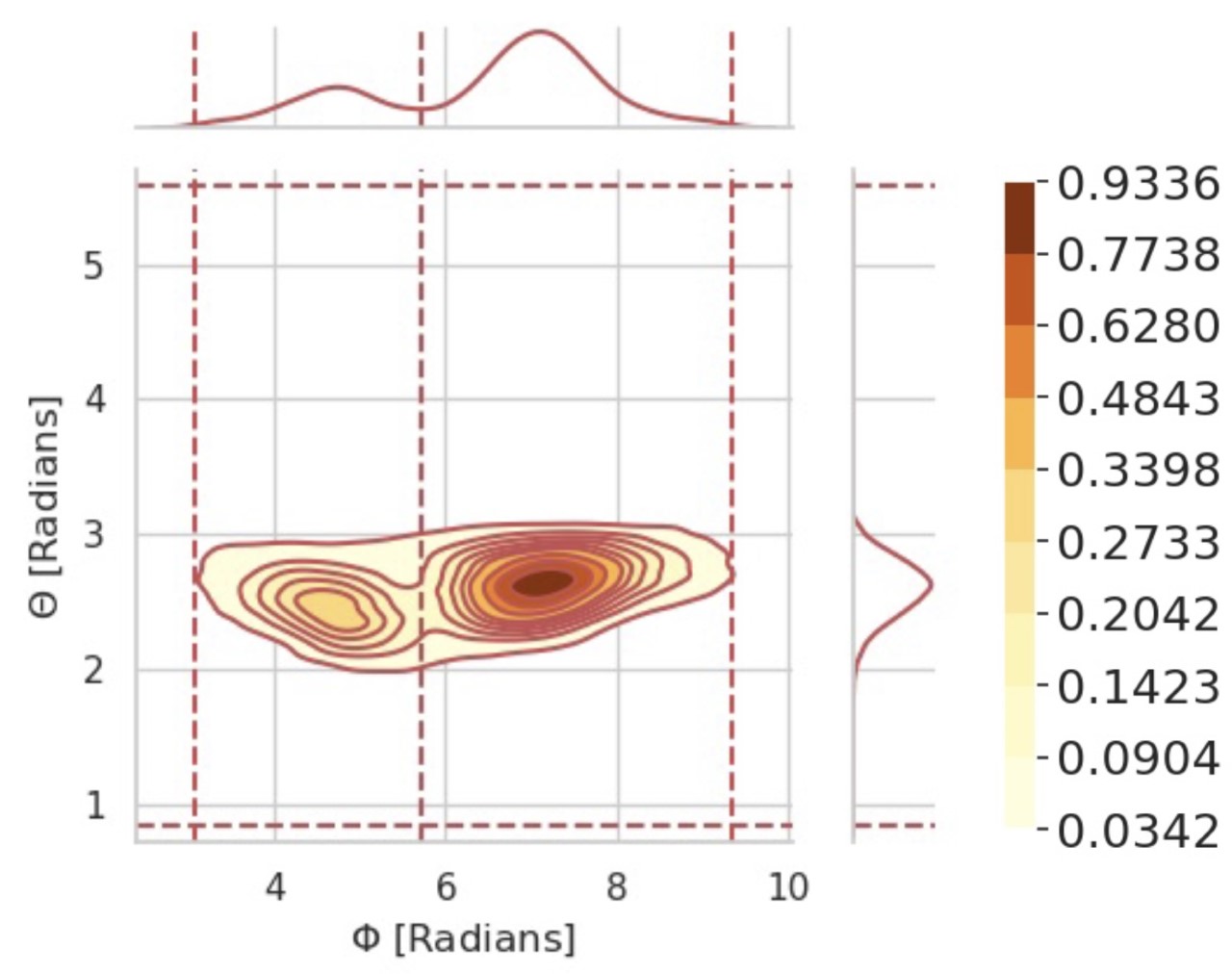
Multivariate Discretization: PENSA offers an automatic discretization of one or multiple combined features into distinct states. For example, amino acid torsions typically oscillate about local minima in populations known as rotamers, often sampling different rotamers in an MD simulation.Dunbrack (2002); Dunbrack and Karplus (1993); Scouras and Daggett (2011) In the Dunbrack library,Dunbrack and Karplus (1993), each local rotamer can be accurately represented by a Gaussian distribution around the minimum energy conformation. Similarly, Gaussians can represent the oscillation of a water molecule’s polarization (orientation of its dipole) within a water cavity, while cavity occupancy changes can be conveniently considered as two discrete populations with zero oscillation. The discrete states of these distributions are generated by applying a multi-modal Gaussian fit. The fit parameters are obtained with a non-linear least squares fit of up to ten Gaussians to each feature distribution using the SciPy library Virtanen et al. (2020). For our purposes, we found this method to be computationally more efficient than alternative methods such as Gaussian Mixture Model. The state limits for each distribution are then defined by the Gaussian intersects. Distributions which have a cyclic periodicity may oscillate about a periodic boundary but show up as two states on different ends of the distribution. To account for this boundary effect, all periodic distributions can be linearly shifted. In general, state limits can be defined for any kind of distribution, even non-Gaussian, using suitable clustering algorithms. For these cases, PENSA allows the manual input of arbitrary state boundaries. In instances where the dynamics of an amino acid are described by a combined view of all side-chain angles, e.g., the five side-chain torsions of arginine, an arbitrary number of N dimensions can be combined into one joint feature. The states of an N-dimensional feature are then the combination of all discretized microstates, defining a grid in the combined feature space, as shown in Fig. 3.
Dimensionality Reduction: Comparisons of multiple complex systems can be made more tractable by representing these systems in terms of low-dimensional, higher-order descriptions that summarize a large number of primary features in a small number of quantities. The most popular algorithm for such dimensionality reduction is Principal Component Analysis (PCA) F.R.S. (1901). Time series, such as those from molecular dynamics simulations, can also be projected via Time-Lagged Independent Component Analysis (TICA) Schultze and Grubmüller (2021). Classically, dimensionality reduction has been applied on single ensembles. But to detect patterns across ensembles and differences between them, we have to define the same representation for all investigated ensembles. PENSA users can perform dimensionality reduction on the combined data of all ensembles included in the analysis and then compare them along the resulting reduced dimensions.
Clustering: The coordinate space defined by the dimensionality reduction methods discussed above can be clustered into discrete states, again using data from all ensembles. Clustering the structures from all ensembles in the resulting lower-dimensional space provides discrete states. PENSA implements k-means clustering Macqueen (1967) and regular-space clustering Prinz et al. (2011), two popular algorithms for this task. Users can calculate populations of the resulting discrete states in each ensemble and compare them.
II.3 Feature-by-Feature Comparison
The local extent of deviations between two ensembles can be quantified by comparing each feature’s probability distribution in one ensemble to its distribution in the other ensemble. For a feature in a simulation trajectory of length , the samples of give an empirical estimation of the distribution , which describes the behaviour of in that ensemble. We want to compare the distribution in ensemble to the corresponding distribution in ensemble . These distributions may have complex functional form, hence comparing their summary statistics (mean and standard deviation) might not be sufficient as they cannot capture more subtle differences in the distributions, e.g., the split-up of one state (unimodal distribution) into two states (bimodal). Instead, PENSA provides comparison measures that are designed to capture differences in probability distributions, namely the Jensen-Shannon Distance (JSD) and the Kolmogorov-Smirnov Statistic (KSS) divergence.
Jensen-Shannon Distance: Two distributions can be compared using the Jensen-Shannon distance , a symmetrized and numerically more stable version of the Kullback-Leibler divergence . For two distributions over a feature from ensemble and ensemble , is defined below
| (1) | |||||
with the Kullback-Leibler divergence. For numerical reasons, we always use its discrete version:
| (2) | ||||
where is the set of possible states. In the case of continuous features, these states are bins along the feature coordinate, obtained by evenly dividing the range of the joint distribution. Note that is not symmetric, i.e., but is. The use of JSD as a comparison metric has been discussed in more detail in previous workLindorff-Larsen and Ferkinghoff-Borg (2009) where the comparison was performed on entire ensembles instead of individual features. Most importantly, in contrast to the unbounded — and in practice often divergent — KL divergence, the Jensen-Shannon distance ranges from 0 to 1 where 0 is obtained for identical distributions and 1 for a pair of completely different distributions.
Kolmogorov-Smirnov Statistic: Alternatively, we can quantify the differences between two distributions of continuous features without the need to define a binning parameter by using the Kolmogorov–Smirnov statistic . It is defined for a feature as
| (3) |
with and the empirical distribution functions of and , respectively. The empirical distribution functions are directly obtained from the calculated features and require no sorting of the data into arbitrary bins. The results of and for the same comparison ideally are very similar which can serve as an important sanity check.
Overall Ensemble Similarity: The overall similarity of two ensembles over all features in a metrics can be quantified by aggregating similarity metrics of all features . For example, an average Kolmogorov-Smirnov statistic of two ensembles and can be computed as:
| (4) |
and analogously. Aggregation functions other than the average, including the maximum and the minimum, are also implemented. Similar to other metrics that quantify the similarity of two ensembles in a single score,Brüschweiler (2003); Lindorff-Larsen and Ferkinghoff-Borg (2009); Tiberti et al. (2015) these aggregated metrics are particularly helpful when we need to evaluate the output of a new method to a reference ensemble, for example comparing a simulation or a generative machine learning model to a ground truth from an experiment or a more accurate level of simulation.
II.4 Mutual Information Analysis
A mutual information analysis can be employed to measure how much the specific value of one feature is coupled to the specific value of another.Shannon (1948) Applying this approach to two conformational ensembles and , one can identify if the specific values of a feature are dependent on the system ensemble – or , and vice versa, how much the value of a feature reveals about whether it stems from ensemble or . The ensemble identifiers and are then equivalent to the values of an additional feature within one (joint) ensemble. PENSA focuses on mutual information shared between a feature’s conformational states (e.g., the multivariate states described in section II.2) and the ensemble states and . To quantify this, we calculate the State-Specific Information (SSI, Fig. 4), a linear, discrete-state adaptation of mutual information that has originally been developed for amino acid torsions that act as molecular switchesThomson et al. (2021) but here is generalized to any feature with a distribution that can be represented as discrete states. It should be noted that an arbitrary number of ensembles could be incorporated into the SSI calculation, simultaneously measuring the mutual information between all conditions and a feature, but it is currently implemented for two. Similarly, SSI can operate on a single ensemble that is partitioned into two sub-ensembles, e.g., along a state boundary.
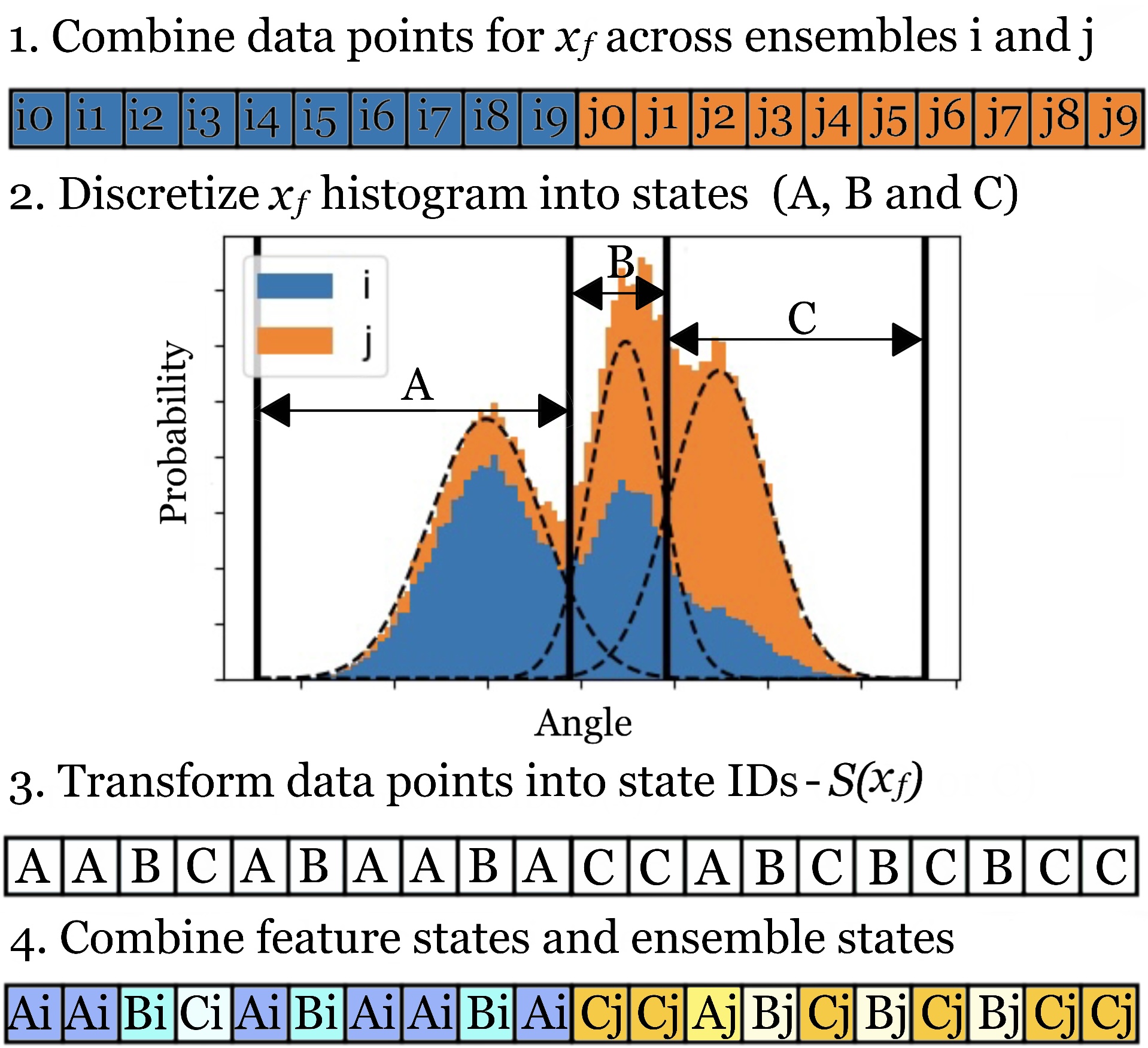
State-Specific Information (SSI): The SSI measure quantifies the degree to which conformational state transitions of feature signal information about the ensembles and or the transitions between them. It is defined as
| (5) |
where is the transformation of the combined data points of across ensembles and into data points referring to a feature state-identifier , derived from the discretized probability distribution of , as seen in figure 4. The variable is a generic representation of the ensemble ID, with possible values or . is the joint distribution of a feature state of and ensemble state (obtained from the probabilities of each element in the list shown in Fig. 4, step 4.), and and are the marginal distributions. The SSI ranges from 0 bits to 1 bit, where 0 bits represents no shared information and 1 bit represents maximal shared information between the ensemble (transitions) and the features.
State-Specific Co-Information (CoSSI): To quantify the degree to which two features interact with one another as they signal information about the ensemble they are in ( or ), State-Specific Co-Information (CoSSI) is employed. This multivariate feature-feature-ensemble metric is the linear, discrete-state adaptation of co-informationBell (2003) that uses Shannon’s discrete entropy formulation Shannon (1948), calculated using
| (6) | ||||
where the transformation is as previously defined. can be positive or negative, indicating whether the switch between ensembles increases (), decreases (), or does not affect () the communication between two features and . In the case of small-molecule ligand binding, for instance, positive between features can represent the turning-on of a signal channel by a ligand.
II.5 Visualization
PENSA includes convenient functions to visualize all stages of the analysis workflow. Primary features as well as processed features (like projections onto PCA eigenvectors) can easily be compared individually using histograms or inferred densities, and combinations of two features using heatmaps, with all functionality based on Matplotlib.Hunter (2007) The water and ion cavity featurizer writes out a structure file with the average position of the molecular ensemble, generated via MDAnalysis,Michaud-Agrawal et al. (2011) with additional atoms added via BiotiteKunzmann and Hamacher (2018) that mark the cavity centers and store the magnitude of the probability maxima. Input structures can be sorted along the values of primary or processed features using MDAnalysisMichaud-Agrawal et al. (2011) which is particularly useful for PCA or tICA to see which component of a molecule’s motion is associated with which eigenvector. Analysis metrics for comparison and mutual information that are related to a single residue (e.g., the maximum JSD of all side-chain torsions in an amino acid) can be stored in structure files using MDAnalysisMichaud-Agrawal et al. (2011) and we provide scripts for PyMolSchrödinger, LLC (2015) and VMDHumphrey et al. (1996) to visualize them via the color or the width of the cartoon representation. Metrics related to two features (e.g., distances) are visualized in square heatmaps, also implemented via Matplotlib.Hunter (2007) These visualization options provide a comprehensive overview of complex systems in one — or very few — figures.
III Example Applications
III.1 Understanding effects of a small chemical modification: loop opening in an oxidoreductase
As a first example, we show how the systematic comparison of protein backbone and side-chain torsions provides a comprehensive overview on the differences in the conformational ensembles induced by a small chemical modification. We consider the oxidation of two cysteine thiols to a disulfide bond in the N-terminal domain of the key bacterial oxidoreductase DsbD (nDsbD). DsbD plays an important role in electron transport across the inner cytoplasmic membrane of gram-negative bacteria and this reaction is an important step in its catalytic cycle.Stelzl et al. (2020)
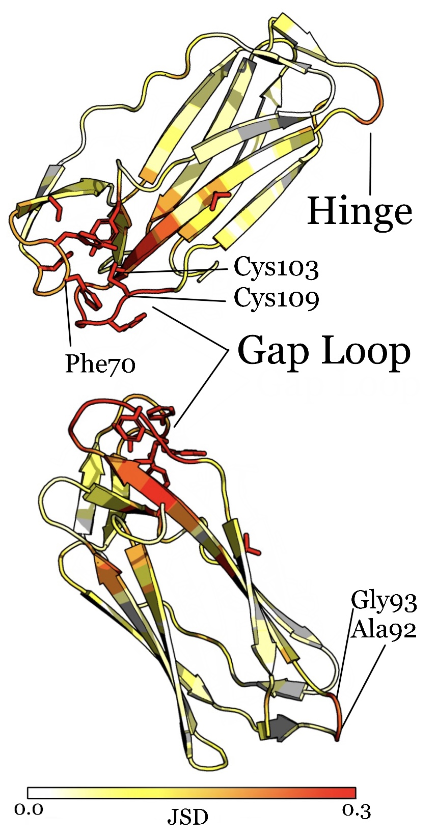
Visualization of the maximum JSD per residue for backbone and sidechain torsions (see Fig. 5) shows at one glance the opposing residues in the gap loop to be the most affected regions. Unsurprisingly, the residues directly involved in the reaction, Cys103 and Cys109, show the highest values. The effects of this reaction on the surrounding residues – showing up in our analysis as medium JSD values – cause a change in the distance of the opposing loop and a corresponding opening of the gap. In particular, we find residues Phe70 and Tyr71 at the neighboring loop to be strongly affected. Indeed, the authors of the original study identified the distance between residues Phe70 and Cys109 to be the characteristic hallmark of loop opening.
Besides the influence on the gap loop, we identify a more subtly involved and previously not discussed region at the opposite side of the protein. This region – mainly the backbones of residues Ala92 and Gly93 – functions as a hinge for the beta-strand region that slightly tilts when the gap loop is pushed outward during the gap opening. This finding demonstrates how our approach picks up differences between simulations that are otherwise easily missed.
III.2 Comparing force field parameters: Interactions of Calcium with DNA
As a second use case, we show how to quantify the effects of small changes in force field parameters on the overall conformational ensemble (Fig. 6). We consider the binding of calcium ions (Ca2+) to DNA Cruz-León et al. (2021). Metal cations play a crucial role in stabilizing the structure of nucleic acid systems. Their force field parameters are usually determined to reproduce bulk properties like the solvation-free energy and thus often not directly transferable to interactions with biomolecules.Panteva et al. (2015) Furthermore, small changes in the interactions can have significant conformational consequences overall.Yoo and Aksimentiev (2012) Studying the effect of such small changes on the overall conformation is an important problem in force field optimization.
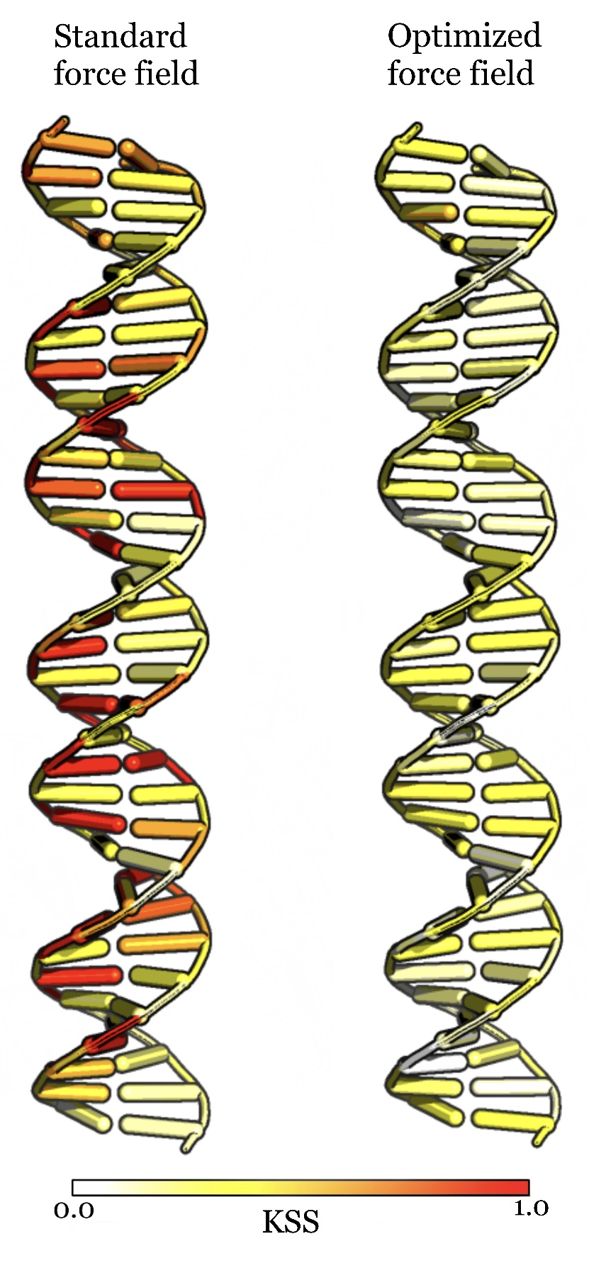
Comparison of DNA backbone torsions from MD simulations using the standard force field parameters for Ca2+-DNA interactions to an experimental reference ensemble reveals a periodic pattern of strong deviations along the entire double strand that are strongly reduced by using optimized force field parameters. We generate the reference distribution from the experimental structure by Gaussian sampling of the relevant coordinates using the uncertainty of the X-ray structure (1.7 Å, PDB: 477D) as the width of the distribution. We then compare the simulations of each parameter set to this reference (Fig. 6) using the Kolmogorov-Smirnov statistic (Eq. 3) because it provides a parameter-free measure for the deviations. The approximate periodicity of the deviations (Fig. 6, left) shows that, using standard parameters, the conformational ensemble as a whole deviates from the reference distribution and has problems reproducing the overall structure of the double strand. The authors of the original study Cruz-León et al. (2021) identified an overestimation of Ca2+-DNA interactions as the main cause of such deviations. It allows the Ca2+ ions to bridge between the phosphate oxygen atoms of opposite backbone strands which causes the minor groove of the DNA strand to shrink and in turn affects the entire structure. Thus, they rescaled the force field parameters to optimize the Ca2+-DNA interactions. This not only improved the local accuracy but the entire conformational ensemble, even though the overall structure was not explicitly optimized for during the rescaling. In our PENSA-based analysis, this improvement is immediately visible by the reduced deviations (Fig. 6, right). The overall ensemble KSS (as in Eq. 4) is reduced from 0.50 (standard force field) to 0.34 (optimized force field). This example shows that our workflow quickly identifies whether and where small changes in local interactions propagate to strong deviations in the overall structure of a biomolecule.
III.3 Tracing information linked to a protonation state: The central aspartic acid in the -opioid receptor
To showcase the usefulness of State-Specific Information (SSI), we investigate the relationship between the protonation state of a central aspartic acid and the -opioid receptor (OR) ensemble. The OR, a G protein–coupled receptor (GPCR), is a transmembrane receptor protein that converts extracellular stimuli into intracellular signalling cascades. The diversity in structure and function among GPCRs underpins complex activation mechanisms that, despite large pharmaceutical interest, remain unresolved. Katritch et al. (2013); Hauser et al. (2018); Eiger et al. (2022) Rotamer changes in residue side chains give rise to larger-scale conformational changes that enable the binding of effector proteins and trigger downstream signaling (receptor activation).Hauser et al. (2021) These rotamers can be understood as molecular micro-switches, making them an ideal use case for our state-based mutual information approach, SSI. Many receptors are influenced by environmental pH changes,Ludwig et al. (2003); Rowe et al. (2021); Sanderlin et al. (2015) and protonation changes of an evolutionarily conserved aspartic acid residue have been hypothesised to represent a key step in receptor activation. Ghanouni et al. (2000); Mahalingam et al. (2008); Vickery et al. (2018) Here, we apply SSI to side-chain and backbone torsions to investigate the effect of protonating this conserved aspartic acid, Asp114 ( protonated, de-protonated) in the antagonist-bound OR (PDB: 4DKL, Fig. 7).
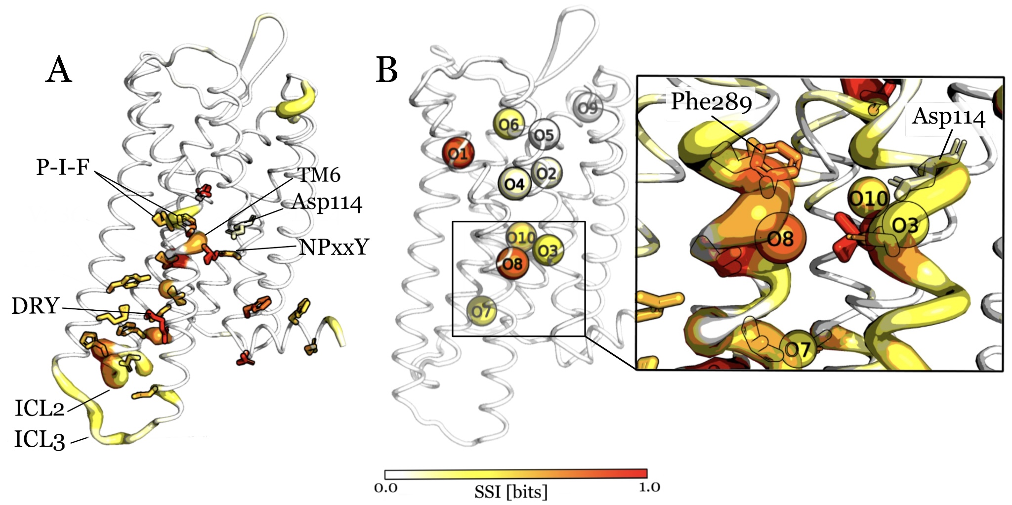
Mutual information analysis of side-chain rotamers: The SSI values calculated for each residue reveal those parts of the receptor that signal information about the protonation state of Asp114 (Fig. 7). Namely, rotameric state changes in the backbone torsions of transmembrane helix TM6, proximal to Asp114, are coupled to the protonation state changes of Asp114. Similarly, the backbone rotamer states of intracellular loops ICL2 and ICL3 couple to the protonation state of Asp114. An outward swing of TM6, enabled by backbone conformational state changes near Phe289 (P-I-F motif), is characteristic of receptor activation,Katritch et al. (2013) and specific conformations of ICL2 and ICL3 are implicated in the binding of signal proteins.Koehl et al. (2018); Mafi et al. (2020) Furthermore, side chain changes are identified on the P-I-F motif, the NPxxY motif, and the DRY motif, three receptor motifs that are known to undergo distinct rotamer changes in the transition from inactive to active receptor states.Katritch et al. (2013) The recognition of receptor regions where conformational changes are associated with activation and signaling suggests that the Asp114 protonation state and GPCR activation are intertwined. This example thus demonstrates how SSI and its visualization help to pinpoint receptor regions where the features’ rotamer states inform about an aspartic acids protonation state.
Mutual information analysis of intracavity water sites: To further demonstrate how SSI can be used to analyze water cavity features, we featurized the ten most well-defined water sites (Fig. 7). The locations of the water cavities were determined using the PENSA water featurizer as the ten sites with the largest probability maxima in the water density grid of the combined ensembles and labelled O1–O10 according to their ranking. The positioning of all ten sites agrees well with water molecules resolved in experimentsGranier et al. (2012); Huang et al. (2020) and predicted by the HomolWat serverMayol et al. (2020) for the inactive OR crystal structure (PDB: 4DKL), confirming the accuracy of PENSA’s water cavity featurization. Three water cavities are within the vicinity of Asp114: O3, O8 and O10. Using SSI (eq. 5), we calculated that water cavities O1–O10 share information with the Asp 114 protonation state on levels between 0.00–0.74 bits. Water cavity O8, for example, shares 0.67 bits of information in coupled conformational state changes linked to the transition between ensemble and , i.e., unprotonated to protonated Asp114. O8 also sits beside Phe289 of the P-I-F motif, where we identified that backbone rotamer state changes are coupled to Asp114 protonation. Surprisingly, the more distant water site O1 shares 0.74 bits of information, 82% of which is due to an occupancy change. Comparing the average ensemble structures reveals an increased packing between TM5 and TM6 in the region about the water O1 cavity, with the distance between surrounding Cs moving over on average 1Å closer in the Asp114-protonated ensemble, suggesting that helix movements on TM6 lead to a collapse of the cavity. This analysis highlights a concerted behaviour of water cavities and TM6, whereby state changes to both are indicative of the protonation state of Asp114. It shows how the combined analysis of multiple different features and a comprehensive visualization help to find interrelations within a receptor and discover signaling pathways.
IV Discussion
With PENSA, we have implemented an open-source library that provides systematic, easy-to-apply methods that make the otherwise often cumbersome exploration of biomolecular systems faster, more reliable and easier to interpret. The data readers implemented in PENSA engage with a range of biomolecular systems via robust featurization implementations. These include readers for interatomic distances and the characteristic torsion angles of amino acids and nucleic acids as well as a novel approach that reliably incorporates water and ion cavities via their occupancy and, in the case of water, the polarization of the cavity. Combined with dimensionality reduction tools, PENSA can handle biomolecular systems on a wide range of scales and resolutions. PENSA includes two comparison measures with slightly different use cases: JSD is a fast way to compare ensembles and pick up relevant features. The sensitivity of JSD depends on — and can be adjusted via — the spacing of the discretization. KSS is a parameter-free metric that picks up every ensemble difference, even the noise. In most practical cases, JSD and KSS provide similar results. In addition, PENSA includes a mutual information measure, State-Specific information (SSI). With no prior knowledge of the data, PENSA performs an automatic discretization of feature distributions into conformational states, and via SSI, quantifies the information that each features’ conformational states signal about the ensemble they are in. The ideal use case involves features that switch between well-defined states, such as molecular switches, however customizable state definitions allow SSI to operate with many kinds of feature discretization. SSI can be further extended to three or more features to quantify information flow within a system but is currently only implemented for two. Combined with PENSA’s convenient visualization tools, these methods allow for a detailed analysis of biomolecular ensembles without the bias of hand-picked metrics, acting as a solid basis for mechanistical interpretations and further, more detailed analysis.
Our example analyses demonstrate the versatility of PENSA on three different biomolecular systems. By investigating the effects of a small chemical modification on loop opening in an oxidoreductase with JSD, we demonstrate the validity of the method in confirming previously discovered results, while additionally reporting novel, more subtle findings within the same system. We demonstrate the applicability of PENSA in the optimization of force field parameters via a comparison of the interactions between Calcium and DNA under different force field parameters with KSS. Finally, we report on a communication channel in the -opioid receptor that transmits information between the intracellular signalling site and the protonation state of a distal aspartic acid, shedding further light on the signal transduction mechanism of this mechanistically complicated system. The major limit to the accuracy of PENSA is the quality of the input ensembles. For example, insufficiently converged MD simulations can cause false positives when an equally probable transition happens only in one of the conditions. Or they can cause false negatives for overall rare events or slow processes. If in doubt, validation by other means may be necessary (experiment, independent/longer simulations). Although non-converged simulations can give useful hints, these cases demand a cautious systematic analysis. Despite these caveats, PENSA has the potential for high-throughput analysis of a large amount of simulations, e.g., those available in GPCRmdRodríguez-Espigares et al. (2020), can be used to independently quantify the quality of force fields or generative machine learning models, and unravel molecular mechanisms and signaling pathways.
V Conclusions
In summary, we present a powerful toolkit to build workflows for the systematic and quantitative analysis of biomolecular systems and their conformational ensembles. PENSA provides flexible options to featurize various biomolecular systems, metrics to compare ensembles and to detect interrelations between different regions of a system, and methods to produce intuitive visualizations. We demonstrate the effectiveness of these methods on three real-world examples from molecular biology, showing how PENSA makes it easier for researchers to analyze large amounts of complex simulation data.
VI Acknowledgments
We thank Lukas Stelzl for the trajectory data of the oxidoreductase example as well as Sergio Cruz-León, Kara Grotz, and Nadine Schwierz for the trajectory data of the DNA-Calcium example. We are grateful to Alexander Powers, Lukas Stelzl, Nicole Ong, Eleanore Ocana, Emma Andrick, Callum Ives, and Bu Tran for beta-testing as well as Maria Karelina, Marc Dämgen, Patricia Suriana, Sergio Cruz-León, Michael Ward, and Ramon Guixà-González for helpful discussions. M.V. was supported by the EMBO long-term fellowship ALTF 235-2019. N.J.T. was supported by a BBSRC EASTBIO PhD studentship. This work was supported by National Institutes of Health grant R01GM127359 (R.O.D.) An award of computer time was provided by the INCITE program. This research used resources of the Oak Ridge Leadership Computing Facility, a DOE Office of Science User Facility supported under contract DE-AC05-00OR22725. Additional computing for this project was performed on the Sherlock cluster and the University of Dundee SLS HPC cluster. We thank Stanford University, the Stanford Research Computing Facility, and the University of Dundee for providing computational resources and support that contributed to these research results.
References
- Hollingsworth and Dror (2018) S. A. Hollingsworth and R. O. Dror, Neuron 99, 1129 (2018).
- Knoverek et al. (2018) C. R. Knoverek, G. K. Amarasinghe, and G. R. Bowman, Trends in Biochemical Sciences 44, 351 (2018).
- Suomivuori et al. (2020) C.-M. Suomivuori, N. R. Latorraca, L. M. Wingler, S. Eismann, M. C. King, A. L. W. Kleinhenz, M. A. Skiba, D. P. Staus, A. C. Kruse, R. J. Lefkowitz, and R. O. Dror, Science 367, 881 (2020).
- Provasi et al. (2011) D. Provasi, M. C. Artacho, A. Negri, J. C. Mobarec, and M. Filizola, PLoS Computational Biology 7, 1 (2011).
- McCorvy et al. (2018) J. D. McCorvy, K. V. Butler, B. Kelly, K. Rechsteiner, J. Karpiak, R. M. Betz, B. L. Kormos, B. K. Shoichet, R. O. Dror, J. Jin, and B. L. Roth, Nature Chemical Biology 14, 126 (2018).
- Cordero-Morales et al. (2007) J. F. Cordero-Morales, V. Jogini, A. Lewis, V. Vásquez, D. M. Cortes, B. Roux, and E. Perozo, Nature Structural and Molecular Biology 14, 1062 (2007).
- Liu et al. (2015) Y. Liu, M. Ke, and H. Gong, Biophysical Journal 109, 542 (2015).
- Thomson et al. (2021) N. J. Thomson, O. N. Vickery, C. M. Ives, and U. Zachariae, bioRxiv (2021), 10.1101/2020.08.28.271510.
- Humphrey et al. (1996) W. Humphrey, A. Dalke, and K. Schulten, Journal of Molecular Graphics 14, 33 (1996).
- DeLano (2002) W. L. DeLano, CCP4 Newsletter on protein crystallography 40, 82 (2002).
- Dror et al. (2011) R. O. Dror, D. H. Arlow, P. Maragakis, T. J. Mildorf, A. C. Pan, H. Xu, D. W. Borhani, and D. E. Shaw, Proceedings of the National Academy of Sciences of the United States of America 108, 18684 (2011).
- Zhou et al. (2019) Q. Zhou, D.-H. Yang, M. Wu, Y. Guo, W. Guo, L. Zhong, X. Cai, A. Dai, W. Jang, E. I. Shakhnovich, Z.-J. Liu, R. C. Stevens, N. A. Lambert, M. M. Babu, M.-W. Wang, and S. Zhao, eLife 8, e50279 (2019).
- Dror et al. (2013) R. O. Dror, H. F. Green, C. Valant, D. W. Borhani, J. R. Valcourt, A. C. Pan, D. H. Arlow, M. Canals, J. R. Lane, R. Rahmani, J. B. Baell, P. M. Sexton, A. Christopoulos, and D. E. Shaw, Nature 503, 295 (2013).
- Bowman et al. (2015) G. R. Bowman, E. R. Bolin, K. M. Hart, B. C. Maguire, and S. Marqusee, Proceedings of the National Academy of Sciences of the United States of America 112, 2734 (2015).
- Zivanovic et al. (2020) S. Zivanovic, G. Bayarri, F. Colizzi, D. Moreno, J. L. Gelpí, R. Soliva, A. Hospital, and M. Orozco, Journal of Chemical Theory and Computation 16, 6586 (2020).
- Brüschweiler (2003) R. Brüschweiler, Proteins: Structure, Function and Genetics 50, 26 (2003).
- Lindorff-Larsen and Ferkinghoff-Borg (2009) K. Lindorff-Larsen and J. Ferkinghoff-Borg, PLOS ONE 4, 1 (2009).
- Tiberti et al. (2015) M. Tiberti, E. Papaleo, T. Bengtsen, W. Boomsma, and K. Lindorff-Larsen, PLoS Computational Biology 11, 1 (2015).
- Noé et al. (2019) F. Noé, S. Olsson, J. Köhler, and H. Wu, Science 365, eaaw1147 (2019).
- Husic and Pande (2018) B. E. Husic and V. S. Pande, Journal of the American Chemical Society 140, 2386 (2018).
- Nüske et al. (2017) F. Nüske, H. Wu, J. H. Prinz, C. Wehmeyer, C. Clementi, and F. Noé, Journal of Chemical Physics 146, 094104 (2017).
- Fraccalvieri et al. (2011) D. Fraccalvieri, A. Pandini, F. Stella, and L. Bonati, BMC Bioinformatics 12, 158 (2011).
- Ward et al. (2021) M. D. Ward, M. I. Zimmerman, A. Meller, M. Chung, S. J. Swamidass, and G. R. Bowman, Nature Communications 12, 1 (2021).
- Malik et al. (2021) M. Malik, M. D. Ward, Y. Fang, J. R. Porter, M. I. Zimmerman, T. Koelblen, M. Roh, A. I. Frolova, T. P. Burris, G. R. Bowman, P. I. Imoukhuede, and S. K. England, ACS Pharmacology and Translational Science 4, 1543 (2021).
- Vögele et al. (2021) M. Vögele, N. Thomson, S. Truong, and J. McAvity, “PENSA,” (2021), doi: 10.5281/ZENODO.4362136.
- Schrödinger, LLC (2015) Schrödinger, LLC, “The PyMOL molecular graphics system, version 1.8,” (2015).
- Kunzmann and Hamacher (2018) P. Kunzmann and K. Hamacher, BMC Bioinformatics 19, 346 (2018).
- Stelzl et al. (2020) L. S. Stelzl, D. A. Mavridou, E. Saridakis, D. Gonzalez, A. J. Baldwin, S. J. Ferguson, M. S. Sansom, and C. Redfield, eLife 9, 1 (2020).
- Cruz-León et al. (2021) S. Cruz-León, K. K. Grotz, and N. Schwierz, Journal of Chemical Physics 154, 171102 (2021).
- Michaud-Agrawal et al. (2011) N. Michaud-Agrawal, E. J. Denning, T. B. Woolf, and O. Beckstein, Journal of Computational Chemistry 32, 2319 (2011).
- Gowers et al. (2016) R. J. Gowers, M. Linke, J. Barnoud, T. J. E. Reddy, M. N. Melo, S. L. Seyler, J. Domański, D. L. Dotson, S. Buchoux, I. M. Kenney, and O. Beckstein, in Proceedings of the 15th Python in Science Conference (2016) pp. 98–105.
- Scherer et al. (2015) M. K. Scherer, B. Trendelkamp-Schroer, F. Paul, G. Pérez-Hernández, M. Hoffmann, N. Plattner, C. Wehmeyer, J. H. Prinz, and F. Noé, Journal of Chemical Theory and Computation 11, 5525 (2015).
- Keating et al. (2011) K. S. Keating, E. L. Humphris, and A. M. Pyle, Quarterly Reviews of Biophysics 44, 433 (2011).
- Venkatakrishnan et al. (2019) A. J. Venkatakrishnan, A. K. Ma, R. Fonseca, N. R. Latorraca, B. Kelly, R. M. Betz, C. Asawa, B. K. Kobilka, and R. O. Dror, Proceedings of the National Academy of Sciences of the United States of America 116, 3288 (2019).
- Yuan et al. (2014) S. Yuan, S. Filipek, K. Palczewski, and H. Vogel, Nature Communications 5, 4733 (2014).
- Pardo et al. (2007) L. Pardo, X. Deupi, N. Dölker, M. L. López-Rodríguez, and M. Campillo, ChemBioChem 8, 19 (2007).
- Levy and Onuchic (2004) Y. Levy and J. N. Onuchic, Proceedings of the National Academy of Sciences of the United States of America 101, 3325 (2004).
- Kajander et al. (2000) T. Kajander, P. C. Kahn, S. H. Passila, D. C. Cohen, L. Lehtiö, W. Adolfsen, J. Warwicker, U. Schell, and A. Goldman, Structure 8, 1203 (2000).
- Zarzycka et al. (2019) B. Zarzycka, S. A. Zaidi, B. L. Roth, and V. Katritch, Pharmacological Reviews 71, 571 (2019).
- Andreini et al. (2008) C. Andreini, I. Bertini, G. Cavallaro, G. L. Holliday, and J. M. Thornton, Journal of Biological Inorganic Chemistry 13, 1205 (2008).
- Ives et al. (2022) C. M. Ives, N. J. Thomson, and U. Zachariae, bioRxiv (2022), 10.1101/2022.04.01.486690.
- Dunbrack (2002) R. L. Dunbrack, Current Opinion in Structural Biology 12, 431 (2002).
- Dunbrack and Karplus (1993) R. L. Dunbrack and M. Karplus, Journal of Molecular Biology 230, 543 (1993).
- Scouras and Daggett (2011) A. D. Scouras and V. Daggett, Protein Science 20, 341 (2011).
- Virtanen et al. (2020) P. Virtanen, R. Gommers, T. E. Oliphant, M. Haberland, T. Reddy, D. Cournapeau, E. Burovski, P. Peterson, W. Weckesser, J. Bright, S. J. van der Walt, M. Brett, J. Wilson, K. J. Millman, N. Mayorov, A. R. J. Nelson, E. Jones, R. Kern, E. Larson, C. J. Carey, İ. Polat, Y. Feng, E. W. Moore, J. VanderPlas, D. Laxalde, J. Perktold, R. Cimrman, I. Henriksen, E. A. Quintero, C. R. Harris, A. M. Archibald, A. H. Ribeiro, F. Pedregosa, P. van Mulbregt, and SciPy 1.0 Contributors, Nature Methods 17, 261 (2020).
- F.R.S. (1901) K. P. F.R.S., The London, Edinburgh, and Dublin Philosophical Magazine and Journal of Science 2, 559 (1901).
- Schultze and Grubmüller (2021) S. Schultze and H. Grubmüller, Journal of Chemical Theory and Computation 17, 5766 (2021).
- Macqueen (1967) J. Macqueen, in In 5-th Berkeley Symposium on Mathematical Statistics and Probability (1967) pp. 281–297.
- Prinz et al. (2011) J.-H. Prinz, H. Wu, M. Sarich, B. Keller, M. Senne, M. Held, J. D. Chodera, C. Schütte, and F. Noé, The Journal of Chemical Physics 134, 174105 (2011).
- Shannon (1948) C. E. Shannon, Bell System Technical Journal 27, 379 (1948).
- Bell (2003) A. J. Bell, 4th International Symposium on Independent Component Analysis and Blind Source Separation (2003).
- Hunter (2007) J. D. Hunter, Computing in Science and Engineering 9, 90 (2007).
- Panteva et al. (2015) M. T. Panteva, G. M. Giambaşu, and D. M. York, Journal of Physical Chemistry B 119, 15460 (2015).
- Yoo and Aksimentiev (2012) J. Yoo and A. Aksimentiev, Journal of Physical Chemistry Letters 3, 45 (2012).
- Katritch et al. (2013) V. Katritch, V. Cherezov, and R. C. Stevens, Annual Review of Pharmacology and Toxicology 53, 531 (2013).
- Hauser et al. (2018) A. S. Hauser, S. Chavali, I. Masuho, L. J. Jahn, K. A. Martemyanov, D. E. Gloriam, and M. M. Babu, Cell 172, 41 (2018).
- Eiger et al. (2022) D. S. Eiger, U. Pham, J. Gardner, C. Hicks, and S. Rajagopal, American Journal of Physiology-Cell Physiology 322, C887 (2022).
- Hauser et al. (2021) A. S. Hauser, A. J. Kooistra, C. Munk, F. M. Heydenreich, D. B. Veprintsev, M. Bouvier, M. M. Babu, and D. E. Gloriam, Nature Structural and Molecular Biology 28, 879 (2021).
- Ludwig et al. (2003) M. G. Ludwig, M. Vanek, D. Guerini, J. A. Gasser, C. E. Jones, U. Junker, H. Hofstetter, R. M. Wolf, and K. Seuwen, Nature 425, 93 (2003).
- Rowe et al. (2021) J. B. Rowe, N. J. Kapolka, G. J. Taghon, W. M. Morgan, and D. G. Isom, Journal of Biological Chemistry 296, 100167 (2021).
- Sanderlin et al. (2015) E. J. Sanderlin, C. R. Justus, E. A. Krewson, and L. V. Yang, Cell Health and Cytoskeleton 7, 99 (2015).
- Ghanouni et al. (2000) P. Ghanouni, H. Schambye, R. Seifert, T. W. Lee, S. G. Rasmussen, U. Gether, and B. K. Kobilka, Journal of Biological Chemistry 275, 3121 (2000).
- Mahalingam et al. (2008) M. Mahalingam, K. Martínez-Mayorga, M. F. Brown, and R. Vogel, Proceedings of the National Academy of Sciences of the United States of America 105, 17795 (2008).
- Vickery et al. (2018) O. N. Vickery, C. A. Carvalheda, S. A. Zaidi, A. V. Pisliakov, V. Katritch, and U. Zachariae, Structure 26, 171 (2018).
- Koehl et al. (2018) A. Koehl, H. Hu, S. Maeda, Y. Zhang, Q. Qu, J. M. Paggi, N. R. Latorraca, D. Hilger, R. Dawson, H. Matile, G. F. X. Schertler, S. Granier, W. I. Weis, R. O. Dror, A. Manglik, G. Skiniotis, B. K. Kobilka, C. J. Draper-Joyce, M. Khoshouei, D. M. Thal, Y.-L. Liang, A. T. N. Nguyen, S. G. B. Furness, H. Venugopal, J.-A. Baltos, J. M. Plitzko, R. Danev, and W. Baumeister, Nature , 547–552 (2018).
- Mafi et al. (2020) A. Mafi, S. K. Kim, and W. A. Goddard, Proceedings of the National Academy of Sciences of the United States of America 117, 16346 (2020).
- Granier et al. (2012) S. Granier, A. Manglik, A. C. Kruse, T. S. Kobilka, F. S. Thian, W. I. Weis, B. K. Kobilka, J. M. Mathiesen, R. K. Sunahara, and L. Pardo, Nature 485, 321–326 (2012).
- Huang et al. (2020) W. Huang, A. Manglik, A. J. Venkatakrishnan, T. Laeremans, E. N. Feinberg, A. L. Sanborn, H. E. Kato, K. E. Livingston, T. S. Thorsen, R. C. Kling, S. Granier, P. Gmeiner, S. M. Husbands, J. R. Traynor, W. I. Weis, J. Steyaert, R. O. Dror, and B. K. Kobilka, Nature, Nature 584, E16 (2020).
- Mayol et al. (2020) E. Mayol, A. Garcia-Recio, J. K. Tiemann, P. W. Hildebrand, R. Guixa-Gonzalez, M. Olivella, and A. Cordomi, Nucleic Acids Research 48, W54 (2020).
- Rodríguez-Espigares et al. (2020) I. Rodríguez-Espigares, M. Torrens-Fontanals, J. K. Tiemann, D. Aranda-García, J. M. Ramírez-Anguita, T. M. Stepniewski, N. Worp, A. Varela-Rial, A. Morales-Pastor, B. Medel-Lacruz, G. Pándy-Szekeres, E. Mayol, T. Giorgino, J. Carlsson, X. Deupi, S. Filipek, M. Filizola, J. C. Gómez-Tamayo, A. Gonzalez, H. Gutiérrez-de Terán, M. Jiménez-Rosés, W. Jespers, J. Kapla, G. Khelashvili, P. Kolb, D. Latek, M. Marti-Solano, P. Matricon, M. T. Matsoukas, P. Miszta, M. Olivella, L. Perez-Benito, D. Provasi, S. Ríos, I. R. Torrecillas, J. Sallander, A. Sztyler, S. Vasile, H. Weinstein, U. Zachariae, P. W. Hildebrand, G. De Fabritiis, F. Sanz, D. E. Gloriam, A. Cordomi, R. Guixà-González, and J. Selent, Nature Methods 17, 777 (2020).