Electron Dynamics at High-Energy Densities in Nickel from Non-linear Resonant X-ray Absorption Spectra
Abstract
The pulse intensity from X-ray free-electron lasers (FELs) can create extreme excitation densities in solids, entering the regime of non-linear X-ray-matter interactions. We show -edge absorption spectra of metallic nickel thin films with fluences entering a regime where several X-ray photons are incident per absorption cross-section. Main features of the observed non-linear spectral changes are described with a predictive rate model for electron population dynamics during the pulse, utilizing a fixed density of states and tabulated ground-state properties.
The modern understanding of complex solid materials relies on appropriate approximations to the unabridged quantum mechanical description of the full, correlated many-body problem. To assess the predictive power of theoretical models and the selected approximations, detailed experimental studies far away from known territory are especially insightful. Absorbing the high power densities available from an X-ray free-electron laser (FEL) in a solid metal generates a very unusual state of warm dense matter far from equilibrium: Individual electronic excitations reach up to hundreds of eV and excitation levels average out to many eV per atom [1, 2, 3, 4, 5, 6, 7, 8, 9]. As the absorption of an intense X-ray pulse depends on the changes it drives in the electronic system [10, 11, 12, 13, 14, 15, 16, 15], a single-pulse non-linear absorption measurement can be used to investigate its evolution on the timescale of the pulse duration.
We present fluence-dependent X-ray absorption spectra recorded with monochromatic X-rays on metallic nickel thin films around the nickel 2 () edge, revealing a changing valence electron system around the Fermi level as a consequence of the high excitation densities from fluences up to 60 J/cm2 (corresponding to 2 W/cm2).
The electronic processes that ensue after the absorption of photons at core levels trigger a complex dynamical process that is challenging to treat in purely ab-initio simulations [17, 18, 19, 20, 21]. Here, we take an alternative approach and develop a simple rate equation model that provides an intuitive understanding of the relevant processes [22]. The resulting picture of the evolution of electronic populations within a fixed ground-state density of states successfully describes the largest part of the non-linear changes in the spectra. This corroborates the dominant impact of electron redistribution from the strong non-equilibrium state towards a thermalized electronic system. Some of the observed changes, especially in the close vicinity of the resonance, deviate from the predictions of the rate model and call for more evolved theories. Here, our work provides a benchmark to identify observations of advanced physical processes and effects. While this letter discusses the experiment and resulting insights, we lay out the framework of the model in detail in a separate publication [22].
Additionally, our straightforward picture of intense core-resonant X-ray pulse interaction with the valence system of a 3 metal lays a solid knowledge-based foundation for the planning and interpretation of non-linear X-ray spectroscopy experiments at FELs; in particular, the relevance of electronic scattering processes observed here is expected to affect methods relying on stimulated emission from core excitations and X-ray or X-ray/optical wave-mixing [23, 24, 25, 26, 27, 28, 29, 30, 31, 32, 33, 34, 35, 36, 37, 38].
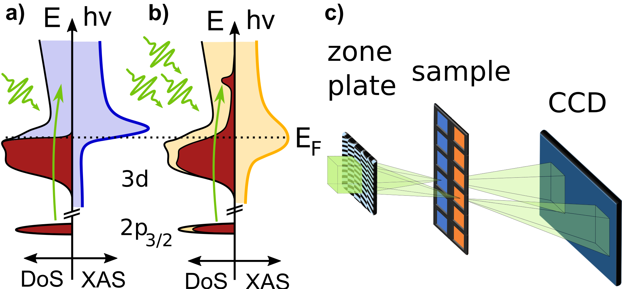
Setup for non-linear XAS (c) The split-beam-normalization scheme uses a special zone plate [39], which generates two adjacent beam foci for transmission through the sample and a reference membrane before the beams impinge on the detector.
X-ray absorption spectra of the nickel 2 () edge were recorded at the Spectroscopy and Coherent Scattering Instrument (SCS) of the European XFEL [40].
The XAS spectra were measured by continuously scanning the SASE3 monochromator [41] (synchronized with the undulator gap) back-and-forth many times in the range 846-856 eV. The photon bandwidth was about 420 meV and the FEL pulse duration on the sample was about 30 fs FWHM. The overall beam intensity was controlled using a gas attenuator filled with nitrogen and monitored using an X-ray gas-monitor (XGM) downstream of the monochromator [42, 43].
For X-ray absorption measurements at FELs based on Self-Amplified Spontaneous Emission (SASE), beam-splitting schemes can deliver optimal normalization of SASE-fluctuations [44, 45, 46]. Here, a focusing and beam-splitting zone plate also creates the required tight focusing to achieve extreme fluences. Figure 1 shows the schematic experimental layout.
The zone plate combines an off-axis Fresnel structure for focusing and a line grating for beam-splitting in a single optical element [39]. It thus produces two m-sized, identical foci in the sample plane, 1.9 mm apart, originating from the first-order diffraction of the zone plate, as well as the positive and negative first orders of the line grating. The sample has a square support of 25 mm size, containing Si3N4 membrane windows (orange in Figure 1) of 0.5 mm size and 200 nm thickness with a distance of 2 mm between adjacent windows. Every second pair of rows (blue in Figure 1) was additionally coated with a 20 sample layer of polycrystalline metallic Ni by sputter deposition, on top of a 2 bonding layer of Ta; a 2 Pt capping layer prevents oxidation during sample-handling.
The sample frame was positioned such that one zone plate focus impinged on a nickel-coated membrane, while the other hit a bare silicon-nitride membrane. Thus, the difference in transmission of both beams can be attributed solely to the nickel film.
The detector was a fast readout-speed charge-coupled device (FastCCD) with high dynamic range, enabling 10 Hz read-out and increasing the fluence range available to the experiment [47, 48, 49]. Due to the unstable detector temperature, significant retroactive calibration of the detector was necessary (see supplement). To prevent detector saturation, an additional aluminum filter of about 13 thickness was used between sample and detector for measurements with the unattenuated beam.
During these high-intensity measurements, sample and reference films were locally damaged by intense individual FEL shots. Thus, the FEL was operated in single-shot mode at 10 Hz repetition rate, and the sample was scanned through the beam continuously at 0.5 , resulting in 10 shots per membrane window.
The shot craters in the reference membranes were later analyzed with scanning electron microscopy (SEM) to determine the effective focal size at specific photon energies. The resulting spot sizes were used to calibrate ray-tracing calculations which delivered the photon-energy-dependent spot size, ranging from 0.4 to about 3 (see supplement for details on the spot size determination).
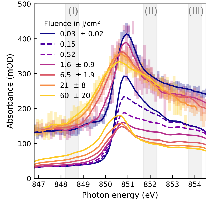
Figure 2 shows the spectra for the nickel -edge next to simulated spectra for increasing X-ray fluence over more than 3 orders or magnitude, from 0.03 to 60 J/cm2. Each measured point represents an average of several FEL shots, sorted by X-ray fluence and photon energy. The varying statistical uncertainty is a result of the pulse intensity fluctuations of monochromatized SASE radiation [50] in combination with photon energy-dependent spot sizes (see supplement for details on the shot sorting).
We observe four main fluence-dependent effects, which we quantify and compare to the simulated results in Figure 3: a) a red-shift of the absorption peak of up to 0.9 0.1 eV in the rising flank; b) an increase of the pre-edge absorbance, as the rising edge of the absorption peak shifts and broadens; c) a reduced peak absorbance and d), e) a reduced post-edge absorbance. The integration regions from which the effects b), d) and e) are derived, are highlighted in Figure 2 as (I), (II) and (III), respectively. The shift of the absorption edge is quantified by the photon energy at which the absorbance reaches half of the peak value; its uncertainty is propagated from the statistical uncertainty of the absorption peak measurement.
Before we analyze these observations in detail, let us quickly paraphrase our modeling approach [22]: In contrast to earlier rate models [51, 12], we describe the evolution of the electronic system with an energy-resolved population of the valence band. Tracking the full non-thermal population history proved crucial to describe the non-linear absorption changes near and around the Fermi level. As coupling between electrons and phonons in metals is typically not yet important on the timescale of 30 fs [52, 53, 54] and we do not account for collective electron correlation effects, we test the approximation that the Density of States (DoS) remains unchanged within the pulse duration.
Transition rates between electronic states are determined by scaling ground state rates with the populations of initial and final states. The relevant process rates are compiled into differentials of electronic populations and photon density in space and time and implemented in a finite-element simulation to derive the electron population history and ultimately the X-ray transmission of a three-dimensional sample.
The model implements the processes of resonant absorption from the 2 core level and non-resonant absorption from other (mostly valence) electrons. Stimulated emission is described as a time-inverted resonant absorption process. Electronic thermalization is modeled with a bulk timescale (essentially quantifying electron-electron scattering) that moves the non-thermal valence electron distribution towards a target Fermi-Dirac distribution that corresponds to the momentary internal energy and population of the valence band. Finally, scattering cascades initiated by fast Auger-electrons and photo-electrons from non-resonant absorption are parameterized by another scattering time .
With this simple description of the underlying processes, we provide a microscopic picture of the electronic system and its interaction with resonant X-rays as a complementary approach to more complex calculations [20, 21].
Solely considering the population dynamics of the electronic system, the simulation already achieves good agreement with the experimental data across more than three orders of magnitude in fluence. This is particularly remarkable since nearly all input parameters are experimental parameters or well-known ground-state properties of the material, such as density, electronic configuration, and ground-state spectrum. Only the valence thermalization time and electron scattering time were varied to achieve the best match to the experimental results. The found value fs compares well to recent estimates for excitations on this energy scale [33, 38, 55, 56].
The time constant fs characterizes the secondary electron scattering cascade which transfers energy (and population) from fast electrons to (unoccupied) valence states. The constant summarizes many individual electron scattering events and compares to the tabulated time between individual collisions in ground-state nickel of roughly 100 attoseconds [57].
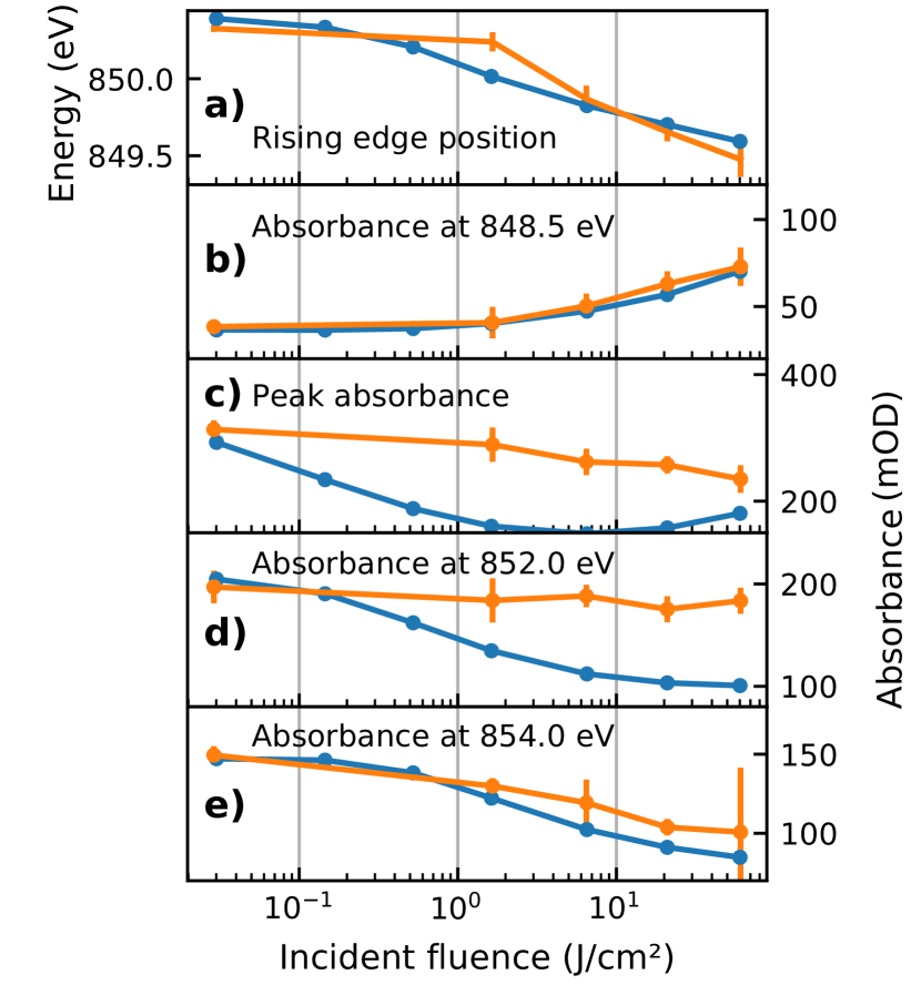
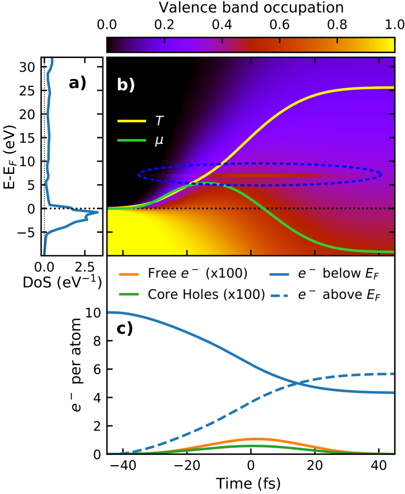
Figure 4 shows an example of a simulated valence band population history, specifically from the uppermost 4 Å thick layer of the sample, excited with a Gaussian pulse profile centered around with 30 fs FWHM duration and 30 J/cm2 fluence. While panel a) shows the calculated DoS as used by the simulation and published in [58, 59], the colormap in b) shows the occupation of these states over time. Panel c) shows the number of electrons per atom in the valence band below and above the Fermi level (blue solid and dashed curves, respectively) as well as the average number of core holes and the number of free electrons over time. Even though the direct interaction with the photons creates core holes via resonant absorption and free electrons via non-resonant absorption, the excitation energy of both processes is so quickly transferred to the valence electrons that only the valence electron distribution ever deviates strongly from the ground state. By the end of the pulse in this example, more than half of the valence electrons are excited to valence states above the Fermi level, while the highest instantaneous number of core holes was only about one per 100 atoms, as shown in Figure 4 c). Due to the small-bandwidth excitation, the core- and resonant valence states operate like a two-level system. Since the number of resonant valence states is small in comparison to the number of core electrons, the resonant absorption process saturates due to occupied valence states long before the core level is depleted. A heated Fermi-Dirac distribution further contributes to the occupation of states above the Fermi level.
Since in our experiment, the same monochromatic pulse excites and probes the sample, the situation is different for energies below the edge: absorption only rises after non-resonant absorption has led to sufficient electronic heating until the tail of the hot hole distribution reaches the probed energy. Only then, additional resonant absorption begins to occur and accelerates further electronic heating and in turn additional pre-edge absorption. Since this process occurs exponentially faster near the absorption edge, it contributes significantly to the observed spectral red-shift (see Figure 3 a) and b)).
Another cause of the observed edge shift is the shift of the chemical potential , which strongly depends on the exact shape of the DoS and is shown in Figure 4 b) as a green line. Initially, increases with absorbed fluence, as thermally excited electrons from the states must spread out in energy to the lower DoS above the Fermi level. With rising electronic temperature, the high DoS of the states becomes less relevant and the chemical potential drops again as expected in regular metals. A similar evolution of the chemical potential and electronic temperature was predicted for optically excited nickel by previous experiments and calculations [60, 61, 62, 4].
A significant deviation between model and experiment can be observed at the resonance peak itself, where the simulated electron dynamics lead us to expect a much stronger saturation effect than observed experimentally (Figure 3 c)). This underestimation may be related to a fluence-dependent decrease of the excited state lifetime and consequent energetic broadening of the resonant core-valence transition, which is not considered in our model. While it is unsurprising to find additional resonant effects in the resonance peak itself, the lack of any significant saturation around 852 eV (Figure 3 d)) is even more surprising. Both disagreements point to additional physical effects and call for more sophisticated models.
We speculatively propose mechanisms which could contribute to these disagreements: The transition matrix elements could get modified at higher excitation densities, especially around the resonance, while we model the absorption only based on the ground-state spectrum. An energy dependence of the electron-electron scattering cross-section could allow for particularly fast scattering of electrons with certain energies, counteracting the saturation. Furthermore, a collective, correlated response of the electronic system could modify the DoS or the transitions even on the fast time scale of the FEL pulse duration [63]. Despite these remaining discrepancies, the main aspects of the spectral changes are covered in our very simple population dynamics model.
We want to point out that substantially smaller spectral red-shifts were observed before in nickel after excitation with optical lasers, albeit at three orders of magnitude lower excitation fluence. These required qualitatively different interpretations [64, 65, 56, 63], where the explanation for time-dependent changes included a variable DoS, calculated using (Time-Dependent) Density Functional Theory (TD)DFT; this dependency is overshadowed in our high-fluence study by the effects of electron population dynamics.
To summarize, we interpret the fluence-dependent near-edge X-ray absorption spectra of the nickel 2 core level at X-ray fluences of up to 60 . We propose a rate-equation model, describing the various excitation and decay processes that connect core- and valence electronic states using differential equations based on scaling of known ground-state properties with the evolving electron populations. For the measured spectra of metallic nickel, the model successfully predicts the increase of absorption before and its decrease beyond the resonance, as well as the observed shift of the absorption peak over more than three orders of magnitude in fluence.
It therefore allows us to identify the most important processes responsible for spectral changes: Heating of valence electrons due to secondary electron cascades from Auger electrons, as well as electrons emitted from the valence band due to non-resonant absorption, appeared particularly relevant. Furthermore, saturation appears dominated to by the heated valence states rather than the core holes.
This study provides the fingerprints of how strong X-ray fluences may alter the electronic system and thus the spectra in studies, where the X-ray pulses were originally assumed to be non-disturbing. It becomes clear that a complete modeling of high-fluence spectra needs to build upon dominant population dynamics and requires special treatment around resonances. This provides an excellent benchmark for sophisticated theories. Our results also apply to the resonant regime which is particularly interesting for pioneering non-linear X-ray studies.
Acknowledgements.
We acknowledge European XFEL in Schenefeld, Germany, for provision of X-ray free-electron laser beamtime at the SCS instrument and would like to thank the staff for their assistance. Funding by the Deutsche Forschungsgemeinschaft (DFG, German Research Foundation) - Project-ID 278162697 - SFB 1242 and the Helmholtz Association (grant VH-NG-1105) is gratefully acknowledged. Access to Synchrotron SOLEIL through proposal ID 20160880 for characterization of static properties of the Ni films is acknowledged. Parts of this work were funded by the Swiss National Science Foundation (Grants No. PZ00P2-179944)Author Contributions
M.B., C.D., F.D., L.L.G., J.L., J.P.M., B.R. and S.T. conceptualized and planned the experiment; M.C., C.D., F.D., N.G., L.L.G., M.I., E.J., A.K., C.-H.L., L.M., B.P., B.R., A.S., K.S., C.S., H.W. and A.Y. prepared the measurement apparatus and samples; O.A., K.A., M.B., J.B., R.C., M.C., V.C., G.S.C., C.D., F.D., R.Y.E., A.E., N.G., L.L.G., O.S.H., M.I., L.M., G.M., P.S.M., B.R., N.R., A.S., J.S., S.T. A.Y. and Z.Y. performed the experiment; O.A., R.Y.E, L.L.G., O.S.H., B.R. and N.R. analyzed and visualized the results; M.B. and R.Y.E. wrote the manuscript; M.B., U.B., C.D., R.Y.E., A.E., L.L.G., O.S.H., M.I., E.J., T.L., P.S.M., K.O., K.R., M.S., J.O.S., S.M.V., H.W. and Z.Y. reviewed and edited the manuscript; M.B. and J.P.M. supervised or administered the project.
References
- Zastrau et al. [2008] U. Zastrau, C. Fortmann, R. R. Fäustlin, L. F. Cao, T. Döppner, S. Düsterer, S. H. Glenzer, G. Gregori, T. Laarmann, H. J. Lee, A. Przystawik, P. Radcliffe, H. Reinholz, G. Röpke, R. Thiele, J. Tiggesbäumker, N. X. Truong, S. Toleikis, I. Uschmann, A. Wierling, T. Tschentscher, E. Förster, and R. Redmer, Bremsstrahlung and line spectroscopy of warm dense aluminum plasma heated by xuv free-electron-laser radiation, Phys. Rev. E 78, 066406 (2008).
- Vinko et al. [2012] S. M. Vinko, O. Ciricosta, B. I. Cho, K. Engelhorn, H.-K. Chung, C. R. D. Brown, T. Burian, J. Chalupský, R. W. Falcone, C. Graves, V. Hájková, A. Higginbotham, L. Juha, J. Krzywinski, H. J. Lee, M. Messerschmidt, C. D. Murphy, Y. Ping, A. Scherz, W. Schlotter, S. Toleikis, J. J. Turner, L. Vysin, T. Wang, B. Wu, U. Zastrau, D. Zhu, R. W. Lee, P. A. Heimann, B. Nagler, and J. S. Wark, Creation and diagnosis of a solid-density plasma with an X-ray free-electron laser, Nature 482, 59 (2012).
- Cho et al. [2012] B. I. Cho, K. Engelhorn, S. M. Vinko, H.-K. Chung, O. Ciricosta, D. S. Rackstraw, R. W. Falcone, C. R. D. Brown, T. Burian, J. Chalupský, C. Graves, V. Hájková, A. Higginbotham, L. Juha, J. Krzywinski, H. J. Lee, M. Messersmidt, C. Murphy, Y. Ping, N. Rohringer, A. Scherz, W. Schlotter, S. Toleikis, J. J. Turner, L. Vysin, T. Wang, B. Wu, U. Zastrau, D. Zhu, R. W. Lee, B. Nagler, J. S. Wark, and P. A. Heimann, Resonant K Spectroscopy of Solid-Density Aluminum Plasmas, Physical Review Letters 109, 245003 (2012).
- Humphries et al. [2020] O. S. Humphries, R. S. Marjoribanks, Q. Y. van den Berg, E. C. Galtier, M. F. Kasim, H. J. Lee, A. J. F. Miscampbell, B. Nagler, R. Royle, J. S. Wark, and S. M. Vinko, Probing the electronic structure of warm dense nickel via resonant inelastic x-ray scattering, Phys. Rev. Lett. 125, 195001 (2020).
- García Saiz et al. [2008] E. García Saiz, G. Gregori, D. O. Gericke, J. Vorberger, B. Barbrel, R. J. Clarke, R. R. Freeman, S. H. Glenzer, F. Y. Khattak, M. Koenig, O. L. Landen, D. Neely, P. Neumayer, M. M. Notley, A. Pelka, D. Price, M. Roth, M. Schollmeier, C. Spindloe, R. L. Weber, L. Van Woerkom, K. Wünsch, and D. Riley, Probing warm dense lithium by inelastic X-ray scattering, Nature Physics 4, 940 (2008).
- Vinko [2015] S. M. Vinko, X-ray free-electron laser studies of dense plasmas, Journal of Plasma Physics 81, 365810501 (2015).
- Bailey et al. [2015] J. E. Bailey, T. Nagayama, G. P. Loisel, G. A. Rochau, C. Blancard, J. Colgan, P. Cosse, G. Faussurier, C. J. Fontes, F. Gilleron, I. Golovkin, S. B. Hansen, C. A. Iglesias, D. P. Kilcrease, J. J. MacFarlane, R. C. Mancini, S. N. Nahar, C. Orban, J. C. Pain, A. K. Pradhan, M. Sherrill, and B. G. Wilson, A higher-than-predicted measurement of iron opacity at solar interior temperatures, Nature 517, 56 (2015).
- Hollebon et al. [2019] P. Hollebon, O. Ciricosta, M. P. Desjarlais, C. Cacho, C. Spindloe, E. Springate, I. C. E. Turcu, J. S. Wark, and S. M. Vinko, Ab initio simulations and measurements of the free-free opacity in aluminum, Physical Review E 100, 043207 (2019), arXiv:1806.02726 .
- Preston et al. [2017] T. R. Preston, S. M. Vinko, O. Ciricosta, P. Hollebon, H.-K. Chung, G. L. Dakovski, J. Krzywinski, M. Minitti, T. Burian, J. Chalupský, V. Hájková, L. Juha, V. Vozda, U. Zastrau, R. W. Lee, and J. S. Wark, Measurements of the K-Shell Opacity of a Solid-Density Magnesium Plasma Heated by an X-Ray Free-Electron Laser, Physical Review Letters 119, 085001 (2017).
- Nagler et al. [2009] B. Nagler, U. Zastrau, R. R. Fäustlin, S. M. Vinko, T. Whitcher, A. J. Nelson, R. Sobierajski, J. Krzywinski, J. Chalupsky, E. Abreu, S. Bajt, T. Bornath, T. Burian, H. Chapman, J. Cihelka, T. Döppner, S. Düsterer, T. Dzelzainis, M. Fajardo, E. Förster, C. Fortmann, E. Galtier, S. H. Glenzer, S. Göde, G. Gregori, V. Hajkova, P. Heimann, L. Juha, M. Jurek, F. Y. Khattak, A. R. Khorsand, D. Klinger, M. Kozlova, T. Laarmann, H. J. Lee, R. W. Lee, K.-H. Meiwes-Broer, P. Mercere, W. J. Murphy, A. Przystawik, R. Redmer, H. Reinholz, D. Riley, G. Röpke, F. Rosmej, K. Saksl, R. Schott, R. Thiele, J. Tiggesbäumker, S. Toleikis, T. Tschentscher, I. Uschmann, H. J. Vollmer, and J. S. Wark, Turning solid aluminium transparent by intense soft x-ray photoionization, Nature Physics 5, 693 (2009).
- Recoules and Mazevet [2009] V. Recoules and S. Mazevet, Temperature and density dependence of XANES spectra in warm dense aluminum plasmas, Physical Review B 80, 064110 (2009).
- Di Cicco et al. [2014] A. Di Cicco, K. Hatada, E. Giangrisostomi, R. Gunnella, F. Bencivenga, E. Principi, C. Masciovecchio, and A. Filipponi, Interplay of electron heating and saturable absorption in ultrafast extreme ultraviolet transmission of condensed matter, Phys. Rev. B 90, 220303(R) (2014).
- Rackstraw et al. [2015] D. S. Rackstraw, O. Ciricosta, S. M. Vinko, B. Barbrel, T. Burian, J. Chalupský, B. I. Cho, H.-K. Chung, G. L. Dakovski, K. Engelhorn, V. Hájková, P. Heimann, M. Holmes, L. Juha, J. Krzywinski, R. W. Lee, S. Toleikis, J. J. Turner, U. Zastrau, and J. S. Wark, Saturable absorption of an x-ray free-electron-laser heated solid-density aluminum plasma, Phys. Rev. Lett. 114, 015003 (2015).
- Principi et al. [2016] E. Principi, E. Giangrisostomi, R. Cucini, F. Bencivenga, A. Battistoni, A. Gessini, R. Mincigrucci, M. Saito, S. Di Fonzo, F. D’Amico, A. Di Cicco, R. Gunnella, A. Filipponi, A. Giglia, S. Nannarone, and C. Masciovecchio, Free electron laser-driven ultrafast rearrangement of the electronic structure in Ti, Structural Dynamics 3, 023604 (2016).
- Chen et al. [2018] Z. Chen, D. J. Higley, M. Beye, M. Hantschmann, V. Mehta, O. Hellwig, A. Mitra, S. Bonetti, M. Bucher, S. Carron, T. Chase, E. Jal, R. Kukreja, T. Liu, A. H. Reid, G. L. Dakovski, A. Föhlisch, W. F. Schlotter, H. A. Dürr, and J. Stöhr, Ultrafast Self-Induced X-Ray Transparency and Loss of Magnetic Diffraction, Physical Review Letters 121, 137403 (2018).
- Yoneda et al. [2014] H. Yoneda, Y. Inubushi, M. Yabashi, T. Katayama, T. Ishikawa, H. Ohashi, H. Yumoto, K. Yamauchi, H. Mimura, and H. Kitamura, Saturable absorption of intense hard x-rays in iron, Nature communications 5, 5080 (2014).
- Chen and Lopata [2020] M. Chen and K. Lopata, First-Principles Simulations of X-ray Transient Absorption for Probing Attosecond Electron Dynamics, Journal of Chemical Theory and Computation 16, 4470 (2020).
- Mo et al. [2020] C. Mo, Z.-G. Fu, P. Zhang, W. Kang, W. Zhang, and X. T. He, First-principles method for x-ray Thomson scattering including both elastic and inelastic features in warm dense matter, Physical Review B 102, 195127 (2020).
- Williams and Fajardo [2020] G. O. Williams and M. Fajardo, Collisional ionization and recombination in degenerate plasmas beyond the free-electron-gas approximation, Phys. Rev. E 102, 063204 (2020).
- Medvedev et al. [2018] N. Medvedev, V. Tkachenko, V. Lipp, Z. Li, and B. Ziaja, Various damage mechanisms in carbon and silicon materials under femtosecond x-ray irradiation, 4open 1, 3 (2018).
- Lipp et al. [2022] V. Lipp, V. Tkachenko, M. Stransky, B. Aradi, T. Frauenheim, and B. Ziaja, Density functional tight binding approach utilized to study x-ray-induced transitions in solid materials, Scientific reports 12, 1551 (2022).
- Engel et al. [2022] R. Y. Engel, M. Scholz, J. O. Schunck, and M. Beye, A rate-model for nonlinear x-ray near edge absorption spectra, Jointly submitted to Phys. Rev. B (2022).
- Mukamel [2005] S. Mukamel, Multiple core-hole coherence in x-ray four-wave-mixing spectroscopies, Phys. Rev. B 72, 235110 (2005).
- Glover et al. [2012] T. E. Glover, D. M. Fritz, M. Cammarata, T. K. Allison, S. Coh, J. M. Feldkamp, H. Lemke, D. Zhu, Y. Feng, R. N. Coffee, M. Fuchs, S. Ghimire, J. Chen, S. Shwartz, D. A. Reis, S. E. Harris, and J. B. Hastings, X-ray and optical wave mixing, Nature 488, 603 (2012).
- Beye et al. [2013] M. Beye, S. Schreck, F. Sorgenfrei, C. Trabant, N. Pontius, C. Schüßler-Langeheine, W. Wurth, and A. Föhlisch, Stimulated x-ray emission for materials science, Nature 501, 191 (2013).
- Weninger et al. [2013] C. Weninger, M. Purvis, D. Ryan, R. A. London, J. D. Bozek, C. Bostedt, A. Graf, G. Brown, J. J. Rocca, and N. Rohringer, Stimulated electronic x-ray raman scattering, Phys. Rev. Lett. 111, 233902 (2013).
- Shwartz et al. [2014] S. Shwartz, M. Fuchs, J. B. Hastings, Y. Inubushi, T. Ishikawa, T. Katayama, D. A. Reis, T. Sato, K. Tono, M. Yabashi, S. Yudovich, and S. E. Harris, X-ray second harmonic generation, Physical Review Letters 112, 163901 (2014).
- Tamasaku et al. [2014] K. Tamasaku, E. Shigemasa, Y. Inubushi, T. Katayama, K. Sawada, H. Yumoto, H. Ohashi, H. Mimura, M. Yabashi, K. Yamauchi, and T. Ishikawa, X-ray two-photon absorption competing against single and sequential multiphoton processes, Nature Photonics 8, 313 (2014).
- Bencivenga et al. [2015] F. Bencivenga, R. Cucini, F. Capotondi, A. Battistoni, R. Mincigrucci, E. Giangrisostomi, A. Gessini, M. Manfredda, I. P. Nikolov, E. Pedersoli, E. Principi, C. Svetina, P. Parisse, F. Casolari, M. B. Danailov, M. Kiskinova, and C. Masciovecchio, Four-wave mixing experiments with extreme ultraviolet transient gratings, Nature 520, 205 (2015).
- Schreck et al. [2015] S. Schreck, M. Beye, and A. Föhlisch, Implications of stimulated resonant x-ray scattering for spectroscopy, imaging, and diffraction in the regime from soft to hard x-rays, Journal of Modern Optics 62, S34 (2015).
- Lam et al. [2018] R. K. Lam, S. L. Raj, T. A. Pascal, C. D. Pemmaraju, L. Foglia, A. Simoncig, N. Fabris, P. Miotti, C. J. Hull, A. M. Rizzuto, J. W. Smith, R. Mincigrucci, C. Masciovecchio, A. Gessini, E. Allaria, G. De Ninno, B. Diviacco, E. Roussel, S. Spampinati, G. Penco, S. Di Mitri, M. Trovò, M. Danailov, S. T. Christensen, D. Sokaras, T. C. Weng, M. Coreno, L. Poletto, W. S. Drisdell, D. Prendergast, L. Giannessi, E. Principi, D. Nordlund, R. J. Saykally, and C. P. Schwartz, Soft x-ray second harmonic generation as an interfacial probe, Physical Review Letters 120, 023901 (2018).
- Tamasaku et al. [2018] K. Tamasaku, E. Shigemasa, Y. Inubushi, I. Inoue, T. Osaka, T. Katayama, M. Yabashi, A. Koide, T. Yokoyama, and T. Ishikawa, Nonlinear spectroscopy with x-ray two-photon absorption in metallic copper, Physical Review Letters 121, 083901 (2018).
- Higley et al. [2019] D. J. Higley, A. H. Reid, Z. Chen, A. A. L. Loïc Le Guyader and, T. Liu, T. Chase, G. L. Dakovski, A. Mitra, E. Yuan, H. A. Dürr, W. F. Schlotter, and J. Stöhr, Femtosecond x-ray induced changes of the electronic and magnetic response of solids from electron redistribution, Nature communications 10, 5289 (2019).
- Rottke et al. [2022] H. Rottke, R. Y. Engel, D. Schick, J. O. Schunck, P. S. Miedema, M. C. Borchert, M. Kuhlmann, N. Ekanayake, S. Dziarzhytski, G. Brenner, U. Eichmann, C. von Korff Schmising, M. Beye, and S. Eisebitt, Probing electron and hole colocalization by resonant four-wave mixing spectroscopy in the extreme ultraviolet, Science Advances 8, eabn5127 (2022).
- Rouxel et al. [2021] J. R. Rouxel, D. Fainozzi, R. Mankowsky, B. Rösner, G. Seniutinas, R. Mincigrucci, S. Catalini, L. Foglia, R. Cucini, F. Döring, A. Kubec, F. Koch, F. Bencivenga, A. A. Haddad, A. Gessini, A. A. Maznev, C. Cirelli, S. Gerber, B. Pedrini, G. F. Mancini, E. Razzoli, M. Burian, H. Ueda, G. Pamfilidis, E. Ferrari, Y. Deng, A. Mozzanica, P. J. M. Johnson, D. Ozerov, M. G. Izzo, C. Bottari, C. Arrell, E. J. Divall, S. Zerdane, M. Sander, G. Knopp, P. Beaud, H. T. Lemke, C. J. Milne, C. David, R. Torre, M. Chergui, K. A. Nelson, C. Masciovecchio, U. Staub, L. Patthey, and C. Svetina, Hard X-ray transient grating spectroscopy on bismuth germanate, Nature Photonics 15, 499 (2021).
- Bencivenga et al. [2021] F. Bencivenga, R. Mincigrucci, F. Capotondi, A. Calvi, R. Cucini, L. Foglia, E. Pedersoli, E. Principi, A. Simoncig, P. Cinquegrana, M. B. Danailov, G. De Ninno, S. Di Mitri, G. Gaio, A. Gessini, L. Giannessi, N. Mahne, M. Manfredda, I. P. Nikolov, G. M. Penco, L. Raimondi, P. R. Ribic, C. Svetina, M. Trovo, M. Zangrando, and C. Masciovecchio, An approach for realizing four-wave-mixing experiments stimulated by two-color extreme ultraviolet pulses, in International Conference on X-Ray Lasers 2020, July 2021, edited by D. Bleiner (SPIE, 2021) p. 28.
- Wirok-Stoletow et al. [2022] S. Wirok-Stoletow, R. Jin, D. Kolbasova, S.-K. Son, A. Aquila, and R. Santra, Nonsequential two-photon absorption in solid Ge irradiated by an intense x-ray free-electron-laser pulse, Physical Review A 106, 023118 (2022).
- Higley et al. [2022] D. J. Higley, Z. Chen, M. Beye, M. Hantschmann, A. H. Reid, V. Mehta, O. Hellwig, G. L. Dakovski, A. Mitra, R. Y. Engel, T. Maxwell, Y. Ding, S. Bonetti, M. Bucher, S. Carron, T. Chase, E. Jal, R. Kukreja, T. Liu, A. Föhlisch, H. A. Dürr, W. F. Schlotter, and J. Stöhr, Stimulated resonant inelastic x-ray scattering in a solid, Communications Physics 5, 1 (2022).
- Döring et al. [2020] F. Döring, B. Rösner, M. Langer, A. Kubec, A. Kleibert, J. Raabe, C. A. F. Vaz, M. Lebugle, and C. David, Multifocus off-axis zone plates for x-ray free-electron laser experiments, Optica 7, 1007 (2020).
- Tschentscher et al. [2017] T. Tschentscher, C. Bressler, J. Grünert, A. Madsen, A. Mancuso, M. Meyer, A. Scherz, H. Sinn, and U. Zastrau, Photon Beam Transport and Scientific Instruments at the European XFEL, Applied Sciences 7, 592 (2017).
- Gerasimova et al. [2022] N. Gerasimova, D. La Civita, L. Samoylova, M. Vannoni, R. Villanueva, D. Hickin, R. Carley, R. Gort, B. E. Van Kuiken, P. Miedema, L. Le Guyarder, L. Mercadier, G. Mercurio, J. Schlappa, M. Teichman, A. Yaroslavtsev, H. Sinn, and A. Scherz, The soft X-ray monochromator at the SASE3 beamline of the European XFEL: from design to operation, Journal of Synchrotron Radiation 29, 1299 (2022).
- Grünert et al. [2019] J. Grünert, M. P. Carbonell, F. Dietrich, T. Falk, W. Freund, A. Koch, N. Kujala, J. Laksman, J. Liu, T. Maltezopoulos, K. Tiedtke, U. F. Jastrow, A. Sorokin, E. Syresin, A. Grebentsov, and O. Brovko, X-ray photon diagnostics at the European XFEL, Journal of Synchrotron Radiation 26, 1422 (2019).
- Maltezopoulos et al. [2019] T. Maltezopoulos, F. Dietrich, W. Freund, U. F. Jastrow, A. Koch, J. Laksman, J. Liu, M. Planas, A. A. Sorokin, K. Tiedtke, and J. Grünert, Operation of X-ray gas monitors at the European XFEL, Journal of Synchrotron Radiation 26, 1045 (2019).
- Engel et al. [2021] R. Y. Engel, M. Ekimova, P. S. Miedema, C. Kleine, J. Ludwig, M. Ochmann, B. Grimm-Lebsanft, R. Ma, M. Teubner, S. Dziarzhytski, G. Brenner, M. K. Czwalinna, B. Rösner, T. K. Kim, C. David, S. Herres-Pawlis, M. Rübhausen, E. T. J. Nibbering, N. Huse, and M. Beye, Shot noise limited soft x-ray absorption spectroscopy in solution at a SASE-FEL using a transmission grating beam splitter, Structural Dynamics 8, 014303 (2021).
- Schlotter et al. [2020] W. F. Schlotter, M. Beye, S. Zohar, G. Coslovich, G. L. Dakovski, M. F. Lin, Y. Liu, A. Reid, S. Stubbs, P. Walter, K. Nakahara, P. Hart, P. S. Miedema, L. Le Guyader, K. Hofhuis, P. T. P. Le, J. E. T. Elshof, H. Hilgenkamp, G. Koster, X. H. Verbeek, S. Smit, M. S. Golden, H. A. Durr, and A. Sakdinawat, Balanced detection in femtosecond x-ray absorption spectroscopy to reach the ultimate sensitivity limit (2020), arXiv:2006.13968 .
- Guyader et al. [2022] L. L. Guyader, A. Eschenlohr, M. Beye, W. Schlotter, F. Döring, C. Carinan, D. Hickin, N. Agarwal, C. Boeglin, U. Bovensiepen, J. Buck, R. Carley, A. Castoldi, A. D’Elia, J.-T. Delitz, W. Ehsan, R. Engel, F. Erdinger, H. Fangohr, P. Fischer, C. Fiorini, A. Föhlisch, L. Gelisio, M. Gensch, N. Gerasimova, R. Gort, K. Hansen, S. Hauf, M. Izquierdo, E. Jal, E. Kamil, S. Karabekyan, T. Kluyver, T. Laarmann, T. Lojewski, D. Lomidze, S. Maffessanti, T. Mamyrbayev, A. Marcelli, L. Mercadier, G. Mercurio, P. S. Miedema, K. Ollefs, K. Rossnagel, B. Rösner, N. Rothenbach, A. Samartsev, J. Schlappa, K. Setoodehnia, G. S. Chiuzbaian, L. Spieker, C. Stamm, F. Stellato, S. Techert, M. Teichmann, M. Turcato, B. Van Kuiken, H. Wende, A. Yaroslavtsev, J. Zhu, S. Molodtsov, C. David, M. Porro, and A. Scherz, Photon shot-noise limited transient absorption soft x-ray spectroscopy at the european xfel (2022), arXiv:2211.0426 .
- Denes et al. [2009] P. Denes, D. Doering, H. A. Padmore, J.-P. Walder, and J. Weizeorick, A fast, direct x-ray detection charge-coupled device, Review of Scientific Instruments 80, 083302 (2009).
- Januschek et al. [2016] F. Januschek, I. Klačkova, N. Andresen, P. Denes, S. Hauf, J. Joseph, M. Kuster, and C. Tindall, Performance of the LBNL FastCCD for the European XFEL, in 2016 IEEE Nuclear Science Symposium, Medical Imaging Conference and Room-Temperature Semiconductor Detector Workshop (NSS/MIC/RTSD) (2016) pp. 1–3.
- Klačková et al. [2019] I. Klačková, G. Blaj, P. Denes, A. Dragone, S. Göde, S. Hauf, F. Januschek, J. Joseph, and M. Kuster, Characterization of the ePix100a and the FastCCD semiconductor detectors for the european XFEL, Journal of Instrumentation 14 (01), C01008.
- Saldin et al. [2006] E. Saldin, E. Schneidmiller, and M. Yurkov, Statistical properties of the radiation from VUV FEL at DESY operating at 30 nm wavelength in the femtosecond regime, Nuclear Instruments and Methods in Physics Research Section A: Accelerators, Spectrometers, Detectors and Associated Equipment 562, 472 (2006).
- Hatada and Di Cicco [2014] K. Hatada and A. Di Cicco, Modeling saturable absorption for ultra short x-ray pulses, Journal of Electron Spectroscopy and Related Phenomena 196, 177 (2014).
- Anisimov et al. [1973] S. Anisimov, B. L. Kapeliovich, and T. L. Perel’man, Electron emission from metal surfaces exposed to ultrashort laser pulses, Sov. Phys. JETP 39, 375 (1973).
- Chen et al. [2006] J. Chen, D. Tzou, and J. Beraun, A semiclassical two-temperature model for ultrafast laser heating, International Journal of Heat and Mass Transfer 49, 307 (2006).
- Hartley et al. [2019] N. Hartley, J. Grenzer, W. Lu, L. Huang, Y. Inubushi, N. Kamimura, K. Katagiri, R. Kodama, A. Kon, V. Lipp, M. Makita, T. Matsuoka, N. Medvedev, S. Nakajima, N. Ozaki, T. Pikuz, A. Rode, K. Rohatsch, D. Sagae, A. Schuster, K. Tono, J. Vorberger, T. Yabuuchi, and D. Kraus, Ultrafast anisotropic disordering in graphite driven by intense hard X-ray pulses, High Energy Density Physics 32, 63 (2019).
- Mueller and Rethfeld [2013] B. Y. Mueller and B. Rethfeld, Relaxation dynamics in laser-excited metals under nonequilibrium conditions, Phys. Rev. B 87, 035139 (2013).
- Chang et al. [2021] H.-T. Chang, A. Guggenmos, S. K. Cushing, Y. Cui, N. U. Din, S. R. Acharya, I. J. Porter, U. Kleineberg, V. Turkowski, T. S. Rahman, D. M. Neumark, and S. R. Leone, Electron thermalization and relaxation in laser-heated nickel by few-femtosecond core-level transient absorption spectroscopy, Phys. Rev. B 103, 064305 (2021).
- Powell and Jablonski [2000] C. Powell and A. Jablonski, NIST electron inelastic-mean-free-path database 71, version 1.1 (2000).
- Jain et al. [2013] A. Jain, S. P. Ong, G. Hautier, W. Chen, W. D. Richards, S. Dacek, S. Cholia, D. Gunter, D. Skinner, G. Ceder, and K. a. Persson, The Materials Project: A materials genome approach to accelerating materials innovation, APL Materials 1, 011002 (2013).
- Persson [2016] K. Persson, Materials data on Ni (sg:225) by materials project (2016).
- Bévillon et al. [2014] E. Bévillon, J. P. Colombier, V. Recoules, and R. Stoian, Free-electron properties of metals under ultrafast laser-induced electron-phonon nonequilibrium: A first-principles study, Phys. Rev. B 89, 115117 (2014).
- Lin et al. [2008] Z. Lin, L. V. Zhigilei, and V. Celli, Electron-phonon coupling and electron heat capacity of metals under conditions of strong electron-phonon nonequilibrium, Phys. Rev. B 77, 075133 (2008).
- Lin and Zhigilei [2007] Z. Lin and L. V. Zhigilei, Temperature dependences of the electron–phonon coupling, electron heat capacity and thermal conductivity in Ni under femtosecond laser irradiation, Applied Surface Science 253, 6295 (2007).
- Lojewski et al. [2022] T. Lojewski, M. F. Elhanoty, L. L. Guyader, O. Grånäs, N. Agarwal, C. Boeglin, R. Carley, A. Castoldi, C. David, C. Deiter, F. Döring, R. Y. Engel, F. Erdinger, H. Fangohr, C. Fiorini, P. Fischer, N. Gerasimova, R. Gort, F. de Groot, K. Hansen, S. Hauf, D. Hickin, M. Izquierdo, B. E. Van Kuiken, Y. Kvashnin, C.-H. Lambert, D. Lomidze, S. Maffessanti, L. Mercadier, G. Mercurio, P. S. Miedema, K. Ollefs, M. Pace, M. Porro, J. Rezvani, B. Rösner, N. Rothenbach, A. Samartsev, A. Scherz, J. Schlappa, C. Stamm, M. Teichmann, P. Thunstrom, M. Turcato, A. Yaroslavtsev, J. Zhu, M. Beye, H. Wende, U. Bovensiepen, O. Eriksson, and A. Eschenlohr, The interplay of local electron correlations and ultrafast spin dynamics in fcc Ni (2022), arXiv:2210.13162 .
- Stamm et al. [2007] C. Stamm, T. Kachel, N. Pontius, R. Mitzner, T. Quast, K. Holldack, S. Khan, C. Lupulescu, E. F. Aziz, M. Wietstruk, H. A. Dürr, and W. Eberhardt, Femtosecond modification of electron localization and transfer of angular momentum in nickel, Nature materials 6, 740 (2007).
- Dürr et al. [2008] H. A. Dürr, C. Stamm, T. Kachel, N. Pontius, R. Mitzner, T. Quast, K. Holldack, S. Khan, C. Lupulescu, E. F. Aziz, M. Wietstruk, and W. Eberhardt, Ultrafast electron and spin dynamics in nickel probed with femtosecond x-ray pulses, IEEE Transactions on Magnetics 44, 1957 (2008).