Twisted carotenoids do not support efficient intramolecular singlet fission in the orange carotenoid protein
Abstract
Singlet exciton fission is the spin-allowed generation of two triplet electronic excited states from a singlet state. Intramolecular singlet fission has been suggested to occur on individual carotenoid molecules within protein complexes, provided the conjugated backbone is twisted out-of-plane (giving a \textnu4 resonance Raman peak). However, this hypothesis has only been forwarded in protein complexes containing multiple carotenoids and bacteriochlorophylls in close contact. To test the hypothesis on twisted carotenoids in a ‘minimal’ one-carotenoid system, we study the orange carotenoid protein (OCP). OCP exists in two forms: in its orange form (OCPo), the single bound carotenoid is twisted, whereas in its red form (OCPr), the carotenoid is planar. To enable room-temperature spectroscopy on canthaxanthin-binding OCPo and OCPr without laser-induced photoconversion, we trap them in trehalose glass. Using transient absorption spectroscopy, we show that there is no evidence of long-lived triplet generation through intramolecular singlet fission, despite the canthaxanthin twist in OCPo.
keywords:
singlet exciton fission, singlet fission, orange carotenoid protein, carotenoid, canthaxanthinThese authors contributed equally. \altaffiliationThese authors contributed equally. \phone+44 (0)114 222 3526 \abbreviationsIR,NMR,UV
1 Introduction
Singlet exciton fission (SF) is the conversion of a spin-0 singlet exciton 1 (or excited singlet state) into a pair of spin-1 triplet excitons 2, 3, 4, 5. This multiexciton generation process has been studied over the past decade primarily because of its promise to improve solar cell efficiency 6, 7, 8, 9, 10; one-high energy photon creates two low-energy excited states, which could be harvested by conventional photovoltaic devices in a process minimizing energetic losses due to thermalization. SF has other potential applications for non-linear optics 11, 12, 13, OLEDs 14 or even quantum technologies 15, 16, 17, 18 by taking advantage of the virtue that a single photon creates a pair of spin-entangled quantum states. However, despite promising results 19, 20, practical applications have yet to be realized, in part due to the limited library of materials that undergo SF, none of which is yet ideal 7, 4.
In the search for other SF materials, the polyenes, ‘class III’ SF materials according to Smith and Michl’s categorization 2, form an intriguing materials class. In these materials, the lowest-lying singlet excited state (S1) has dominant triplet-pair character, denoted 1(TT) (see Refs. 21, 22, 3) and thus demonstrates negligible one-photon absorption from the ground-state. S1 is instead accessed by internal conversion following excitation to the strongly absorbing S2 state.
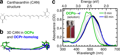
This SF class includes conjugated polymers such as polydiacetylene23, 24, 25, poly(alkyl-thienylenevinylene)26, 27, 28, a new generation of donor-acceptor singlet fission polymers29, 30, 31, 32, quinoidal thiophenes 33, 34, 35, 36, 37, carbene-based diradicaloids 38 and antiaromatic core-structured molecules 39, 40. The polyene family also includes the carotenoids, a large class of over 1000 naturally occurring molecules,41, 42 represented here by canthaxanthin (CAN) which forms the subject of this work (see structure in Fig. 1a).
In comparison with better-studied ‘class I’ SF materials 2, 46, 47, 48, 49, mostly based on molecules such as pentacene50, 51 or tetracene 52, 53, 54, 55, SF in polyenes is less well understood. This is attributable in part to their complex manifold of low-lying triplet-pair states 56, 57, 58 and strong vibronic coupling59, 60, and also partly due to the sensitivity of the photophysics to conjugation length and molecular geometry. In polyenes, the lowest-lying 1(TT) state that makes up the dominant contribution of S1 21, 22, 61, 62 contains tightly-bound triplets that are unlikely to easily separate into free triplets 63 without additional energy 56.
Indeed, while intramolecular singlet fission (iSF) has been observed in a variety of long-chain polyenes in solution 23, 24, 25, 26, 27, 28, 29, 30, 31, 32, unlike the recently designed ‘class I’ iSF systems 49, 48, 47, 64, the triplet-pairs in polyenes decay rapidly (-) to the S0 ground-state 3. Even in carotenoid aggregates, where intermolecular SF occurs between neighboring chromophores 65, 66, 67, 68, 69, 70, the majority of triplet excited states decay to S0 surprisingly quickly (within a nanosecond) 65, 3, 70. In isolated carotenoids in solution, the dominant deactivation channel from the photoexcited S2 state is internal conversion to S1. To our knowledge, there is no evidence that isolated carotenoids in solution demonstrate intramolecular SF (iSF).
Nevertheless, similarly to recent reports that torsion or twisting along a molecular backbone can allow both rapid iSF and formation of long-lived triplets in ‘class I’ SF materials 48, 64, iSF along a single twisted carotenoid chain to produce long-lived () triplets has been suggested to occur in some photosynthetic light-harvesting complexes 71, 72, 73, 74, 75. In these systems, the protein binds the carotenoid so that it is constrained in a twisted geometry. This twist reportedly stabilizes a triplet at either end of the molecule 71, 73.
This hypothesis was initially proposed to explain the presence of SF in the light-harvesting antenna (LH1) from Rhodospirillum rubrum, because of the large intermolecular distances between neighboring carotenoids () 71. More recently, Yu et al.73 observed a correlation between the presence of SF and the so-called \textnu4 resonance Raman peak () in light-harvesting complexes (LH1-RC and LH2) from Thermochromatium tepidum and Rhodobacter sphaeroides 2.4.1. The intensity of \textnu4 is related to carotenoid backbone twisting,73, 76 so this finding led to the conclusion that backbone twisting of the carotenoid directly enables iSF.
To test the hypothesis that singlet fission (SF) can occur along a single twisted carotenoid chain, we examine a protein that binds a single carotenoid: the orange carotenoid protein (OCP). In OCP the protein exists in two forms, orange and red (OCPo and OCPr) with the carotenoid in either a twisted or planar conformation, respectively (see Fig. 1). By studying both forms with the protein fixed in a trehalose-sucrose glass, we demonstrate that a twisted backbone is not sufficient to enable iSF in a protein-bound carotenoid. In light of recent work understanding magnetic field effects (MFEs) in SF systems 77, 78, we also discuss published reports of MFE in light-harvesting complexes from purple bacteria 79, 80, 81, 82, 83, 84 and find that the reported MFEs are also inconsistent with iSF. Overall, we conclude that iSF is not supported on carotenoids bound to the OCP and unlikely to occur in light-harvesting complexes.
2 Results
In this study, the OCP was produced in Escherichia (E.) coli by virtue of a dual plasmid system comprised of pET28a with the Synechocystis sp. PCC 6803 OCP gene (slr1963) and pAC-CANTHipi, which provides near-100% accumulation of CAN.85 Carotenoid-containing protein was isolated according to the method described in the methods Section.
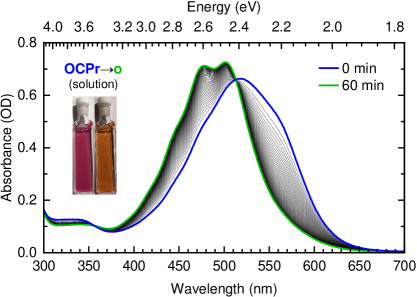
In solution, upon illumination with white light, the dark-adapted OCPo form undergoes a conformational switch to the OCPr form, with a concomitant red-shift of its steady-state absorbance spectrum due to the effective conjugation length extension of the bound carotenoid 86, 87, 76, 88, see Fig. 1c. The change is reversible, with back-conversion from OCPr to OCPo occurring in the dark, see SI Fig. 6.
Previously published X-ray diffraction structures by Leverenz, Sutter, and co-workers 43 show that the conjugated backbone of the bound carotenoid is twisted out of the plane of conjugation in OCPo (PDB 4XB5 44), while in OCPr N-terminal domain homologs such as red carotenoid protein (RCP) it is relatively planar (PDB 4XB4 45). The difference between the two conformations of CAN is depicted in Fig. 1b using data from X-ray diffraction structures 43. The different protein conformations containing a twisted and non-twisted form of CAN provide an uncomplicated model system to study the role of carotenoid geometry on iSF.
To avoid the problems associated with using spectroscopy to probe a light-activated conformational switch, we prevent the conformational change by trapping the protein in either its OCPo or OCPr conformations in a trehalose-sucrose glass as previously described70. This glass matrix prevents OCPo OCPr conversion, as demonstrated in Fig. 2(a,b), and allows us to probe each conformation in isolation at room temperature, without altering its conformation or photophysics89, 90.
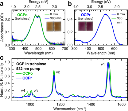
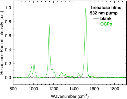
To confirm the twisted/planar conformations of CAN in OCPo/OCPr glass films, we turn to resonance Raman spectroscopy. As described above73, 76, the presence of a so-called \textnu4 peak at in the resonance Raman spectrum of carotenoids (due to out-of-plane \ceC-H wagging modes 91) is generally associated with a backbone twist of the carotenoid 73, 76. Fig. 2c shows the resonance Raman spectrum of OCPo (blue) and OCPr (green). Consistent with previous measurements on an echinenone-binding OCP 76, we observe a larger twist-induced \textnu4 peak in OCPo than in OCPr, confirming native geometry is maintained in trehalose-encapsulated OCPo and OCPr.
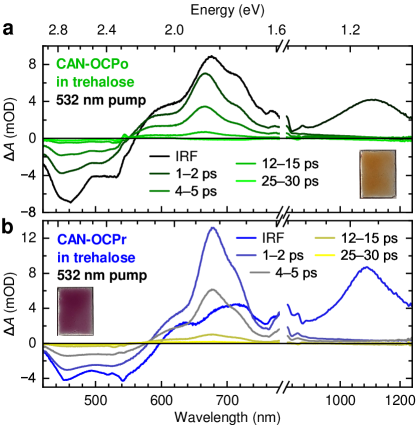
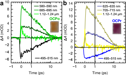
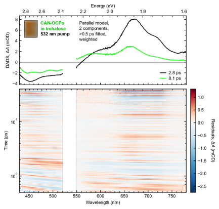
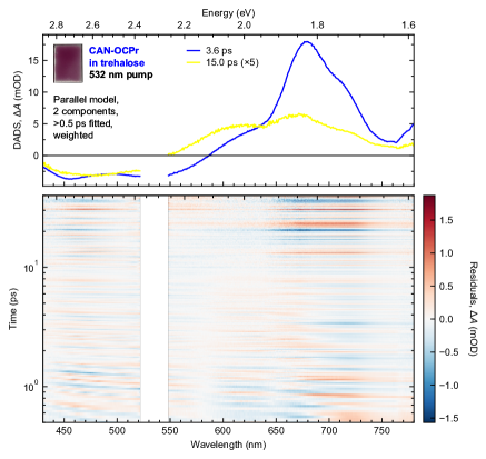
Having established that the CAN backbone is more twisted in OCPo than OCPr, we test the suggestion that such a twist is the determinant for iSF reactivity72, 73, 75. Picosecond transient absorption spectroscopy spectra and dynamics are shown in Figs. 3 and 4. Global lifetime analysis of the data is shown in SI Fig. 9 and 10, but simply from inspection of the raw data in Fig. 3, we see that all spectral features in both OCPo (green) and OCPr (blue) decay to 1% of the initial population within . Importantly, we observe no obvious formation of SF-generated triplets, as reported in light-harvesting complexes in purple bacteria 73, 71. Instead both OCPo and OCPr broadly demonstrate the expected isolated carotenoid behavior characterized by rapid internal conversion from S2 to S1 (evidenced by the instrument-limited decay of an excited-state absorption (ESA) in the near infrared region), and subsequent decay of S1-like states to the ground-state. Therefore, a twist along the carotenoid backbone is not sufficient to enable iSF.
3 Discussion
The lack of intramolecular singlet fission (iSF) in the protein-twisted carotenoid in OCPo appears to counter the currently accepted explanation for singlet fission (SF) in light-harvesting complexes (LHCs) in purple bacteria 71, 72, 73, 74, 75. We therefore return to the original studies of SF in these LHC systems and discuss them in light of recent work on the nature of intermediate triplet-pair states involved in singlet fission 77, 78.

Singlet fission in LHCs was first observed in a series of experiments that probed their magnetic field-dependent fluorescence 81, 80, 79. Representative data for oxidized cells from Rhodobacter sphaeroides 2.4.1 from Ref. 79 are reproduced in Fig. 5; similar behavior has been reported for whole cells and isolated LHCs from several strains of purple bacteria 80, 79, 84, 81. The shape of the magnetic field effect (MFE in Fig. 5), with an initial dip in fluorescence as the field increases from 0 to , and then a rise in fluorescence to saturation beyond , is a characteristic signature of SF.
This behavior is very well described by the kinetic model of SF by Johnson and Merrifield92, 93, 51, 94, 95, published in the 1960s and 70s. Recent work shows 77, 78 that this low-field Merrifield-type MFE behavior can only be observed when the inter-triplet exchange interaction is negligible, or more precisely when 96, where is the intra-triplet dipolar zero-field splitting parameter. In carotenoids, and indeed most organic chromophores, is relatively small, on the order of – 97, 98. If increases beyond , the MFE has a different behavior, showing dips in fluorescence at much higher field strengths 77, 78, 99, 100. Therefore, to determine whether SF along a single carotenoid chain is capable of producing the measured MFEs in LHCs, we must estimate the values of and .
Before doing so we make several observations about the carotenoids involved in SF in LHCs of purple bacteria: (1) the S0S2 absorbance spectra of the carotenoids in light-harvesting antenna are similar to their all-trans forms in organic solvent and depend sensitively on carotenoid conjugation length 74, 101. A full break in conjugation along the chain would lead to a dramatic blue-shift of the carotenoid absorption feature that is not observed. (2) The carotenoid T1Tn excited-state absorption feature seen in transient absorption of LHCs71, 72, 74, 75 is very similar to that seen in aggregated carotenoids of comparable conjugation lengths forming triplets by intermolecular SF65, 66, 67, 70. The T1Tn feature is also sensitive to carotenoid conjugation length,102, 103, 74 and a conjugation break along the chain would similarly lead to a blue-shift that is not observed. (3) The dipolar and parameters of the SF-generated triplets in LHCs from transient electron parametric resonance (EPR) spectroscopy are similar to full-chain triplet and parameters, rather than to their half-chain alternatives 84. These observations suggest that the conjugation along the chain is not broken, even in the protein, and therefore that the triplets at either end of the chain maintain orbital overlap and, presumably, non-negligible .
The exchange interaction, , between triplets within a pair is equal to one sixth of the energy difference between the pure singlet triplet-pair, denoted 1(TT), and the pure quintet, 5(TT). In addition, to first approximation, the energy of 5(TT) is equal to twice the free triplet energy 104, 3, 56. In carotenoids, as described above, the lowest energy singlet state (S1) is predominantly a pure singlet 1(TT) state. Therefore, comparison between twice the energy of a triplet on half a chain against the energy of S1 on a full chain provides an indication of the exchange interaction.
Recent high-level density matrix renormalization group (DMRG) calculations of the Pariser-Parr-Pople-Peierls Hamiltonian56 calculate that for a half chain is higher in energy than S1 (1(TT)) for a full chain at all conjugation lengths. This is supported by experimentally determined energies: for diphenylexatriene with conjugated double bonds, 102, 105, while for spheroidene with twice the number of double bonds (), 106. This would indicate an exchange interaction of , which is orders of magnitude larger than the dipolar parameter – 97, 98. These energies indicate that the triplets within 1(TT) should be strongly exchange coupled.
The triplets within a single carotenoid chain are therefore exchange coupled (), even in a protein that twists the carotenoid backbone 73, as no breaks in conjugation along the carotenoid chain have been observed (i.e. no observable shifts in absorption spectra 74 or changes in dipolar and parameters84). Therefore, MFEs such as those reproduced in Fig. 5, that were the initial proof of SF in purple bacteria, cannot be explained with an intramolecular model of SF.
4 Conclusions
We conclude that singlet fission (SF) to produce long-lived triplets does not occur along a single twisted carotenoid chain in the OCP and is unlikely to occur in purple bacterial LHCs, contrary to the current notion 71, 72, 74, 73, 75. We conclude this because (1) immobilized OCPo – an uncomplicated, minimal carotenoprotein – shows similar twisted carotenoid geometry to LHCs, but shows no evidence of SF, and (2) the MFEs that identified SF in purple bacteria are irreconcilable with iSF without a significant break in conjugation (which is not observed). These findings therefore call into question the mechanism of SF that is observed in LHCs.
5 Supplementary Information
5.1 Materials and Methods
5.1.1 Sample preparation
OCP containing 100% canthaxanthin (CAN) was produced from BL21(DE3) Escherichia coli (E. coli) using a dual-plasmid system comprised of pAC-CANTHipi 85 and pET28a containing the gene encoding OCP (slr1963) from Synechocystis sp. PCC 6803. Briefly, cultures were grown at ( agitation) in baffled Erlenmeyer flasks using lysogeny broth (LB) medium containing the appropriate concentrations of antibiotics. When the optical density of the medium at (OD600) had reached a value of 0.6, protein production was induced by addition of isopropyl \textbeta-D-1-thiogalatopyranoside and the cultures incubated for at .
Cells were harvested by centrifugation (4,400, , ) and resuspended in binding buffer ( HEPES, pH 7.4, NaCl, imidazole). Cells were lysed by sonication and then centrifuged (53,000, , ). The supernatant was collected and filtered ( filter pores) and applied to a Chelating Sepharose Fast Flow column (GE Healthcare) pre-equilibrated with NiSO4. The column was washed with binding buffer, wash buffer ( HEPES, pH 7.4, NaCl, imidazole) and elution buffer ( HEPES, pH 7.4, NaCl, imidazole) with the elution pooled for further purification. The protein sample was buffer exchanged into buffer A ( HEPES, pH 7.4) loaded onto a Fast Flow Q-Sepharose column (GE Healthcare) and a linear gradient of 0– NaCl was applied. Fractions were analyzed by SDS-PAGE and appropriate samples taken forward for size exclusion chromatography on a Superdex 200 Increase column (GE Healthcare) in buffer B ( HEPES, pH 7.4, NaCl). Where necessary OCP samples were concentrated using centrifugal dialysis (VivaSpin, Sartorius).
OCPo samples were fixed in sucrose-trehalose glasses in strict darkness by mixing of concentrated protein solution (ODmax ) in aqueous buffer ( HEPES, NaCl, pH 7.4) with of a trehalose-sucrose mixture ( trehalose, sucrose). of the protein-trehalose mixture was drop-cast in the center of a quartz-coated glass substrate (S151, Ossila; 1520). The substrate was incubated under vacuum () with an excess of calcium sulfate desiccant (Drierite) at room temperature for at least 48 hours. OCPr trehalose glasses were made in an identical manner, with samples illuminated for () prior to the addition of the trehalose-sucrose solution and constant weaker illumination () for the duration of the desiccation.
OCPo and OCPr samples measured with the resonance Raman setup were additionally encapsulated with imaging spacers and a cover slip to protect the trehalose against atmospheric rehydration. For these samples, a stack of two imaging spacers (SecureSeal, Grace BioLabs; diameter, thickness) were attached to the quartz-coated glass substrate (S151, Ossila; 1520) and of the protein-trehalose mixture drop-cast in the center of the imaging spacer. The substrate was then placed in vacuum as above; pressure was released under a continuous flow of ultra-pure nitrogen gas and a glass microscope cover slip (ThermoScientific; 22, No.1 thickness) was attached to the upper imaging spacer.
5.1.2 Transient absorption spectroscopy
Short-time transient absorption spectroscopy was undertaken with a commercial spectrometer (Helios, Ultrafast Systems) outfitted with a Ti:Sapphire seed laser (MaiTai, Spectra Physics) providing pulses (, nominal FWHM) and a Ti:Sapphire chirped-pulse amplifier (Spitfire Ace PA-40) amplifying pulses (, average power, nominal FWHM). Tuneable pump pulses for excitation were generated by seeding a part of the beam in an optical parametric amplifier (TOPAS Prime, Light Conversion). An optical chopper was used to modulate the pump frequency to . An intensity spectrum for the pump used in visible-probe measurements is shown in Fig. 8. Pump beam spot sizes were measured at the sample position with a CCD beam profiler (BC106N-VIS/M, Thorlabs), and used in subsequent calculations to tune a pump fluence. Supercontinuum probes were generated with a part of the pulse focused on either a sapphire crystal for visible probes (450–) or a YAG crystal for NIR probes (800–). Pump-probe delay was controlled with a motorized delay stage with a random stepping order. The signal was dispersed with a grating and detected with CMOS or InGaAs sensors for visible or NIR probes, respectively. The pump and probe polarizations were set to the magic angle.
Surface Xplorer 4.3.0 (Ultrafast Systems) was used in processing the transient absorption datasets. Noisy edges of the spectra were trimmed, and the program’s bad spectra replacement procedure was applied. A background correction (‘subtract scattered light’) was then applied using the spectra before any apparent response from the sample. Chirp correction was applied, choosing points at the first apparent signal for a given kinetic. Time zero is adjusted to the time of maximum initial signal. Further processing and some analysis was performed with original Python code.
5.1.3 Resonance Raman spectroscopy
Resonance Raman measurements were performed with a Renishaw inVia Raman system (Renishaw plc., Wotton-Under-Edge, UK) in a backscattering configuration. A laser (150– power) and a 50 objective were used (NA 0.50, spot size ). Acquisition times used were in the range 5–.
5.2 Figure preparation
OriginPro 9.6.0.172 (OriginLab) and home-built Python code was used to prepare the plots.
5.3 Additional steady-state absorbance data
5.4 Additional resonance Raman data
5.5 Additional transient absorption data
5.5.1 Pump spectrum

5.5.2 Additional note: global lifetime analysis
Global lifetime analysis on the visible-probe transient absorption data of OCPo and OCPr was performed. This was done using the Glotaran 1.5.1 software package(http://glotaran.org) 107, a GUI for the R package TIMP108. Data used had already been processed with the steps outlined in Section 5.1.2; in particular, a chirp correction had already been applied, so that a term to account for chirp did not need to be included in the fitting. Noisy regions in the data due to pump scatter were excluded for all times to ensure a good fit of the rest of the data. Noisy red and blue ends in the data associated with tails of the probe were also excluded, so that the fitted wavelengths were to . The fitting was weighted favourably at later delay times for good fits of any long-lived features; Table 1 shows the weighting applied. Due to the strong coherent artifact feature in the first , only data beyond that time was fitted. Thus, in the model, terms to account for the coherent artifact and S2 states were not included. This left a relatively simple fitted model of a number of wavelength-dependent decay-associated difference spectra (DADS) decaying exponentially in parallel.
| Time range () | Weighting |
|---|---|
| 0.5 – 10 | 1 |
| 10 – 20 | 2 |
| 20 – 30 | 3 |
| 30 – 35 | 4 |
| 35 | 5 |
Global lifetime analysis of the OCP data does not indicate any significant population of longer-lived features. 2-component global lifetime analysis of the CAN-binding OCPo and OCPr data are shown in Fig. 9 and 10, respectively. Fitting a 2-component parallel decay model in an artifact-free region of the visible-probe data beyond the initial coherent artifact and S2-associated response gives two decay-associated difference spectra (DADS) for both the OCPo data and OCPr data, with the longer time-constant DADS relatively weaker and blueshifted in both cases. Both components are likely associated with mixed S1 decay, internal vibrational redistribution, and vibrational cooling.60 A single component is not sufficient to adequately fit the region of the data, and a fitting a third component gives results with spurious DADS profiles.
We note that sample degradation (caused the pump fluence) likely affects the fitted time constants and DADS profiles, and that the maximum time delay used was about .
The authors thank James D. Shipp, Sayantan Bhattacharya and David G. Bossanyi for assistance with transient absorption measurements.
Funding: G.A.S. and C.N.H. acknowledge ERC Synergy Grant 854126. J.P.P. thanks the EPSRC for support through a Doctoral Training Partnership Scholarship (EP/R513313/1). The authors thank the EPSRC for a Capital Equipment Award (EP/L022613/1 and EP/R042802/1) which funded the Lord Porter Laser facility used in this study. J.C., C.N.H, G.A.S. and S.W. thank the EPSRC for funding through EP/S002103/1. J.C. and S.W. also thank the EPSRC for funding through EP/N014022/1. M.S.P. and M.P.J. were supported by Leverhulme Trust award RPG-2019-045. A.H. acknowledges The Royal Society (award URF\R1\191548). H.K.H.L. and W.C.T. acknowledge the SPECIFIC Innovation and Knowledge Centre (EP/N020863/1) grant for providing financial support.
Author contributions: G.A.S. conceived the study. G.A.S., J.P.P., S.W. and J.C. designed the experiments. G.A.S., M.S.P. and A.H. prepared protein samples under the supervision of M.P.J. and C.N.H. Steady-state measurements were performed by G.A.S. Resonance Raman measurements were performed by H.K.H.L. under the supervision of W.C.T. Transient absorption was conducted by J.P.P. and S.W. within the Lord Porter Laser, with D.C. providing facility management. J.P.P. and G.A.S. analyzed the data. J.P.P., G.A.S. and J.C. wrote the manuscript and prepared the figures with input from all authors.
-
•
Supplementary information (SI): Canthaxanthin-binding OCP production, OCPo and OCPr in sugar glasses preparation, spectroscopic setups, and data analysis procedures are detailed. A figure showing back-conversion of solution OCPr to OCPo in the dark is included. Figures showing a short-time transient absorption pump spectrum and a resonance Raman spectrum of blank trehalose are included. The results of global lifetime analysis on the transient absorption data is detailed.
-
•
Data will be available on Sheffield University’s repository, ORDA, once the paper has been accepted for publication.
References
- Bardeen 2014 Bardeen, C. J. The Structure and Dynamics of Molecular Excitons. Annual Review of Physical Chemistry 2014, 65, 127–148
- Smith and Michl 2013 Smith, M. B.; Michl, J. Recent Advances in Singlet Fission. Annual Review of Physical Chemistry 2013, 64, 361–386
- Musser and Clark 2019 Musser, A. J.; Clark, J. Triplet-Pair States in Organic Semiconductors. Annual Review of Physical Chemistry 2019, 70, 323–351
- Ullrich et al. 2021 Ullrich, T.; Munz, D.; Guldi, D. M. Unconventional singlet fission materials. Chemical Society Reviews 2021, 50, 3485–3518
- Kim and Zimmerman 2018 Kim, H.; Zimmerman, P. M. Coupled double triplet state in singlet fission. Physical Chemistry Chemical Physics 2018, 20, 30083–30094
- Hanna and Nozik 2006 Hanna, M. C.; Nozik, A. J. Solar conversion efficiency of photovoltaic and photoelectrolysis cells with carrier multiplication absorbers. Journal of Applied Physics 2006, 100, 074510
- Rao and Friend 2017 Rao, A.; Friend, R. H. Harnessing singlet exciton fission to break the Shockley–Queisser limit. Nature Reviews Materials 2017, 2, 17063
- Ehrler et al. 2021 Ehrler, B.; Yanai, N.; Nienhaus, L. Up- and down-conversion in molecules and materials. The Journal of Chemical Physics 2021, 154, 070401
- Daiber et al. 2021 Daiber, B.; van den Hoven, K.; Futscher, M. H.; Ehrler, B. Realistic Efficiency Limits for Singlet-Fission Silicon Solar Cells. ACS Energy Letters 2021, 6, 2800–2808
- Ehrler et al. 2022 Ehrler, B.; W. Y. Ho-Baillie, A.; Hutter, E. M.; Milić, J. V.; Tayebjee, M. J. Y.; Wilson, M. W. B. Scalable ways to break the efficiency limit of single-junction solar cells. Applied Physics Letters 2022, 120, 010402
- Liu et al. 2015 Liu, Y.; Zhang, C.; Wang, R.; Zhang, B.; Tan, Z.; Wang, X.; Xiao, M. Large Optical Nonlinearity Induced by Singlet Fission in Pentacene Films. Angewandte Chemie International Edition 2015, 54, 6222–6226
- Zhao et al. 2015 Zhao, M.; Liu, K.; Zhang, Y.-D.; Wang, Q.; Li, Z.-G.; Song, Y.-L.; Zhang, H.-L. Singlet fission induced giant optical limiting responses of pentacene derivatives. Materials Horizons 2015, 2, 619–624
- Tonami et al. 2019 Tonami, T.; Nagami, T.; Okada, K.; Yoshida, W.; Nakano, M. Singlet-Fission-Induced Enhancement of Third-Order Nonlinear Optical Properties of Pentacene Dimers. ACS Omega 2019, 4, 16181–16190
- Nagata et al. 2018 Nagata, R.; Nakanotani, H.; Potscavage, W. J.; Adachi, C. Exploiting Singlet Fission in Organic Light-Emitting Diodes. Advanced Materials 2018, 30, 1801484
- Teichen and Eaves 2015 Teichen, P. E.; Eaves, J. D. Collective aspects of singlet fission in molecular crystals. The Journal of Chemical Physics 2015, 143, 044118
- Bardeen 2019 Bardeen, C. J. Time dependent correlations of entangled states with nondegenerate branches and possible experimental realization using singlet fission. The Journal of Chemical Physics 2019, 151, 124503
- Marcus and Barford 2020 Marcus, M.; Barford, W. Triplet-triplet decoherence in singlet fission. Physical Review B 2020, 102, 035134
- Smyser and Eaves 2020 Smyser, K. E.; Eaves, J. D. Singlet fission for quantum information and quantum computing: the parallel JDE model. Scientific Reports 2020, 10, 18480
- Einzinger et al. 2019 Einzinger, M.; Wu, T.; Kompalla, J. F.; Smith, H. L.; Perkinson, C. F.; Nienhaus, L.; Wieghold, S.; Congreve, D. N.; Kahn, A.; Bawendi, M. G.; Baldo, M. A. Sensitization of silicon by singlet exciton fission in tetracene. Nature 2019, 571, 90–94
- Daiber et al. 2020 Daiber, B.; Maiti, S.; Ferro, S. M.; Bodin, J.; van den Boom, A. F. J.; Luxembourg, S. L.; Kinge, S.; Pujari, S. P.; Zuilhof, H.; Siebbeles, L. D. A.; Ehrler, B. Change in Tetracene Polymorphism Facilitates Triplet Transfer in Singlet Fission-Sensitized Silicon Solar Cells. The Journal of Physical Chemistry Letters 2020, 11, 8703–8709
- Tavan and Schulten 1987 Tavan, P.; Schulten, K. Electronic excitations in finite and infinite polyenes. Physical Review B 1987, 36, 4337–4358
- Schmidt and Tavan 2012 Schmidt, M.; Tavan, P. Electronic excitations in long polyenes revisited. The Journal of Chemical Physics 2012, 136, 124309
- Kraabel et al. 1998 Kraabel, B.; Hulin, D.; Aslangul, C.; Lapersonne-Meyer, C.; Schott, M. Triplet exciton generation, transport and relaxation in isolated polydiacetylene chains: Subpicosecond pump-probe experiments. Chemical Physics 1998, 227, 83–98
- Pandya et al. 2020 Pandya, R.; Gu, Q.; Cheminal, A.; Chen, R. Y.; Booker, E. P.; Soucek, R.; Schott, M.; Legrand, L.; Mathevet, F.; Greenham, N. C.; Barisien, T.; Musser, A. J.; Chin, A. W.; Rao, A. Optical Projection and Spatial Separation of Spin-Entangled Triplet Pairs from the S1 (21 Ag–) State of Pi-Conjugated Systems. Chem 2020, 6, 2826–2851
- Lanzani et al. 2001 Lanzani, G.; Cerullo, G.; Zavelani-Rossi, M.; De Silvestri, S.; Comoretto, D.; Musso, G.; Dellepiane, G. Triplet-Exciton Generation Mechanism in a New Soluble (Red-Phase) Polydiacetylene. Physical Review Letters 2001, 87, 187402
- Musser et al. 2013 Musser, A. J.; Al-Hashimi, M.; Maiuri, M.; Brida, D.; Heeney, M.; Cerullo, G.; Friend, R. H.; Clark, J. Activated Singlet Exciton Fission in a Semiconducting Polymer. Journal of the American Chemical Society 2013, 135, 12747–12754
- Lafalce et al. 2011 Lafalce, E.; Jiang, X.; Zhang, C. Generation and Recombination Kinetics of Optical Excitations in Poly(3-dodecylthienylenevinylene) with Controlled Regioregularity. The Journal of Physical Chemistry B 2011, 115, 13139–13148
- Musser et al. 2019 Musser, A. J.; Al-Hashimi, M.; Heeney, M.; Clark, J. Heavy-atom effects on intramolecular singlet fission in a conjugated polymer. The Journal of Chemical Physics 2019, 151, 044902
- Busby et al. 2015 Busby, E.; Xia, J.; Wu, Q.; Low, J. Z.; Song, R.; Miller, J. R.; Zhu, X.-Y.; Campos, L. M.; Sfeir, M. Y. A design strategy for intramolecular singlet fission mediated by charge-transfer states in donor-acceptor organic materials. Nature Materials 2015, 14, 426–433
- Kasai et al. 2015 Kasai, Y.; Tamai, Y.; Ohkita, H.; Benten, H.; Ito, S. Ultrafast Singlet Fission in a Push–Pull Low-Bandgap Polymer Film. Journal of the American Chemical Society 2015, 137, 15980–15983
- Fallon et al. 2019 Fallon, K. J. et al. Exploiting Excited-State Aromaticity To Design Highly Stable Singlet Fission Materials. Journal of the American Chemical Society 2019, 141, 13867–13876
- Huynh et al. 2017 Huynh, U. N. V.; Basel, T. P.; Ehrenfreund, E.; Li, G.; Yang, Y.; Mazumdar, S.; Vardeny, Z. V. Transient Magnetophotoinduced Absorption Studies of Photoexcitations in -Conjugated Donor-Acceptor Copolymers. Physical Review Letters 2017, 119, 017401
- Casado et al. 2012 Casado, J.; Ponce Ortiz, R.; López Navarrete, J. T. Quinoidal oligothiophenes: new properties behind an unconventional electronic structure. Chemical Society Reviews 2012, 41, 5672–5686
- Varnavski et al. 2015 Varnavski, O.; Abeyasinghe, N.; Aragó, J.; Serrano-Pérez, J. J.; Ortí, E.; López Navarrete, J. T.; Takimiya, K.; Casanova, D.; Casado, J.; Goodson, T. High Yield Ultrafast Intramolecular Singlet Exciton Fission in a Quinoidal Bithiophene. The Journal of Physical Chemistry Letters 2015, 6, 1375–1384
- Chien et al. 2015 Chien, A. D.; Molina, A. R.; Abeyasinghe, N.; Varnavski, O. P.; Goodson, T.; Zimmerman, P. M. Structure and Dynamics of the 1(TT) State in a Quinoidal Bithiophene: Characterizing a Promising Intramolecular Singlet Fission Candidate. The Journal of Physical Chemistry C 2015, 119, 28258–28268
- Kim et al. 2018 Kim, H.; Keller, B.; Ho-Wu, R.; Abeyasinghe, N.; Vázquez, R. J.; Goodson, T.; Zimmerman, P. M. Enacting Two-Electron Transfer from a Double-Triplet State of Intramolecular Singlet Fission. Journal of the American Chemical Society 2018, 140, 7760–7763
- Kawata et al. 2016 Kawata, S.; Pu, Y.-J.; Saito, A.; Kurashige, Y.; Beppu, T.; Katagiri, H.; Hada, M.; Kido, J. Singlet Fission of Non-polycyclic Aromatic Molecules in Organic Photovoltaics. Advanced Materials 2016, 28, 1585–1590
- Ullrich et al. 2020 Ullrich, T.; Pinter, P.; Messelberger, J.; Haines, P.; Kaur, R.; Hansmann, M. M.; Munz, D.; Guldi, D. M. Singlet Fission in Carbene-Derived Diradicaloids. Angewandte Chemie International Edition 2020, 59, 7906–7914
- Wu et al. 2017 Wu, Y.; Wang, Y.; Chen, J.; Zhang, G.; Yao, J.; Zhang, D.; Fu, H. Intramolecular Singlet Fission in an Antiaromatic Polycyclic Hydrocarbon. Angewandte Chemie International Edition 2017, 56, 9400–9404
- Liu et al. 2019 Liu, Y.; Wu, Y.; Wang, L.; Wang, L.; Yao, J.; Fu, H. Efficient triplet pair separation from intramolecular singlet fission in dibenzopentalene derivatives. Science China Chemistry 2019, 62, 1037–1043
- Canniffe and Hitchcock 2021 Canniffe, D. P.; Hitchcock, A. In Encyclopedia of Biological Chemistry III, 3rd ed.; Jez, J., Ed.; Elsevier: Oxford, 2021; Vol. 2; pp 163–185
- Yabuzaki 2017 Yabuzaki, J. Carotenoids Database: structures, chemical fingerprints and distribution among organisms. Database (Oxford) 2017, 2017, bax004
- Leverenz et al. 2015 Leverenz, R. L.; Sutter, M.; Wilson, A.; Gupta, S.; Thurotte, A.; Bourcier de Carbon, C.; Petzold, C. J.; Ralston, C.; Perreau, F.; Kirilovsky, D.; Kerfeld, C. A. A 12 Å carotenoid translocation in a photoswitch associated with cyanobacterial photoprotection. Science 2015, 348, 1463–1466
- Kerfeld et al. 2014 Kerfeld, C. A.; Sutter, M.; Leverenz, R. L. Structure of orange carotenoid protein binding canthaxanthin. 2014; \url10.2210/pdb4XB5/pdb
- Kerfeld et al. 2014 Kerfeld, C. A.; Sutter, M.; Leverenz, R. L. Structure of the N-terminal domain of OCP binding canthaxanthin. 2014; \url10.2210/pdb4XB4/pdb
- Yong et al. 2017 Yong, C. K. et al. The entangled triplet pair state in acene and heteroacene materials. Nature Communications 2017, 8, 15953
- Pun et al. 2019 Pun, A. B.; Asadpoordarvish, A.; Kumarasamy, E.; Tayebjee, M. J. Y.; Niesner, D.; McCamey, D. R.; Sanders, S. N.; Campos, L. M.; Sfeir, M. Y. Ultra-fast intramolecular singlet fission to persistent multiexcitons by molecular design. Nature Chemistry 2019, 11, 821–828
- Korovina et al. 2020 Korovina, N. V.; Chang, C. H.; Johnson, J. C. Spatial separation of triplet excitons drives endothermic singlet fission. Nature Chemistry 2020, 12, 391–398
- Wang et al. 2021 Wang, Z.; Liu, H.; Xie, X.; Zhang, C.; Wang, R.; Chen, L.; Xu, Y.; Ma, H.; Fang, W.; Yao, Y.; Sang, H.; Wang, X.; Li, X.; Xiao, M. Free-triplet generation with improved efficiency in tetracene oligomers through spatially separated triplet pair states. Nature Chemistry 2021, 13, 559–567
- Wilson et al. 2011 Wilson, M. W. B.; Rao, A.; Clark, J.; Kumar, R. S. S.; Brida, D.; Cerullo, G.; Friend, R. H. Ultrafast Dynamics of Exciton Fission in Polycrystalline Pentacene. Journal of the American Chemical Society 2011, 133, 11830–11833
- Bossanyi et al. 2021 Bossanyi, D. G.; Matthiesen, M.; Wang, S.; Smith, J. A.; Kilbride, R. C.; Shipp, J. D.; Chekulaev, D.; Holland, E.; Anthony, J. E.; Zaumseil, J.; Musser, A. J.; Clark, J. Emissive spin-0 triplet-pairs are a direct product of triplet–triplet annihilation in pentacene single crystals and anthradithiophene films. Nature Chemistry 2021, 13, 163–171
- Burdett et al. 2013 Burdett, J. J.; Piland, G. B.; Bardeen, C. J. Magnetic field effects and the role of spin states in singlet fission. Chemical Physics Letters 2013, 585, 1–10
- Piland and Bardeen 2015 Piland, G. B.; Bardeen, C. J. How Morphology Affects Singlet Fission in Crystalline Tetracene. The Journal of Physical Chemistry Letters 2015, 6, 1841–1846
- Tayebjee et al. 2013 Tayebjee, M. J. Y.; Clady, R. G. C. R.; Schmidt, T. W. The exciton dynamics in tetracene thin films. Physical Chemistry Chemical Physics 2013, 15, 14797–14805
- Wilson et al. 2013 Wilson, M. W. B.; Rao, A.; Johnson, K.; Gélinas, S.; di Pietro, R.; Clark, J.; Friend, R. H. Temperature-Independent Singlet Exciton Fission in Tetracene. Journal of the American Chemical Society 2013, 135, 16680–16688
- Valentine et al. 2020 Valentine, D. J.; Manawadu, D.; Barford, W. Higher-energy triplet-pair states in polyenes and their role in intramolecular singlet fission. Physical Review B 2020, 102, 125107
- Manawadu et al. 2022 Manawadu, D.; Valentine, D. J.; Marcus, M.; Barford, W. Singlet Triplet-Pair Production and Possible Singlet-Fission in Carotenoids. The Journal of Physical Chemistry Letters 2022, 13, 1344–1349
- Barford 2022 Barford, W. Theory of the dark state of polyenes and carotenoids. 2022; \urlhttps://arxiv.org/abs/2203.15520
- Balevičius et al. 2016 Balevičius, V.; Abramavicius, D.; Polívka, T.; Galestian Pour, A.; Hauer, J. A Unified Picture of S* in Carotenoids. The Journal of Physical Chemistry Letters 2016, 7, 3347–3352
- Balevičius et al. 2019 Balevičius, V.; Wei, T.; Di Tommaso, D.; Abramavicius, D.; Hauer, J.; Polívka, T.; Duffy, C. D. P. The full dynamics of energy relaxation in large organic molecules: from photo-excitation to solvent heating. Chemical Science 2019, 10, 4792–4804
- Taffet et al. 2020 Taffet, E. J.; Fassioli, F.; Toa, Z. S. D.; Beljonne, D.; Scholes, G. D. Uncovering dark multichromophoric states in Peridinin–Chlorophyll–Protein. Journal of The Royal Society Interface 2020, 17, 20190736
- Barford et al. 2001 Barford, W.; Bursill, R. J.; Lavrentiev, M. Y. Density-matrix renormalization-group calculations of excited states of linear polyenes. Physical Review B 2001, 63, 195108
- Polak et al. 2019 Polak, D. W.; Musser, A. J.; Sutherland, G. A.; Auty, A.; Branchi, F.; Dzurnak, B.; Chidgey, J.; Cerullo, G.; Hunter, C. N.; Clark, J. Band-edge Excitation of Carotenoids Removes S* Revealing Triplet-pair Contributions to the S1 Absorption Spectrum. 2019; \urlhttp://arxiv.org/abs/1901.04900
- Yablon et al. 2022 Yablon, L. M.; Sanders, S. N.; Miyazaki, K.; Kumarasamy, E.; He, G.; Choi, B.; Ananth, N.; Sfeir, M. Y.; Campos, L. M. Singlet fission and triplet pair recombination in bipentacenes with a twist. Materials Horizons 2022, 9, 462–470
- Musser et al. 2015 Musser, A. J.; Maiuri, M.; Brida, D.; Cerullo, G.; Friend, R. H.; Clark, J. The Nature of Singlet Exciton Fission in Carotenoid Aggregates. Journal of the American Chemical Society 2015, 137, 5130–5139
- Zhang et al. 2018 Zhang, D.; Tan, L.; Dong, J.; Yi, J.; Wang, P.; Zhang, J. Structure and Excitation Dynamics of -Carotene Aggregates in Cetyltrimethylammonium Bromide Micelle. Chemical Research in Chinese Universities 2018, 34, 643–648
- Chang et al. 2017 Chang, H.-T.; Chang, Y.-Q.; Han, R.-M.; Wang, P.; Zhang, J.-P.; Skibsted, L. H. Singlet Fission Reaction of Light-Exposed -Carotene Bound to Bovine Serum Albumin. A Novel Mechanism in Protection of Light-Exposed Tissue by Dietary Carotenoids. Journal of Agricultural and Food Chemistry 2017, 65, 6058–6062
- Wang and Tauber 2010 Wang, C.; Tauber, M. J. High-Yield Singlet Fission in a Zeaxanthin Aggregate Observed by Picosecond Resonance Raman Spectroscopy. Journal of the American Chemical Society 2010, 132, 13988–13991
- Wang et al. 2011 Wang, C.; Schlamadinger, D. E.; Desai, V.; Tauber, M. J. Triplet Excitons of Carotenoids Formed by Singlet Fission in a Membrane. ChemPhysChem 2011, 12, 2891–2894
- Sutherland et al. 2020 Sutherland, G. A.; Polak, D.; Swainsbury, D. J. K.; Wang, S.; Spano, F. C.; Auman, D. B.; Bossanyi, D. G.; Pidgeon, J. P.; Hitchcock, A.; Musser, A. J.; Anthony, J. E.; Dutton, P. L.; Clark, J.; Hunter, C. N. A Thermostable Protein Matrix for Spectroscopic Analysis of Organic Semiconductors. Journal of the American Chemical Society 2020, 142, 13898–13907
- Gradinaru et al. 2001 Gradinaru, C. C.; Kennis, J. T. M.; Papagiannakis, E.; van Stokkum, I. H. M.; Cogdell, R. J.; Fleming, G. R.; Niederman, R. A.; van Grondelle, R. An unusual pathway of excitation energy deactivation in carotenoids: Singlet-to-triplet conversion on an ultrafast timescale in a photosynthetic antenna. Proceedings of the National Academy of Sciences 2001, 98, 2364–2369
- Papagiannakis et al. 2002 Papagiannakis, E.; Kennis, J. T. M.; van Stokkum, I. H. M.; Cogdell, R. J.; van Grondelle, R. An alternative carotenoid-to-bacteriochlorophyll energy transfer pathway in photosynthetic light harvesting. Proceedings of the National Academy of Sciences 2002, 99, 6017–6022
- Yu et al. 2017 Yu, J.; Fu, L.-M.; Yu, L.-J.; Shi, Y.; Wang, P.; Wang-Otomo, Z.-Y.; Zhang, J.-P. Carotenoid Singlet Fission Reactions in Bacterial Light Harvesting Complexes As Revealed by Triplet Excitation Profiles. Journal of the American Chemical Society 2017, 139, 15984–15993
- Niedzwiedzki et al. 2017 Niedzwiedzki, D. M.; Swainsbury, D. J. K.; Martin, E. C.; Hunter, C. N.; Blankenship, R. E. Origin of the S* Excited State Feature of Carotenoids in Light-Harvesting Complex 1 from Purple Photosynthetic Bacteria. The Journal of Physical Chemistry B 2017, 121, 7571–7585
- Zhang et al. 2022 Zhang, Y.; Qi, C.-H.; Yamano, N.; Wang, P.; Yu, L.-J.; Wang-Otomo, Z.-Y.; Zhang, J.-P. Carotenoid Single-Molecular Singlet Fission and the Photoprotection of a Bacteriochlorophyll b-Type Core Light-Harvesting Antenna. The Journal of Physical Chemistry Letters 2022, 13, 3534–3541
- Kish et al. 2015 Kish, E.; Pinto, M. M. M.; Kirilovsky, D.; Spezia, R.; Robert, B. Echinenone vibrational properties: From solvents to the orange carotenoid protein. Biochimica et Biophysica Acta (BBA) - Bioenergetics 2015, 1847, 1044–1054
- Bayliss et al. 2016 Bayliss, S. L.; Weiss, L. R.; Rao, A.; Friend, R. H.; Chepelianskii, A. D.; Greenham, N. C. Spin signatures of exchange-coupled triplet pairs formed by singlet fission. Physical Review B 2016, 94, 045204
- Bossanyi et al. 2021 Bossanyi, D. G.; Sasaki, Y.; Wang, S.; Chekulaev, D.; Kimizuka, N.; Yanai, N.; Clark, J. Spin Statistics for Triplet–Triplet Annihilation Upconversion: Exchange Coupling, Intermolecular Orientation, and Reverse Intersystem Crossing. JACS Au 2021, 1, 2188–2201
- Kingma et al. 1985 Kingma, H.; van Grondelle, R.; Duysens, L. Magnetic-field effects in photosynthetic bacteria. I. Magnetic-field-induced bacteriochlorophyll emission changes in the reaction center and the antenna of Rhodospirillum rubrum, Rhodopseudomonas sphaeroides and Prosthecochloris aestuarii. Biochimica et Biophysica Acta (BBA) - Bioenergetics 1985, 808, 363–382
- Kingma et al. 1985 Kingma, H.; van Grondelle, R.; Duysens, L. Magnetic-field effects in photosynthetic bacteria. II. Formation of triplet states in the reaction center and the antenna of Rhodospirillum rubrum and Rhodopseudomonas sphaeroides. Magnetic-field effects. Biochimica et Biophysica Acta (BBA) - Bioenergetics 1985, 808, 383–399
- Rademaker et al. 1980 Rademaker, H.; Hoff, A. J.; Van Grondelle, R.; Duysens, L. N. Carotenoid triplet yields in normal and deuterated Rhodospirillum rubrum. Biochimica et Biophysica Acta (BBA) - Bioenergetics 1980, 592, 240–257
- Klenina et al. 2013 Klenina, I. B.; Makhneva, Z. K.; Moskalenko, A. A.; Kuzmin, A. N.; Proskuryakov, I. I. Singlet-triplet excitation fission in light-harvesting complexes of photosynthetic bacteria and in isolated carotenoids. Biophysics 2013, 58, 43–50
- Klenina et al. 2014 Klenina, I. B.; Makhneva, Z. K.; Moskalenko, A. A.; Gudkov, N. D.; Bolshakov, M. A.; Pavlova, E. A.; Proskuryakov, I. I. Singlet-triplet fission of carotenoid excitation in light-harvesting LH2 complexes of purple phototrophic bacteria. Biochemistry (Moscow) 2014, 79, 235–241
- Gryaznov et al. 2019 Gryaznov, A. A.; Klenina, I. B.; Makhneva, Z. K.; Moskalenko, A. A.; Proskuryakov, I. I. The Singlet–Triplet Fission of Carotenoid Excitation in Light-Harvesting Complexes from Thermochromatium tepidum. Biophysics 2019, 64, 847–852
- Cunningham and Gantt 2007 Cunningham, F. X.; Gantt, E. A portfolio of plasmids for identification and analysis of carotenoid pathway enzymes: Adonis aestivalis as a case study. Photosynthesis Research 2007, 92, 245–259
- Wilson et al. 2008 Wilson, A.; Punginelli, C.; Gall, A.; Bonetti, C.; Alexandre, M.; Routaboul, J.-M.; Kerfeld, C. A.; van Grondelle, R.; Robert, B.; Kennis, J. T. M.; Kirilovsky, D. A photoactive carotenoid protein acting as light intensity sensor. Proceedings of the National Academy of Sciences 2008, 105, 12075–12080
- Niedzwiedzki et al. 2014 Niedzwiedzki, D. M.; Liu, H.; Blankenship, R. E. Excited State Properties of 3′-Hydroxyechinenone in Solvents and in the Orange Carotenoid Protein from Synechocystis sp. PCC 6803. The Journal of Physical Chemistry B 2014, 118, 6141–6149
- Bondanza et al. 2020 Bondanza, M.; Cupellini, L.; Faccioli, P.; Mennucci, B. Molecular Mechanisms of Activation in the Orange Carotenoid Protein Revealed by Molecular Dynamics. Journal of the American Chemical Society 2020, 142, 21829–21841
- Pidgeon et al. 2022 Pidgeon, J. P.; Sutherland, G. A.; Proctor, M. S.; Hitchcock, A.; Wang, S.; Chekulaev, D.; Jayaprakash, R.; Venkatraman, R. K.; Johnson, M. P.; Hunter, C. N.; Clark, J. Transient spectroscopy on orange carotenoid protein trapped in trehalose glasses show that photoswitching is not triggered by dark singlet states. In preparation 2022,
- Kurashov et al. 2018 Kurashov, V.; Gorka, M.; Milanovsky, G. E.; Johnson, T. W.; Cherepanov, D. A.; Semenov, A. Y.; Golbeck, J. H. Critical evaluation of electron transfer kinetics in P700–FA/FB, P700–FX, and P700–A1 Photosystem I core complexes in liquid and in trehalose glass. Biochimica et Biophysica Acta (BBA) - Bioenergetics 2018, 1859, 1288–1301
- Saito and Tasumi 1983 Saito, S.; Tasumi, M. Normal-coordinate analysis of retinal isomers and assignments of Raman and infrared bands. Journal of Raman Spectroscopy 1983, 14, 236–245
- Merrifield 1968 Merrifield, R. E. Diffusion and mutual annihilation of triplet excitons in organic crystals. Accounts of Chemical Research 1968, 1, 129–135
- Groff et al. 1970 Groff, R. P.; Avakian, P.; Merrifield, R. E. Coexistence of Exciton Fission and Fusion in Tetracene Crystals. Physical Review B 1970, 1, 815–817
- Tapping and Huang 2016 Tapping, P. C.; Huang, D. M. Comment on “Magnetic Field Effects on Singlet Fission and Fluorescence Decay Dynamics in Amorphous Rubrene”. The Journal of Physical Chemistry C 2016, 120, 25151–25157
- Piland et al. 2013 Piland, G. B.; Burdett, J. J.; Kurunthu, D.; Bardeen, C. J. Magnetic Field Effects on Singlet Fission and Fluorescence Decay Dynamics in Amorphous Rubrene. The Journal of Physical Chemistry C 2013, 117, 1224–1236
- Benk and Sixl 1981 Benk, H.; Sixl, H. Theory of two coupled triplet states. Molecular Physics 1981, 42, 779–801
- Frick et al. 1990 Frick, J.; Schütz, J. U. V.; Wolf, H. C.; Kothe, G. First Detection of the (Nonphosphorescent) Triplet State in Single Crystals of -Carotene. Molecular Crystals and Liquid Crystals Incorporating Nonlinear Optics 1990, 183, 269–272
- Teki et al. 1994 Teki, Y.; von Schütz, J.; Wachtel, H.; Weiss, V.; Wolf, H. Triplet excitons in diphenylbutadiene and diphenylhexatriene single crystals by zero-field delayed fluorescence ODMR. Chemical Physics Letters 1994, 225, 124–130
- Bayliss et al. 2018 Bayliss, S. L. et al. Site-selective measurement of coupled spin pairs in an organic semiconductor. Proceedings of the National Academy of Sciences 2018, 115, 5077–5082
- Ishikawa et al. 2018 Ishikawa, K.; Yago, T.; Wakasa, M. Exploring the Structure of an Exchange-Coupled Triplet Pair Generated by Singlet Fission in Crystalline Diphenylhexatriene: Anisotropic Magnetic Field Effects on Fluorescence in High Fields. The Journal of Physical Chemistry C 2018, 122, 22264–22272
- Chynwat and Frank 1995 Chynwat, V.; Frank, H. A. The application of the energy gap law to the S1 energies and dynamics of carotenoids. Chemical Physics 1995, 194, 237–244
- Bensasson et al. 1976 Bensasson, R.; Land, E. J.; Maudinas, B. Triplet States of Carotenoids from Photosynthetic Bacteria Studied by Nanosecond Ultraviolet and Electron Pulse Irradiation. Photochemistry and Photobiology 1976, 23, 189–193
- Bensasson et al. 1977 Bensasson, R.; Dawe, E. A.; Long, D. A.; Land, E. J. Singlet → triplet intersystem crossing quantum yields of photosynthetic and related polyenes. Journal of the Chemical Society, Faraday Transactions 1: Physical Chemistry in Condensed Phases 1977, 73, 1319–1325
- Kollmar 1993 Kollmar, C. Electronic structure of diradical and dicarbene intermediates in short-chain polydiacetylene oligomers. The Journal of Chemical Physics 1993, 98, 7210–7228
- Chattopadhyay et al. 1983 Chattopadhyay, S. K.; Das, P. K.; Hug, G. L. Photoprocesses in diphenylpolyenes. 2. Excited-state interactions with stable free radicals. Journal of the American Chemical Society 1983, 105, 6205–6210
- Fujii et al. 1998 Fujii, R.; Onaka, K.; Kuki, M.; Koyama, Y.; Watanabe, Y. The 2Ag- energies of all-trans-neurosporene and spheroidene as determined by fluorescence spectroscopy. Chemical Physics Letters 1998, 288, 847–853
- Snellenburg et al. 2012 Snellenburg, J.; Laptenok, S.; Seger, R.; Mullen, K. Glotaran: A Java-based graphical user interface for the R package TIMP. Journal of Statistical Software 2012, 49
- Mullen and van Stokkum 2007 Mullen, K. M.; van Stokkum, I. H. M. TIMP: An R Package for Modeling Multi-way Spectroscopic Measurements. Journal of Statistical Software 2007, 18, 1–46